Library › Heart failure › Acute decompensated heart failure
Acute decompensated heart failure and acute pulmonary edema
Hospitalized and emergency acute heart failure in adults, from precipitants and hemodynamic profiles through pulmonary edema, decongestion, in-hospital guideline-directed therapy, discharge, and the transition to outpatient care.
Acute decompensated heart failure is new or worsening heart failure that requires urgent, usually intravenous, therapy. Every admission requires two diagnoses: the hemodynamic profile (congestion and perfusion) and the precipitant. Congested patients receive prompt intravenous loop diuretics (Class 1), the response is measured within hours by spot urine sodium or urine output, and diuresis continues until clinical congestion has resolved (Class 1). Guideline-directed medical therapy (GDMT) is continued and, in heart failure with reduced ejection fraction (HFrEF), started before discharge (Class 1). It is titrated rapidly after discharge with early follow-up, generally within 7 days (Class 2a).
Definition and clinical trajectories of acute heart failure
Acute heart failure is the rapid onset or worsening of symptoms and signs of heart failure that leads to an unplanned hospitalization or an urgent visit for intravenous therapy. The 2022 American Heart Association/American College of Cardiology/Heart Failure Society of America (AHA/ACC/HFSA) guideline uses the term acute decompensated heart failure for patients hospitalized with worsening heart failure; the European Society of Cardiology (ESC) uses acute heart failure. Both describe the same syndrome, and both specify that it is a clinical diagnosis supported by natriuretic peptides and imaging, not a single test result.
Two trajectories account for nearly all admissions. Acute-on-chronic, or decompensated chronic, heart failure is the more common: a patient with established heart failure, usually with a known ejection fraction, accumulates sodium and water over days to weeks until symptoms become intolerable. The guideline notes that most hospitalizations are therefore not truly acute but follow a gradual rise in filling pressure on preexisting structural heart disease, often with an identifiable precipitant. De novo acute heart failure is the first presentation of the syndrome. It is less frequent and more often caused by acute myocardial infarction, acute valvular regurgitation, myocarditis, peripartum cardiomyopathy, tachyarrhythmia, or severe hypertension. It carries a different diagnostic workload because the cause of the cardiomyopathy has not yet been established.
The ESC describes four clinical presentations that overlap with these trajectories:
| Presentation | Dominant mechanism | Typical findings | Main treatment |
|---|---|---|---|
| Acutely decompensated heart failure | Progressive fluid accumulation and rising filling pressures | Weight gain, edema, elevated jugular venous pressure, orthopnea; gradual onset | Intravenous loop diuretics; correction of the precipitant; continuation of GDMT |
| Acute pulmonary edema | Rapid rise in left atrial pressure with fluid redistribution to the lungs | Severe dyspnea, orthopnea, hypoxemia, crackles, often hypertension; onset over minutes to hours | Oxygen or noninvasive ventilation, intravenous vasodilators when blood pressure allows, intravenous diuretics |
| Isolated right ventricular failure | Right ventricular dysfunction or raised pulmonary vascular resistance | Raised jugular venous pressure, hepatomegaly, ascites, edema; clear lungs | Diuretics for congestion; treatment of the cause (pulmonary embolism, right ventricular infarction, pulmonary hypertension); support of right ventricular perfusion |
| Cardiogenic shock | Primary cardiac dysfunction with tissue hypoperfusion | Cool extremities, oliguria, confusion, rising lactate, hypotension often present | Inotropes, vasopressors, and mechanical circulatory support; urgent treatment of the cause |
Clinical presentations of acute heart failure, adapted from the 2021 ESC guideline. A patient can move between categories during one admission.
Cardiogenic shock is the extreme of the low-output spectrum and is covered on the cardiogenic shock page.
Worsening heart failure is the broader term for any deterioration that requires intensification of diuretic therapy. It includes episodes treated with intravenous diuretics in an emergency department, an infusion clinic, or the home, and episodes treated with an oral diuretic increase in the outpatient clinic. These outpatient events carry an adverse prognosis and are counted as end points in contemporary trials such as SOLOIST-WHF.
Epidemiology and outcomes of heart failure hospitalization
Acute decompensated heart failure accounts for more than 1 million hospitalizations in the United States each year, and heart failure is the leading cause of hospitalization in adults older than 65 years. Nearly half of patients admitted with heart failure have a preserved ejection fraction (46% of records with a documented ejection fraction in the Acute Decompensated Heart Failure National Registry, ADHERE). Patients are typically older adults, and comorbidity is the rule: hypertension, coronary disease, atrial fibrillation, diabetes, chronic kidney disease, and chronic lung disease are each present in a large fraction of patients.
In-hospital mortality is modest compared with what follows discharge. In the ADHERE registry, in-hospital mortality was 4.2% in the derivation cohort and 4.0% in the validation cohort; in the Get With The Guidelines–Heart Failure (GWTG-HF) program, it was 2.86% among 39,783 admissions. The early postdischarge period carries the greatest risk. Mortality and readmission within 60 to 90 days of discharge approach 15% and 30%, respectively, and 21.3% of Medicare beneficiaries were readmitted within 30 days.
A heart failure hospitalization is a sentinel event. It marks a change in the trajectory of the disease, it identifies patients in whom GDMT is incomplete, and it allows therapy that changes prognosis to be started under daily supervision. The 2022 guideline describes it as a critical opportunity to continue, start, and optimize GDMT. Registry data show how often that opportunity is missed: 42% of patients with HFrEF were not prescribed any GDMT within 30 days of discharge, and fewer than 1% were taking target doses of all classes 12 months after the index admission.
Pathophysiology of decompensation: congestion, redistribution, and the kidney
Hemodynamic congestion and clinical congestion. Decompensation begins with a rise in ventricular filling pressures, which can precede symptoms and weight gain by days to weeks. Hemodynamic congestion becomes clinical congestion when capillary hydrostatic pressure exceeds the capacity of the lymphatics: pulmonary interstitial and alveolar fluid, pleural effusions, hepatic and splanchnic congestion, and peripheral edema. In chronic heart failure, lymphatic capacity increases, so a high pulmonary capillary wedge pressure can be present with clear lung fields and no crackles.
Accumulation and redistribution. Two mechanisms, often combined, raise filling pressures. Fluid accumulation reflects renal sodium retention from neurohormonal activation, dietary sodium excess, missed diuretics, or sodium-retaining drugs, with weight gain over days. Fluid redistribution reflects a shift of blood from the splanchnic venous reservoir into the effective circulating volume through sympathetic venoconstriction, together with arterial vasoconstriction that raises afterload. Redistribution produces acute pulmonary edema with little or no weight gain and explains why patients with hypertensive pulmonary edema can be euvolemic in total body terms and respond quickly to vasodilators. When accumulation predominates, decongestion requires net sodium and water removal.
Afterload mismatch. A failing ventricle is afterload sensitive: a modest rise in systemic vascular resistance causes a large fall in stroke volume and a large rise in filling pressure. In a ventricle with impaired relaxation and a steep diastolic pressure–volume relation, typical of hypertensive heart disease and heart failure with preserved ejection fraction (HFpEF), a small increase in volume produces a large increase in left atrial pressure. Both mechanisms explain the abrupt pulmonary edema that follows a hypertensive surge.
Venous congestion and the kidney. Renal perfusion pressure is the difference between mean arterial pressure and renal venous pressure. A raised central venous pressure lowers the gradient, raises renal interstitial pressure inside the renal capsule, and reduces glomerular filtration. In hemodynamic studies of decompensated heart failure, a raised central venous pressure has been more closely associated with worsening kidney function than a low cardiac index. Raised intra-abdominal pressure from ascites has a similar effect. Congestion, not low output, is therefore the usual cause of kidney dysfunction at admission, and decongestion often improves it.
Neurohormonal activation and diuretic resistance. Activation of the renin-angiotensin-aldosterone system, the sympathetic nervous system, and vasopressin promotes proximal and distal sodium reabsorption, which blunts the response to diuretics. Loop diuretics must be secreted into the tubular lumen by the organic anion transporters of the proximal tubule. Reduced kidney blood flow, hypoalbuminemia, competing organic anions in kidney dysfunction, and gut edema that delays oral absorption all reduce the amount of drug reaching the thick ascending limb. Chronic loop exposure causes hypertrophy of the distal tubule, which reclaims sodium delivered downstream. These mechanisms are the physiologic basis of each step in the decongestion strategy below.
Myocardial injury. Raised wall stress, subendocardial ischemia from a high left ventricular diastolic pressure and low coronary perfusion pressure, tachycardia, and neurohormonal toxicity cause myocyte injury in most patients with acute heart failure.
Precipitants of decompensation and their recognition
The 2022 guideline recommends assessment of common precipitating factors and the overall patient trajectory in every patient hospitalized with heart failure (Class 1), and treatment directed at reversible factors (Class 1). Several precipitants often coexist. When none is found, progression of the underlying disease is the working diagnosis.
| Precipitant | Clues | Tests | Specific management |
|---|---|---|---|
| Acute coronary syndrome or ischemia | Chest pain, ischemic ST changes, new wall motion abnormality, rising and falling troponin | Serial high-sensitivity troponin, electrocardiography (ECG), echocardiography, coronary angiography | Management as acute coronary syndrome; urgent revascularization in ST-elevation myocardial infarction (STEMI) and in high-risk non-ST-elevation acute coronary syndrome (NSTE-ACS) with acute heart failure |
| Atrial fibrillation or other tachyarrhythmia | Irregular or rapid pulse; tachycardia out of proportion to congestion | ECG, telemetry, device interrogation | Rate or rhythm control; cardioversion when the arrhythmia causes instability; anticoagulation |
| Bradyarrhythmia or loss of pacing | Slow pulse, new conduction block, loss of biventricular pacing | ECG, device interrogation (percentage biventricular pacing) | Pacing; correction of drug toxicity; device reprogramming |
| Uncontrolled hypertension | Systolic pressure often above 160 mm Hg; abrupt pulmonary edema | Blood pressure trend; renal artery evaluation when pulmonary edema recurs without another cause | Intravenous vasodilators; long-term antihypertensive therapy |
| Nonadherence to medication or diet | Missed diuretics, high-sodium meals, cost barriers, cognitive impairment | Medication history and reconciliation; pharmacy fill data | Education, regimen simplification, social work, pharmacist review |
| Infection | Fever, leukocytosis, pneumonia, urinary infection | Chest radiograph, cultures, procalcitonin when the cause of infiltrates is uncertain | Antimicrobial therapy; vaccination after recovery |
| Sodium-retaining or negative inotropic drugs | Nonsteroidal anti-inflammatory drugs (NSAIDs), thiazolidinediones, nondihydropyridine calcium channel blockers in HFrEF, some antiarrhythmics, cancer therapies | Medication review | Withdrawal of the offending drug |
| Worsening kidney function | Rising creatinine before admission; reduced diuretic response | Creatinine, urea, urinalysis | Decongestion; review of nephrotoxins and contrast |
| Anemia or bleeding | Pallor, melena, high-output physiology | Hemoglobin, iron studies | Transfusion when hemoglobin is very low or ischemia is present; treatment of the bleeding source; iron repletion |
| Thyroid disease | Atrial fibrillation, amiodarone exposure, weight change | Thyroid-stimulating hormone and free thyroxine | Treatment of hyperthyroidism or hypothyroidism |
| Pulmonary embolism | Sudden dyspnea, hypoxemia out of proportion to congestion, right ventricular strain | Computed tomography (CT) pulmonary angiography, echocardiography | Anticoagulation; reperfusion in high-risk embolism |
| Acute valvular disease or endocarditis | New murmur (which may be soft in acute regurgitation), fever | Transthoracic and transesophageal echocardiography, blood cultures | Urgent surgical or transcatheter correction; antimicrobials |
| Alcohol, stimulants, or other toxins | History, intoxication | Toxicology | Abstinence; treatment of withdrawal |
Common precipitants of heart failure hospitalization (Table 21 of the 2022 AHA/ACC/HFSA guideline), with the tests and specific treatment for each.
The guideline calls for recognition of an acute coronary syndrome at triage, because urgent revascularization may be indicated. A recent onset with accelerating hemodynamic decompensation and no ischemic disease suggests inflammatory heart disease, particularly with conduction block or ventricular arrhythmias, and directs the evaluation toward myocarditis and sarcoidosis. Device data are part of the assessment. Loss of biventricular pacing (from atrial fibrillation, ventricular ectopy, or lead problems) and recent defibrillator therapies are themselves precipitants.
Clinical presentation: congestion and hypoperfusion at the bedside
Symptoms. Dyspnea on exertion progressing to dyspnea at rest, orthopnea, paroxysmal nocturnal dyspnea, bendopnea, abdominal fullness, early satiety, and weight gain describe congestion. Fatigue, reduced exercise tolerance, anorexia, nausea, and confusion describe low output. Orthopnea is the symptom most closely related to a high left-sided filling pressure.
Signs of congestion. The jugular venous pressure is the most useful sign. It is measured as the vertical height of the internal jugular pulsation above the sternal angle, with about 5 cm added to estimate right atrial pressure. A sustained rise of the jugular venous pressure during 10 seconds or more of abdominal compression (positive abdominojugular test) indicates raised filling pressure. Crackles, edema, an S3, ascites, hepatomegaly, and pleural effusions complete the examination. In chronic heart failure, crackles, edema, and an elevated jugular venous pressure were all absent in 18 of 43 patients with a wedge pressure of 22 mm Hg or higher. Presence of at least one of the three signs therefore had a sensitivity of 58% and a specificity of 100%.
Signs of hypoperfusion. Cool extremities, a narrow pulse pressure, low urine output, confusion, and intolerance of neurohormonal antagonists suggest low output. The proportional pulse pressure, defined as (systolic − diastolic) ÷ systolic pressure, correlated with cardiac index (r = 0.82). A value below 25% had a sensitivity of 91% and a specificity of 83% for a cardiac index below 2.2 L/min/m². A preserved systolic pressure does not exclude low output, because vasoconstriction maintains arterial pressure while flow falls. Resting sinus tachycardia in a congested patient is often compensatory. A rising lactate indicates hypoperfusion and possible impending cardiogenic shock.
| Finding | What it indicates | Limitations |
|---|---|---|
| Orthopnea | Raised left-sided filling pressure; correlates with wedge pressure better than crackles | Also present with lung disease, obesity, ascites |
| Elevated jugular venous pressure | Raised right atrial pressure; in most patients with HFrEF, also raised left-sided pressure | Hard to see with obesity or a short neck |
| Positive abdominojugular test | Raised filling pressure; specific when resting jugular pressure is borderline | Requires a cooperative patient and correct technique |
| Crackles | Alveolar fluid; extensive crackles indicate pulmonary edema | Often absent in chronic heart failure despite high wedge pressure; present in pneumonia and fibrosis |
| Peripheral edema | Total body sodium excess | Nonspecific: venous insufficiency, hypoalbuminemia, calcium channel blockers |
| S3 gallop | Rapid early filling into a noncompliant ventricle with high filling pressure | Poor interobserver agreement |
| Proportional pulse pressure <25% | Cardiac index <2.2 L/min/m² | Altered by aortic regurgitation, arterial stiffness, arrhythmia |
| Cool extremities, mottling | Peripheral vasoconstriction from low output | Peripheral artery disease, cold environment |
| Altered mentation, oliguria | End-organ hypoperfusion | Late findings; other causes common in older patients |
| Hypotension with angiotensin-converting enzyme (ACE) inhibitor or angiotensin receptor-neprilysin inhibitor (ARNI) | Low output with dependence on vasoconstriction | Also seen with overdiuresis |
Bedside findings of congestion and hypoperfusion. Physical signs and invasive values agree best when combined; no single sign is sufficient.
Right-left mismatch. In decompensated chronic HFrEF, right- and left-sided filling pressures usually rise together. The guideline notes that up to 1 in 4 patients have a mismatch. A disproportionately raised right atrial pressure, typically with severe tricuspid regurgitation, hinders decongestion and causes kidney and hepatic congestion with clear lungs. A disproportionately raised left-sided pressure, in a patient with no jugular distention or edema, can go unrecognized as the cause of dyspnea.
Hemodynamic profiles: warm or cold, wet or dry
The 2022 guideline recommends that the severity of congestion and the adequacy of perfusion be assessed in every patient hospitalized with heart failure to guide triage and initial therapy (Class 1). The bedside two-by-two classification describes congestion as wet or dry and perfusion as warm or cold. It derives from the invasive hemodynamic subsets defined after myocardial infarction (the Forrester classification, by cardiac index above or below 2.2 L/min/m² and wedge pressure above or below 18 mm Hg), translated to clinical findings.
| Profile | Congestion | Perfusion | Share of admissions in the derivation cohort | Initial therapy |
|---|---|---|---|---|
| A: warm and dry | Absent | Adequate | 27% | Search for a noncardiac cause or transient ischemia or arrhythmia; oral GDMT optimized |
| B: warm and wet | Present | Adequate | 49% | Intravenous loop diuretic; vasodilator when blood pressure is high; GDMT continued |
| C: cold and wet | Present | Reduced | 20% | Evaluation for cardiogenic shock; vasodilator when systolic pressure is adequate and resistance is high; inotrope when hypoperfusion persists; diuretic once perfusion is supported; invasive hemodynamics when the response is uncertain |
| L: cold and dry | Absent | Reduced | 4% | Assessment of filling pressures; cautious fluid if underfilled; inotrope or advanced therapies if output is low despite adequate filling |
Clinical hemodynamic profiles. Proportions are from 452 patients admitted to a US cardiomyopathy service; profiles B and C predicted death or urgent transplantation at 1 year (multivariate hazard ratio 2.48). In unselected admissions, profile B is more common still and profile C less common.
Profile B. Most patients have congestion with adequate perfusion. Treatment is intravenous diuretics, with vasodilators added when blood pressure is high and dyspnea is severe. Decongestion lowers wall stress and functional mitral regurgitation and usually increases forward output.
Profile C. Congestion with hypoperfusion carries the highest mortality. Systemic vascular resistance is high, and the failing ventricle is on the flat portion of its Frank–Starling curve, so diuresis alone lowers filling pressure with little gain in stroke volume and can worsen renal perfusion. When systolic pressure permits, afterload reduction with nitroprusside raises cardiac output. When it does not, or when hypoperfusion persists, an inotrope is added, and the patient is assessed for cardiogenic shock and mechanical support. A poor response to an adequate diuretic dose is itself a clue to low output. Diuretic response often returns once perfusion is restored.
Profile L. Hypoperfusion without congestion is uncommon. It occurs after overdiuresis, with poor oral intake, or in advanced heart failure with a small, stiff ventricle. A cold-and-dry profile does not establish that the left ventricle is underfilled. Blood pressure, jugular venous pressure, recent diuresis, kidney function, and, when uncertainty remains, invasive filling pressures are assessed before fluid is given. An underfilled patient improves with a small monitored fluid challenge; a patient with a high wedge pressure and little peripheral edema does not.
Profile A. A warm and dry patient admitted with dyspnea has another explanation for symptoms: transient ischemia or arrhythmia, lung disease, pneumonia, or anemia. The guideline notes that more focused hemodynamic assessment may be warranted when the initial assessment does not suggest congestion or hypoperfusion.
Patients move between profiles during treatment. A warm-and-wet patient who is overdiuresed, or who receives a vasodilator, can become cold and dry; a cold-and-wet patient treated with an inotrope can become warm and wet, at which point diuretics are effective. The profile is reassessed at least daily, and every few hours during escalation.
Triage, level of care, and risk stratification
The level of care is set by respiratory status, perfusion, rhythm, and the need for titrated intravenous drugs. The ESC lists criteria for admission to an intensive or coronary care unit: current or imminent need for intubation, signs or symptoms of hypoperfusion, oxygen saturation below 90% despite supplemental oxygen, use of accessory muscles with a respiratory rate above 25/min, heart rate below 40/min or above 130/min, and systolic pressure below 90 mm Hg. Patients receiving noninvasive ventilation, nitroprusside, or inotropes, and those with suspected acute coronary syndrome or a mechanical complication, are managed where continuous monitoring and rapid escalation are available. A minority of low-risk patients with a known precipitant, a good early diuretic response, normal troponin, and stable kidney function are discharged from an emergency department observation unit, provided early follow-up is assured.
| Model | Population | Variables | Performance |
|---|---|---|---|
| ADHERE classification tree | 65,275 admissions in a US registry | Blood urea nitrogen ≥43 mg/dL, then systolic pressure <115 mm Hg, then creatinine ≥2.75 mg/dL | In-hospital mortality from 2.1% (low risk) to 21.9% (high risk); odds ratio 12.9 between extreme groups |
| GWTG-HF in-hospital mortality score | 39,783 admissions at 198 US hospitals | Age, systolic pressure, blood urea nitrogen, heart rate, sodium, chronic obstructive pulmonary disease, and race | C-index 0.75; predicted mortality 0.4% to 9.7% across deciles |
| Admission natriuretic peptide | Multiple cohorts | B-type natriuretic peptide (BNP) or N-terminal pro-BNP (NT-proBNP) at admission | Higher values predict in-hospital and postdischarge death (Class 1 recommendation for prognostic use) |
| Predischarge natriuretic peptide | Multiple cohorts | BNP or NT-proBNP before discharge, and its change from admission | Informs trajectory and postdischarge prognosis (Class 2a) |
| Troponin | Multiple cohorts | Any detectable or rising high-sensitivity troponin | Associated with higher in-hospital and postdischarge mortality |
Risk stratification at admission. The variables shared by most models are low blood pressure, high urea and creatinine, hyponatremia, and older age. The GWTG-HF score assigns points by race, a variable whose inclusion in clinical algorithms is under reassessment.
Risk models support the choice of care setting and the intensity of follow-up; they do not replace the hemodynamic profile, which decides initial therapy. The 2024 ACC decision pathway emphasizes the trajectory over the first 24 to 48 hours. Response to diuretics, change in kidney function, and blood pressure tolerance identify patients whose course diverges from the expected one. These patients need escalation, invasive assessment, or referral for advanced therapies.
Natriuretic peptides, troponin, and laboratory tests at presentation
Natriuretic peptides. In patients presenting with dyspnea, BNP or NT-proBNP is useful to support or exclude the diagnosis of heart failure (Class 1). At admission, either peptide is recommended to establish prognosis (Class 1), and a predischarge value can be useful to inform the trajectory and postdischarge prognosis (Class 2a). The two assays are not interchangeable, and each is interpreted against its own cut points.
| Test | Threshold | Performance in acute dyspnea |
|---|---|---|
| BNP, rule-out | <100 pg/mL | Diagnostic accuracy 83.4% at 100 pg/mL; negative predictive value 96% below 50 pg/mL (Breathing Not Properly) |
| NT-proBNP, rule-out | <300 pg/mL | Sensitivity 93.9%, negative predictive value 98.0% (ICON-RELOADED) |
| NT-proBNP, rule-in, age <50 years | >450 pg/mL | Sensitivity 85.7%, specificity 93.9% |
| NT-proBNP, rule-in, age 50–75 years | >900 pg/mL | Sensitivity 79.3%, specificity 84.0% |
| NT-proBNP, rule-in, age >75 years | >1,800 pg/mL | Sensitivity 75.9%, specificity 75.0% |
Natriuretic peptide cut points for acute heart failure. The ESC uses BNP below 100 pg/mL, NT-proBNP below 300 pg/mL, and mid-regional pro-atrial natriuretic peptide (MR-proANP) below 120 pmol/L to make acute heart failure unlikely.
Natriuretic peptides are lower in obesity, which reduces sensitivity, and in flash pulmonary edema sampled within the first hour, before secretion has risen. They are higher with age, atrial fibrillation, kidney dysfunction, pulmonary embolism, sepsis, and acute coronary syndromes, which reduces specificity. Sacubitril/valsartan raises BNP, a neprilysin substrate, but not NT-proBNP, so NT-proBNP is used in patients taking an ARNI. Values in the intermediate range are interpreted with the clinical picture, imaging, and lung ultrasound.
Troponin. High-sensitivity troponin is measured at presentation in every patient. Most patients with acute heart failure have detectable troponin, and many exceed the 99th percentile without an acute coronary syndrome. A type 1 myocardial infarction is recognized by a rise and fall of troponin with ischemic symptoms, ECG changes, or a new regional wall motion abnormality. A flat or slowly changing elevation without these features is consistent with myocardial injury from wall stress. Both carry prognostic weight.
Chemistry and other blood tests. Creatinine, urea, sodium, potassium, magnesium, glucose, liver tests, and a complete blood count are measured at presentation and daily during intravenous therapy. A rise in urea out of proportion to creatinine reflects neurohormonal activation and is associated with worse outcomes. Hepatic congestion raises bilirubin and alkaline phosphatase; a marked rise in aminotransferases indicates hypoperfusion (ischemic hepatitis). Hyponatremia indicates neurohormonal activation and predicts mortality. Lactate is measured when hypoperfusion is suspected. Thyroid-stimulating hormone is measured in new heart failure and new atrial fibrillation. Iron studies (ferritin and transferrin saturation) are measured during the admission; iron deficiency is defined in heart failure trials as a ferritin below 100 µg/L, or 100 to 299 µg/L with a transferrin saturation below 20%. Procalcitonin helps separate pneumonia from pulmonary edema when chest imaging is indeterminate. An arterial or venous blood gas is obtained in respiratory distress, mainly to identify hypercapnia and acidosis.
Electrocardiogram, chest radiograph, and lung ultrasound
Electrocardiogram. The ECG identifies arrhythmia (atrial fibrillation, flutter, ventricular tachycardia, heart block), ischemia or infarction, left ventricular hypertrophy, low voltage suggesting infiltration or effusion, conduction disease relevant to resynchronization, and QT prolongation that affects drug selection. A completely normal ECG makes HFrEF unlikely. Paced rhythm prompts device interrogation.
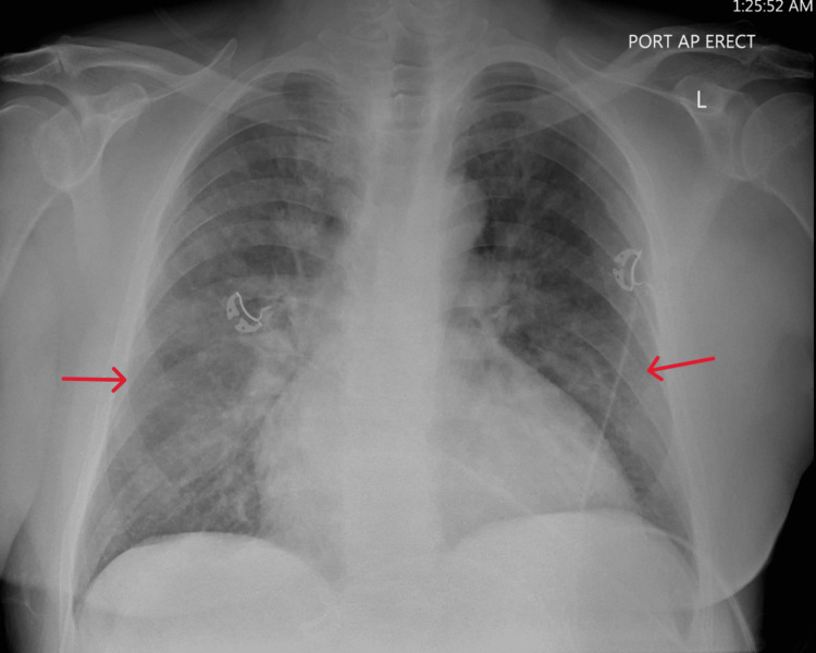
Chest radiograph. The radiograph shows the sequence of pulmonary venous hypertension: upper-lobe vascular redistribution (cephalization), then interstitial edema with Kerley B lines, peribronchial cuffing, and indistinct hila, then alveolar edema with perihilar (bat-wing) opacities, and pleural effusions, usually bilateral or right-sided. Cardiomegaly is common in chronic heart failure but absent in many patients with HFpEF or acute pulmonary edema from a sudden insult. The radiograph also identifies pneumonia, pneumothorax, and other causes of dyspnea. Its sensitivity for acute heart failure is limited: a normal radiograph does not exclude elevated filling pressure.
Lung ultrasound. Vertical hyperechoic artifacts arising from the pleural line (B-lines) reflect thickened interlobular septa and alveolar fluid. A diffuse bilateral B-line pattern, conventionally three or more B-lines in two or more zones on each side, supports cardiogenic pulmonary edema. In a meta-analysis of six studies with 1,827 patients with dyspnea, lung ultrasound had a pooled sensitivity of 88% and specificity of 90% for acute heart failure, against 73% and 90% for the chest radiograph. Lung ultrasound also identifies pleural effusions, and B-line counts fall with decongestion, so serial examinations track pulmonary congestion. Focal B-lines, an irregular pleural line, or subpleural consolidation suggest pneumonia or interstitial lung disease.
| Modality | Findings of congestion | Strengths | Limitations |
|---|---|---|---|
| Chest radiograph | Cephalization, Kerley B lines, peribronchial cuffing, alveolar edema, effusions, cardiomegaly | Universally available; identifies alternative diagnoses | Pooled sensitivity 73%; supine portable films underestimate effusions and cephalization |
| Lung ultrasound | Diffuse bilateral B-lines; pleural effusions | Pooled sensitivity 88%; repeatable at the bedside; tracks decongestion | Operator dependent; B-lines also in fibrosis, pneumonia, and acute respiratory distress syndrome |
| Focused echocardiography | Ventricular function, valve disease, raised filling pressures, dilated inferior vena cava | Identifies cause and mechanical complications | Comprehensive Doppler assessment requires expertise |
| Venous excess ultrasound (VExUS) Doppler | Pulsatile portal flow, hepatic vein systolic reversal, discontinuous intrarenal venous flow with a dilated inferior vena cava | Grades systemic venous congestion; associated with kidney injury | Validation mainly after cardiac surgery; confounded by tricuspid regurgitation and liver disease |
| CT of the chest | Ground-glass opacity, septal thickening, effusions | Excludes pulmonary embolism and other lung disease | Contrast and radiation; not needed for most patients |
Imaging of congestion at presentation. Sensitivities for lung ultrasound and chest radiograph are pooled values from a meta-analysis of patients with symptoms suggestive of acute decompensated heart failure.
Echocardiography and venous congestion assessment
Transthoracic echocardiography is performed in every patient with de novo heart failure and in patients with known heart failure whose ventricular or valvular function is unknown or may have changed. It establishes the ejection fraction category, regional wall motion abnormalities, valve disease (including acute or severe regurgitation and prosthetic dysfunction), right ventricular size and function, pericardial effusion, and features of specific cardiomyopathies. In amyloidosis, these include left ventricular wall thickening with a strain pattern that spares the apex. In unstable patients, focused echocardiography at the bedside is done immediately to identify mechanical complications, tamponade, severe valve disease, and right ventricular failure from pulmonary embolism.
Filling pressures. Doppler indices estimate left atrial pressure: an E/A ratio of 2 or more, an average E/e′ above 14, a tricuspid regurgitant velocity above 2.8 m/s, and a left atrial volume index above 34 mL/m² support raised left atrial pressure in a patient with abnormal myocardium. The assessment and its algorithm are covered in detail on the HFpEF and diastolic function page. A restrictive mitral inflow pattern that persists after decongestion indicates a poor prognosis.
Right atrial pressure. The inferior vena cava diameter and its inspiratory collapse estimate right atrial pressure. A diameter of 2.1 cm or less that collapses by more than 50% with a sniff corresponds to a right atrial pressure of about 3 mm Hg (range 0 to 5). A diameter above 2.1 cm that collapses by less than 50% corresponds to about 15 mm Hg (range 10 to 20). Other combinations correspond to about 8 mm Hg. Mechanical ventilation, athletic training, and severe tricuspid regurgitation limit the estimate.
Venous excess ultrasound. The VExUS system grades systemic venous congestion from the inferior vena cava diameter and Doppler patterns in the hepatic vein, portal vein, and intrarenal veins. In 706 assessments after cardiac surgery, severe congestion, defined as a dilated inferior vena cava (2 cm or more) with severe flow abnormalities in multiple Doppler patterns, was associated with subsequent acute kidney injury (hazard ratio 3.69). Its use to guide decongestion in acute heart failure is under study; it is most informative when right-sided congestion is suspected and the jugular venous pressure cannot be seen.
Differential diagnosis of acute dyspnea with congestion
| Condition | Features that overlap with heart failure | Features that distinguish it |
|---|---|---|
| Pneumonia | Dyspnea, crackles, infiltrates, raised natriuretic peptide in older patients | Fever, focal consolidation, raised procalcitonin, focal B-lines with consolidation on ultrasound |
| Exacerbation of chronic obstructive pulmonary disease | Dyspnea, orthopnea, edema with cor pulmonale | Wheeze, hyperinflation, hypercapnia, low natriuretic peptide unless heart failure coexists |
| Acute respiratory distress syndrome and noncardiogenic edema | Bilateral alveolar opacities, hypoxemia | Sepsis, aspiration, or other insult; normal or low filling pressures; normal left atrial size; patchy peripheral opacities |
| Pulmonary embolism | Dyspnea, tachycardia, right ventricular dysfunction, raised troponin and natriuretic peptide | Clear lungs, risk factors, CT pulmonary angiography |
| Acute coronary syndrome without heart failure | Dyspnea as anginal equivalent | No congestion; regional wall motion abnormality; troponin dynamics |
| Kidney failure with volume overload | Edema, pulmonary congestion, raised natriuretic peptide | Oliguria, very high creatinine; normal ventricular function; responds to dialysis |
| Nephrotic syndrome and cirrhosis | Edema, ascites, effusions | Low albumin, proteinuria or liver disease, normal jugular venous pressure |
| Cardiac tamponade or constriction | Raised jugular venous pressure, edema, dyspnea | Clear lungs, pulsus paradoxus, echocardiographic findings; see the constrictive pericarditis page |
| High-altitude or neurogenic pulmonary edema, transfusion-related overload, and lung injury | Alveolar edema | Clinical setting; normal ventricular function |
Differential diagnosis of dyspnea with congestion or pulmonary infiltrates. Two diagnoses frequently coexist, most often heart failure with pneumonia or with chronic obstructive pulmonary disease.
Acute cardiogenic pulmonary edema and hypertensive acute heart failure
Acute cardiogenic pulmonary edema is the rapid accumulation of fluid in the pulmonary interstitium and alveoli from a sudden rise in pulmonary capillary pressure. Patients present with severe dyspnea, orthopnea, tachypnea, diaphoresis, hypoxemia, diffuse crackles, and sometimes frothy pink sputum, often with hypertension and tachycardia. Onset over minutes to hours is called flash pulmonary edema.
Hypertensive acute heart failure. A large group of patients present with a systolic pressure above 140 mm Hg, often above 180 mm Hg, pulmonary edema, and a preserved or mildly reduced ejection fraction. Emergency medicine literature calls the most abrupt form sympathetic crash acute pulmonary edema (SCAPE). Treatment is rapid afterload and preload reduction with intravenous nitrates, given as high-dose infusions or as repeated boluses, together with noninvasive ventilation, which reduces left ventricular afterload and work of breathing. A diuretic is added, often at a lower dose than in fluid accumulation. Improvement often occurs within hours. General hypertensive emergency guidance limits the fall in blood pressure to no more than about 25% within the first hour, because a precipitous drop can cause cerebral, coronary, or renal ischemia.
Causes of flash pulmonary edema. Recurrent or unexplained flash pulmonary edema leads to a search for a mechanical or intermittent cause:
| Cause | Mechanism | Diagnosis |
|---|---|---|
| Acute ischemia, including transient ischemia without infarction | Diastolic and systolic dysfunction; ischemic mitral regurgitation | ECG during symptoms, troponin, stress testing or coronary angiography |
| Acute severe mitral regurgitation (papillary muscle rupture, chordal rupture, endocarditis) | Large volume load into a small, noncompliant left atrium; murmur may be soft or absent | Transthoracic and transesophageal echocardiography; see the mitral regurgitation page |
| Acute severe aortic regurgitation (endocarditis, aortic dissection) | Abrupt rise in left ventricular diastolic pressure; early mitral closure | Echocardiography; CT for dissection |
| Dynamic mitral regurgitation | Exercise- or ischemia-induced regurgitation in ischemic or dilated cardiomyopathy | Exercise echocardiography |
| Bilateral renal artery stenosis, or stenosis to a solitary kidney | Renin-mediated hypertension and sodium retention | Duplex ultrasound, CT or magnetic resonance angiography |
| Tachyarrhythmia in a stiff ventricle or mitral stenosis | Shortened diastolic filling time | ECG, monitoring |
| Dynamic left ventricular outflow obstruction | Hypertrophic cardiomyopathy, apical ballooning with basal hyperkinesis | Echocardiography with provocation |
| Prosthetic valve thrombosis or leaflet dysfunction | Acute obstruction or regurgitation | Echocardiography, fluoroscopy, CT |
| Stress (takotsubo) cardiomyopathy | Acute regional dysfunction after emotional or physical stress | Echocardiography, coronary angiography, cardiac magnetic resonance |
Causes of recurrent or flash pulmonary edema that require specific treatment.
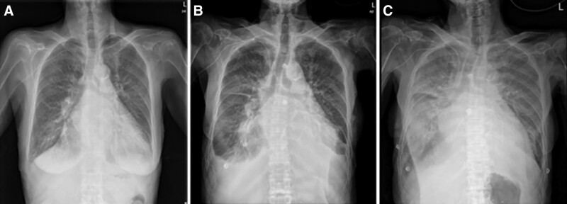
Renovascular flash pulmonary edema, sometimes called Pickering syndrome, occurs with bilateral renal artery stenosis or stenosis to a solitary functioning kidney. Randomized trials of renal artery stenting in hypertension and kidney disease did not show benefit and enrolled few patients with recurrent pulmonary edema. In this subgroup, revascularization of a hemodynamically significant stenosis can stop recurrences, and ACC/AHA peripheral artery disease guidance has supported it.
Oxygen, noninvasive ventilation, and intubation
Oxygen. Supplemental oxygen corrects hypoxemia. The ESC recommends oxygen when oxygen saturation is below 90% or arterial oxygen tension is below 60 mm Hg (Class 1) and advises against routine oxygen in patients who are not hypoxemic, because hyperoxia causes vasoconstriction and reduces cardiac output. The 2022 AHA/ACC/HFSA guideline does not make a specific recommendation.
Noninvasive ventilation. Continuous positive airway pressure (CPAP) and bilevel noninvasive positive pressure ventilation (NIPPV) recruit flooded alveoli, reduce work of breathing, and raise intrathoracic pressure, which lowers venous return and left ventricular transmural pressure and afterload. In the 3CPO trial of 1,069 patients with acute cardiogenic pulmonary edema, noninvasive ventilation did not reduce 7-day mortality compared with standard oxygen therapy (9.5% and 9.8%). The settings were CPAP at 5 to 15 cm H₂O or NIPPV at inspiratory pressures of 8 to 20 cm H₂O and expiratory pressures of 4 to 10 cm H₂O. Noninvasive ventilation produced faster improvement in dyspnea, heart rate, acidosis, and hypercapnia at 1 hour. Death or intubation did not differ between CPAP and NIPPV (11.7% and 11.1%). A Cochrane review of 24 studies with 2,664 participants found that noninvasive ventilation may reduce hospital mortality (risk ratio 0.65; 95% CI 0.51 to 0.82; low-quality evidence) and probably reduces intubation (risk ratio 0.49; 95% CI 0.38 to 0.62). The ESC recommends noninvasive ventilation in respiratory distress (respiratory rate above 25/min, oxygen saturation below 90%), started as soon as possible (Class 2a).
Noninvasive ventilation lowers preload and can cause hypotension in patients who are preload dependent, such as those with right ventricular infarction or hypovolemia. Contraindications include inability to protect the airway, depressed consciousness, vomiting, facial trauma, and shock. High-flow nasal oxygen is an alternative in patients who do not tolerate a mask; comparative trials in cardiogenic pulmonary edema are small.
Intubation. Intubation and mechanical ventilation are indicated for progressive respiratory failure despite oxygen or noninvasive ventilation (ESC Class 1), for inability to protect the airway, and for cardiogenic shock with respiratory failure. Positive end-expiratory pressure benefits the failing left ventricle but can reduce right ventricular output, and induction agents can cause profound hypotension in a patient dependent on sympathetic tone. Measured wedge and right atrial pressures rise with positive end-expiratory pressure without an equivalent rise in transmural filling pressure.
Intravenous loop diuretics: dose, route, and timing
Patients with heart failure admitted with evidence of significant fluid overload should be promptly treated with intravenous loop diuretics to improve symptoms and reduce morbidity (Class 1). Diuretics and other GDMT are titrated with the goal of resolving clinical congestion (Class 1). Intravenous administration bypasses the unpredictable absorption of oral furosemide (bioavailability 10% to 90%, mean about 50%), which falls further with gut edema. Loop diuretics also cause early venodilation that relieves pulmonary congestion before natriuresis begins.
Timing. In the REALITY-AHF registry of 1,291 patients treated with intravenous furosemide within 24 hours of emergency department arrival, the median door-to-furosemide time was 90 minutes. Treatment within 60 minutes was associated with lower in-hospital mortality (2.3% and 6.0%; adjusted odds ratio 0.39; 95% CI 0.20 to 0.76). The association is observational, but it supports giving the first dose in the emergency department once congestion is recognized.
Dose. The initial intravenous dose is set from the home dose, kidney function, and the severity of congestion.
| Situation | Initial intravenous furosemide | Source |
|---|---|---|
| No outpatient loop diuretic | 20–40 mg bolus; higher when kidney function is reduced | 2021 ESC guideline; 2019 Heart Failure Association (HFA) position statement |
| Outpatient loop diuretic, standard strategy | Total daily dose 1 to 2.5 times the oral furosemide-equivalent dose, milligram for milligram, divided into two or three boluses or given as an infusion | DOSE (1× and 2.5×); 2022 AHA/ACC/HFSA guideline text (often at least 2×); ESC (1 to 2×) |
| Acetazolamide trial protocol | Twice the oral maintenance dose, intravenously | ADVOR |
| Kidney dysfunction | Higher single doses, because tubular secretion is reduced and a higher dose is needed to reach the natriuretic threshold | 2019 HFA position statement |
Initial intravenous loop diuretic dose. Approximate equivalence: furosemide 40 mg orally = furosemide 20 mg intravenously = torsemide 20 mg = bumetanide 1 mg. Torsemide and bumetanide have oral bioavailability above 80%.
The DOSE trial randomized 308 patients with acute decompensated heart failure in a 2-by-2 factorial design to furosemide by bolus every 12 hours or continuous infusion, and at a low dose (equal to the previous oral dose) or a high dose (2.5 times the oral dose). Bolus and infusion did not differ in global symptom assessment over 72 hours (area under the curve 4,236 and 4,373; P = 0.47) or in creatinine change (0.05 and 0.07 mg/dL). The high-dose strategy produced a nonsignificant trend toward greater symptom relief (4,430 and 4,171; P = 0.06). In secondary analyses it produced more diuresis, weight loss, and relief of dyspnea, and more transient rises in creatinine, without a difference in creatinine change at 72 hours (0.08 and 0.04 mg/dL; P = 0.21) or in 60-day outcomes. Patients in the low-dose group more often needed a 50% dose increase at 48 hours. The trial established that both routes are acceptable and that a higher initial dose is safe.
Route and frequency. Furosemide has a short half-life. After each dose, natriuresis lasts about 6 hours and is followed by avid sodium reabsorption (postdiuretic sodium retention). Once-daily intravenous dosing is therefore inadequate in most patients; boluses are given two or three times a day, or a continuous infusion follows a loading bolus. An infusion without a loading bolus takes hours to reach a natriuretic concentration.
The treatment goal. The goal of intravenous diuretic therapy is complete resolution of clinical congestion: no orthopnea, a jugular venous pressure that is not elevated, no or minimal edema, and no hepatojugular reflux. Weight is a supporting measure, but the target weight is an estimate. Persistent congestion at discharge, reported in 25% to 50% of patients, is associated with higher mortality and readmission.
Natriuresis and urine output monitoring
The diuretic response is measured early, because the response to the first dose predicts the response to subsequent doses, and a poor response identified at 2 to 6 hours allows the dose to be escalated the same day. Urine output and weight are standard measures, but intake and output records are often incomplete and weight changes are delayed.
Spot urine sodium. A spot urine sample obtained 1 or 2 hours after a loop diuretic dose predicts cumulative sodium excretion over the following 6 hours. In a validation study of 50 patients, an equation based on the spot urine sodium and creatinine predicted measured 6-hour sodium output with a correlation of 0.91. It identified patients with a poor natriuretic response, defined as less than 50 mmol of sodium excreted. The 2019 HFA position statement and the 2021 ESC guideline define an inadequate response as a spot urine sodium below 50 to 70 mmol/L at 2 hours, or a urine output below 100 to 150 mL/h during the first 6 hours. The next dose is then doubled, up to a total daily intravenous furosemide dose of 400 to 600 mg, and the response is reassessed.
Natriuresis-guided protocols. In PUSH-AHF, 310 patients were randomized to natriuresis-guided therapy, in which treatment was intensified whenever the spot urine sodium was below 70 mmol/L, or to usual care. Natriuresis over 24 hours was greater with guided therapy (409 and 345 mmol; P = 0.0061), but all-cause death or heart failure rehospitalization at 180 days did not differ (31% in both groups; hazard ratio 0.92; 95% CI 0.62 to 1.38). In the nonrandomized ENACT-HF study of 401 patients at 29 centers, a standardized natriuresis-guided protocol increased natriuresis at day 1 (282 and 174 mmol) and day 2 and shortened length of stay (5.8 and 7.0 days). Natriuresis-guided escalation increases sodium excretion in the first days of treatment. A shorter hospital stay has been shown only in the nonrandomized ENACT-HF comparison, not in PUSH-AHF, and no effect on outcomes after discharge has been shown.
Oral furosemide equivalents: torsemide × 2, bumetanide × 40. Published torsemide equivalents range from 10 to 20 mg; the calculator uses the conservative 20 mg. Intravenous furosemide is dosed milligram for milligram against the oral furosemide-equivalent dose, as in DOSE and ADVOR. Values are rounded to the nearest 10 mg.
| Step | Timing | Action |
|---|---|---|
| 1. First dose | On recognition of congestion, in the emergency department | Intravenous loop diuretic at the dose above; bladder emptied before the dose so the 2-hour sample reflects the response |
| 2. Early response | Spot urine sodium at 2 hours; urine output over 6 hours | Adequate: same dose repeated two or three times daily. Inadequate: dose doubled |
| 3. Daily reassessment | Every 24 hours | Congestion examination, weight, net fluid balance, creatinine, potassium, sodium, magnesium; dose adjusted to maintain a net negative balance (often 3 to 5 L/day in trial protocols) |
| 4. Diuretic resistance | Inadequate response after dose escalation, often by day 2 or 3 | Second diuretic class added: thiazide-type diuretic or acetazolamide (Class 2a for a second diuretic); mineralocorticoid receptor antagonist (MRA) and sodium-glucose cotransporter 2 (SGLT2) inhibitor given if not already taken |
| 5. Persistent resistance | Despite combination therapy | Reassessment of perfusion and of the hemodynamic profile; invasive hemodynamics; inotrope if low output; ultrafiltration in selected patients; referral to an advanced heart failure team |
| 6. Transition | Clinical congestion resolved | Oral diuretic at an estimated maintenance dose, observed for stability; discharge plan for diuretic adjustment (Class 1) |
Stepped decongestion protocol, based on the 2022 AHA/ACC/HFSA guideline, the 2024 ACC decision pathway, the 2019 HFA position statement, and the protocols of DOSE, CARRESS-HF, ADVOR, and PUSH-AHF.
Diuretic resistance and sequential nephron blockade
Diuretic resistance is an inadequate natriuretic response despite an adequate loop diuretic dose, or persistent congestion despite escalating doses. Postdiuretic sodium retention between doses, dietary sodium, and NSAIDs add to the nephron-level mechanisms described under pathophysiology.
When doubling the loop dose does not produce an adequate response, it is reasonable to intensify the regimen by adding a second diuretic (Class 2a). Each class blocks a different nephron segment:
| Agent | Site of action | Dose in acute heart failure | Evidence | Principal adverse effects |
|---|---|---|---|---|
| Acetazolamide | Proximal tubule (carbonic anhydrase) | 500 mg intravenously once daily, with the loop diuretic | ADVOR: successful decongestion at 3 days 42.2% and 30.5% (risk ratio 1.46; 95% CI 1.17 to 1.82); death or heart failure readmission at 3 months 29.7% and 27.8% (hazard ratio 1.07) | Metabolic acidosis; hypokalemia; patients taking SGLT2 inhibitors were excluded |
| Metolazone | Distal convoluted tubule | 2.5–10 mg orally, given before the loop dose | Observational data and trial protocols (CARRESS-HF stepped care) | Hypokalemia, hyponatremia, hypomagnesemia, kidney dysfunction; long half-life prolongs effect |
| Chlorothiazide | Distal convoluted tubule | 500–1,000 mg intravenously once or twice daily | Retrospective comparisons with metolazone show similar urine output | As for metolazone; cost |
| Hydrochlorothiazide | Distal convoluted tubule | 25–100 mg orally, adjusted to kidney function | CLOROTIC: weight loss at 72 hours 2.3 and 1.5 kg (P = 0.002); no difference in dyspnea; impaired kidney function 46.5% and 17.2% | Kidney dysfunction, hypokalemia |
| Spironolactone or eplerenone | Collecting duct (aldosterone) | Standard GDMT doses | ATHENA-HF: spironolactone 100 mg did not reduce NT-proBNP more than usual care | Hyperkalemia; slow onset |
| SGLT2 inhibitor | Proximal tubule (glucose-linked sodium reabsorption) | Empagliflozin 10 mg or dapagliflozin 10 mg daily | DICTATE-AHF: diuretic efficiency not significantly improved (odds ratio 0.65; P = 0.06), lower cumulative loop dose and more natriuresis at 24 hours | Genital infection, volume depletion; ketoacidosis rare |
| Tolvaptan | Collecting duct (vasopressin V2 receptor) | 15 mg orally once daily, started in hospital | EVEREST: weight and dyspnea improved early; no effect on mortality or heart failure readmission | Overly rapid sodium correction; liver injury (treatment limited to 30 days) |
Agents added to intravenous loop diuretics. Combinations cause electrolyte losses, so potassium, magnesium, sodium, and creatinine are measured at least daily, and more often with thiazide-type diuretics.
Acetazolamide blocks proximal sodium reabsorption, so more sodium reaches the loop of Henle. In ADVOR, 519 patients with volume overload and an NT-proBNP above 1,000 pg/mL or BNP above 250 pg/mL received intravenous loop diuretics at twice the oral maintenance dose with acetazolamide or placebo. Acetazolamide increased urine output and natriuresis, shortened length of stay, and had similar rates of worsening kidney function, hypokalemia, and hypotension. It did not change death or readmission at 3 months.
Thiazide-type diuretics block the distal tubule, where chronic loop exposure has increased sodium reabsorption. They produce a large natriuresis in loop-resistant patients but cause more electrolyte disturbance and kidney dysfunction than either drug alone. In CLOROTIC, 230 patients with a mean age of 83 years received hydrochlorothiazide or placebo with intravenous furosemide. Weight loss and 24-hour diuresis were greater with hydrochlorothiazide, but dyspnea, mortality, and rehospitalization did not differ. Impaired kidney function was nearly three times as frequent. The choice between metolazone and intravenous chlorothiazide rests on cost and route; their effects on urine output were similar in retrospective comparisons.
MRAs have modest diuretic effect at standard doses, and their main role is prognostic in HFrEF. SGLT2 inhibitors cause a glycosuric natriuresis that adds to loop diuretics without activating the renin-angiotensin system. In DICTATE-AHF, 240 patients (71% with type 2 diabetes) were randomized in an open-label design to dapagliflozin, started within 24 hours of admission, or to structured usual care. Diuretic efficiency did not improve significantly, but patients needed less loop diuretic and fewer dose escalations to achieve the same weight loss. Natriuresis and urine output were higher.
Ultrafiltration, hypertonic saline, and other decongestive strategies
Ultrafiltration. Venovenous ultrafiltration removes isotonic fluid at a set rate without neurohormonal activation. In UNLOAD, early ultrafiltration increased fluid loss at 48 hours and reduced rehospitalization compared with intravenous diuretics without systematic escalation. In CARRESS-HF, 188 patients with worsening kidney function and persistent congestion were randomized to ultrafiltration or a stepped pharmacologic algorithm. Ultrafiltration was inferior for the combined end point of creatinine and weight change at 96 hours because creatinine rose (+0.23 and −0.04 mg/dL) with similar weight loss (5.7 and 5.5 kg). Serious adverse events were more frequent (72% and 57%). The 2022 guideline states that patient selection, fluid removal rates, venous access, prevention of complications, and cost require further investigation, and it makes no recommendation. European guidance limits ultrafiltration to refractory congestion that has not responded to diuretic-based strategies, particularly with advanced kidney disease and in centers with experience.
Hypertonic saline. Small trials, mostly from single centers, have combined small volumes of hypertonic saline with high-dose furosemide to draw interstitial fluid into the vascular space and increase delivery of sodium to the loop. The evidence is limited, US guidelines do not recommend it, and the tolvaptan label advises against combining the two.
Low-dose dopamine and nesiritide for kidney protection. In the ROSE trial, neither low-dose dopamine nor low-dose nesiritide added to diuretics improved 72-hour urine volume or cystatin C in patients with acute heart failure and kidney dysfunction. The 2022 guideline notes a subgroup with an ejection fraction below 40% in whom urine output and weight loss increased with dopamine.
Torsemide after discharge. In TRANSFORM-HF, 2,859 patients hospitalized with heart failure were randomized to a torsemide or furosemide strategy at discharge. Death occurred in 26.1% and 26.2% over a median of 17.4 months (hazard ratio 1.02; 95% CI 0.89 to 1.18), and all-cause death or hospitalization at 12 months did not differ. The loop diuretic is chosen for absorption, prior response, and cost.
Outpatient and home intravenous decongestion. Infusion clinics and heart failure day units give intravenous loop diuretics to patients with worsening congestion who are hemodynamically stable, with same-day laboratory testing and follow-up. Subcutaneous furosemide delivered by an on-body infusor over about 5 hours is approved by the US Food and Drug Administration for congestion in chronic heart failure. Hospital-at-home programs extend intravenous therapy to the home under daily clinician review. These models avoid admission in selected patients; the evidence is observational, and none has been compared with hospitalization in a large randomized trial.
Vasodilators: nitroglycerin, nitroprusside, and nesiritide
In patients admitted with decompensated heart failure, in the absence of systemic hypotension, intravenous nitroglycerin or nitroprusside may be considered as an adjuvant to diuretic therapy for relief of dyspnea (Class 2b). Vasodilators relieve pulmonary congestion by lowering preload and afterload, but no trial has shown a durable effect on rehospitalization or survival, and their use is limited to symptom relief in patients with intact or high blood pressure. Patients with hypertension, coronary ischemia, or significant mitral regurgitation are the most suitable candidates.
| Drug | Mechanism | Dose | Cautions |
|---|---|---|---|
| Nitroglycerin | Venodilator at low doses; arterial dilator at higher doses; coronary vasodilator | Infusion starting at 10–20 µg/min, titrated to symptoms and blood pressure up to 200 µg/min; higher-dose boluses are used in hypertensive pulmonary edema | Tachyphylaxis within 24 hours; up to 20% of patients resistant to high doses; headache; hypotension; contraindicated after phosphodiesterase-5 inhibitors |
| Nitroprusside | Balanced arterial and venous dilator | 0.3 µg/kg/min, titrated up to 5 µg/kg/min | Arterial line and intensive care setting; thiocyanate and cyanide toxicity with prolonged infusion, kidney or hepatic dysfunction; rebound on abrupt withdrawal |
| Nesiritide (recombinant BNP) | Arterial and venous dilator; modest natriuresis | 2 µg/kg bolus, then 0.01 µg/kg/min | Hypotension; no effect on death or readmission in ASCEND-HF |
Intravenous vasodilators in acute heart failure. Doses from the 2016 ESC guideline and US prescribing information. The ESC limits vasodilators to patients with a systolic pressure above 110 mm Hg (Class 2b).
Nitroglycerin is the first-line vasodilator for acute pulmonary edema with hypertension. Sublingual nitroglycerin is given while intravenous access is established.
Nitroprusside is the most potent afterload-reducing drug and is used in severely congested patients with hypertension, high systemic vascular resistance, or severe mitral or aortic regurgitation complicating left ventricular dysfunction. In profile C with an adequate blood pressure, nitroprusside guided by invasive hemodynamics raises cardiac output and lowers filling pressure. It is also used as a bridge in acute severe valvular regurgitation. Patients with severe aortic stenosis tolerate it only with invasive monitoring because a fixed outflow limits the rise in output.
Nesiritide reduced dyspnea modestly and lowered wedge pressure in earlier trials. ASCEND-HF randomized 7,141 patients. Dyspnea improved slightly more at 6 hours (44.5% and 42.1%) and 24 hours (68.2% and 66.1%), short of the prespecified significance level. Rehospitalization for heart failure or death at 30 days was 9.4% and 10.1% (P = 0.31), and worsening kidney function did not differ. Nesiritide is not recommended for routine use.
Inotropes and vasopressors when perfusion is inadequate
Inotropes increase cardiac output and improve hemodynamics but have not been shown to improve survival in patients hospitalized with heart failure, and their routine use in patients without hypoperfusion causes harm. In OPTIME-CHF, 951 patients hospitalized with decompensated HFrEF who did not need inotropes were randomized to a 48-hour milrinone infusion or placebo. Days hospitalized for cardiovascular causes within 60 days did not differ (median 6 and 7), and sustained hypotension requiring intervention (10.7% and 3.2%) and new atrial arrhythmias (4.6% and 1.5%) were more frequent with milrinone. The trial shows that inotropes do not belong in profile B; it does not apply to patients with documented low output and end-organ hypoperfusion.
In patients with cardiogenic shock, intravenous inotropic support is recommended to maintain systemic perfusion and preserve end-organ performance (Class 1). The ESC reserves inotropes for patients with a systolic pressure below 90 mm Hg and hypoperfusion who do not respond to standard treatment (Class 2b) and recommends against routine use (Class 3). Dobutamine stimulates beta-1 receptors and its effect is attenuated by beta-blockade. Milrinone, a phosphodiesterase-3 inhibitor, acts downstream of the beta receptor, causes more vasodilation and hypotension, and accumulates in kidney dysfunction. Norepinephrine is the usual vasopressor when hypotension persists. Levosimendan, a calcium sensitizer used in Europe, is not available in the United States.
Starting an inotrope in acute decompensated heart failure raises the question of cardiogenic shock, temporary mechanical support, and candidacy for advanced therapies, and it involves the multidisciplinary shock or advanced heart failure team. Vasoactive drug choice, doses, staging by the Society for Cardiovascular Angiography and Interventions (SCAI), and mechanical circulatory support are covered on the cardiogenic shock page.
Therapies without benefit in acute heart failure
Many agents improved hemodynamics or symptoms in early studies and failed in outcome trials. Several remain in use for narrow indications.
| Therapy | Trial | Result | Current role |
|---|---|---|---|
| Milrinone in patients without hypoperfusion | OPTIME-CHF (951 patients) | No reduction in hospital days; more hypotension and atrial arrhythmias | Low output with hypoperfusion; bridge to advanced therapies; palliation |
| Nesiritide | ASCEND-HF (7,141 patients) | No effect on 30-day death or rehospitalization; more hypotension | Not recommended for routine use |
| Routine pulmonary artery catheter | ESCAPE (433 patients) | Days alive and out of hospital at 6 months 133 and 135; more in-hospital adverse events (21.9% and 11.5%) | Uncertain hemodynamics or failure of empiric therapy (Class 2a); routine use not recommended (Class 3: No Benefit) |
| Tolvaptan | EVEREST (4,133 patients) | Mortality 25.9% and 26.3% over a median of 9.9 months; cardiovascular death or heart failure hospitalization 42.0% and 40.2% | Severe symptomatic hyponatremia despite fluid restriction |
| Low-dose dopamine | ROSE (renal dysfunction) | No change in 72-hour urine volume or cystatin C | None for kidney protection |
| High-dose spironolactone (100 mg) | ATHENA-HF (360 patients) | No greater NT-proBNP reduction; no change in clinical end points | Standard-dose MRA as GDMT |
| Serelaxin | RELAX-AHF-2 (6,545 patients) | Cardiovascular death at 180 days 8.7% and 8.9%; worsening heart failure at day 5 6.9% and 7.7% | Not approved |
| Ultrafiltration as initial therapy in cardiorenal syndrome | CARRESS-HF (188 patients) | Creatinine rose; similar weight loss; more serious adverse events | Refractory congestion in experienced centers |
| Morphine | MIMO (111 patients, stopped early); ADHERE registry | More serious adverse events than midazolam; independent association with mortality in the registry | Not used routinely |
Therapies tested in acute heart failure without improvement in outcomes. Omecamtiv mecarbil, a cardiac myosin activator, modestly reduced heart failure events or cardiovascular death in GALACTIC-HF (hazard ratio 0.92; 95% CI 0.86 to 0.99), a chronic HFrEF trial that also enrolled hospitalized patients. It had no effect on cardiovascular death alone. Istaroxime remains investigational.
Worsening kidney function during decongestion
A rise in creatinine of more than 0.3 mg/dL is common during decongestion; in DOSE it occurred within 72 hours in 23% of patients in the high-dose group and 14% in the low-dose group. Rising creatinine has different causes and different meanings. The 2022 guideline states that diuresis should not be stopped prematurely because of small changes in creatinine: rises in the range of 0.3 mg/dL do not predict worse outcomes except when patients are discharged with persistent congestion (Class 1).
| Pattern | Interpretation | Management |
|---|---|---|
| Creatinine rise with effective decongestion, rising hemoglobin and albumin (hemoconcentration), good urine output | Hemodynamic change in filtration; not tubular injury; associated with better survival | Diuresis continued until congestion resolves |
| Creatinine rise with persistent congestion and poor diuretic response | Venous congestion, low output, or both; poor prognosis | Reassessment of perfusion and filling pressures; diuretic intensification or combination therapy; invasive hemodynamics; inotrope if low output |
| Creatinine rise with hypotension, cool extremities, or rising lactate | Low output or overdiuresis | Diuretic paused; filling pressures assessed; inotrope or advanced therapies when output is low |
| Creatinine rise after starting or increasing an ARNI, ACE inhibitor, angiotensin receptor blocker (ARB), or SGLT2 inhibitor | Expected hemodynamic effect, usually small | Therapy continued unless the rise is large, progressive, or accompanied by hyperkalemia |
| Creatinine rise with contrast, nephrotoxins, obstruction, or infection | Intrinsic or postrenal injury | Cause removed; urinalysis and kidney ultrasound |
Interpretation of rising creatinine during treatment of acute heart failure. Persistent congestion at discharge, not the creatinine change, is the stronger predictor of death and readmission.
Hemoconcentration, a rise in hemoglobin, hematocrit, or albumin during treatment, indicates that diuresis is outpacing refill from the interstitium. In a cohort from the ESCAPE trial, it was associated with worsening kidney function but with better survival.
Electrolytes, hyponatremia, and hyperkalemia during treatment
Potassium and magnesium. Loop and thiazide-type diuretics cause hypokalemia and hypomagnesemia, which increase the risk of ventricular arrhythmias and digoxin toxicity. Both are measured at least daily during intravenous therapy and replaced. Potassium is kept in the normal range; MRAs and renin-angiotensin inhibitors reduce losses.
Hyponatremia. Hyponatremia in heart failure is usually hypervolemic, the result of vasopressin release and reduced free water excretion, and it predicts mortality. Thiazide-type diuretics worsen it. Fluid restriction modestly raises sodium; the 2022 guideline states that the benefit of fluid restriction to reduce congestive symptoms in advanced heart failure with hyponatremia is uncertain (Class 2b). Tolvaptan is indicated for clinically significant hypervolemic or euvolemic hyponatremia (sodium below 125 mEq/L, or less marked hyponatremia that is symptomatic and has resisted fluid restriction), including in heart failure. It is started and restarted only in hospital at 15 mg once daily, increased at intervals of at least 24 hours to 30 mg and then to a maximum of 60 mg. Treatment is limited to 30 days because of liver injury, and it is avoided in liver disease. Correction faster than 12 mEq/L in 24 hours can cause osmotic demyelination. Severe hyponatremia with neurologic symptoms is treated with hypertonic saline under nephrology guidance.
Hyperkalemia. Hyperkalemia is most often caused by kidney dysfunction combined with an MRA or renin-angiotensin inhibitor. An MRA is held when potassium exceeds 5.5 mEq/L and resumed at a lower dose once potassium is controlled. In patients who develop hyperkalemia (5.5 mEq/L or higher) while taking a renin-angiotensin inhibitor, the effectiveness of potassium binders (patiromer, sodium zirconium cyclosilicate) in improving outcomes by facilitating continuation of therapy is uncertain (Class 2b). Management of potassium during titration is detailed on the HFrEF page.
Metabolic alkalosis. Contraction alkalosis develops with vigorous loop and thiazide diuresis and impairs ventilatory drive in patients with lung disease. Acetazolamide corrects it.
Supportive care: morphine, thromboprophylaxis, sodium, and monitoring
Morphine and anxiolysis. Morphine was used for decades for dyspnea and anxiety in pulmonary edema. In 147,362 hospitalizations in the ADHERE registry, the 14.1% of patients who received morphine more often needed mechanical ventilation (15.4% and 2.8%) and intensive care (38.7% and 14.4%) and had higher mortality (13.0% and 2.4%). After risk adjustment, morphine remained independently associated with death (odds ratio 4.84). In MIMO, 111 patients with acute cardiogenic pulmonary edema, dyspnea, and anxiety were randomized to intravenous midazolam or morphine. In-hospital mortality did not differ significantly (12.7% and 17.9%), and the trial was stopped early. Opioids depress respiration, cause nausea and vomiting, and increase the need for intubation. The ESC recommends against routine use of opiates except in selected patients with severe or intractable pain or anxiety (Class 3).
Thromboprophylaxis. Prophylaxis for venous thromboembolism is recommended in patients hospitalized with heart failure (Class 1). In patients admitted for decompensated heart failure with a creatinine clearance above 30 mL/min, enoxaparin 40 mg subcutaneously once daily, unfractionated heparin 5,000 units subcutaneously every 8 or 12 hours, or rivaroxaban 10 mg once daily reduces radiographically demonstrable venous thrombosis. Extending prophylaxis after discharge reduces venous thromboembolism but increases bleeding and does not provide net benefit. Patients already taking an oral anticoagulant for atrial fibrillation continue it, with the dose adjusted to kidney function.
Sodium and fluid. Dietary sodium restriction during admission is common practice, but restriction to very low intakes has not improved outcomes in trials of outpatients, and the 2022 guideline frames sodium advice as avoidance of excessive intake. Fluid restriction is reserved for hyponatremia and refractory congestion. Malnutrition and cachexia are common in advanced heart failure, and dietitian review addresses protein and calorie intake as well as sodium.
Monitoring. Daily measurements during intravenous therapy include standing weight at the same time each morning, fluid intake and output, vital signs including orthostatic pressure before discharge, and the clinical signs of congestion and hypoperfusion. Serum electrolytes, urea, and creatinine are measured daily during active medication changes. Continuous ECG monitoring is used for patients with arrhythmia, ischemia, QT-prolonging drugs, or severe electrolyte disturbance.
Atrial fibrillation and other arrhythmias during decompensation
Atrial fibrillation is both a cause and a consequence of decompensation. A rapid ventricular rate shortens diastolic filling and raises left atrial pressure, and loss of atrial contraction reduces stroke volume in a stiff ventricle. In patients with hemodynamic instability attributable to atrial fibrillation, urgent electrical cardioversion is performed. In stable patients, rate control relies on beta-blockers when tolerated; nondihydropyridine calcium channel blockers are avoided in HFrEF because of their negative inotropic effect. Intravenous digoxin or amiodarone controls the rate when beta-blockers are limited by hypotension or decompensation. Tachycardia is often partly compensatory, and the rate falls as congestion resolves, so the rate target is approached gradually. Rhythm control, catheter ablation, and anticoagulation follow the principles on the atrial fibrillation page.
Ventricular tachycardia and frequent defibrillator shocks indicate advanced disease, ischemia, or electrolyte disturbance; they are treated by correcting the trigger, beta-blockade, and antiarrhythmic drugs. Bradycardia and high-grade block from drug toxicity, ischemia, or conduction disease are treated by withdrawal of the offending drug and pacing.
Right heart failure and isolated systemic congestion
Predominant right heart failure presents with a high jugular venous pressure, hepatomegaly, ascites, edema, and clear lungs, often with severe tricuspid regurgitation. Causes include left heart failure with secondary pulmonary hypertension, pulmonary arterial hypertension, chronic thromboembolic disease and acute pulmonary embolism, right ventricular infarction, arrhythmogenic cardiomyopathy, tricuspid valve disease, and constrictive pericarditis. Kidney and hepatic congestion are pronounced, and the abdominal compartment contributes to kidney dysfunction. Diuretic response is often poor because of gut edema and low kidney perfusion pressure, and high-dose intravenous diuretics, combination therapy, or ultrafiltration are frequently needed.
The right ventricle tolerates pressure poorly. Excess volume dilates it, shifts the septum toward the left ventricle, and lowers left ventricular filling and output. Hypotension in right heart failure is treated by restoring coronary perfusion pressure with norepinephrine, reducing right ventricular afterload (pulmonary vasodilators in pulmonary arterial hypertension, reperfusion in pulmonary embolism, correction of hypoxemia and acidosis), and inotropic support. Fluid loading helps only when right-sided filling pressures are low, as in some patients with right ventricular infarction. Pulmonary hypertension is covered on the pulmonary hypertension page, and acute right ventricular failure from embolism on the pulmonary embolism page.
Pulmonary artery catheter and invasive hemodynamics
In selected patients with persistent or worsening symptoms, signs, or diagnostic parameters, and in whom hemodynamics are uncertain, invasive hemodynamic monitoring can be useful to guide management (Class 2a). Routine invasive hemodynamic monitoring is not recommended (Class 3: No Benefit). In ESCAPE, 433 patients with severe recurrent heart failure were randomized to therapy guided by clinical assessment and a pulmonary artery catheter, with targets of a wedge pressure of 15 mm Hg and right atrial pressure of 8 mm Hg, or to clinical assessment alone. Mortality did not differ, and the hazard ratio for days alive and out of hospital over 6 months was 1.00. Both groups had substantial reductions in symptoms, jugular venous pressure, and edema.
The catheter is used when the hemodynamic profile is uncertain (for example, congestion with apparent low output, or hypotension with unclear filling pressures), when kidney function worsens with uncertain volume status, when the patient does not respond to empiric therapy, when vasoactive drugs need titration, and when candidacy for mechanical support or transplantation is being assessed. Measured cardiac output, wedge pressure, right atrial pressure, systemic and pulmonary vascular resistance, and the right atrial to wedge pressure ratio define the phenotype and guide vasodilator, inotrope, and diuretic therapy. Measurement technique and normal values are on the right heart catheterization and hemodynamics page.
Continuing and starting GDMT in hospital
The 2022 guideline makes four Class 1 recommendations for patients with HFrEF who require hospitalization. Preexisting GDMT should be continued and optimized unless contraindicated. Diuresis and other GDMT should not routinely be discontinued for a mild decrease in kidney function or an asymptomatic reduction in blood pressure. GDMT should be started during hospitalization after clinical stability is achieved. If GDMT must be stopped, it should be restarted and further optimized as soon as possible. True contraindications are uncommon: advanced atrioventricular block without a pacemaker (beta-blockers), cardiogenic shock (until it resolves), and angioedema (ACE inhibitors and ARNIs).
In the CHAMP-HF registry of 2,588 US outpatients with chronic HFrEF, each of the beta-blocker, renin-angiotensin inhibitor, ARNI, and MRA classes was started or increased in 10% or fewer of patients over 12 months. Fewer than 1% were taking target doses of all three classes at once. The 2024 ACC decision pathways for HFrEF and for hospitalized heart failure place increased emphasis on establishing all four drug classes during the admission when possible, with follow-up to monitor tolerance and continue titration. The hospitalized heart failure pathway states that SGLT2 inhibitors can be started at any point during hospitalization, regardless of ejection fraction.
| Drug class | At admission | Starting in hospital | Evidence in hospitalized patients |
|---|---|---|---|
| Beta-blocker | Continued; dose reduced or held only for cardiogenic shock, marked hypoperfusion, symptomatic bradycardia, or high-grade block | Started once euvolemic or near euvolemic and off intravenous inotropes, at a low dose; caution after inotrope use | OPTIMIZE-HF: withdrawal associated with higher adjusted mortality (hazard ratio 2.3) than continuation; B-CONVINCED: continuation did not delay improvement |
| ARNI (or ACE inhibitor or ARB) | Continued unless hypotension with hypoperfusion, angioedema, or severe hyperkalemia | Sacubitril/valsartan started after stabilization; 36-hour washout after an ACE inhibitor | PIONEER-HF: greater NT-proBNP reduction than enalapril (−46.7% and −25.3%), with similar rates of worsening kidney function, hyperkalemia, symptomatic hypotension, and angioedema |
| MRA | Continued unless potassium is above 5.5 mEq/L or kidney function is rapidly worsening | Started when potassium is below 5.0 mEq/L and estimated glomerular filtration rate (eGFR) above 30 mL/min/1.73 m² | Registry data: continuation and initiation associated with lower mortality and readmission |
| SGLT2 inhibitor | Continued; held when oral intake is poor or ketoacidosis risk is high | Started during admission when stable, regardless of ejection fraction or diabetes | EMPULSE: clinical benefit at 90 days (win ratio 1.36; 95% CI 1.09 to 1.68); SOLOIST-WHF: total cardiovascular deaths and heart failure events reduced (hazard ratio 0.67) |
| Intravenous iron | Iron studies measured | Ferric carboxymaltose before or soon after discharge in HFrEF with iron deficiency | AFFIRM-AHF: total heart failure hospitalizations reduced (rate ratio 0.74); primary composite not significant (rate ratio 0.79; P = 0.059) |
Guideline-directed medical therapy during a heart failure hospitalization. Doses, sequencing, and titration targets for chronic HFrEF are on the HFrEF page.
Beta-blockers. In OPTIMIZE-HF, among 2,373 patients eligible for beta-blockers at discharge, continuation was associated with lower postdischarge mortality than no beta-blocker (hazard ratio 0.60), and withdrawal was associated with higher mortality than continuation (hazard ratio 2.3; 95% CI 1.2 to 4.6). The guideline states that withholding or reducing beta-blocker therapy should be considered in patients with marked volume overload or marginal low cardiac output. A common practice is to halve the dose in patients with borderline perfusion and to stop it only in shock or symptomatic bradycardia.
Sacubitril/valsartan. PIONEER-HF enrolled 881 patients with HFrEF hospitalized for acute decompensated heart failure at 129 US sites. Randomization followed hemodynamic stabilization: systolic pressure of at least 100 mm Hg for 6 hours, no increase in intravenous diuretic dose for 6 hours, no intravenous vasodilators for 6 hours, and no intravenous inotropes for 24 hours. The time-averaged reduction in NT-proBNP was greater with sacubitril/valsartan (ratio of change 0.71; 95% CI 0.63 to 0.81), evident by week 1, without more adverse events. Patients who had never taken an ACE inhibitor or ARB also benefited, so an ARNI can be the first renin-angiotensin inhibitor started in hospital.
SGLT2 inhibitors. In EMPULSE, 530 patients hospitalized for acute de novo or decompensated chronic heart failure, regardless of ejection fraction, were randomized to empagliflozin 10 mg or placebo at a median of 3 days after admission. Empagliflozin produced a clinical benefit on a hierarchical composite of death, heart failure events, and symptoms at 90 days, consistent across de novo and chronic heart failure, ejection fraction, and diabetes status. Serious adverse events were less frequent (32.3% and 43.6%). In SOLOIST-WHF, 1,222 patients with diabetes received sotagliflozin, which was started before discharge in most patients and within a median of 2 days after discharge in the rest. It reduced the rate of cardiovascular death and heart failure hospitalizations and urgent visits (51.0 and 76.3 per 100 patient-years; hazard ratio 0.67; 95% CI 0.52 to 0.85).
Iron. In patients with HFrEF and iron deficiency, intravenous iron is reasonable to improve functional status and quality of life (Class 2a). AFFIRM-AHF randomized 1,108 patients with an ejection fraction below 50% and iron deficiency who had been stabilized after acute heart failure. Ferric carboxymaltose reduced total heart failure hospitalizations (rate ratio 0.74; 95% CI 0.58 to 0.94) but not cardiovascular death, and the primary composite fell just short of significance.
Other drugs. Digoxin is continued when it controls ventricular rate, with the dose adjusted to kidney function and levels measured when toxicity is suspected. Hydralazine and isosorbide dinitrate are continued or started in Black patients with HFrEF and in patients who cannot take renin-angiotensin inhibitors. Drugs that worsen heart failure (NSAIDs, thiazolidinediones, nondihydropyridine calcium channel blockers in HFrEF, dronedarone in recent decompensation, and many antiarrhythmics) are stopped. The full drug evidence base for chronic HFrEF, including doses and sequencing, is on the HFrEF page.
Preserved or mildly reduced ejection fraction and de novo heart failure in hospital
HFpEF and heart failure with mildly reduced ejection fraction (HFmrEF). Patients with a preserved or mildly reduced ejection fraction make up nearly half of admissions. They are older, more often women, and more often hypertensive, obese, and in atrial fibrillation, and they present more often with hypertensive pulmonary edema. Decongestion follows the same principles. Blood pressure control, rate or rhythm control of atrial fibrillation, and treatment of ischemia address the common precipitants. SGLT2 inhibitors are started during the admission. An MRA or ARNI is reasonable in selected patients, particularly with an ejection fraction toward the lower end of the range. Specific causes that present as HFpEF, such as transthyretin amyloidosis, hypertrophic cardiomyopathy, constriction, and high-output states, are sought. The diagnosis and chronic management of HFpEF are on the HFpEF and diastolic function page.
De novo heart failure. The first presentation requires a diagnosis of cause during the admission. Echocardiography establishes the phenotype. Ischemic heart disease is evaluated by coronary angiography or CT angiography in most patients with new HFrEF. Cardiac magnetic resonance characterizes myocarditis, sarcoidosis, amyloidosis, hemochromatosis, and the extent of scar. Thyroid function, iron studies, HIV and other targeted tests, a history of alcohol, stimulant, chemotherapy, and radiation exposure, and a three-generation family history complete the evaluation. Tachycardia-mediated cardiomyopathy is suspected when a sustained arrhythmia preceded the decline. Fulminant myocarditis presents with rapid deterioration, arrhythmias, and conduction block, and it is managed in a center with mechanical support and endomyocardial biopsy.
Acute heart failure in older adults, kidney disease, pregnancy, amyloidosis, and valve disease
Older adults. Frailty, cognitive impairment, delirium, falls, orthostatic hypotension, polypharmacy, and multimorbidity shape care. Decongestion is pursued to the same goal, with closer attention to electrolytes, kidney function, and orthostatic blood pressure. Deprescribing nonessential drugs, physical therapy, and early mobilization reduce functional decline. Goals of care are discussed early.
Chronic kidney disease and dialysis. Higher loop doses are needed to reach the natriuretic threshold, combination diuretics are used earlier, and residual kidney function is preserved where possible. In patients receiving dialysis, volume is removed by ultrafiltration during dialysis, and the dry weight is reassessed. MRAs and renin-angiotensin inhibitors are used with close potassium monitoring.
Pregnancy and the postpartum period. Acute heart failure in pregnancy or after delivery arises from peripartum cardiomyopathy, preexisting cardiomyopathy, valve disease (particularly mitral stenosis), preeclampsia, and pulmonary hypertension. Loop diuretics, nitroglycerin, hydralazine, and beta-blockers (metoprolol) are used in pregnancy. ACE inhibitors, ARBs, ARNIs, MRAs, and SGLT2 inhibitors are contraindicated during pregnancy because of fetal toxicity, and nitroprusside is avoided because of fetal cyanide exposure. After delivery, standard GDMT is started, with drug choice adjusted for breastfeeding. Anticoagulation is weighed in severe ventricular dysfunction because the peripartum state is prothrombotic. Bromocriptine is used in some countries for peripartum cardiomyopathy but is not approved for this indication in the United States. Care is coordinated with obstetrics in a cardio-obstetrics team.
Cardiac amyloidosis. Transthyretin amyloidosis is common in older adults admitted with HFpEF, particularly with aortic stenosis, carpal tunnel syndrome, or low QRS voltage relative to wall thickness. Patients have a small, stiff ventricle with a narrow window between congestion and hypotension, tolerate vasodilators and beta-blockers poorly, and need careful diuresis. Diagnosis and disease-modifying therapy are on the cardiac amyloidosis page.
Valvular disease. Severe aortic stenosis with decompensation requires cautious diuresis, avoidance of hypotension, and evaluation for urgent aortic valve replacement; balloon valvuloplasty is a bridge in unstable patients. Acute severe mitral regurgitation is treated with afterload reduction and urgent repair. Decompensation in hypertrophic cardiomyopathy with outflow obstruction worsens with vasodilators, inotropes, and overdiuresis, and is treated with volume, beta-blockade, and phenylephrine when hypotensive. These are covered on the aortic stenosis, mitral regurgitation, and hypertrophic cardiomyopathy pages.
Acute coronary syndromes. Acute heart failure complicating myocardial infarction is a high-risk feature that favors an early invasive strategy; management is covered on the STEMI and NSTE-ACS pages.
Discharge readiness and the transition to oral diuretics
Discharge is planned from admission. The goals are resolution of congestion, a stable oral regimen, initiation of GDMT, and a transitional plan. Persistent congestion at discharge predicts readmission and death.
| Domain | Criterion before discharge |
|---|---|
| Congestion | Orthopnea resolved; jugular venous pressure not elevated; edema resolved or minimal; no hepatojugular reflux; weight near the estimated dry weight |
| Diuretic regimen | Transition from intravenous to oral diuretic completed, with stable weight and symptoms on the oral dose, commonly over about 24 hours; plan for diuretic adjustment documented (Class 1) |
| Hemodynamics | No intravenous vasodilators or inotropes; blood pressure and heart rate stable; no symptomatic orthostasis |
| Laboratory | Kidney function stable or improving; potassium and sodium acceptable; predischarge natriuretic peptide measured |
| GDMT in HFrEF | Beta-blocker, ARNI (or ACE inhibitor or ARB), MRA, and SGLT2 inhibitor started or continued unless contraindicated; reasons for omission documented; titration plan written |
| GDMT in HFmrEF and HFpEF | SGLT2 inhibitor started; blood pressure and atrial fibrillation treated |
| Precipitant | Identified and treated; ischemia evaluated in new HFrEF; harmful drugs stopped |
| Devices and procedures | Implantable cardioverter-defibrillator (ICD) and resynchronization eligibility reassessed or scheduled after GDMT optimization; valve intervention planned where indicated |
| Education and self-care | Daily weights, sodium intake, symptom recognition, action plan for weight gain, medication purpose and schedule; teach-back confirmed |
| Medication access | Reconciliation by a pharmacist where available; prior authorizations and cost addressed; prescriptions filled |
| Follow-up | Visit within 7 days (in person or by telehealth); laboratory tests within 1 to 2 weeks; referral to a heart failure program; cardiac rehabilitation when eligible |
| Goals of care | Prognosis discussed; advance directive reviewed; palliative care referral when appropriate |
Discharge readiness and in-hospital GDMT checklist, based on the 2022 AHA/ACC/HFSA guideline (Section 9 and Table 25), the 2024 ACC decision pathway for hospitalized heart failure, and the 2021 ESC guideline.
The oral diuretic dose at discharge is estimated from the intravenous dose that achieved euvolemia, the oral bioavailability of the chosen agent, and the expected fall in requirement as GDMT takes effect. Most patients who needed intravenous diuretics require a loop diuretic at discharge, and dose increases are frequently needed early after discharge. A written plan tells the patient and clinicians how to adjust the dose for weight change.
Rapid uptitration after discharge: STRONG-HF and the early visit
STRONG-HF randomized 1,078 patients aged 18 to 85 years hospitalized for acute heart failure, not taking full doses of GDMT, to usual care or a high-intensity strategy before discharge. High-intensity care started beta-blockers, renin-angiotensin inhibitors, and MRAs at half the recommended dose before discharge and increased them to full doses within 2 weeks. It included four scheduled outpatient visits over the 2 months after discharge, with clinical status, laboratory values, and NT-proBNP reviewed at each. By day 90, full doses had been reached in 55% and 2% for renin-angiotensin inhibitors, 49% and 4% for beta-blockers, and 84% and 46% for MRAs. Heart failure readmission or death at 180 days occurred in 15.2% and 23.3% (adjusted risk difference 8.1%; 95% CI 2.9 to 13.2; risk ratio 0.66), and the trial was stopped early for efficacy. Adverse events by 90 days were more frequent with high-intensity care (41% and 29%), but serious and fatal adverse events were similar. Blood pressure, heart rate, New York Heart Association (NYHA) class, weight, and NT-proBNP fell more with rapid uptitration.
The 2023 ESC focused update recommends an intensive strategy of initiation and rapid uptitration of evidence-based treatment before discharge and during frequent and careful follow-up visits in the first 6 weeks after a heart failure hospitalization (Class 1). US guidance reaches the same practice through the Class 1 recommendation for in-hospital initiation and the Class 2a recommendation for early follow-up. The 2024 ACC decision pathways call for follow-up after discharge to monitor tolerance and continue titration. Laboratory monitoring of potassium and creatinine accompanies each step.
The early visit. In patients discharged after hospitalization for worsening heart failure, an early follow-up, generally within 7 days of discharge, is reasonable to optimize care and reduce rehospitalization (Class 2a). In 30,136 Medicare beneficiaries, patients discharged from hospitals in the lowest quartile of early follow-up had a 30-day readmission rate of 23.3%, against 20.5% to 20.9% in the higher quartiles (risk-adjusted hazard ratios 0.85 to 0.91). The visit reassesses congestion, adjusts diuretics, measures electrolytes and creatinine, reconciles medications, and advances GDMT. A telephone or video visit with a nurse or pharmacist can substitute when in-person visits are constrained.
Transitions of care, disease-management programs, and readmission
The weeks after discharge are the vulnerable phase. Filling pressures rise again in patients discharged with residual congestion, GDMT is often incomplete, and medication changes made in hospital are not always communicated.
The 2022 guideline makes four recommendations for transitions. Referral to multidisciplinary heart failure disease-management programs is recommended for patients with high-risk heart failure, particularly those with recurrent hospitalizations for HFrEF (Class 1). Patient-centered discharge instructions with a clear plan for transitional care are provided before discharge (Class 1). Participation in systems that allow benchmarking to performance measures is reasonable (Class 2a). Early follow-up within 7 days is reasonable (Class 2a).
| Component of the transitional care plan | Content |
|---|---|
| Precipitant | Plan for any precipitating cause identified in hospital |
| Diuretics | Adjustment based on volume status, weight, and electrolytes |
| Safety laboratory tests | Electrolytes and creatinine after GDMT is started or increased |
| GDMT | Plans for resuming drugs held in hospital, starting new drugs, and titrating to target doses as tolerated |
| Education | Reinforcement of heart failure education; assessment of adherence to medication, diet, and activity |
| High-risk features | Comorbid conditions (kidney, lung, diabetes, mental health, substance use), limited psychosocial support, low health literacy, cognitive impairment |
| Future procedures | Device or surgical therapy; referral to cardiac rehabilitation |
| Palliative care | Referral to palliative care specialists or hospice in selected patients |
Components of a transitional care plan communicated to the patient and outpatient clinicians before discharge, from Table 25 of the 2022 AHA/ACC/HFSA guideline.
Disease-management programs. Programs organize care across settings with heart failure nurses, pharmacists, dietitians, social workers, and physicians. They provide education, self-management support, medication titration, weight monitoring, rapid access during decompensation, and psychosocial support. A Cochrane review of 47 randomized trials found that case management and multidisciplinary interventions after discharge may reduce all-cause mortality and readmission. The evidence is strongest in HFrEF.
Pharmacists and medication reconciliation. Medication errors at discharge are common, and pharmacist-led reconciliation, counseling, and titration clinics improve GDMT use. Cost and insurance barriers to ARNIs and SGLT2 inhibitors are addressed before discharge so that prescriptions are filled.
Remote monitoring. Structured telephone support and telemonitoring reduce readmission in some trials. Implanted pulmonary artery pressure monitoring (CardioMEMS) allows diuretic adjustment before symptoms develop in selected patients with recurrent hospitalization; its evidence and guideline status are on the HFrEF page.
Readmission and quality measurement. The Centers for Medicare & Medicaid Services Hospital Readmissions Reduction Program penalizes hospitals with excess 30-day heart failure readmissions. Readmission rates reflect social risk, access, and the competing risk of death as well as clinical care. Programs such as GWTG-HF measure prescription of each GDMT class at discharge, ICD counseling, and timely follow-up.
Palliative care, goals of care, and referral for advanced therapies
A hospitalization for heart failure prompts reassessment of the disease stage. For all patients with heart failure, palliative and supportive care, including communication of prognosis, clarification of goals of care, shared decision-making, symptom management, and caregiver support, should be provided (Class 1). Specialist palliative care consultation can be useful for patients with stage D heart failure being evaluated for advanced therapies, those requiring inotropic or temporary mechanical support, those with uncontrolled symptoms or major medical decisions, and those with multimorbidity, frailty, or cognitive impairment (Class 2a). Diuretics, including subcutaneous and home intravenous regimens, are a component of palliative care because they relieve dyspnea and edema. Deactivation of defibrillator therapies is discussed when goals change.
| Clinical indicators of advanced heart failure |
|---|
| Repeated hospitalizations or emergency department visits for heart failure in the past 12 months |
| Need for intravenous inotropic therapy |
| Persistent NYHA class III to IV symptoms despite therapy |
| Severely reduced exercise capacity (peak oxygen uptake below 14 mL/kg/min or below 50% predicted, 6-minute walk below 300 m, or inability to walk one block on level ground) |
| Intolerance of renin-angiotensin-aldosterone inhibitors because of hypotension or worsening kidney function, or of beta-blockers because of worsening heart failure or hypotension |
| Recent need to escalate diuretics to maintain volume status, often above a daily furosemide-equivalent dose of 160 mg or with supplemental metolazone |
| Refractory clinical congestion |
| Progressive deterioration in kidney or hepatic function; worsening right heart failure or secondary pulmonary hypertension |
| Frequent systolic blood pressure of 90 mm Hg or lower; cardiac cachexia; persistent hyponatremia (sodium below 134 mEq/L) |
| Refractory or recurrent ventricular arrhythmias; frequent ICD shocks |
| Predicted 1-year mortality above about 20% by heart failure survival models |
Clinical indicators of advanced heart failure, from Table 18 of the 2022 AHA/ACC/HFSA guideline. Any of these during an admission prompts referral to an advanced heart failure team for evaluation of transplantation or durable mechanical circulatory support, before end-organ dysfunction precludes candidacy.
Continuous intravenous inotropic support is reasonable as a bridge in stage D HFrEF in patients awaiting mechanical support or transplantation (Class 2a), and may be considered as palliative therapy for symptom control in patients ineligible for either (Class 2b). Long-term inotropes for other reasons are potentially harmful (Class 3: Harm).
Prognosis after hospitalization. Short-term mortality after a heart failure hospitalization is several times higher than in-hospital mortality, and each further hospitalization marks a further decline. Markers of risk at discharge are residual congestion, a natriuretic peptide that has not fallen, worsening kidney function with persistent congestion, intolerance of GDMT, and the need for inotropes. Postdischarge risk is modified by GDMT: in claims data, outcomes improved in a graded fashion with monotherapy, dual therapy, and triple therapy compared with none. Rapid uptitration reduced death or readmission in STRONG-HF.
How European guidance differs
The 2021 ESC guideline and its 2023 focused update agree with US guidance on prompt intravenous loop diuretics, continuation of GDMT, and early follow-up. They differ in diuretic dosing and response thresholds, oxygen, noninvasive ventilation, vasodilators, opiates, inotropes, and rapid uptitration, each noted with the relevant therapy above. The ESC recommends intravenous iron in symptomatic HFrEF and HFmrEF with iron deficiency to relieve symptoms and improve quality of life (Class 1) and, as ferric carboxymaltose or ferric derisomaltose, to reduce heart failure hospitalization (Class 2a). It also recommends an early follow-up visit at 1 to 2 weeks after discharge (Class 1).
Sources of error in managing acute decompensated heart failure
- Treating congestion without assessing perfusion: a patient in profile C given high-dose diuretics alone develops worsening kidney function and hypotension. A narrow pulse pressure, cool extremities, and a poor diuretic response indicate low output.
- Reading a normal systolic pressure as adequate perfusion: vasoconstriction maintains pressure while cardiac output falls.
- Missing the precipitant: an acute coronary syndrome, arrhythmia, infection, or NSAID use that is not identified causes early readmission.
- Underdosing the first intravenous diuretic: an intravenous dose below the home oral dose, or a single daily bolus of a short-acting loop diuretic, often produces no net natriuresis.
- Waiting a day to judge the diuretic response: a spot urine sodium at 2 hours or urine output over 6 hours identifies a poor response the same day.
- Stopping diuresis for a small creatinine rise while congestion persists, or discharging on weight alone: a rise of about 0.3 mg/dL during effective decongestion does not predict worse outcomes, whereas discharge with residual congestion does, and target weight is only an estimate.
- Stopping beta-blockers routinely at admission: withdrawal is associated with higher mortality; dose reduction is reserved for marked hypoperfusion, shock, or symptomatic bradycardia.
- Deferring GDMT to the outpatient clinic: drugs not started in hospital are often never started.
- Using routine oxygen in normoxic patients or routine morphine: neither helps, and morphine is associated with more ventilation and death.
- Giving vasodilators in preload-dependent or obstructive physiology: severe aortic stenosis, dynamic outflow obstruction, and right ventricular infarction can respond to nitrates with profound hypotension.
- Combining diuretics without daily electrolytes: thiazide-type diuretics added to loop diuretics cause severe hypokalemia, hyponatremia, and hypomagnesemia within days.
- Delaying referral: repeated hospitalizations, inotrope need, intolerance of GDMT, and escalating diuretic doses indicate advanced heart failure, and referral before end-organ dysfunction preserves options.
- Discharging without follow-up: the visit within 7 days and the laboratory tests after GDMT changes are scheduled before the patient leaves.
Self-assessment
A 71-year-old man with ischemic cardiomyopathy (LVEF 25%) taking furosemide 80 mg orally twice daily, carvedilol, sacubitril/valsartan, and spironolactone is admitted with orthopnea, jugular venous pressure of 15 cm H₂O, bilateral edema, warm extremities, blood pressure 128/74 mm Hg, and creatinine 1.6 mg/dL. He receives furosemide 160 mg intravenously. Two hours later the spot urine sodium is 41 mmol/L. What is his hemodynamic profile, should his carvedilol be stopped, and what is the next diuretic step?
Cardiovascular Medicine Board ReviewReferences
- Heidenreich PA, Bozkurt B, Aguilar D, et al. 2022 AHA/ACC/HFSA guideline for the management of heart failure: a report of the American College of Cardiology/American Heart Association Joint Committee on Clinical Practice Guidelines. Circulation. 2022;145:e895–e1032. Corrections in Circulation. 2022;145:e1033, 2022;146:e185, and 2023;147:e674. doi:10.1161/CIR.0000000000001063
- Hollenberg SM, Stevenson LW, Ahmad T, et al. 2024 ACC expert consensus decision pathway on clinical assessment, management, and trajectory of patients hospitalized with heart failure focused update. J Am Coll Cardiol. 2024;84:1241–1267. doi:10.1016/j.jacc.2024.06.002
- Hollenberg SM, Warner Stevenson L, Ahmad T, et al. 2019 ACC expert consensus decision pathway on risk assessment, management, and clinical trajectory of patients hospitalized with heart failure. J Am Coll Cardiol. 2019;74:1966–2011. doi:10.1016/j.jacc.2019.08.001
- Maddox TM, Januzzi JL Jr, Allen LA, et al. 2024 ACC expert consensus decision pathway for treatment of heart failure with reduced ejection fraction. J Am Coll Cardiol. 2024;83:1444–1488. doi:10.1016/j.jacc.2023.12.024
- McDonagh TA, Metra M, Adamo M, et al. 2021 ESC guidelines for the diagnosis and treatment of acute and chronic heart failure. Eur Heart J. 2021;42:3599–3726. doi:10.1093/eurheartj/ehab368
- McDonagh TA, Metra M, Adamo M, et al. 2023 focused update of the 2021 ESC guidelines for the diagnosis and treatment of acute and chronic heart failure. Eur Heart J. 2023;44:3627–3639. doi:10.1093/eurheartj/ehad195
- Mullens W, Damman K, Harjola VP, et al. The use of diuretics in heart failure with congestion: a position statement from the Heart Failure Association of the European Society of Cardiology. Eur J Heart Fail. 2019;21:137–155. doi:10.1002/ejhf.1369
- Nohria A, Tsang SW, Fang JC, et al. Clinical assessment identifies hemodynamic profiles that predict outcomes in patients admitted with heart failure. J Am Coll Cardiol. 2003;41:1797–1804. doi:10.1016/S0735-1097(03)00309-7
- Stevenson LW, Perloff JK. The limited reliability of physical signs for estimating hemodynamics in chronic heart failure. JAMA. 1989;261:884–888. doi:10.1001/jama.1989.03420060100040
- Greene SJ, Fonarow GC, Vaduganathan M, et al. The vulnerable phase after hospitalization for heart failure. Nat Rev Cardiol. 2015;12:220–229. doi:10.1038/nrcardio.2015.14
- Fonarow GC, Adams KF Jr, Abraham WT, et al. Risk stratification for in-hospital mortality in acutely decompensated heart failure: classification and regression tree analysis. JAMA. 2005;293:572–580. doi:10.1001/jama.293.5.572
- Peterson PN, Rumsfeld JS, Liang L, et al. A validated risk score for in-hospital mortality in patients with heart failure from the American Heart Association Get With The Guidelines program. Circ Cardiovasc Qual Outcomes. 2010;3:25–32. doi:10.1161/CIRCOUTCOMES.109.854877
- Maisel AS, Krishnaswamy P, Nowak RM, et al. Rapid measurement of B-type natriuretic peptide in the emergency diagnosis of heart failure. N Engl J Med. 2002;347:161–167. doi:10.1056/NEJMoa020233
- Januzzi JL Jr, Chen-Tournoux AA, Christenson RH, et al. N-terminal pro-B-type natriuretic peptide in the emergency department: the ICON-RELOADED study. J Am Coll Cardiol. 2018;71:1191–1200. doi:10.1016/j.jacc.2018.01.021
- Maw AM, Hassanin A, Ho PM, et al. Diagnostic accuracy of point-of-care lung ultrasonography and chest radiography in adults with symptoms suggestive of acute decompensated heart failure: a systematic review and meta-analysis. JAMA Netw Open. 2019;2:e190703. doi:10.1001/jamanetworkopen.2019.0703
- Beaubien-Souligny W, Rola P, Haycock K, et al. Quantifying systemic congestion with point-of-care ultrasound: development of the venous excess ultrasound grading system. Ultrasound J. 2020;12:16. doi:10.1186/s13089-020-00163-w
- Matsue Y, Damman K, Voors AA, et al. Time-to-furosemide treatment and mortality in patients hospitalized with acute heart failure. J Am Coll Cardiol. 2017;69:3042–3051. doi:10.1016/j.jacc.2017.04.042
- Felker GM, Lee KL, Bull DA, et al. Diuretic strategies in patients with acute decompensated heart failure. N Engl J Med. 2011;364:797–805. doi:10.1056/NEJMoa1005419
- Testani JM, Hanberg JS, Cheng S, et al. Rapid and highly accurate prediction of poor loop diuretic natriuretic response in patients with heart failure. Circ Heart Fail. 2016;9:e002370. doi:10.1161/CIRCHEARTFAILURE.115.002370
- ter Maaten JM, Beldhuis IE, van der Meer P, et al. Natriuresis-guided diuretic therapy in acute heart failure: a pragmatic randomized trial. Nat Med. 2023;29:2625–2632. doi:10.1038/s41591-023-02532-z
- Dauw J, Charaya K, Lelonek M, et al. Protocolized natriuresis-guided decongestion improves diuretic response: the multicenter ENACT-HF study. Circ Heart Fail. 2024;17:e011105. doi:10.1161/CIRCHEARTFAILURE.123.011105
- Mullens W, Dauw J, Martens P, et al. Acetazolamide in acute decompensated heart failure with volume overload. N Engl J Med. 2022;387:1185–1195. doi:10.1056/NEJMoa2203094
- Trullàs JC, Morales-Rull JL, Casado J, et al. Combining loop with thiazide diuretics for decompensated heart failure: the CLOROTIC trial. Eur Heart J. 2023;44:411–421. doi:10.1093/eurheartj/ehac689
- Cox ZL, Collins SP, Hernandez GA, et al. Efficacy and safety of dapagliflozin in patients with acute heart failure. J Am Coll Cardiol. 2024;83:1295–1306. doi:10.1016/j.jacc.2024.02.009
- Voors AA, Angermann CE, Teerlink JR, et al. The SGLT2 inhibitor empagliflozin in patients hospitalized for acute heart failure: a multinational randomized trial. Nat Med. 2022;28:568–574. doi:10.1038/s41591-021-01659-1
- Bhatt DL, Szarek M, Steg PG, et al. Sotagliflozin in patients with diabetes and recent worsening heart failure. N Engl J Med. 2021;384:117–128. doi:10.1056/NEJMoa2030183
- Mentz RJ, Anstrom KJ, Eisenstein EL, et al. Effect of torsemide vs furosemide after discharge on all-cause mortality in patients hospitalized with heart failure: the TRANSFORM-HF randomized clinical trial. JAMA. 2023;329:214–223. doi:10.1001/jama.2022.23924
- Bart BA, Goldsmith SR, Lee KL, et al. Ultrafiltration in decompensated heart failure with cardiorenal syndrome. N Engl J Med. 2012;367:2296–2304. doi:10.1056/NEJMoa1210357
- Costanzo MR, Guglin ME, Saltzberg MT, et al. Ultrafiltration versus intravenous diuretics for patients hospitalized for acute decompensated heart failure. J Am Coll Cardiol. 2007;49:675–683. doi:10.1016/j.jacc.2006.07.073
- Gray A, Goodacre S, Newby DE, et al. Noninvasive ventilation in acute cardiogenic pulmonary edema. N Engl J Med. 2008;359:142–151. doi:10.1056/NEJMoa0707992
- Berbenetz N, Wang Y, Brown J, et al. Non-invasive positive pressure ventilation (CPAP or bilevel NPPV) for cardiogenic pulmonary oedema. Cochrane Database Syst Rev. 2019;4:CD005351. doi:10.1002/14651858.CD005351.pub4
- O'Connor CM, Starling RC, Hernandez AF, et al. Effect of nesiritide in patients with acute decompensated heart failure. N Engl J Med. 2011;365:32–43. doi:10.1056/NEJMoa1100171
- Cuffe MS, Califf RM, Adams KF Jr, et al. Short-term intravenous milrinone for acute exacerbation of chronic heart failure: a randomized controlled trial. JAMA. 2002;287:1541–1547. doi:10.1001/jama.287.12.1541
- Binanay C, Califf RM, Hasselblad V, et al. Evaluation study of congestive heart failure and pulmonary artery catheterization effectiveness: the ESCAPE trial. JAMA. 2005;294:1625–1633. doi:10.1001/jama.294.13.1625
- Konstam MA, Gheorghiade M, Burnett JC Jr, et al. Effects of oral tolvaptan in patients hospitalized for worsening heart failure: the EVEREST Outcome Trial. JAMA. 2007;297:1319–1331. doi:10.1001/jama.297.12.1319
- Metra M, Teerlink JR, Cotter G, et al. Effects of serelaxin in patients with acute heart failure. N Engl J Med. 2019;381:716–726. doi:10.1056/NEJMoa1801291
- Peacock WF, Hollander JE, Diercks DB, et al. Morphine and outcomes in acute decompensated heart failure: an ADHERE analysis. Emerg Med J. 2008;25:205–209. doi:10.1136/emj.2007.050419
- Domínguez-Rodríguez A, Suero-Mendez C, Burillo-Putze G, et al. Midazolam versus morphine in acute cardiogenic pulmonary oedema: results of a multicentre, open-label, randomized controlled trial. Eur J Heart Fail. 2022;24:1953–1962. doi:10.1002/ejhf.2602
- Fonarow GC, Abraham WT, Albert NM, et al. Influence of beta-blocker continuation or withdrawal on outcomes in patients hospitalized with heart failure: findings from the OPTIMIZE-HF program. J Am Coll Cardiol. 2008;52:190–199. doi:10.1016/j.jacc.2008.03.048
- Jondeau G, Neuder Y, Eicher JC, et al. B-CONVINCED: beta-blocker CONtinuation Vs. INterruption in patients with Congestive heart failure hospitalizED for a decompensation episode. Eur Heart J. 2009;30:2186–2192. doi:10.1093/eurheartj/ehp323
- Velazquez EJ, Morrow DA, DeVore AD, et al. Angiotensin-neprilysin inhibition in acute decompensated heart failure. N Engl J Med. 2019;380:539–548. doi:10.1056/NEJMoa1812851
- Mebazaa A, Davison B, Chioncel O, et al. Safety, tolerability and efficacy of up-titration of guideline-directed medical therapies for acute heart failure (STRONG-HF): a multinational, open-label, randomised, trial. Lancet. 2022;400:1938–1952. doi:10.1016/S0140-6736(22)02076-1
- Ponikowski P, Kirwan BA, Anker SD, et al. Ferric carboxymaltose for iron deficiency at discharge after acute heart failure: a multicentre, double-blind, randomised, controlled trial. Lancet. 2020;396:1895–1904. doi:10.1016/S0140-6736(20)32339-4
- Hernandez AF, Greiner MA, Fonarow GC, et al. Relationship between early physician follow-up and 30-day readmission among Medicare beneficiaries hospitalized for heart failure. JAMA. 2010;303:1716–1722. doi:10.1001/jama.2010.533
- Butler J, Anstrom KJ, Felker GM, et al. Efficacy and safety of spironolactone in acute heart failure: the ATHENA-HF randomized clinical trial. JAMA Cardiol. 2017;2:950–958. doi:10.1001/jamacardio.2017.2198
- Ponikowski P, Voors AA, Anker SD, et al. 2016 ESC guidelines for the diagnosis and treatment of acute and chronic heart failure. Eur Heart J. 2016;37:2129–2200. doi:10.1093/eurheartj/ehw128
- Rudski LG, Lai WW, Afilalo J, et al. Guidelines for the echocardiographic assessment of the right heart in adults: a report from the American Society of Echocardiography. J Am Soc Echocardiogr. 2010;23:685–713. doi:10.1016/j.echo.2010.05.010
- Greene SJ, Fonarow GC, DeVore AD, et al. Titration of medical therapy for heart failure with reduced ejection fraction. J Am Coll Cardiol. 2019;73:2365–2383. doi:10.1016/j.jacc.2019.02.015