Library › Heart failure › HFpEF and diastolic function
Heart failure with preserved ejection fraction and diastolic dysfunction
Heart failure with preserved and mildly reduced ejection fraction, from mechanisms and phenotypes through echocardiographic diastolic assessment, diagnostic scores, invasive testing, drug and device therapy, and prognosis.
Heart failure with preserved ejection fraction (HFpEF) is the clinical syndrome of heart failure with a left ventricular ejection fraction of 50% or more and objective evidence of elevated left-sided filling pressure at rest or with exercise. The 2025 American Society of Echocardiography (ASE) algorithm estimates left atrial pressure from early diastolic mitral annular velocity (e′), the ratio of early mitral inflow velocity to e′ (E/e′), and tricuspid regurgitant (TR) velocity, and it resolves mixed findings with left atrial strain, pulmonary vein flow, left atrial volume, or isovolumic relaxation time. Diastolic dysfunction is graded by the ratio of early to late mitral inflow velocity (E/A) only after the pressure state is known. A sodium-glucose cotransporter 2 (SGLT2) inhibitor is the first disease-modifying therapy, finerenone is the preferred mineralocorticoid receptor antagonist in the 2026 American College of Cardiology (ACC) pathway, and incretin-based therapy treats obesity-related HFpEF.
Definition and classification of HFpEF, HFmrEF, and HFimpEF
Heart failure is a clinical syndrome of symptoms and signs caused by a structural or functional cardiac abnormality and corroborated by elevated natriuretic peptides or objective evidence of pulmonary or systemic congestion. The 2022 American Heart Association/American College of Cardiology/Heart Failure Society of America (AHA/ACC/HFSA) guideline and the universal definition of heart failure classify the syndrome by left ventricular ejection fraction (LVEF), because the major randomized trials enrolled patients by ejection fraction and the treatment evidence follows those categories.
| Category | LVEF | Additional criteria | Evidence base for therapy |
|---|---|---|---|
| HF with reduced EF (HFrEF) | ≤40% | Clinical heart failure | Four drug classes reduce mortality |
| HF with mildly reduced EF (HFmrEF) | 41%–49% | Evidence of spontaneous or provokable increased left ventricular filling pressure (elevated natriuretic peptide, noninvasive or invasive hemodynamic measurement) | SGLT2 inhibitors and finerenone in dedicated trials; HFrEF therapies by subgroup analysis |
| HF with preserved EF (HFpEF) | ≥50% | Evidence of spontaneous or provokable increased filling pressure | SGLT2 inhibitors, finerenone, and incretin therapy in obesity |
| HF with improved EF (HFimpEF) | Previous LVEF ≤40%, follow-up LVEF >40% | Universal definition: also a rise of 10 percentage points or more from baseline | HFrEF therapy is continued |
Ejection fraction categories in the 2022 AHA/ACC/HFSA guideline and the 2021 universal definition of heart failure. The 2021 European Society of Cardiology (ESC) guideline uses the same LVEF bands and defines HFpEF by LVEF of 50% or more with structural or functional abnormalities consistent with diastolic dysfunction or raised filling pressure, including raised natriuretic peptides.
Ejection fraction is a load-dependent ratio. A small, concentrically remodeled ventricle can eject a low absolute stroke volume at a normal ejection fraction, and longitudinal shortening measured by global longitudinal strain is often reduced in HFpEF despite a normal LVEF. The 50% boundary is therefore a convention for classification and trial eligibility. HFmrEF behaves as a transitional group. Many patients have ischemic heart disease and features shared with HFrEF, and the benefit of renin-angiotensin and mineralocorticoid antagonism in trials of the broad preserved range was concentrated at the lower end of the LVEF spectrum. Ejection fraction is reassessed during follow-up, and a patient moves between categories when the ejection fraction changes.
Diastolic dysfunction, diastolic heart failure, and HFpEF. Diastolic dysfunction is a myocardial abnormality: slowed relaxation, increased chamber stiffness, or both, measured by echocardiography or invasively. It is common in older adults with hypertension, diabetes, or obesity and is often asymptomatic. Diastolic heart failure was the older term for the clinical syndrome with a normal ejection fraction. It was abandoned because the syndrome involves more than ventricular filling: abnormal systolic reserve, chronotropic incompetence, left atrial myopathy, pulmonary vascular disease, right ventricular dysfunction, and peripheral limitation all contribute. HFpEF is the clinical diagnosis. A grade of diastolic dysfunction on an echocardiogram describes physiology and does not by itself establish heart failure.
Stages of heart failure. The 2022 guideline stages apply across ejection fraction. Stage A denotes patients at risk (hypertension, diabetes, obesity, atherosclerotic disease), stage B pre–heart failure with structural disease, abnormal filling pressure, or elevated natriuretic peptides or troponin without symptoms, stage C symptomatic heart failure, and stage D advanced heart failure. An asymptomatic patient with grade 2 diastolic dysfunction and left ventricular hypertrophy is in stage B.
Epidemiology of HFpEF
HFpEF accounts for about half of patients with heart failure and about half of heart failure hospitalizations, and its proportion is rising relative to HFrEF as the population ages and the prevalence of obesity, diabetes, and hypertension increases. In a national cohort of nearly 40,000 Medicare beneficiaries hospitalized for heart failure, 46% had HFpEF, 8% had an ejection fraction of 41% to 49%, and 46% had HFrEF.
The typical patient is older, more often a woman, and carries several of the following: hypertension, obesity, type 2 diabetes, chronic kidney disease, atrial fibrillation, coronary artery disease, obstructive sleep apnea, anemia, and chronic lung disease. Age older than 60 years, a body mass index above 30 kg/m², treated hypertension, diabetes, and atrial fibrillation are the comorbidities that most raise the pretest probability of HFpEF in a patient with exertional dyspnea. Atrial fibrillation is present in a large minority of patients and is frequently paroxysmal or occult at presentation.
Sex differences. Women have smaller, stiffer ventricles and arteries, greater age-related increases in vascular and ventricular stiffness, and a higher lifetime risk of HFpEF relative to HFrEF. Menopause, preeclampsia and other hypertensive disorders of pregnancy, and autoimmune disease are associated with later HFpEF. In PARAGON-HF, the possible benefit of sacubitril/valsartan was greater in women. Normal ejection fraction is a few points higher in women, so a fixed threshold classifies some women with mildly reduced systolic function as having a preserved ejection fraction.
Pathophysiology: relaxation, stiffness, and left atrial pressure
Diastole has an active and a passive phase. Active relaxation is energy dependent: calcium is pumped back into the sarcoplasmic reticulum by sarcoplasmic reticulum calcium ATPase 2a (SERCA2a), troponin C releases calcium, and actin-myosin cross-bridges detach. Relaxation governs the fall of left ventricular pressure during isovolumic relaxation and generates the early diastolic suction that draws blood across the mitral valve. The time constant of isovolumic pressure decline, tau (τ), quantifies relaxation. A tau above 48 ms is abnormal, and relaxation is incomplete when tau is prolonged and diastole is short, as in tachycardia.
Passive stiffness dominates late diastole. Chamber stiffness is the slope of the end-diastolic pressure-volume relationship (EDPVR), and a stiffness constant above 0.015 is abnormal. Stiffness has a myocyte component and an extracellular component. Titin, the giant sarcomeric spring, determines myocyte passive tension; in HFpEF, a shift toward the stiffer N2B isoform and reduced titin phosphorylation by protein kinases A and G increase resting tension. The extracellular component reflects collagen quantity, cross-linking, and type, together with infiltrates such as amyloid. A stiff ventricle operates on the steep portion of its EDPVR. A small volume increment (a salt load, missed diuretic doses, atrial fibrillation with loss of atrial contraction, or tachycardia that shortens filling time) therefore produces a large rise in left atrial pressure and pulmonary congestion.
The comorbidity-inflammation pathway. Obesity, diabetes, hypertension, chronic kidney disease, and chronic lung disease produce a systemic proinflammatory state. Coronary microvascular endothelial inflammation reduces nitric oxide bioavailability and cyclic guanosine monophosphate (cGMP) and protein kinase G activity in adjacent cardiomyocytes, which promotes hypertrophy, titin hypophosphorylation, and interstitial fibrosis. Coronary microvascular dysfunction, measured as reduced coronary flow reserve, is present in most patients with HFpEF and predicts outcome. The paradigm explains why drugs that act on the nitric oxide–cGMP pathway were expected to help; isosorbide mononitrate and sildenafil did not improve exercise capacity in randomized trials.
Ventricular-arterial coupling. HFpEF couples a stiff ventricle to stiff arteries. Arterial elastance and end-systolic elastance are both high, so blood pressure swings widely with small changes in volume. Patients become hypertensive with modest volume loads and hypotensive with modest diuresis or vasodilation, and a narrow therapeutic window for diuretics and antihypertensive drugs is characteristic.
Impaired systolic and chronotropic reserve. Resting systolic function measured by ejection fraction is normal, but longitudinal strain, contractile reserve, and stroke volume augmentation during exercise are reduced. Chronotropic incompetence, a failure to raise heart rate appropriately during exercise, limits cardiac output when stroke volume cannot rise. Exercise studies in HFpEF showed blunted increases in heart rate, systemic vasodilation, and cardiac output, together with an excessive rise in filling pressure.
Left atrial myopathy. The left atrium acts as a reservoir during ventricular systole, a conduit during early diastole, and a booster pump at end diastole. Chronic pressure exposure dilates and stiffens it, reservoir strain falls, and atrial fibrillation follows. A stiff atrium produces large v waves and high mean pressure out of proportion to volume. Atrial myopathy is both a consequence and a cause of HFpEF and explains part of the overlap between atrial fibrillation and HFpEF.
Pulmonary vascular disease and the right ventricle. Chronic elevation of left atrial pressure causes postcapillary pulmonary hypertension, and in a subset pulmonary vascular remodeling adds a precapillary component (combined pre- and postcapillary pulmonary hypertension). Right ventricular dysfunction and right ventricular–pulmonary arterial uncoupling mark advanced disease and carry the worst prognosis. Pulmonary vascular disease also changes the response to therapy: in REDUCE LAP-HF II, interatrial shunting was associated with harm in patients with latent pulmonary vascular disease.
Peripheral and skeletal muscle limitation. Reduced muscle mass, intramuscular fat, capillary density, and mitochondrial oxidative capacity lower peripheral oxygen extraction. Peak oxygen consumption in HFpEF is limited by peripheral as well as central factors, which is the basis for the benefit of exercise training and caloric restriction on exercise capacity without measurable change in cardiac function.
| Mechanism | Measurement | Clinical consequence |
|---|---|---|
| Slowed active relaxation | Tau >48 ms; reduced e′; long IVRT | Filling pressure rises steeply with tachycardia |
| Increased passive stiffness | Steep EDPVR; stiffness constant >0.015 | Large pressure rise for a small volume increment |
| Ventricular-arterial stiffening | High arterial and end-systolic elastance; wide pulse pressure | Labile blood pressure; sensitivity to preload and afterload |
| Chronotropic incompetence | Low heart rate reserve on exercise testing | Exercise intolerance; beta-blockers worsen it |
| Left atrial myopathy | Reduced LA reservoir strain; LA enlargement; atrial fibrillation | Large v waves; atrial fibrillation; pulmonary venous hypertension |
| Pulmonary vascular disease and RV dysfunction | High PVR; reduced TAPSE/PASP ratio | Worst prognosis; harm from interatrial shunting |
| Coronary microvascular dysfunction | Reduced coronary flow reserve | Exertional symptoms; subendocardial ischemia |
| Skeletal muscle and peripheral limitation | Low peripheral oxygen extraction | Exercise intolerance responsive to training |
Mechanisms of HFpEF and how each is measured. e′, early diastolic mitral annular velocity; LA, left atrial; LV, left ventricular; RV, right ventricular; PASP, pulmonary artery systolic pressure; IVRT, isovolumic relaxation time; EDPVR, end-diastolic pressure-volume relationship; PVR, pulmonary vascular resistance; TAPSE, tricuspid annular plane systolic excursion.
Clinical phenotypes of HFpEF
HFpEF is heterogeneous, and the dominant mechanism differs between patients. Unsupervised clustering of clinical, laboratory, and echocardiographic variables (phenomapping) identified groups with distinct outcomes, and current consensus documents describe phenotypes that guide testing and therapy. A patient often belongs to more than one.
Obesity-related HFpEF. Compared with HFpEF without obesity, obesity-related HFpEF shows increased plasma volume, more concentric remodeling, greater right ventricular dilation and dysfunction, thicker epicardial fat, and greater total heart volume, despite lower N-terminal pro–B-type natriuretic peptide (NT-proBNP) levels. The enlarged heart within the pericardium produces pericardial restraint and enhanced ventricular interdependence, with a higher ratio of right- to left-sided filling pressures. Natriuretic peptides are lowered by obesity, so a normal value does not exclude the diagnosis. Weight loss with caloric restriction, incretin-based therapy, or bariatric surgery treats the mechanism.
Hypertensive and cardiometabolic HFpEF. Long-standing hypertension produces concentric hypertrophy or remodeling, impaired relaxation, and arterial stiffness. Diabetes adds microvascular dysfunction, advanced glycation of collagen, and myocyte hypertrophy. Blood pressure control, SGLT2 inhibition, and finerenone act on this phenotype.
Atrial myopathy phenotype. Left atrial enlargement, low reservoir strain, and atrial fibrillation dominate, sometimes with only mild ventricular abnormality. Rhythm control, rate control, and anticoagulation address the atrial substrate.
Chronotropic incompetence. A low heart rate reserve limits exercise output. The finding is sought on exercise testing, beta-blockers prescribed without a firm indication are reconsidered, and rate-adaptive atrial pacing did not improve exercise capacity in the RAPID-HF trial.
Pulmonary vascular disease and right heart failure. Combined pre- and postcapillary pulmonary hypertension, right ventricular dysfunction, and systemic venous congestion define a late phenotype with high mortality. Diuretic needs are high, renal congestion is common, and interatrial shunting was associated with harm in this phenotype.
Cardiorenal and older, frail phenotypes. Chronic kidney disease, anemia, sarcopenia, and frailty dominate in the oldest patients. Exercise and physical rehabilitation, careful diuresis, and management of polypharmacy are the main tools.
| Phenotype | Typical findings | Therapy directed at the mechanism |
|---|---|---|
| Obesity and cardiometabolic | BMI ≥30 kg/m²; plasma volume expansion; epicardial fat; low natriuretic peptides; RV enlargement | Semaglutide or tirzepatide; caloric restriction; SGLT2 inhibitor; bariatric surgery in selected patients |
| Hypertensive concentric remodeling | LV hypertrophy; reduced e′; arterial stiffness; labile blood pressure | Blood pressure control; finerenone; ARNI or ARB at the lower end of the LVEF range |
| Atrial myopathy | LA enlargement; LA reservoir strain ≤18%; atrial fibrillation | Rhythm control including ablation in selected patients; anticoagulation |
| Chronotropic incompetence | Low heart rate reserve on exercise testing | Withdrawal of nonessential beta-blockade; exercise training |
| Pulmonary vascular disease and RV dysfunction | High PVR; RV dilation; systemic venous congestion | Diuretics; treatment of sleep apnea and lung disease; no interatrial shunt |
| Cardiorenal and frail older adult | CKD; anemia; sarcopenia; multimorbidity | Physical rehabilitation; careful decongestion; SGLT2 inhibitor; deprescribing |
Common HFpEF phenotypes and the therapies matched to them, after the 2026 ACC expert consensus decision pathway. BMI, body mass index; ARNI, angiotensin receptor–neprilysin inhibitor; ARB, angiotensin receptor blocker; CKD, chronic kidney disease.
Symptoms, examination, electrocardiogram, and chest radiograph in HFpEF
Early HFpEF presents with exertional dyspnea, exercise intolerance, and fatigue. Orthopnea, paroxysmal nocturnal dyspnea, bendopnea, edema, and weight gain appear as congestion develops. Because the first symptoms are exertional and the resting examination may be normal, many patients are labeled as deconditioned, obese, or limited by lung disease. The Framingham criteria are specific but insensitive for early HFpEF, particularly in older adults who have not had an acute decompensation.
Examination findings of congestion include elevated jugular venous pressure, a positive abdominojugular reflux test, pulmonary crackles, an S3 (less common than in HFrEF), an S4, hepatomegaly, ascites, and edema. A loud P2 and a tricuspid regurgitant murmur suggest pulmonary hypertension. Blood pressure is often elevated at presentation. In obese patients, jugular venous pressure is difficult to assess, and edema can reflect venous insufficiency, calcium channel blockers, or lymphedema.
The electrocardiogram is obtained in every patient. Common findings are left ventricular hypertrophy, left atrial abnormality, atrial fibrillation or flutter, and conduction disease. Low limb-lead voltage despite increased wall thickness, a pseudoinfarct pattern, and atrioventricular block suggest cardiac amyloidosis. Ambulatory monitoring uncovers paroxysmal atrial fibrillation in patients with unexplained dyspnea and left atrial enlargement.
The chest radiograph shows pulmonary venous congestion, interstitial edema with Kerley B lines, pleural effusions, or cardiomegaly in decompensated patients and identifies noncardiac causes of dyspnea. Radiographic signs of congestion are insensitive compared with invasively measured wedge pressure. Lung ultrasound B-lines increase with exercise in HFpEF and are being studied as part of diastolic stress testing.
| Feature | HFpEF | HFrEF |
|---|---|---|
| Typical patient | Older; more often a woman; hypertension, obesity, diabetes, atrial fibrillation | Younger on average; more often a man; prior myocardial infarction or dilated cardiomyopathy |
| Left ventricle | Normal or small cavity; concentric remodeling or hypertrophy | Dilated; eccentric remodeling |
| Left atrium | Enlarged; reduced reservoir strain | Enlarged |
| Natriuretic peptides | Lower for the same filling pressure; normal in up to about 30% | Usually elevated |
| Blood pressure | Often high; labile with volume shifts | Normal or low |
| Third heart sound | Less common | Common |
| Mortality-reducing drug classes | No therapy has reduced all-cause mortality | ARNI, beta-blocker, MRA, SGLT2 inhibitor |
| Cause of death | A larger share noncardiovascular | Predominantly cardiovascular, including sudden death |
Clinical differences between HFpEF and HFrEF. MRA, mineralocorticoid receptor antagonist.
Natriuretic peptides in HFpEF
B-type natriuretic peptide (BNP) and NT-proBNP are released in response to myocardial wall stress. In HFpEF, levels are lower than in HFrEF for a comparable filling pressure, because a thick-walled, small ventricle has lower end-diastolic wall stress (by the law of Laplace, wall stress rises with radius and falls with thickness). The 2025 ASE recommendations note that up to 30% of patients with HFpEF have normal natriuretic peptide levels despite symptoms, echocardiographic abnormalities, and invasively confirmed elevated filling pressure.
| Setting | BNP | NT-proBNP | Source |
|---|---|---|---|
| Ambulatory, supports heart failure | ≥35 pg/mL | ≥125 pg/mL | Universal definition of heart failure |
| Hospitalized or decompensated, supports heart failure | ≥100 pg/mL | ≥300 pg/mL | Universal definition of heart failure |
| HFA-PEFF major criterion, sinus rhythm | >80 pg/mL | >220 pg/mL | HFA-PEFF algorithm |
| HFA-PEFF major criterion, atrial fibrillation | >240 pg/mL | >660 pg/mL | HFA-PEFF algorithm |
| Trial entry, SUMMIT (tirzepatide) | — | >200 pg/mL (>600 pg/mL with atrial fibrillation) | Trial eligibility |
Natriuretic peptide thresholds used in the diagnosis of HFpEF. Thresholds for exclusion of heart failure in acute dyspnea differ by age and assay. HFA-PEFF, Heart Failure Association of the ESC diagnostic algorithm for HFpEF.
Factors that lower natriuretic peptides are obesity, concentric remodeling, constrictive pericarditis, and early or exercise-only disease. Factors that raise them independently of congestion are age, atrial fibrillation (which roughly triples the HFA-PEFF thresholds), chronic kidney disease, pulmonary hypertension and right ventricular strain, pulmonary embolism, sepsis, and acute coronary syndrome. Sacubitril/valsartan raises BNP, a neprilysin substrate, and does not raise NT-proBNP, so NT-proBNP is the peptide used to follow patients taking an angiotensin receptor–neprilysin inhibitor (ARNI). Natriuretic peptides measured at admission and before discharge also carry prognostic information, and a falling value after decongestion indicates a lower risk of readmission.
Echocardiographic variables of diastolic function
The echocardiographic assessment of diastolic function rests on a small set of Doppler and two-dimensional measurements, each with a physiological basis and a characteristic failure mode. Heart rate, rhythm, and blood pressure are recorded with every study because each changes the variables.
Mitral inflow. Pulsed-wave Doppler at the mitral leaflet tips in the apical four-chamber view records the early (E) and late (A) diastolic velocities. E reflects the early left atrial–left ventricular pressure gradient: it rises with left atrial pressure and falls when relaxation slows. A reflects atrial contractility and late-diastolic ventricular compliance. Deceleration time (DT) is measured from the E peak to the extrapolated return of its downslope to baseline. With impaired relaxation, E falls, E/A is below 1, and DT and isovolumic relaxation time (IVRT) lengthen. As left atrial pressure rises, E increases and the pattern returns toward normal (pseudonormal filling). Restrictive filling shows a tall E, short DT, and short IVRT because high atrial pressure opens the mitral valve early and a stiff ventricle halts filling abruptly. E/A falls with normal aging, and an E/A below 1 is common in healthy older adults; in young adults with vigorous relaxation, E/A can exceed 2 with a short DT and normal pressure.
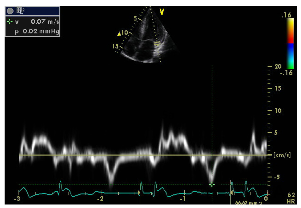
Mitral annular tissue Doppler (e′). Pulsed tissue Doppler at the septal and lateral mitral annulus records the early diastolic annular velocity e′, the primary noninvasive marker of relaxation. The sample volume is placed at or within 1 cm of the insertion of the leaflets, the beam is aligned with annular motion, and the velocity scale is low. Septal e′ is normally lower than lateral e′, so each site has its own threshold. e′ depends on relaxation, restoring forces, and lengthening load; when relaxation is impaired, e′ is delayed until left ventricular pressure falls to left atrial pressure, which makes it relatively independent of preload.
E/e′. Because e′ is less preload dependent than E, the E/e′ ratio corrects mitral inflow for relaxation and rises with left atrial pressure. The average of septal and lateral ratios is preferred; when only one site is technically adequate, the site-specific threshold applies (septal 15 or more, lateral 13 or more, average 14 or more). E/e′ is unreliable in normal hearts with vigorous relaxation, in constrictive pericarditis, with moderate or severe mitral annular calcification, mitral stenosis, a mitral prosthesis or repair, significant mitral regurgitation, regional basal dysfunction, and left bundle branch block or ventricular pacing.
Tricuspid regurgitant velocity and pulmonary artery systolic pressure. In the absence of pulmonary vascular or parenchymal disease, pulmonary artery systolic pressure (PASP) rises passively with left atrial pressure. PASP is 4 × (TR velocity)² plus estimated right atrial pressure. A TR velocity of 2.8 m/s or more, or a PASP of 35 mm Hg or more when right atrial pressure can be estimated, supports elevated left atrial pressure. The TR jet is recorded from several windows, and agitated saline or an ultrasound-enhancing agent completes an incomplete envelope.
Left atrial maximal volume index (LAVi). LAVi, measured by the biplane disk summation or area-length method at end systole and indexed to body surface area, integrates the effect of filling pressure over months to years. A value above 34 mL/m² supports elevated pressure but does not measure pressure at the time of the study. Athletic training, anemia, high-output states, atrial fibrillation or flutter, and mitral valve disease enlarge the atrium without current pressure elevation.
Pulmonary vein flow. Pulsed-wave Doppler 1 to 2 cm into the right upper pulmonary vein records systolic (S), diastolic (D), and atrial reversal (Ar) waves. With elevated left atrial pressure, systolic filling of the atrium is blunted and D dominates; an S/D ratio of 0.67 or less, equivalent to a systolic filling fraction of 40% or less, supports elevated pressure. The ratio is most reliable with reduced LVEF and can be 0.67 or less in healthy young adults. An Ar velocity above 35 cm/s with an Ar duration more than 30 ms longer than mitral A duration indicates elevated left ventricular end-diastolic pressure, even when mean left atrial pressure is normal.
Left atrial reservoir strain (LARS). Speckle-tracking strain of the left atrium during ventricular systole measures atrial lengthening. A LARS of 18% or less supports elevated left atrial pressure with high specificity and lower sensitivity, particularly with a normal LVEF. Values of 19% to 24% lie in the low normal range. LARS is also reduced by atrial myopathy and fibrosis.
Isovolumic relaxation time, L wave, Valsalva, and pulmonary regurgitation. An IVRT of 70 ms or less is consistent with elevated left atrial pressure when relaxation is impaired. A mitral L wave (a mid-diastolic inflow velocity during diastasis) of 50 cm/s or more indicates both impaired relaxation and elevated pressure. A decrease in E/A of 50% or more during an adequate Valsalva strain, with E and A remaining separate, is specific for elevated pressure. A pulmonary regurgitant end-diastolic velocity of 2 m/s or more, or a calculated pulmonary artery diastolic pressure of 16 mm Hg or more, supports elevated left atrial pressure in the absence of pulmonary disease.
| Variable | Abnormal value | Role in the 2025 ASE algorithm |
|---|---|---|
| Septal e′ | ≤6 cm/s | Primary: relaxation |
| Lateral e′ | ≤7 cm/s | Primary: relaxation |
| Average e′ | ≤6.5 cm/s | Primary: relaxation |
| Average / septal / lateral E/e′ | ≥14 / ≥15 / ≥13 | Primary: pressure |
| TR velocity or PASP | ≥2.8 m/s or ≥35 mm Hg | Primary: pressure |
| LA reservoir strain | ≤18% | Supplemental |
| Pulmonary vein S/D | ≤0.67 | Supplemental |
| LAVi | >34 mL/m² | Supplemental |
| IVRT | ≤70 ms | Alternative supplemental |
| PR end-diastolic velocity or PA diastolic pressure | ≥2 m/s or ≥16 mm Hg | Additional |
| Mitral L-wave velocity | ≥50 cm/s | Additional |
| Ar − A duration | >30 ms | Additional |
| Valsalva decrease in E/A | ≥50% | Additional |
| E/A ratio | ≤0.8; <2; ≥2 | Separates grade 1 when only e′ is reduced; grades 2 and 3 once pressure is elevated |
Variables and thresholds of the 2025 ASE general pathway. TR, tricuspid regurgitation; PASP, pulmonary artery systolic pressure; LAVi, left atrial maximal volume index; PR, pulmonary regurgitation; PA, pulmonary artery.
Age and e′. e′ falls and E/e′ rises with age, so the 2025 document provides age-specific limits. The default thresholds (septal ≤6, lateral ≤7, average ≤6.5 cm/s) correspond to the lower limits of normal after 65 years and to prognostically low-risk values in older adults. Age-adjusted limits can replace them in younger patients.
| Annular velocity | 20–39 years | 40–65 years | >65 years |
|---|---|---|---|
| Septal e′ | <7 cm/s | <6 cm/s | <6 cm/s |
| Lateral e′ | <10 cm/s | <8 cm/s | <7 cm/s |
| Average e′ | <9 cm/s | <7 cm/s | <6.5 cm/s |
Age-specific mitral annular e′ values indicating impaired relaxation (2025 ASE recommendations, Table 6).

The 2025 ASE algorithm for left atrial pressure and diastolic grading
The 2025 ASE recommendations supersede the 2016 ASE and European Association of Cardiovascular Imaging (EACVI) document. They separate two questions: whether diastolic dysfunction is present, and whether mean left atrial pressure is elevated. The pressure algorithm applies to adults in sinus rhythm, irrespective of LVEF. It excludes severe primary mitral regurgitation, any degree of mitral stenosis, moderate or severe mitral annular calcification, mitral repair, replacement, or transcatheter edge-to-edge repair, atrial fibrillation, heart transplantation, noncardiac pulmonary hypertension, pericardial constriction, and left ventricular assist devices, which have their own pathways. The recommendations do not apply to children, normal pregnancy, or intraoperative studies.
- Primary variables are assessed together: reduced e′ (septal ≤6, lateral ≤7, or average ≤6.5 cm/s), increased E/e′ (septal ≥15, lateral ≥13, or average ≥14), and increased TR velocity (≥2.8 m/s) or PASP (≥35 mm Hg). PASP is used when right atrial pressure can be estimated; TR velocity is used otherwise.
- When all three primary variables are normal, left atrial pressure is normal and diastolic function is normal.
- When all three are abnormal, left atrial pressure is elevated, and diastolic dysfunction is graded by E/A: grade 2 when E/A is below 2 and grade 3 when E/A is 2 or more.
- When e′ is the only abnormal variable and E/A is 0.8 or less, left atrial pressure is normal and the grade is 1 (impaired relaxation).
- Supplemental variables are assessed when e′ is the only abnormal variable with E/A above 0.8, when only E/e′ or only TR velocity or PASP is abnormal, or when any two of the three are abnormal. They are pulmonary vein S/D 0.67 or less, LA reservoir strain 18% or less, and LAVi above 34 mL/m², with IVRT 70 ms or less as an alternative.
- One or more abnormal supplemental variables indicate elevated left atrial pressure, graded 2 or 3 by E/A. When none is abnormal, left atrial pressure is normal (grade 1 if e′ is reduced).
- When supplemental variables are unavailable or unreliable, additional methods are used: PR end-diastolic velocity, mitral L wave, Ar − A duration, and the Valsalva response. If none can be obtained, left atrial pressure is reported as indeterminate with the limiting variables named.
- Symptomatic patients with normal left atrial pressure at rest, including grade 1 dysfunction, are referred for diastolic exercise echocardiography.
The algorithm does not count abnormal variables. One abnormal pressure marker does not establish elevated pressure, and two normal variables do not make a study normal when the third is abnormal; both situations go to the supplemental variables. E/A grades a pressure state that has already been established. An E/A of 2 or more with normal primary variables describes the vigorous filling of a young heart and is not grade 3.
Diagnosis of diastolic dysfunction. Separately from pressure estimation, diastolic dysfunction is present when e′ is reduced and at least one marker of remodeling or elevated pressure is present (average E/e′ above 14, LA reservoir strain 18% or less, E/A 0.8 or less or 2 or more, LAVi above 34 mL/m², or left ventricular mass index above 95 g/m² in women or 115 g/m² in men). Diastolic dysfunction is also present when e′ is preserved and two or more of these markers are present. Left atrial enlargement from athletic training, anemia, atrial arrhythmias, or mitral valve disease is excluded before it is counted.
| Feature | 2016 ASE/EACVI | 2025 ASE |
|---|---|---|
| Initial variables | Four equivalent variables: e′, average E/e′ >14, LAVi >34 mL/m², TR velocity >2.8 m/s | Three primary variables: e′, E/e′ (site-specific), TR velocity ≥2.8 m/s or PASP ≥35 mm Hg |
| Decision rule | Majority of abnormal variables; half abnormal was indeterminate | All normal, all abnormal, or e′ alone with E/A ≤0.8 decides; mixed findings go to supplemental variables |
| Left atrial volume | Primary variable | Supplemental variable |
| Left atrial strain | Not in the algorithm | Supplemental variable (≤18%) |
| Age | Fixed e′ thresholds | Default thresholds plus age-specific limits |
| Indeterminate result | Common | Reduced by supplemental and additional variables |
| HFpEF diagnosis | Not addressed | Dedicated diagnostic algorithm including exercise testing and catheterization |
| Atrial fibrillation | List of individual variables | Multiparametric algorithm with supplemental variables |
Changes from the 2016 ASE/EACVI recommendations to the 2025 ASE update.
Performance of the 2025 algorithm. In 756 ambulatory patients with HFpEF confirmed by invasive exercise hemodynamics, about two-thirds (68%) were classified as having normal diastolic function or grade 1 dysfunction at rest by the 2025 criteria. The finding reflects the definition of HFpEF itself, in which filling pressure is often normal at rest, and it is the reason the algorithm routes symptomatic patients with normal resting pressure to exercise testing. A resting grade of normal or grade 1 does not exclude HFpEF.
Diastolic function and left atrial pressure calculator
The calculator applies the 2025 ASE general pathway for adults in sinus rhythm. It is not used in atrial fibrillation, mitral stenosis, moderate or severe mitral annular calcification, severe primary mitral regurgitation, after mitral valve intervention, in heart transplant recipients, with a left ventricular assist device, in noncardiac pulmonary hypertension, or in constriction. Fields left blank are treated as unavailable.
Left atrial pressure and diastolic grade (sinus rhythm)
2025 ASE general pathwayE/e′ is the average of septal and lateral ratios when both e′ values are entered, and the site-specific ratio otherwise. PASP = 4 × (TR velocity)² + right atrial (RA) pressure; when RA pressure is left blank, TR velocity ≥2.8 m/s is the criterion. The example values (septal e′ 5, lateral e′ 7, E 90, A 80, TR 2.5 m/s, LA reservoir strain 16%) give reduced e′ and an average E/e′ of 15 with a normal TR velocity. The reduced LA strain resolves the mixed findings as elevated pressure, and E/A of 1.1 gives grade 2.
Grades of diastolic dysfunction and the diastolic function report
| Grade | Left atrial pressure | Typical findings | Clinical meaning |
|---|---|---|---|
| Normal | Normal | Normal e′, E/e′, and TR velocity | No diastolic dysfunction |
| Grade 1 (impaired relaxation) | Normal at rest | Reduced e′; E/A ≤0.8; long DT and IVRT; normal E/e′ and TR velocity | Common with age, hypertension, and hypertrophy; exercise testing when symptomatic |
| Grade 2 | Mild to moderately elevated | Reduced e′; E/A 0.8 to <2 (pseudonormal); increased E/e′, TR velocity, or LA markers | Elevated filling pressure; supports HFpEF in the clinical setting |
| Grade 3 | Markedly elevated | E/A ≥2; short DT and IVRT; increased E/e′ and TR velocity | Restrictive filling; high risk of death and hospitalization |
Grades of diastolic dysfunction in the 2025 ASE recommendations. DT, deceleration time.
Every echocardiographic report states whether diastolic function is normal or abnormal and, when abnormal, the grade: grade 1, grade 2 (mild to moderate increase in left atrial pressure), or grade 3 (marked increase). When a grade cannot be assigned, the report states whether left atrial pressure is normal, elevated, or indeterminate. An isolated increase in left ventricular end-diastolic pressure (identified by Ar − A duration) is reported, because it predisposes to elevated mean left atrial pressure with exercise, tachycardia, or increased afterload. Reports include mitral E and A, e′, peak TR velocity, E/A, and average E/e′, and they include LA strain, pulmonary vein flow, IVRT, and A and Ar durations when those variables were used to reach the conclusion. A change in grade from the previous study is stated.
The older label grade 3 "reversible" or "irreversible" restrictive filling, based on the response to preload reduction, is not part of the 2025 classification. Persistent restrictive filling despite decongestion, however, carries a poor prognosis in both HFpEF and HFrEF.
Diastolic assessment in atrial fibrillation
Atrial fibrillation removes the A wave and atrial contraction, varies cycle length, and enlarges the left atrium. E/A, Ar − A, the Valsalva response, and grading by E/A are therefore unavailable, and LAVi loses specificity. Values are averaged from several cycles whose length reflects the average heart rate, markedly dissimilar cycles are avoided, and E and e′ are ideally recorded in the same beat (a dual-Doppler probe improves accuracy). Less beat-to-beat variability of mitral inflow with varying cycle length indicates elevated filling pressure. No single variable is strong enough to stand alone; the 2025 ASE document uses a multiparametric algorithm with moderate accuracy.
| Step | Variables | Conclusion |
|---|---|---|
| 1. Primary variables | Mitral E ≥100 cm/s; septal E/e′ >11; TR velocity >2.8 m/s or PASP >35 mm Hg; DT ≤160 ms | 3 or more abnormal: elevated left atrial pressure. None or 1 abnormal: normal left atrial pressure. 2 abnormal: step 2 |
| 2. Supplemental variables | LA reservoir strain <18%; pulmonary vein S/D <1; body mass index >30 kg/m² | 2 or 3 present: elevated. None present: normal. Only 1, or unavailable or unreliable: indeterminate |
Estimation of mean left atrial pressure in atrial fibrillation (2025 ASE recommendations, Figure 8). No diastolic grade is assigned in atrial fibrillation. Other indicators listed by the document are peak acceleration rate of mitral E ≥1,900 cm/s², IVRT ≤65 ms, pulmonary venous D-wave deceleration time ≤220 ms, and E/Vp (ratio of mitral E velocity to color M-mode flow propagation velocity) ≥1.4.
Sinus tachycardia and first-degree atrioventricular block fuse E and A. A fused pattern is not labeled as any grade. In tachycardia, an IVRT of 70 ms or less and a pulmonary vein systolic filling fraction of 40% or less are specific for elevated pressure, and average E/e′ above 14 is specific but insensitive. The compensatory pause after a premature beat often separates E and A for measurement.
Diastolic assessment in mitral disease, cardiomyopathy, pulmonary hypertension, transplantation, and devices
Conditions that alter the annulus, the mitral valve, the pulmonary circulation, or ventricular activation change the relationship between Doppler variables and filling pressure. The 2025 ASE document provides specific indicators for each.
| Condition | Indicators of elevated filling pressure | Variables not used |
|---|---|---|
| Moderate or severe mitral annular calcification | E/A <0.8 normal; E/A >1.8 elevated; for E/A 0.8–1.8, IVRT ≥80 ms normal and <80 ms elevated | Annular e′ and E/e′ |
| Mitral stenosis | IVRT <60 ms; mitral A >1.5 m/s; IVRT/TE−e′ <4.2 | E/e′ |
| Significant mitral regurgitation | IVRT <60 ms; Ar − A ≥30 ms; IVRT/TE−e′ <5.6; average E/e′ >14 only with depressed EF | High E and E/A as evidence of grade 3; pulmonary vein S/D |
| Hypertrophic cardiomyopathy | Average E/e′ >14; Ar − A ≥30 ms; TR velocity >2.8 m/s; LAVi >34 mL/m² | E/A alone |
| Restrictive cardiomyopathy | Average E/e′ >14; DT <140 ms; E/A >2.5; IVRT <50 ms (specific, insensitive) | Inflow alone for diagnosis |
| Pulmonary hypertension | E/A ≥2 favors postcapillary and ≤0.8 precapillary; for E/A between, lateral E/e′ >13, LAVi >34 mL/m², and LA strain <18% favor postcapillary | TR velocity as a marker of left atrial pressure |
| Heart transplant | Average E/e′ <7 normal and >14 elevated; between, E/SRIVR >200 cm elevated, or TR velocity >2.8 m/s elevated | General pathway |
| Left ventricular assist device | E/A >2; RA pressure >10 mm Hg; PASP >40 mm Hg; average E/e′ >14 or septal ≥15; LAVi >33 mL/m²; interatrial septal position | General pathway without device context |
Indicators of elevated left ventricular filling pressure in special populations (2025 ASE recommendations, Table 7). TE−e′, interval between the onsets of mitral E and annular e′; SRIVR, global strain rate during isovolumic relaxation.
In mitral stenosis and other inflow obstruction (left atrial myxoma, cor triatriatum), left ventricular diastolic pressure is usually normal or low, and timing variables estimate atrial pressure. In primary mitral regurgitation, a compliant enlarged atrium attenuates the pressure rise until late; high E and E/A reflect regurgitant flow. With a left ventricular assist device (LVAD), a neutral interatrial septum indicates similar right and left atrial pressures, a septum bowing to the right indicates left atrial pressure about 5 mm Hg higher than right atrial pressure, and a septum bowing to the left indicates the reverse.
Constriction and restriction. Both show restrictive inflow. In constrictive pericarditis the myocardium relaxes normally, septal e′ is preserved or increased and often exceeds lateral e′ (annulus reversus), mitral E varies markedly with respiration, and hepatic vein diastolic flow reverses in expiration. In restrictive cardiomyopathy, septal e′ is reduced. E/e′ does not track filling pressure in constriction (annulus paradoxus). The general pressure algorithm is not applied in constriction.
Left bundle branch block, ventricular pacing, and cardiac resynchronization. Abnormal activation distorts septal motion and IVRT and lowers septal e′. Lateral e′ and lateral E/e′, TR velocity, pulmonary vein flow, and LA strain carry more weight, and the conclusion is stated with lower confidence.
Left atrial strain, global longitudinal strain, and other supplementary measures
Left atrial strain. Speckle tracking of the left atrium yields reservoir, conduit, and pump (contractile) strain. Reservoir strain is the most robust and the one used in the algorithm. The 2025 ASE document gives age-related lower limits of normal for reservoir strain of about 30% at 20 to 39 years, 27% at 40 to 60 years, and 24% at 60 to 80 years (fifth percentiles), with vendor differences. A value of 18% or less indicates elevated left atrial pressure, and reduced LARS identifies patients more likely to develop elevated filling pressure with exercise. Atrial strain is measured from nonforeshortened apical four- and two-chamber views focused on the atrium, with the zero reference at ventricular end diastole (QRS onset). The left atrial stiffness index (E/e′ divided by LARS) carries prognostic information in HFpEF but has no validated diagnostic threshold.
Global longitudinal strain. Left ventricular global longitudinal strain (GLS) is reduced in many patients with HFpEF despite a normal LVEF, reflecting subendocardial longitudinal fiber dysfunction. A GLS below 16% in absolute value is a minor functional criterion in the HFA-PEFF score. A regional pattern with relative apical sparing suggests cardiac amyloidosis; a reduced septal or basal inferolateral strain suggests hypertrophic cardiomyopathy, Fabry disease, or sarcoidosis. GLS values differ between vendors, so serial comparisons use the same platform.
Ventricular mass and geometry. Left ventricular mass index above 95 g/m² in women or 115 g/m² in men, relative wall thickness above 0.42, and end-diastolic wall thickness of 12 mm or more support structural heart disease. Concentric remodeling (normal mass with increased relative wall thickness) and concentric hypertrophy are the typical geometries of HFpEF.
Right heart. Right ventricular size and function (TAPSE, fractional area change, free-wall strain), the TAPSE/PASP ratio as an index of right ventricular–pulmonary arterial coupling, inferior vena cava size and collapse, and hepatic vein flow complete the evaluation and carry prognostic weight.
Magnetic resonance, CT, and artificial intelligence. Cardiac magnetic resonance and cardiac CT measure left ventricular filling rates and left atrial volumes and function. Artificial-intelligence models that grade diastolic function from echocardiograms or from the 12-lead electrocardiogram are under development; they were trained against the 2016 algorithm and require validation against invasive hemodynamics and outcomes.
Diastolic exercise echocardiography
Up to half of patients with HFpEF have normal left ventricular filling pressure at rest that rises rapidly with exercise. Diastolic exercise echocardiography is indicated in patients with exertional dyspnea and grade 1 diastolic dysfunction at rest and in symptomatic patients with indeterminate filling pressure at rest. Patients with entirely normal diastolic function and normal e′ at rest are unlikely to develop elevated pressure with exercise, and patients with elevated pressure at rest already have a cardiac explanation for dyspnea; neither group is referred for diastolic stress testing.
The test is performed with supine bicycle or treadmill exercise; dobutamine stress is strongly discouraged for diastolic assessment. Mitral E, septal and lateral e′, and peak TR velocity are recorded at rest, during each stage of supine bicycle exercise, and in early recovery once E and A separate. Elevated filling pressure persists for several minutes after exercise. With treadmill testing, wall motion images are acquired first, then TR velocity, then annular and inflow velocities once E and A separate.
| Result | Criteria during or immediately after exercise |
|---|---|
| Definitely abnormal (elevated filling pressure) | Average E/e′ ≥14 or septal E/e′ ≥15, and peak TR velocity >3.2 m/s |
| HFpEF likely | E/e′ >14 with peak TR velocity >2.8 and <3.2 m/s |
| Does not support HFpEF | E/e′ <14, even with TR velocity >3.2 m/s |
| Normal | Average (or septal) E/e′ <10 and peak TR velocity <2.8 m/s |
Interpretation of diastolic exercise echocardiography (2025 ASE recommendations). The HFA-PEFF algorithm (step 3) uses a different cutoff: average E/e′ of 15 or more at peak stress scores 2 points, and a peak TR velocity above 3.4 m/s adds a third point. The superseded 2016 ASE/EACVI test was positive with average E/e′ above 14 or septal E/e′ above 15, peak TR velocity above 2.8 m/s, and septal e′ below 7 cm/s.
An isolated rise in TR velocity is not diagnostic, because pulmonary blood flow increases with exercise in normal subjects. In a series of more than 14,000 exercise echocardiograms, 17% of patients developed elevated filling pressure with exercise and 28% had ischemia, and patients with elevated filling pressure without ischemia had a worse prognosis than those with isolated ischemia. A PASP above 50 mm Hg or a TR velocity above 3.2 m/s during exercise is associated with adverse outcome. Exercise echocardiography also identifies ischemia, dynamic mitral regurgitation, and chronotropic incompetence in the same study. When the exercise study is negative or inconclusive and clinical suspicion persists, invasive exercise hemodynamics is the next test.
H2FPEF, HFA-PEFF, and HFpEF-ABA scores
Diagnostic scores estimate the probability of HFpEF in a patient with unexplained dyspnea and a preserved ejection fraction. They do not replace confirmation of elevated filling pressure: a high score supports the diagnosis when the clinical picture is concordant, a low score redirects the evaluation, and an intermediate score is resolved by exercise testing. The 2026 ACC expert consensus decision pathway lists three scores: H2FPEF, HFA-PEFF, and HFpEF-ABA.
H2FPEF. The H2FPEF score was derived in 414 patients with unexplained dyspnea who underwent invasive exercise hemodynamic testing, with HFpEF defined by elevated filling pressure at rest or with exercise, and validated in a separate cohort. It uses six variables that are routinely available.
| Letter | Variable | Criterion | Points |
|---|---|---|---|
| H | Heavy | Body mass index >30 kg/m² | 2 |
| H | Hypertensive | Two or more antihypertensive medications | 1 |
| F | Atrial fibrillation | Paroxysmal or persistent | 3 |
| P | Pulmonary hypertension | Doppler-estimated PASP >35 mm Hg | 1 |
| E | Elder | Age >60 years | 1 |
| F | Filling pressure | E/e′ >9 | 1 |
H2FPEF score (range 0 to 9). Scores of 0 to 1 indicate low probability, 2 to 5 intermediate probability, and 6 to 9 high probability of HFpEF. The 2025 ASE document specifies septal E/e′ for the filling-pressure criterion.
A continuous version of the model uses age, body mass index, E/e′, PASP, and atrial fibrillation as continuous or binary inputs and returns a probability. It is more precise than the integer score near the category boundaries.
HFA-PEFF. The Heart Failure Association of the ESC algorithm has four steps: pretest assessment (P), echocardiographic and natriuretic peptide score (E), functional testing in cases of uncertainty (F1), and final etiology (F2). The score in step E assigns up to 2 points in each of three domains: 2 points when any major criterion is met in that domain and 1 point when only minor criteria are met. Points are not added within a domain.
| Domain | Major criteria (2 points) | Minor criteria (1 point) |
|---|---|---|
| Functional | Septal e′ <7 cm/s or lateral e′ <10 cm/s (age ≥75: septal <5 or lateral <7 cm/s); average E/e′ ≥15; TR velocity >2.8 m/s; PASP >35 mm Hg | Average E/e′ 9–14; GLS <16% |
| Morphological | LAVi >34 mL/m² (sinus rhythm) or >40 mL/m² (atrial fibrillation); LV mass index ≥149 g/m² (men) or ≥122 g/m² (women) with relative wall thickness >0.42 | LAVi 29–34 mL/m² (sinus) or 34–40 mL/m² (AF); LV mass index ≥115 g/m² (men) or ≥95 g/m² (women); relative wall thickness >0.42; LV wall thickness ≥12 mm |
| Biomarker, sinus rhythm | NT-proBNP >220 pg/mL or BNP >80 pg/mL | NT-proBNP 125–220 pg/mL or BNP 35–80 pg/mL |
| Biomarker, atrial fibrillation | NT-proBNP >660 pg/mL or BNP >240 pg/mL | NT-proBNP 365–660 pg/mL or BNP 105–240 pg/mL |
HFA-PEFF step E scoring. A total of 5 or 6 points is diagnostic of HFpEF, 2 to 4 points is intermediate and leads to a diastolic stress test or invasive hemodynamics, and 0 or 1 point makes HFpEF unlikely.
HFpEF-ABA. The HFpEF-ABA score, derived from the same invasive cohort and validated in an international multicenter cohort of 736 patients, uses only age, body mass index, and a history of atrial fibrillation. It was designed as a screening tool for undiagnosed HFpEF in patients with dyspnea when echocardiographic data are not yet available, and it showed good discrimination (area under the curve 0.81 in validation).
The scores overlap but are not interchangeable. H2FPEF weighs atrial fibrillation and obesity heavily and needs only basic echocardiographic data. HFA-PEFF depends on natriuretic peptides and detailed structural measurements and classifies more patients as intermediate. Both perform less well in populations unlike their derivation cohorts, and both are applied only after valve disease, infiltrative and hypertrophic cardiomyopathy, constriction, and pulmonary arterial hypertension have been considered.
H2FPEF score
Points score and continuous probabilityPoints: body mass index >30 kg/m², 2; two or more antihypertensive drugs, 1; atrial fibrillation, 3; PASP >35 mm Hg, 1; age >60 years, 1; E/e′ >9, 1. Continuous model: y = −9.1917 + 0.0451 × age + 0.1307 × BMI + 0.0859 × E/e′ + 0.0520 × PASP + 1.6997 × AF (1 if present); probability = ey ÷ (1 + ey). The example (72 years, BMI 36, two drugs, no AF, PASP 40 mm Hg, E/e′ 13) scores 6 points, high probability.
HFA-PEFF score (step E)
Functional, morphological, and biomarker domainsEach domain scores 2 points for any major criterion or 1 point for minor criteria only. The e′ criteria use the thresholds for age 75 years and older when the patient is 75 or older. The example (septal e′ 6 cm/s, average E/e′ 13, LAVi 36 mL/m², relative wall thickness 0.44, NT-proBNP 180 pg/mL in sinus rhythm) scores 2 + 2 + 1 = 5, diagnostic of HFpEF.
Invasive and exercise hemodynamics in suspected HFpEF
Right heart catheterization is the reference standard for the diagnosis of HFpEF. It is indicated when noninvasive testing is inconclusive in a patient whose symptoms suggest HFpEF, when pulmonary hypertension needs classification, and when the hemodynamic profile will change treatment. Pressures are measured at end expiration with the transducer zeroed at the phlebostatic level. In obesity and lung disease, large respiratory swings in intrathoracic pressure make the end-expiratory value higher than the value averaged over the respiratory cycle, and both are recorded.
| Measurement | Value consistent with HFpEF or diastolic dysfunction |
|---|---|
| Time constant of LV relaxation (tau) | >48 ms |
| LV chamber stiffness constant | >0.015 |
| Mean PCWP at rest | >15 mm Hg (≥15 mm Hg in HFA-PEFF) |
| LV end-diastolic pressure at rest | >16 mm Hg (≥16 mm Hg in HFA-PEFF) |
| Mean PCWP during supine exercise | ≥25 mm Hg |
| LV end-diastolic pressure during exercise | ≥23 mm Hg |
| PCWP/cardiac output slope during supine exercise | >2 mm Hg/L/min |
Invasive values that diagnose left ventricular diastolic dysfunction and HFpEF (2025 ASE recommendations, Table 1). PCWP, pulmonary capillary wedge pressure. A mean pulmonary artery pressure/cardiac output slope above 3 mm Hg/L/min defines an abnormal pulmonary pressure response to exercise.
Exercise right heart catheterization. Supine cycle ergometry with simultaneous measurement of right atrial, pulmonary artery, and wedge pressures, cardiac output (direct Fick or thermodilution), and arterial and mixed venous oxygen saturation unmasks HFpEF in patients with normal resting hemodynamics. In a referral series of patients with exertional dyspnea, normal natriuretic peptides, and normal resting pressures, an exercise wedge pressure of 25 mm Hg or more identified HFpEF in more than half. Those patients had blunted increases in heart rate, systemic vasodilation, and cardiac output, and 88% of them had exercise-induced pulmonary hypertension. Flow-normalized indices (PCWP/cardiac output slope above 2 mm Hg/L/min) separate an abnormal rise in filling pressure from the physiological rise at high workloads. Exercise catheterization also measures peak exercise pulmonary vascular resistance, which identifies latent pulmonary vascular disease.
Saline loading. When exercise is not feasible, rapid infusion of saline raises filling pressure more in HFpEF than in control subjects. It is less sensitive than exercise.
Interpretation in treated patients. A normal resting wedge pressure does not exclude HFpEF in a patient receiving diuretics, because decongestion is the intended effect of treatment. A resting study performed after diuresis for decompensation likewise does not exclude the diagnosis. Large v waves raise mean wedge pressure; the wedge is read at mid-a wave or as the mean, and a large v wave is reported with its cause (a stiff atrium, mitral regurgitation, or both). Left ventricular end-diastolic pressure measured directly confirms the left-sided origin when wedge tracings are damped or uncertain. Coronary angiography or CT angiography is added when ischemia is suspected.
Pulmonary hypertension in HFpEF. In the 2022 ESC/ERS pulmonary hypertension guideline, pulmonary hypertension is defined by a mean pulmonary artery pressure above 20 mm Hg. It is postcapillary when wedge pressure exceeds 15 mm Hg, and combined pre- and postcapillary when pulmonary vascular resistance also exceeds 2 Wood units. Combined disease carries a worse prognosis. Pulmonary arterial hypertension–specific vasodilators are not used for group 2 pulmonary hypertension.
Cardiac magnetic resonance, CT, and nuclear imaging in HFpEF
Cardiac magnetic resonance (CMR) is used when echocardiographic images are inadequate or when an infiltrative or inflammatory cardiomyopathy is suspected. Cine imaging measures volumes, mass, and wall thickness with high reproducibility. Late gadolinium enhancement patterns separate causes. Amyloidosis shows diffuse subendocardial or transmural enhancement with abnormal myocardial and blood-pool nulling. Hypertrophic cardiomyopathy shows patchy midwall enhancement at the right ventricular insertion points or in the regions of greatest hypertrophy. Fabry disease shows basal inferolateral midwall enhancement, sarcoidosis patchy midwall or epicardial enhancement, and infarction subendocardial enhancement in a coronary territory. Native T1 and extracellular volume fraction (ECV) quantify diffuse interstitial expansion. ECV is markedly increased in amyloidosis, native T1 is low in Fabry disease, and T2* is low in iron overload. Diffuse fibrosis burden adds prognostic information to the echocardiographic assessment of diastolic function. CMR phase-contrast imaging and feature tracking measure filling rates and left atrial strain.
Bone scintigraphy with technetium-99m pyrophosphate, 3,3-diphosphono-1,2-propanodicarboxylic acid (DPD), or hydroxymethylene diphosphonate confirms transthyretin cardiac amyloidosis noninvasively when grade 2 or 3 myocardial uptake is confirmed by single-photon emission CT (SPECT). A monoclonal protein must first be excluded by serum free light chains and serum and urine immunofixation. Myocardial perfusion imaging with positron emission tomography (PET) measures myocardial blood flow and coronary flow reserve, which is reduced in coronary microvascular dysfunction. Coronary CT angiography excludes obstructive coronary disease in patients with intermediate risk, and CT measures pericardial thickness and calcification when constriction is suspected.
Diseases that present as HFpEF and how they are identified
The diagnosis of HFpEF requires exclusion of conditions that present identically but are treated differently. The 2026 ACC pathway added high-output heart failure, frailty, deconditioning, and obesity to the noncardiac mimics and ischemic heart disease to the cardiac mimics.
| Condition | Clues | Confirmatory test |
|---|---|---|
| Transthyretin cardiac amyloidosis | Older man; carpal tunnel syndrome, lumbar stenosis, or biceps tendon rupture; low voltage for wall thickness; intolerance of vasodilators; wall thickness ≥12 mm; apical-sparing strain; atrioventricular block | Free light chains and immunofixation; bone scintigraphy with SPECT; CMR; TTR genotyping |
| Light-chain amyloidosis | Nephrotic proteinuria; neuropathy; macroglossia; periorbital purpura; rapidly progressive symptoms | Free light chains and immunofixation; tissue biopsy |
| Hypertrophic cardiomyopathy | Wall thickness ≥15 mm (≥13 mm with family history); systolic anterior motion; outflow gradient; family history | Echocardiography with provocation; CMR; genetic testing |
| Constrictive pericarditis | Prior cardiac surgery, radiation, or tuberculosis; Kussmaul sign; pericardial knock; septal bounce; preserved septal e′; respiratory variation | Echocardiography; CMR or CT; simultaneous left and right heart catheterization |
| Valve disease | More than mild stenosis or at least moderate regurgitation; mitral annular calcification with stenosis | Comprehensive echocardiography; transesophageal or stress echocardiography |
| Pulmonary arterial hypertension and lung disease | Normal LA size; E/A ≤0.8; high TR velocity; low DLCO | Right heart catheterization (PCWP ≤15 mm Hg); pulmonary function tests; CT |
| High-output heart failure | Anemia, arteriovenous fistula, cirrhosis, thyrotoxicosis, Paget disease, thiamine deficiency, obesity | Cardiac output and systemic vascular resistance; laboratory tests |
| Coronary artery disease | Angina; regional wall motion abnormality; ischemic electrocardiogram | Stress imaging or coronary CT angiography; coronary flow reserve |
| Fabry disease | Young adult; renal disease, neuropathic pain, angiokeratoma; short PR interval; basal inferolateral fibrosis | Alpha-galactosidase A activity (men); GLA genotyping; low native T1 on CMR |
| Cardiac sarcoidosis | Atrioventricular block; ventricular arrhythmia; basal septal thinning; extracardiac sarcoidosis | CMR; fluorodeoxyglucose PET |
| Hemochromatosis and iron overload | Transfusion dependence; high ferritin and transferrin saturation; diabetes; liver disease | CMR T2* (<20 ms at 1.5 T indicates myocardial iron) |
| Radiation-associated heart disease | Prior mediastinal radiation; valve, coronary, pericardial, and conduction disease | Multimodality imaging |
Differential diagnosis of HFpEF and the tests that establish each alternative. DLCO, diffusing capacity for carbon monoxide.
Transthyretin cardiac amyloidosis is the most important specific cause to identify, because tafamidis and other transthyretin-directed therapies change its course. In a prospective study of patients aged 60 years or older admitted with HFpEF and a wall thickness of 12 mm or more, 13.3% had moderate-to-severe myocardial uptake on DPD scintigraphy, all wild-type. Estimates of its prevalence in HFpEF range from about 5% to 13%, and in a prospective endomyocardial biopsy series 14% of patients with HFpEF had cardiac amyloidosis. The 2022 AHA/ACC/HFSA guideline recommends screening for monoclonal light chains with serum and urine immunofixation and serum free light chains when amyloidosis is suspected, followed by bone scintigraphy when no monoclonal protein is found (Class 1).
Diagnostic pathway for suspected HFpEF
The 2025 ASE recommendations and the 2026 ACC pathway describe a stepwise approach that combines clinical assessment, natriuretic peptides, comprehensive echocardiography, probability scores, and stress or invasive testing.
- Clinical assessment: symptoms and signs of heart failure, comorbid risk factors, electrocardiogram, chest radiograph, natriuretic peptide, hemoglobin, renal and thyroid function, and evaluation for noncardiac causes of dyspnea (lung disease, anemia, obesity and deconditioning, kidney and liver disease, venous insufficiency).
- Comprehensive echocardiography: LVEF of 50% or more (41% to 49% for HFmrEF), wall thickness and mass, left atrial volume and strain, left ventricular GLS, right ventricular function, and exclusion of significant valve disease, hypertrophic or infiltrative cardiomyopathy, constriction, and noncardiac pulmonary hypertension. Multimodality imaging and catheterization are used as needed to exclude alternatives.
- Left atrial pressure estimation by the 2025 algorithm. Elevated left atrial pressure at rest in a symptomatic patient establishes the diagnosis of HFpEF.
- When left atrial pressure is normal at rest in a symptomatic patient, diastolic exercise echocardiography is performed. A positive test establishes HFpEF; a negative test points to a noncardiac cause of dyspnea.
- When the exercise echocardiogram is inconclusive or clinical suspicion persists, right heart catheterization with exercise establishes or excludes the diagnosis.
- Once HFpEF is established, the phenotype and comorbid drivers are defined, because they determine therapy.
US and European definitions differ in emphasis. The 2022 AHA/ACC/HFSA guideline requires an LVEF of 50% or more with symptoms or signs of heart failure and evidence of diastolic dysfunction or elevated filling pressure. The 2021 ESC guideline requires structural or functional abnormalities consistent with diastolic dysfunction or raised filling pressure, including raised natriuretic peptides. It lists LAVi above 34 mL/m², LV mass index of 115 g/m² or more in men or 95 g/m² or more in women, E/e′ of 13 or more at rest, and mean e′ below 9 cm/s as key echocardiographic abnormalities.
Decongestion and diuretic therapy in HFpEF
Loop diuretics relieve congestion and improve symptoms, and the 2022 AHA/ACC/HFSA guideline recommends diuretics in patients with heart failure and fluid retention (Class 1). They have no proven effect on mortality. The dose is the lowest that maintains euvolemia, and it is adjusted for weight, symptoms, and blood pressure. Because the stiff ventricle operates on a steep pressure-volume curve, overdiuresis produces hypotension, prerenal azotemia, and reduced stroke volume, while small volume gains produce congestion. In TRANSFORM-HF, torsemide and furosemide after discharge produced similar all-cause mortality (26.1% versus 26.2%; hazard ratio 1.02), with consistent results across ejection fraction categories. Thiazide-type diuretics added to a loop diuretic (sequential nephron blockade) treat diuretic resistance, with close monitoring of potassium, sodium, and renal function. Hemodynamic monitoring with an implanted pulmonary artery sensor allows diuretic adjustment before symptoms appear in selected patients.
SGLT2 inhibitors in HFpEF and HFmrEF
Two large trials established SGLT2 inhibitors as the first disease-modifying therapy across the preserved and mildly reduced ejection fraction range.
EMPEROR-Preserved. In 5,988 patients with class II to IV heart failure and LVEF above 40%, empagliflozin 10 mg daily reduced the composite of cardiovascular death or hospitalization for heart failure over a median of 26.2 months (13.8% versus 17.1%; hazard ratio 0.79; 95% CI 0.69 to 0.90). The effect was driven by fewer heart failure hospitalizations (total hospitalizations, hazard ratio 0.73), and results were consistent in patients with and without diabetes.
DELIVER. The trial included 6,263 patients with LVEF above 40%, among them patients whose ejection fraction had improved from 40% or less and patients enrolled during or soon after hospitalization. Dapagliflozin 10 mg daily reduced cardiovascular death or worsening heart failure over a median of 2.3 years (16.4% versus 19.5%; hazard ratio 0.82; 95% CI 0.73 to 0.92). Worsening heart failure was reduced (hazard ratio 0.79), cardiovascular death was not significantly reduced (hazard ratio 0.88; 95% CI 0.74 to 1.05), and results were similar above and below an LVEF of 60%.
Pooled analysis. In 12,251 patients from both trials, SGLT2 inhibitors reduced cardiovascular death or first heart failure hospitalization (hazard ratio 0.80; 95% CI 0.73 to 0.87), with reductions in both cardiovascular death (0.88; 0.77 to 1.00) and first hospitalization (0.74; 0.67 to 0.83), consistent across ejection fraction.
The 2022 AHA/ACC/HFSA guideline rates SGLT2 inhibitors as reasonable in HFmrEF and HFpEF to decrease heart failure hospitalization and cardiovascular mortality (Class 2a, B-R). The 2023 ESC focused update recommends empagliflozin or dapagliflozin in HFmrEF and HFpEF (Class I, A), and the 2026 ACC pathway recommends an SGLT2 inhibitor in all patients with HFpEF unless contraindicated. Both drugs are given at 10 mg once daily without titration. An initial small fall in estimated glomerular filtration rate (eGFR) is expected and does not require discontinuation. Genital mycotic infection, volume depletion when combined with diuretics, and euglycemic ketoacidosis (in patients with diabetes, particularly during acute illness, surgery, or fasting) are the principal adverse effects, and the drugs are held before major surgery.
Mineralocorticoid receptor antagonists: spironolactone and finerenone
TOPCAT. In 3,445 patients with LVEF of 45% or more, spironolactone did not significantly reduce the composite of cardiovascular death, aborted cardiac arrest, or heart failure hospitalization over a mean of 3.3 years (18.6% versus 20.4%; hazard ratio 0.89; 95% CI 0.77 to 1.04). Heart failure hospitalization was reduced (12.0% versus 14.2%; hazard ratio 0.83; 95% CI 0.69 to 0.99). Hyperkalemia doubled (18.7% versus 9.1%), and serum creatinine rose. A post hoc analysis found an approximately fourfold difference in event rates between patients enrolled in Russia and Georgia and those enrolled in the Americas. Russia and Georgia had lower event rates and evidence of lower drug exposure; in the Americas, spironolactone was associated with benefit. The 2022 AHA/ACC/HFSA guideline rates mineralocorticoid receptor antagonists as possibly useful in selected patients with HFpEF, particularly at the lower end of the ejection fraction spectrum (Class 2b, B-R).
FINEARTS-HF. In 6,001 patients with LVEF of 40% or more, the nonsteroidal mineralocorticoid receptor antagonist finerenone reduced the composite of total worsening heart failure events and cardiovascular death over a median of 32 months (rate ratio 0.84; 95% CI 0.74 to 0.95). Total worsening heart failure events were reduced (rate ratio 0.82; 95% CI 0.71 to 0.94), while cardiovascular death was not significantly different (8.1% versus 8.7%; hazard ratio 0.93; 95% CI 0.78 to 1.11). Finerenone increased hyperkalemia and reduced hypokalemia. The US Food and Drug Administration (FDA) approved finerenone in 2025 to reduce the risk of cardiovascular death, heart failure hospitalization, and urgent heart failure visits in adults with heart failure and LVEF of 40% or more. The labeled target dose is 40 mg once daily when eGFR at initiation is 60 mL/min/1.73 m² or more and 20 mg once daily when eGFR is 25 to less than 60. Treatment is not started when serum potassium exceeds 5.0 mEq/L, and potassium and eGFR are measured 4 weeks after initiation and after dose changes. In FINEARTS-HF, hyperkalemia (9.7% versus 4.2%) and investigator-reported hypotension (7.6% versus 4.7%) were more frequent than with placebo, and the prescribing information reports adverse reactions related to worsening renal function in 18% versus 12%.
The 2026 ACC pathway recommends a mineralocorticoid receptor antagonist in addition to an SGLT2 inhibitor unless contraindicated, and favors finerenone over spironolactone because finerenone has direct outcome evidence in this population. Spironolactone remains an alternative when finerenone is unavailable. The two drugs are not combined. Spironolactone is started at 12.5 to 25 mg daily with the same potassium and renal precautions, and gynecomastia is a recognized adverse effect.
Angiotensin receptor–neprilysin inhibitors, angiotensin receptor blockers, and ACE inhibitors
PARAGON-HF. In 4,822 patients with LVEF of 45% or more, sacubitril/valsartan did not significantly reduce total heart failure hospitalizations and cardiovascular death compared with valsartan (rate ratio 0.87; 95% CI 0.75 to 1.01; P = 0.06). Total heart failure hospitalizations had a rate ratio of 0.85 (95% CI 0.72 to 1.00), and cardiovascular death did not differ (8.5% versus 8.9%). Among 12 prespecified subgroups, the possible benefit was greater in patients with lower ejection fraction and in women. Sacubitril/valsartan caused more hypotension and angioedema, less hyperkalemia, and less worsening of renal function. The FDA label for sacubitril/valsartan indicates that the benefit is most evident in patients with LVEF below normal.
CHARM-Preserved and I-PRESERVE. In CHARM-Preserved (LVEF above 40%), candesartan did not significantly reduce cardiovascular death or heart failure hospitalization (22% versus 24%; unadjusted hazard ratio 0.89; 95% CI 0.77 to 1.03) but reduced heart failure hospitalization. In I-PRESERVE (LVEF of 45% or more), irbesartan had no effect on the primary outcome (hazard ratio 0.95) or mortality.
The 2022 AHA/ACC/HFSA guideline rates ARNI (Class 2b, B-R) and angiotensin receptor blockers (Class 2b, B-R) as possibly useful in selected patients with HFpEF to decrease hospitalizations, particularly at the lower end of the ejection fraction spectrum. These drugs also treat hypertension, which is present in most patients. Sacubitril/valsartan is started at 24/26 mg or 49/51 mg twice daily and titrated to 97/103 mg twice daily as blood pressure, potassium, and renal function permit, with a 36-hour washout after an angiotensin-converting enzyme (ACE) inhibitor.
GLP-1 receptor agonists and tirzepatide in obesity-related HFpEF
STEP-HFpEF and STEP-HFpEF DM. In 529 patients with HFpEF and a body mass index of 30 kg/m² or more without diabetes, semaglutide 2.4 mg weekly for 52 weeks improved the Kansas City Cardiomyopathy Questionnaire clinical summary score (KCCQ-CSS) by 16.6 points versus 8.7 with placebo (difference 7.8 points). It also reduced body weight by 13.3% versus 2.6% and improved 6-minute walk distance, C-reactive protein, and NT-proBNP. In 616 patients with obesity-related HFpEF and type 2 diabetes, semaglutide improved KCCQ-CSS by 13.7 versus 6.4 points (difference 7.3) and reduced weight by 9.8% versus 3.4%. Neither trial was powered for clinical events; heart failure events were fewer with semaglutide in exploratory analyses.
SUMMIT. In 731 patients with HFpEF (LVEF of 50% or more), body mass index of 30 kg/m² or more, and elevated NT-proBNP, tirzepatide titrated to 15 mg weekly reduced cardiovascular death or worsening heart failure over a median of 104 weeks (9.9% versus 15.3%; hazard ratio 0.62; 95% CI 0.41 to 0.95). The effect was driven by worsening heart failure events (8.0% versus 14.2%; hazard ratio 0.54); cardiovascular deaths were few (2.2% versus 1.4%). KCCQ-CSS improved by 19.5 versus 12.7 points at 52 weeks (difference 6.9), body weight fell by 13.9% versus 2.2%, and 6-minute walk distance and high-sensitivity C-reactive protein improved. Gastrointestinal adverse events led to discontinuation in 6.3% versus 1.4%.
The 2026 ACC pathway recommends incretin-based therapy with semaglutide or tirzepatide for patients with HFpEF and a body mass index of 30 kg/m² or more. Weight loss reduces plasma volume, epicardial fat, pericardial restraint, and systemic inflammation, which are the mechanisms of the obesity phenotype. Doses are escalated gradually to limit nausea and vomiting, and loop diuretic doses are often reduced as weight falls.
Caloric restriction and bariatric surgery. In older patients with obesity and HFpEF, 20 weeks of caloric restriction or aerobic exercise each increased peak oxygen consumption by about 1.2 to 1.3 mL/kg/min, and the effects were additive (joint effect 2.5 mL/kg/min). Bariatric surgery is associated with lower incident heart failure and improved cardiac structure in observational studies, and it is an option in patients with severe obesity.
Guideline recommendations and drug doses in HFpEF and HFmrEF
| Recommendation | 2022 AHA/ACC/HFSA | 2023 ESC focused update | 2026 ACC pathway |
|---|---|---|---|
| Diuretics for congestion | 1 | I | Recommended |
| Blood pressure treated to guideline targets | 1 (C-LD) | Comorbidity treatment | Recommended |
| SGLT2 inhibitor, HFpEF and HFmrEF | 2a (B-R) | I (A) | Recommended for all unless contraindicated |
| Management of atrial fibrillation to improve symptoms | 2a (C-EO) | — | Recommended |
| MRA, HFpEF | 2b (B-R) | — | Recommended; finerenone preferred |
| ARNI, HFpEF | 2b (B-R) | — | Selected patients |
| ARB, HFpEF | 2b (B-R) | — | Selected patients |
| Incretin therapy, BMI ≥30 kg/m² | Not addressed | Not addressed | Recommended |
| Evidence-based beta-blocker, ARNI, ACEi or ARB, and MRA in HFmrEF | 2b (B-NR) | IIb (C) | — |
| Routine nitrates or PDE5 inhibitors to improve activity or quality of life | 3: No benefit (B-R) | — | Not recommended |
| GDMT continued in HFimpEF | 1 (B-R) | — | — |
Recommendations for HFpEF and HFmrEF. The 2026 ACC expert consensus decision pathway is not a guideline and does not assign classes of recommendation. ACEi, angiotensin-converting enzyme inhibitor; GDMT, guideline-directed medical therapy; PDE5, phosphodiesterase-5.
| Drug | Starting dose | Target or usual dose | Monitoring |
|---|---|---|---|
| Empagliflozin | 10 mg once daily | 10 mg once daily | Volume status; genital hygiene; ketoacidosis risk in diabetes |
| Dapagliflozin | 10 mg once daily | 10 mg once daily | As above |
| Finerenone | By eGFR | 40 mg daily (eGFR ≥60) or 20 mg daily (eGFR 25 to <60) | Potassium and eGFR at 4 weeks and after dose changes; not started if potassium >5.0 mEq/L |
| Spironolactone | 12.5–25 mg daily | 25–50 mg daily | Potassium and creatinine at 1 week, 4 weeks, then periodically |
| Sacubitril/valsartan | 24/26 or 49/51 mg twice daily | 97/103 mg twice daily | Blood pressure; potassium; renal function; 36-hour ACE inhibitor washout |
| Candesartan | 4–8 mg daily | 32 mg daily | Blood pressure; potassium; renal function |
| Semaglutide | 0.25 mg weekly | 2.4 mg weekly | Gastrointestinal tolerance; diuretic dose as weight falls |
| Tirzepatide | 2.5 mg weekly | Up to 15 mg weekly | As above |
Doses of drugs with outcome or health-status evidence in HFpEF and HFmrEF. Incretin doses are escalated at 4-week intervals.
Therapies without benefit in HFpEF
Beta-blockers. No adequately powered trial has shown benefit of beta-blockers in HFpEF. Beta-blockade worsens chronotropic incompetence and can limit exercise output. The 2026 ACC pathway reserves beta-blockers for specific indications such as angina, rate control in atrial fibrillation, recent myocardial infarction, or ventricular arrhythmia. In HFmrEF, evidence-based beta-blockers may be considered (Class 2b).
Nitrates and phosphodiesterase-5 inhibitors. In NEAT-HFpEF, isosorbide mononitrate reduced daily activity and hours of activity per day and did not improve 6-minute walk distance, quality of life, or NT-proBNP. In RELAX, sildenafil for 24 weeks did not improve peak oxygen consumption, clinical status, or 6-minute walk distance. Routine use of either class to improve activity or quality of life provides no benefit (Class 3: No Benefit), although nitrates remain appropriate for angina.
Digoxin. In the ancillary Digitalis Investigation Group trial (LVEF above 45%), digoxin did not significantly reduce heart failure hospitalization or heart failure death (hazard ratio 0.82; 95% CI 0.63 to 1.07) and had no effect on mortality.
Calcium channel blockers, pulmonary vasodilators, and intravenous iron. Calcium channel blockers that lower heart rate (verapamil, diltiazem) are used for rate control or angina but have no outcome benefit. Pulmonary arterial hypertension–specific vasodilators are not used for postcapillary pulmonary hypertension. Intravenous iron improves symptoms in HFrEF with iron deficiency; data in HFpEF are limited, and iron deficiency is managed independently of the HFpEF diagnosis.
Blood pressure, atrial fibrillation, and comorbidity management in HFpEF
Hypertension. Blood pressure is titrated to guideline targets (Class 1). The 2025 AHA/ACC high blood pressure guideline sets a goal below 130/80 mm Hg for most adults and encourages a systolic pressure below 120 mm Hg when tolerated. Drugs with heart failure benefit (SGLT2 inhibitors, finerenone or spironolactone, ARNI or ARB) are preferred. Orthostatic hypotension, falls, and renal function limit intensification in older patients, and blood pressure is checked standing.
Atrial fibrillation. Atrial fibrillation worsens HFpEF by removing atrial contraction, shortening diastole at rapid rates, and adding irregularity, and HFpEF promotes atrial fibrillation through atrial stretch and fibrosis. Management of atrial fibrillation to improve symptoms is reasonable in HFpEF (Class 2a). Anticoagulation follows the stroke risk score, and a direct oral anticoagulant is preferred over warfarin except with moderate or severe mitral stenosis or a mechanical valve. Rate control uses beta-blockers or nondihydropyridine calcium channel blockers with attention to chronotropic incompetence. Rhythm control, including catheter ablation in selected patients, is used when symptoms or heart failure are attributed to the arrhythmia. Evidence for ablation in HFpEF comes from subgroup and observational analyses, whereas randomized evidence of outcome benefit is strongest in HFrEF.
Coronary artery disease. Coronary disease is common, and ischemia contributes to exertional symptoms and diastolic dysfunction. Patients with angina or ischemic findings undergo evaluation and treatment according to chronic coronary disease guidelines; revascularization treats angina, but improvement in HFpEF outcomes has not been shown in randomized trials. Coronary microvascular dysfunction, measured by PET or invasive coronary function testing, is common in HFpEF.
Diabetes. SGLT2 inhibitors and incretin-based therapies treat diabetes and HFpEF together. Thiazolidinediones cause fluid retention and are avoided, and saxagliptin increased heart failure hospitalization in a cardiovascular outcomes trial.
Chronic kidney disease. Kidney disease is common and limits drug titration. SGLT2 inhibitors and finerenone slow kidney disease progression in their respective trial populations. Venous congestion contributes to renal dysfunction (congestive nephropathy), and decongestion can improve eGFR.
Sleep apnea. Obstructive sleep apnea is common in obesity-related HFpEF. A formal sleep assessment is reasonable in patients with heart failure and suspected sleep-disordered breathing (Class 2a), and continuous positive airway pressure may be reasonable to improve sleep quality and daytime sleepiness (Class 2b).
Anemia and iron deficiency. Anemia reduces exercise capacity and is associated with worse outcome. Its cause is sought, and iron deficiency is identified by ferritin and transferrin saturation.
Chronic lung disease. Chronic obstructive pulmonary disease, interstitial lung disease, and pulmonary embolism coexist with or mimic HFpEF. Pulmonary function testing separates the contributions, and cardioselective beta-blockers are tolerated in chronic obstructive pulmonary disease when indicated.
Exercise training and cardiac rehabilitation in HFpEF
Exercise training is recommended in patients with heart failure who are able to participate, to improve functional status, exercise performance, and quality of life (Class 1, A), and a cardiac rehabilitation program is reasonable (Class 2a, B-NR). In HFpEF, aerobic training increases peak oxygen consumption, largely through peripheral adaptations in skeletal muscle and vascular function, without measurable changes in resting diastolic function. REHAB-HF enrolled 349 older patients, 97% of whom were frail or prefrail, in a progressive multidomain physical rehabilitation program begun during or soon after hospitalization. The program improved the Short Physical Performance Battery score at 3 months (8.3 versus 6.9; difference 1.5 points) across ejection fraction categories, without a significant difference in rehospitalization. Medicare covers cardiac rehabilitation for chronic heart failure only when LVEF is 35% or less.
HFmrEF and HFimpEF management
HFmrEF (LVEF 41% to 49%) shares features of both HFrEF and HFpEF. Coronary artery disease is more common than in HFpEF, and many patients are recovering from or progressing toward HFrEF. SGLT2 inhibitors (DELIVER and EMPEROR-Preserved included HFmrEF) and finerenone (FINEARTS-HF, LVEF 40% or more) have direct trial evidence. Subgroup analyses of PARAGON-HF, TOPCAT, and CHARM-Preserved suggest that ARNI, spironolactone, and candesartan are more effective at the lower end of the preserved range. The 2022 guideline rates evidence-based beta-blockers, ARNI, ACE inhibitors or ARBs, and MRAs as possibly useful in HFmrEF (Class 2b, B-NR). Ejection fraction is reassessed after treatment, because a fall to 40% or less reclassifies the patient as HFrEF with a full set of mortality-reducing therapies.
HFimpEF denotes a previous LVEF of 40% or less with a follow-up LVEF above 40%. It describes a trajectory: the myocardial disease usually persists, and in the TRED-HF trial withdrawal of therapy in patients with recovered dilated cardiomyopathy led to relapse in 44% within 6 months. Guideline-directed medical therapy is continued in HFimpEF to prevent relapse, even in asymptomatic patients (Class 1, B-R).
Interatrial shunts, remote pressure monitoring, and other device approaches
Implantable pulmonary artery pressure monitoring. A wireless pressure sensor implanted in a branch pulmonary artery transmits daily pressures, allowing diuretic and vasodilator adjustment before congestion becomes clinically evident. In CHAMPION, hemodynamically guided management in 550 patients with class III heart failure reduced heart failure hospitalization over 6 months (hazard ratio 0.70) and over the full follow-up (hazard ratio 0.64). In GUIDE-HF (1,000 patients across ejection fraction, class II to IV), the primary endpoint was not significantly reduced (hazard ratio 0.88; 95% CI 0.74 to 1.05), and a prespecified analysis before the COVID-19 pandemic suggested benefit (hazard ratio 0.81; 95% CI 0.66 to 1.00). The 2022 AHA/ACC/HFSA guideline rates the usefulness of wireless pulmonary artery pressure monitoring as uncertain in selected patients with class III heart failure and a recent hospitalization or elevated natriuretic peptides (Class 2b, B-R).
Interatrial shunt devices. A shunt from left to right atrium was designed to decompress the left atrium during exercise. In REDUCE LAP-HF II, 626 patients with LVEF of 40% or more and an exercise wedge pressure of 25 mm Hg or more were randomized to a shunt or a sham procedure, and the primary endpoint did not differ (win ratio 1.0; 95% CI 0.8 to 1.2). In a secondary analysis, latent pulmonary vascular disease, defined as peak exercise pulmonary vascular resistance of 1.74 Wood units or more, identified patients harmed by the shunt (win ratio 0.60), while patients without latent pulmonary vascular disease and without a pacemaker showed a signal of benefit (win ratio 1.51). This responder group is being tested prospectively. In RELIEVE-HF (508 patients across ejection fraction), a different shunt was safe but did not improve the primary effectiveness outcome. In a prespecified exploratory analysis, cardiovascular events were fewer with the shunt in reduced LVEF and more frequent in preserved LVEF (annualized rate 60.2% versus 35.9%; relative risk 1.68). Interatrial shunting is investigational in HFpEF, and the 2026 ACC pathway treats it as an unresolved question.
Pacing for chronotropic incompetence. In the RAPID-HF crossover trial of 29 patients with HFpEF and chronotropic incompetence, rate-adaptive atrial pacing raised heart rate during exercise but did not increase cardiac output, because stroke volume fell, and did not improve oxygen consumption, symptoms, or NT-proBNP; device-related adverse events occurred in 21%.
Investigational procedures and devices. Greater splanchnic nerve ablation, which targets the splanchnic venous reservoir that shifts volume into the central circulation during exercise, cardiac contractility modulation, baroreflex activation, and left atrial pacing strategies are under investigation.
HFpEF in older adults, women, and patients with kidney disease
Older adults and frailty. Most patients with HFpEF are older than 65 years, and frailty, sarcopenia, cognitive impairment, falls, and polypharmacy shape care. Frailty is assessed with a validated instrument, orthostatic blood pressure is measured before antihypertensive or diuretic intensification, and drug burden is reviewed at every visit. Physical rehabilitation (as in REHAB-HF) improves physical function in frail patients after hospitalization. Goals of care and advance care planning are addressed as disease advances.
Women. Women with HFpEF are older, have smaller ventricles with higher relative wall thickness, more often have pulmonary vascular and right ventricular involvement, and, in HFpEF trial populations such as PARAGON-HF, have lower NT-proBNP levels than men, although natriuretic peptide levels in healthy women are higher than in men. They report more severe symptoms and worse quality of life at similar hemodynamic severity. The possible benefit of sacubitril/valsartan in PARAGON-HF was greater in women. Hypertensive disorders of pregnancy, preterm delivery, and premature menopause are sex-specific risk factors recorded in the history.
Chronic kidney disease. Drug choice and doses follow eGFR: SGLT2 inhibitors are started down to the eGFR thresholds used in their trials, finerenone has eGFR-based target doses, and spironolactone is avoided when eGFR is below 30 mL/min/1.73 m² or potassium is above 5.0 mEq/L. Natriuretic peptides rise as eGFR falls, which reduces their specificity.
Pregnancy. HFpEF in pregnancy is uncommon. Preeclampsia can cause pulmonary edema with a preserved ejection fraction and diastolic dysfunction, and women with hypertensive disorders of pregnancy carry a higher long-term risk of HFpEF. The 2025 ASE diastolic recommendations do not apply to normal pregnancy.
Prognosis, hospitalization, and follow-up in HFpEF
After hospitalization for heart failure, prognosis is poor regardless of ejection fraction. In a national cohort of Medicare beneficiaries hospitalized for heart failure, risk-adjusted 5-year mortality was 75.7% for HFpEF and 75.3% for HFrEF, the composite of death and rehospitalization was similar across categories, and median survival was 2.1 years. Cardiovascular and heart failure readmission rates were higher in HFrEF, while all-cause readmission in HFpEF reflected the burden of comorbidity. Noncardiovascular deaths make up a larger share of deaths in HFpEF than in HFrEF, and sudden death accounts for a smaller share.
Markers of higher risk include older age, prior heart failure hospitalization, elevated or rising natriuretic peptides and troponin, atrial fibrillation, chronic kidney disease, anemia, low body mass index or cachexia, diabetes, chronic lung disease, pulmonary hypertension, right ventricular dysfunction, restrictive filling (grade 3), reduced GLS and LA strain, low peak oxygen consumption, a steep ventilatory equivalent for carbon dioxide slope, and frailty.
| Situation | Follow-up |
|---|---|
| After hospitalization for heart failure | Clinical review within 7 to 14 days; volume status, weight, renal function, electrolytes, and medication reconciliation |
| Initiation or titration of finerenone or spironolactone | Potassium and eGFR within 1 to 4 weeks and after each dose change, then periodically |
| Stable ambulatory HFpEF | Clinical review every 3 to 6 months; natriuretic peptide when the clinical course changes |
| Change in clinical status, new murmur, or suspected change in LVEF | Repeat echocardiography |
| HFmrEF or HFimpEF | Repeat LVEF after therapy is optimized and when status changes, because reclassification changes therapy |
| Implanted pulmonary artery pressure sensor | Pressure review against target with diuretic adjustment, often weekly at first |
Follow-up in HFpEF and HFmrEF. Intervals reflect common practice; the 2022 AHA/ACC/HFSA guideline rates an early follow-up visit, generally within 7 days of discharge, as reasonable (Class 2a).
Self-care. Patients and caregivers receive education on daily weights, recognition of worsening symptoms, adherence, and when to adjust diuretics. Avoiding excessive sodium intake is reasonable (Class 2a). Vaccination against respiratory illness is reasonable (Class 2a), because respiratory infection is a common trigger of decompensation. Nonsteroidal anti-inflammatory drugs, thiazolidinediones, and nondihydropyridine calcium channel blockers at doses beyond those needed for rate control are avoided. Multidisciplinary heart failure programs and transitional care reduce readmission.
Advanced HFpEF. Patients with persistent class IV symptoms, recurrent hospitalizations despite optimal therapy, diuretic resistance, cardiorenal syndrome, or right heart failure have advanced disease. Mechanical circulatory support has a limited role in HFpEF because of small ventricular cavities, and heart transplantation is considered in selected patients with restrictive physiology. Palliative care improves symptoms and quality of life.
Sources of error in diastolic assessment and HFpEF diagnosis
- Taking a preserved ejection fraction as evidence that a dyspneic patient does not have heart failure: HFpEF accounts for about half of heart failure and is diagnosed by filling pressure, not by systolic function.
- Taking a normal natriuretic peptide in a patient with obesity to exclude HFpEF: obesity lowers natriuretic peptides, and up to 30% of patients with confirmed HFpEF have normal levels.
- Taking a normal resting echocardiogram or resting wedge pressure to exclude HFpEF: filling pressure rises only with exercise in early disease, and diuretic treatment lowers resting pressure.
- Counting abnormal variables by the superseded 2016 rule: the 2025 algorithm sends any mixed primary findings to the supplemental variables, and one abnormal pressure marker does not establish elevated pressure.
- Grading by E/A before pressure is established: an E/A of 2 or more in a young athlete with normal e′ is normal filling, not grade 3.
- Applying the average E/e′ threshold to a single site: septal E/e′ is compared with 15, lateral with 13, and average with 14.
- Using annular velocities with moderate or severe mitral annular calcification, mitral stenosis, a mitral prosthesis or repair, or significant mitral regurgitation: E/A and IVRT or timing indices replace them.
- Reading an elevated TR velocity as left atrial pressure in precapillary pulmonary hypertension or lung disease: TR velocity is not used as a marker of left atrial pressure in noncardiac pulmonary hypertension.
- Measuring fused E and A waves in tachycardia or first-degree atrioventricular block: fusion invalidates E, E/A, DT, and E/e′, and a fused pattern is not graded.
- Attributing left atrial enlargement to elevated pressure in athletes, atrial fibrillation, mitral disease, anemia, or high-output states.
- Labeling hypertrophy as hypertensive without screening for amyloidosis, hypertrophic cardiomyopathy, or Fabry disease when the electrocardiogram, strain pattern, or extracardiac features do not fit.
- Diagnosing HFpEF when constriction, significant valve disease, high-output failure, or pulmonary arterial hypertension explains the symptoms.
- Reading an isolated rise in exercise TR velocity as a positive diastolic stress test: the definitely abnormal response requires an elevated E/e′ together with TR velocity above 3.2 m/s.
- Prescribing a beta-blocker for HFpEF without a separate indication: beta-blockade can worsen chronotropic incompetence.
Self-assessment
A 68-year-old woman in sinus rhythm has exertional dyspnea and an LVEF of 62%. Septal e′ is 5 cm/s, lateral e′ 7 cm/s, mitral E 90 cm/s, mitral A 80 cm/s, peak tricuspid regurgitant velocity 2.5 m/s, and left atrial reservoir strain 16%. Under the 2025 ASE recommendations, what are the left atrial pressure and the diastolic grade?
Echocardiography Board ReviewReferences
- Heidenreich PA, Bozkurt B, Aguilar D, et al. 2022 AHA/ACC/HFSA guideline for the management of heart failure: a report of the American College of Cardiology/American Heart Association Joint Committee on Clinical Practice Guidelines. Circulation. 2022;145:e895–e1032. doi:10.1161/CIR.0000000000001063
- Kittleson MM, Panjrath GS, Bates K, et al. Management of heart failure with preserved ejection fraction: 2026 ACC expert consensus decision pathway. J Am Coll Cardiol. 2026;88:1817–1846. doi:10.1016/j.jacc.2026.06.018
- Nagueh SF, Sanborn DY, Oh JK, et al. Recommendations for the evaluation of left ventricular diastolic function by echocardiography and for heart failure with preserved ejection fraction diagnosis: an update from the American Society of Echocardiography. J Am Soc Echocardiogr. 2025;38:537–569. doi:10.1016/j.echo.2025.03.011
- Nagueh SF, Smiseth OA, Appleton CP, et al. Recommendations for the evaluation of left ventricular diastolic function by echocardiography: an update from the American Society of Echocardiography and the European Association of Cardiovascular Imaging. J Am Soc Echocardiogr. 2016;29:277–314. doi:10.1016/j.echo.2016.01.011
- Harada T, Sorimachi H, Obokata M, et al. Echocardiographic diastolic function grading in HFpEF: testing the updated 2025 ASE criteria. J Am Coll Cardiol. 2026;87:1261–1275. doi:10.1016/j.jacc.2025.11.024
- Bozkurt B, Coats AJS, Tsutsui H, et al. Universal definition and classification of heart failure: a report of the Heart Failure Society of America, Heart Failure Association of the European Society of Cardiology, Japanese Heart Failure Society and Writing Committee of the Universal Definition of Heart Failure. J Card Fail. 2021;27:387–413. doi:10.1016/j.cardfail.2021.01.022
- McDonagh TA, Metra M, Adamo M, et al. 2023 focused update of the 2021 ESC guidelines for the diagnosis and treatment of acute and chronic heart failure. Eur Heart J. 2023;44:3627–3639. doi:10.1093/eurheartj/ehad195
- Reddy YNV, Carter RE, Obokata M, Redfield MM, Borlaug BA. A simple, evidence-based approach to help guide diagnosis of heart failure with preserved ejection fraction. Circulation. 2018;138:861–870. doi:10.1161/CIRCULATIONAHA.118.034646
- Pieske B, Tschöpe C, de Boer RA, et al. How to diagnose heart failure with preserved ejection fraction: the HFA–PEFF diagnostic algorithm: a consensus recommendation from the Heart Failure Association (HFA) of the European Society of Cardiology (ESC). Eur Heart J. 2019;40:3297–3317. doi:10.1093/eurheartj/ehz641
- Reddy YNV, Carter RE, Sundaram V, et al. An evidence-based screening tool for heart failure with preserved ejection fraction: the HFpEF-ABA score. Nat Med. 2024;30:2258–2264. doi:10.1038/s41591-024-03140-1
- Borlaug BA, Nishimura RA, Sorajja P, Lam CSP, Redfield MM. Exercise hemodynamics enhance diagnosis of early heart failure with preserved ejection fraction. Circ Heart Fail. 2010;3:588–595. doi:10.1161/CIRCHEARTFAILURE.109.930701
- Borlaug BA, Melenovsky V, Russell SD, et al. Impaired chronotropic and vasodilator reserves limit exercise capacity in patients with heart failure and a preserved ejection fraction. Circulation. 2006;114:2138–2147. doi:10.1161/CIRCULATIONAHA.106.632745
- Paulus WJ, Tschöpe C. A novel paradigm for heart failure with preserved ejection fraction: comorbidities drive myocardial dysfunction and remodeling through coronary microvascular endothelial inflammation. J Am Coll Cardiol. 2013;62:263–271. doi:10.1016/j.jacc.2013.02.092
- Obokata M, Reddy YNV, Pislaru SV, Melenovsky V, Borlaug BA. Evidence supporting the existence of a distinct obese phenotype of heart failure with preserved ejection fraction. Circulation. 2017;136:6–19. doi:10.1161/CIRCULATIONAHA.116.026807
- Shah SJ, Katz DH, Selvaraj S, et al. Phenomapping for novel classification of heart failure with preserved ejection fraction. Circulation. 2015;131:269–279. doi:10.1161/CIRCULATIONAHA.114.010637
- González-López E, Gallego-Delgado M, Guzzo-Merello G, et al. Wild-type transthyretin amyloidosis as a cause of heart failure with preserved ejection fraction. Eur Heart J. 2015;36:2585–2594. doi:10.1093/eurheartj/ehv338
- Humbert M, Kovacs G, Hoeper MM, et al. 2022 ESC/ERS guidelines for the diagnosis and treatment of pulmonary hypertension. Eur Heart J. 2022;43:3618–3731. doi:10.1093/eurheartj/ehac237
- Shah KS, Xu H, Matsouaka RA, et al. Heart failure with preserved, borderline, and reduced ejection fraction: 5-year outcomes. J Am Coll Cardiol. 2017;70:2476–2486. doi:10.1016/j.jacc.2017.08.074
- Anker SD, Butler J, Filippatos G, et al. Empagliflozin in heart failure with a preserved ejection fraction (EMPEROR-Preserved). N Engl J Med. 2021;385:1451–1461. doi:10.1056/NEJMoa2107038
- Solomon SD, McMurray JJV, Claggett B, et al. Dapagliflozin in heart failure with mildly reduced or preserved ejection fraction (DELIVER). N Engl J Med. 2022;387:1089–1098. doi:10.1056/NEJMoa2206286
- Vaduganathan M, Docherty KF, Claggett BL, et al. SGLT-2 inhibitors in patients with heart failure: a comprehensive meta-analysis of five randomised controlled trials. Lancet. 2022;400:757–767. doi:10.1016/S0140-6736(22)01429-5
- Pitt B, Pfeffer MA, Assmann SF, et al. Spironolactone for heart failure with preserved ejection fraction (TOPCAT). N Engl J Med. 2014;370:1383–1392. doi:10.1056/NEJMoa1313731
- Pfeffer MA, Claggett B, Assmann SF, et al. Regional variation in patients and outcomes in the Treatment of Preserved Cardiac Function Heart Failure With an Aldosterone Antagonist (TOPCAT) trial. Circulation. 2015;131:34–42. doi:10.1161/CIRCULATIONAHA.114.013255
- Solomon SD, McMurray JJV, Vaduganathan M, et al. Finerenone in heart failure with mildly reduced or preserved ejection fraction (FINEARTS-HF). N Engl J Med. 2024;391:1475–1485. doi:10.1056/NEJMoa2407107
- Solomon SD, McMurray JJV, Anand IS, et al. Angiotensin–neprilysin inhibition in heart failure with preserved ejection fraction (PARAGON-HF). N Engl J Med. 2019;381:1609–1620. doi:10.1056/NEJMoa1908655
- Yusuf S, Pfeffer MA, Swedberg K, et al. Effects of candesartan in patients with chronic heart failure and preserved left-ventricular ejection fraction: the CHARM-Preserved Trial. Lancet. 2003;362:777–781. doi:10.1016/S0140-6736(03)14285-7
- Massie BM, Carson PE, McMurray JJ, et al. Irbesartan in patients with heart failure and preserved ejection fraction (I-PRESERVE). N Engl J Med. 2008;359:2456–2467. doi:10.1056/NEJMoa0805450
- Kosiborod MN, Abildstrøm SZ, Borlaug BA, et al. Semaglutide in patients with heart failure with preserved ejection fraction and obesity (STEP-HFpEF). N Engl J Med. 2023;389:1069–1084. doi:10.1056/NEJMoa2306963
- Kosiborod MN, Petrie MC, Borlaug BA, et al. Semaglutide in patients with obesity-related heart failure and type 2 diabetes (STEP-HFpEF DM). N Engl J Med. 2024;390:1394–1407. doi:10.1056/NEJMoa2313917
- Packer M, Zile MR, Kramer CM, et al. Tirzepatide for heart failure with preserved ejection fraction and obesity (SUMMIT). N Engl J Med. 2025;392:427–437. doi:10.1056/NEJMoa2410027
- Kitzman DW, Brubaker P, Morgan T, et al. Effect of caloric restriction or aerobic exercise training on peak oxygen consumption and quality of life in obese older patients with heart failure with preserved ejection fraction: a randomized clinical trial. JAMA. 2016;315:36–46. doi:10.1001/jama.2015.17346
- Kitzman DW, Whellan DJ, Duncan P, et al. Physical rehabilitation for older patients hospitalized for heart failure (REHAB-HF). N Engl J Med. 2021;385:203–216. doi:10.1056/NEJMoa2026141
- Redfield MM, Anstrom KJ, Levine JA, et al. Isosorbide mononitrate in heart failure with preserved ejection fraction (NEAT-HFpEF). N Engl J Med. 2015;373:2314–2324. doi:10.1056/NEJMoa1510774
- Redfield MM, Chen HH, Borlaug BA, et al. Effect of phosphodiesterase-5 inhibition on exercise capacity and clinical status in heart failure with preserved ejection fraction: a randomized clinical trial (RELAX). JAMA. 2013;309:1268–1277. doi:10.1001/jama.2013.2024
- Ahmed A, Rich MW, Fleg JL, et al. Effects of digoxin on morbidity and mortality in diastolic heart failure: the ancillary Digitalis Investigation Group trial. Circulation. 2006;114:397–403. doi:10.1161/CIRCULATIONAHA.106.628347
- Mentz RJ, Anstrom KJ, Eisenstein EL, et al. Effect of torsemide vs furosemide after discharge on all-cause mortality in patients hospitalized with heart failure: the TRANSFORM-HF randomized clinical trial. JAMA. 2023;329:214–223. doi:10.1001/jama.2022.23924
- Joglar JA, Chung MK, Armbruster AL, et al. 2023 ACC/AHA/ACCP/HRS guideline for the diagnosis and management of atrial fibrillation. Circulation. 2024;149:e1–e156. doi:10.1161/CIR.0000000000001193
- Jones DW, Ferdinand KC, Taler SJ, et al. 2025 AHA/ACC/AANP/AAPA/ABC/ACCP/ACPM/AGS/AMA/ASPC/NMA/PCNA/SGIM guideline for the prevention, detection, evaluation and management of high blood pressure in adults. Circulation. 2025;152:e114–e218. doi:10.1161/CIR.0000000000001356
- Abraham WT, Adamson PB, Bourge RC, et al. Wireless pulmonary artery haemodynamic monitoring in chronic heart failure: a randomised controlled trial (CHAMPION). Lancet. 2011;377:658–666. doi:10.1016/S0140-6736(11)60101-3
- Lindenfeld J, Zile MR, Desai AS, et al. Haemodynamic-guided management of heart failure (GUIDE-HF): a randomised controlled trial. Lancet. 2021;398:991–1001. doi:10.1016/S0140-6736(21)01754-2
- Shah SJ, Borlaug BA, Chung ES, et al. Atrial shunt device for heart failure with preserved and mildly reduced ejection fraction (REDUCE LAP-HF II): a randomised, multicentre, blinded, sham-controlled trial. Lancet. 2022;399:1130–1140. doi:10.1016/S0140-6736(22)00016-2
- Borlaug BA, Blair J, Bergmann MW, et al. Latent pulmonary vascular disease may alter the response to therapeutic atrial shunt device in heart failure. Circulation. 2022;145:1592–1604. doi:10.1161/CIRCULATIONAHA.122.059486
- Stone GW, Lindenfeld J, Rodés-Cabau J, et al. Interatrial shunt treatment for heart failure: the randomized RELIEVE-HF trial. Circulation. 2024;150:1931–1943. doi:10.1161/CIRCULATIONAHA.124.070870
- Reddy YNV, Koepp KE, Carter R, et al. Rate-adaptive atrial pacing for heart failure with preserved ejection fraction: the RAPID-HF randomized clinical trial. JAMA. 2023;329:801–809. doi:10.1001/jama.2023.0675
- Halliday BP, Wassall R, Lota AS, et al. Withdrawal of pharmacological treatment for heart failure in patients with recovered dilated cardiomyopathy (TRED-HF): an open-label, pilot, randomised trial. Lancet. 2019;393:61–73. doi:10.1016/S0140-6736(18)32484-X
- McMurray JJV, Jackson AM, Lam CSP, et al. Effects of sacubitril-valsartan versus valsartan in women compared with men with heart failure and preserved ejection fraction: insights from PARAGON-HF. Circulation. 2020;141:338–351. doi:10.1161/CIRCULATIONAHA.119.044491