Library › Cardiomyopathy › Hypertrophic cardiomyopathy
Hypertrophic cardiomyopathy
Hypertrophic cardiomyopathy in adults from genetics and obstruction physiology through imaging, family screening, sudden death prevention, drug and septal reduction therapy, atrial fibrillation, advanced heart failure, exercise, and pregnancy.
Hypertrophic cardiomyopathy is diagnosed in an adult by a maximal end-diastolic wall thickness of 15 mm or more, or 13 to 14 mm in a relative of an affected patient or a carrier of a pathogenic variant, when no other cardiac or systemic disease explains it. A peak outflow gradient of 30 mm Hg or more at rest or with provocation defines obstruction, and 50 mm Hg or more is the threshold for advanced therapy when symptoms persist. Sudden death risk, outflow obstruction, atrial fibrillation, and heart failure are assessed and treated as separate problems.
Definition, nomenclature, and diagnostic thresholds
Hypertrophic cardiomyopathy (HCM) is left ventricular hypertrophy (LVH) that occurs in the absence of another cardiac, systemic, or metabolic disease capable of producing the magnitude of hypertrophy present. In most cases a disease-causing variant in a sarcomere or sarcomere-related gene is found; in the remainder the genetic cause is unresolved. The 2024 AHA/ACC/AMSSM/HRS/PACES/SCMR guideline recommends the single term hypertrophic cardiomyopathy, qualified as obstructive or nonobstructive, in place of older names such as idiopathic hypertrophic subaortic stenosis and hypertrophic obstructive cardiomyopathy, because outflow obstruction is not always present.
In adults, the diagnosis is established by echocardiography or cardiovascular magnetic resonance (CMR) showing a maximal end-diastolic wall thickness of 15 mm or more in any left ventricular segment. A thickness of 13 to 14 mm can be diagnostic in a first-degree relative of a patient with HCM or in a person carrying a pathogenic or likely pathogenic variant. For reference, 15 mm lies about 6 standard deviations above the mean in adults. The threshold applies to a nondilated ventricle and is interpreted against loading conditions: a 16-mm concentric wall in long-standing severe hypertension carries a different meaning from a 17-mm asymmetric septum in a normotensive patient.
Any pattern and distribution of hypertrophy can occur. The basal anterior septum in continuity with the anterior free wall is the most common site, and hypertrophy can be focal, confined to one or two segments with a normal total left ventricular mass. Systolic anterior motion (SAM) of the mitral valve and a hyperdynamic ventricle are common but are neither required for the diagnosis nor specific for it. Several other features belong to the phenotype without being diagnostic: hypertrophied and apically displaced papillary muscles, anomalous insertion of a papillary muscle directly into the anterior mitral leaflet, elongated mitral leaflets, myocardial crypts, myocardial bridging, and right ventricular hypertrophy.
The guideline treats as separate diseases the syndromic, infiltrative, and storage disorders that produce a similar degree of wall thickening (RASopathies, mitochondrial myopathies, Fabry disease, Danon disease, amyloidosis, and others), because their mechanisms, natural history, and treatment differ. Secondary hypertrophy from athletic training, hypertension, and fixed left-sided obstruction is also distinguished from HCM, although the conditions can coexist.
Prevalence, natural history, and prognosis
HCM is among the most common inherited heart diseases worldwide. In the CARDIA echocardiographic study of 4,111 young US adults, unexplained hypertrophy was present in about 1 in 500. Counting genotype-positive carriers without hypertrophy and patients missed by routine practice, the prevalence of the disease and its genetic substrate has been estimated at up to 1 in 200. Clinically recognized disease is far less frequent; medical claims data identify fewer than 1 in 3,000 US adults with a diagnosis. Men and women are affected equally, but women are diagnosed less often and later. In one US cohort, Black patients were diagnosed younger, more often had heart failure, and less often underwent genetic testing.
The course is heterogeneous. Many patients live a normal life span with no or mild symptoms, and HCM is increasingly first recognized after the age of 60 in patients with little disability. Four disease pathways account for most morbidity: sudden death, progressive limiting symptoms from outflow obstruction or diastolic dysfunction, heart failure with systolic dysfunction, and atrial fibrillation (AF) with thromboembolic stroke. Some patients follow more than one pathway over a lifetime.
Contemporary management has changed the prognosis. In 1,000 consecutive adults aged 30 to 59 years followed for a mean of 7 years at two referral centers, HCM-related mortality was 0.53% per year; deaths were divided between progressive heart failure and arrhythmic sudden death, and a further 5.6% survived life-threatening events, most often through implantable cardioverter-defibrillator (ICD) therapy or heart transplantation. Older series reporting annual mortality of up to 6% reflected referral bias and an era before defibrillators. As ICDs have reduced sudden death, heart failure and the complications of AF have become the leading causes of disease-related morbidity in adults.
Outcomes depend on genotype and age at diagnosis. In the Sarcomeric Human Cardiomyopathy Registry (SHaRe), patients with a pathogenic sarcomere variant had about twice the risk of adverse outcomes of variant-negative patients, and younger age at diagnosis predicted a greater lifetime burden of disease. Outflow obstruction is independently adverse: a resting gradient of 30 mm Hg or more doubled the risk of HCM-related death and quadrupled the risk of progression to New York Heart Association (NYHA) class III or IV, heart failure death, or stroke in a cohort of 1,101 patients. Cardiometabolic comorbidity (obesity, hypertension, diabetes, and sleep apnea) is common and worsens prognosis.
Genetic basis of hypertrophic cardiomyopathy
HCM is inherited in an autosomal dominant pattern in most families, and each child of an affected parent has a 50% chance of inheriting the family variant. Penetrance and expressivity vary widely, even within a family: the age at which hypertrophy appears, its extent, and the complications that follow cannot be predicted from the variant alone. Most rare variants are private to a single family.
Eight genes have strong evidence of causing HCM: MYH7 (beta-myosin heavy chain), MYBPC3 (myosin-binding protein C), TNNI3, TNNT2, TPM1, MYL2, MYL3, and ACTC1. MYH7 and MYBPC3 together account for most variant-positive patients, and each of the others for 1% to 5%. A panel of these genes identifies a pathogenic or likely pathogenic variant in about 30% of patients with no family history and about 60% of familial cases; larger panels rarely add diagnostic yield. In up to 40% of patients no sarcomere variant is found and no relative is affected. Polygenic susceptibility, hypertension, and obesity contribute to this nonfamilial disease, and diastolic hypertension has been associated with a 4-fold risk of developing HCM in genotype-negative people.
Sarcomere variants increase actin-myosin cross-bridge formation and energy consumption. The result is hypercontractility, impaired relaxation, myocyte disarray, interstitial and replacement fibrosis, and a small, stiff ventricle with preserved ejection fraction. Several phenotypic elements, including abnormal intramural coronary arteries, elongated mitral leaflets, and anomalies of the subvalvular apparatus, have no known direct link to sarcomere variants. The hypercontractile mechanism is the target of the cardiac myosin inhibitors.
Phenocopies and metabolic mimics
Phenocopies are diseases that reproduce the hypertrophic phenotype through a different mechanism. They are identified because many have specific treatment, different inheritance, or a different prognosis. Clues include multisystem involvement, conduction disease or pre-excitation, an ECG voltage that does not fit the wall thickness, very early or very late onset, and an inheritance pattern other than autosomal dominant. The 2024 guideline recommends genetic testing that includes phenocopy genes when the presentation is atypical or another genetic condition is suspected (Class 1). Amyloid typing and treatment are detailed in cardiac amyloidosis.
| Condition | Gene or cause | Distinguishing features | Confirmatory testing and specific therapy |
|---|---|---|---|
| Transthyretin cardiac amyloidosis | TTR variant or wild-type | Older patient; concentric, often biventricular thickening; low or normal voltage for the wall thickness; thick valves and atrial septum; relative apical sparing of longitudinal strain; carpal tunnel syndrome; diffuse subendocardial or transmural late gadolinium enhancement (LGE) with very high extracellular volume | Monoclonal protein excluded, then bone-tracer scintigraphy grade 2 or 3; TTR genotyping; stabilizer or silencer therapy |
| Light-chain amyloidosis | Plasma-cell clone | As above, with proteinuria, neuropathy, macroglossia, or periorbital purpura | Serum free light chains and immunofixation; tissue biopsy; urgent hematologic therapy |
| Fabry disease | GLA, X-linked | Short PR interval early, later conduction disease; low native T1 before fibrosis; basal inferolateral midwall LGE; renal disease, neuropathic pain, angiokeratoma | Alpha-galactosidase A activity (men), GLA sequencing (women); enzyme replacement or chaperone therapy |
| Danon disease | LAMP2, X-linked | Young men; massive hypertrophy; ventricular pre-excitation; skeletal myopathy, raised creatine kinase, intellectual impairment; rapid progression to systolic failure | Genetic testing; early referral for advanced heart failure care |
| PRKAG2 cardiomyopathy | PRKAG2, autosomal dominant | Pre-excitation, progressive conduction disease, and atrial arrhythmias with hypertrophy | Genetic testing; pacing as required |
| RASopathies | PTPN11, RAF1, and other RAS-MAPK genes | Noonan or related facial features, short stature, pulmonary valve stenosis; usually recognized in infancy or childhood | Genetic testing |
| Mitochondrial disease and Friedreich ataxia | Mitochondrial DNA; FXN | Maternal inheritance, deafness, diabetes, myopathy, lactic acidosis; or progressive ataxia | Genetic and neuromuscular evaluation |
Hypertrophic phenocopies in adults. The 2024 guideline lists movement disorder, peripheral neuropathy, renal dysfunction, and skeletal muscle weakness among the systemic features that prompt biochemical screening, neuromuscular assessment, and genetic testing.
Morphologic variants: septal, apical, and midventricular hypertrophy
Asymmetric septal hypertrophy is the classic form. A reverse-curvature septum, convex toward the left ventricular cavity and producing a crescent-shaped cavity, is associated with sarcomere variants. A sigmoid septum with a discrete basal bulge is common in older hypertensive patients; it can produce SAM and obstruction but, without other features or a family history, does not establish HCM. Concentric hypertrophy overlaps with hypertension and infiltrative disease and requires the most careful exclusion of mimics.
Apical HCM. Hypertrophy confined to or predominating at the apex produces a spade-shaped ("ace of spades") cavity in diastole and giant negative T waves in the precordial leads. In a direct comparison, the apical form accounted for 15% of HCM in a Japanese cohort and 3% in a US cohort. In a Canadian series, apical HCM carried a cardiovascular mortality lower than classic HCM, but morbidity from AF, myocardial infarction, and heart failure was substantial. Apical hypertrophy is easily missed when the apex is foreshortened or poorly opacified. An ultrasound-enhancing agent is reasonable when apical hypertrophy, an apical aneurysm, or an atypical pattern is inconclusive on transthoracic imaging (Class 2a), and CMR is indicated when echocardiography is inconclusive (Class 1). Transapical myectomy at a comprehensive center may be considered in highly selected patients (Class 2b): those with apical HCM, NYHA class III or IV symptoms despite maximal therapy, preserved ejection fraction, and a small cavity (end-diastolic volume below 50 mL/m² and stroke volume below 30 mL/m²).
Midventricular obstruction and apical aneurysm. Midventricular hypertrophy or anomalous papillary muscles can bring the walls together at the papillary level in systole, dividing the ventricle into a basal chamber and a high-pressure apical chamber. A peak gradient of 30 mm Hg or more with midventricular systolic obliteration, unrelated to SAM, defines midventricular obstruction. Apical ischemia from high intracavitary pressure and abnormal small vessels can thin the apex into an aneurysm. The guideline defines an apical aneurysm as a discrete thin-walled akinetic or dyskinetic segment of the most distal ventricle with transmural scar or LGE, regardless of size. In 1,940 patients at two centers, 93 (4.8%) had an aneurysm. Their sudden death event rate (including appropriate ICD therapy) was 4.7% per year, and their combined rate of HCM-related death and aborted events was 6.4% per year, three times that of patients without an aneurysm. Aneurysms are also a source of monomorphic ventricular tachycardia (VT), apical thrombus, and heart failure.
Mechanisms of outflow obstruction and systolic anterior motion
Left ventricular outflow tract obstruction (LVOTO) is dynamic. In most patients it results from SAM of the anterior mitral leaflet, or of chordae, into a narrowed outflow tract, with mid-systolic mitral-septal contact. Septal thickness alone does not determine whether obstruction occurs. Anteriorly displaced or hypertrophied papillary muscles, elongated leaflets, slack chordae, and anomalous papillary insertion place the coaptation point in the ejection stream. Flow striking the posterior surface of the protruding leaflet then pushes it toward the septum. SAM begins at a low outflow velocity, before a Venturi effect could develop, so flow drag is the principal force and Venturi forces add to it once the outflow tract narrows. Obstruction can also arise from posterior leaflet elongation, accessory papillary muscles, or muscular apposition at the midventricle.
Obstruction is present when the peak instantaneous gradient is 30 mm Hg or more. A resting or provoked gradient of 50 mm Hg or more is generally capable of causing symptoms and is the threshold for advanced drug therapy or septal reduction when symptoms persist on standard treatment. Obstruction is the predominant physiology. In 320 consecutive patients assessed at rest, with Valsalva strain, and with exercise, 70% had an outflow gradient of 30 mm Hg or more, about one-third at rest and the remainder only with provocation.
Anything that shrinks the ventricular cavity or increases contractility increases the gradient. Reduced preload (dehydration, diuretics, standing, Valsalva strain, venodilators), reduced afterload (vasodilators, sepsis, anesthesia), and increased contractility (catecholamines, exercise, the beat after a premature contraction) worsen obstruction. Increased preload or afterload (squatting, passive leg raising, isometric handgrip, phenylephrine) and slower heart rates relieve it. Gradients also vary with meals, alcohol, and time of day, so a single resting value describes one loading state.
Mitral regurgitation. SAM pulls the anterior leaflet out of coaptation and produces mitral regurgitation (MR) that is typically mid-to-late systolic and directed posteriorly or laterally, and that waxes and wanes with the gradient. Central or anterior jets can also occur with SAM. A jet that is central or anterior, regurgitation out of proportion to SAM, or regurgitation that persists after obstruction is relieved indicates an intrinsic leaflet lesion such as prolapse, flail, or annular calcification. The distinction determines whether septal reduction alone will correct the regurgitation or the valve requires repair.
Diastolic dysfunction, ischemia, and autonomic abnormalities
Diastolic dysfunction is present in HCM with or without obstruction and is the main source of symptoms in nonobstructive disease. High intracavitary pressure, hypertrophy and fibrosis, abnormal calcium reuptake, altered energetics, and microvascular ischemia slow relaxation and stiffen the ventricle. Relaxation abnormalities are detectable in young sarcomere variant carriers before wall thickening appears. Severe hypertrophy can reduce cavity size and stroke volume enough to produce restrictive physiology. Dependence on atrial contraction explains why the onset of AF often causes abrupt decompensation.
Myocardial ischemia occurs without epicardial coronary disease. Increased mass, hyperdynamic contraction, high systolic intracavitary pressure, medial hypertrophy and reduced density of intramural arterioles, and microvascular dysfunction reduce coronary flow reserve. Perfusion defects on single-photon or positron emission tomography appear in more than half of patients, most of whom have no significant epicardial stenosis, so nuclear stress imaging has a high false-positive rate for coronary disease in HCM. Coexisting atherosclerosis worsens the supply-demand mismatch and the prognosis. Myocardial bridging is common and rarely causes ischemia. Recurrent ischemia contributes to replacement fibrosis, to the progression of some patients to systolic dysfunction, and probably to apical aneurysm formation.
An abnormal blood pressure response to exercise (systolic pressure failing to rise by at least 20 mm Hg from rest to peak exercise, or falling by more than 20 mm Hg from its peak) occurs in about one-quarter of patients. It reflects inappropriate vasodilation, obstruction, or both, and impaired heart rate recovery points to autonomic dysfunction. No specific test or treatment for autonomic dysfunction is recommended.
Symptoms and physical examination
Many patients are asymptomatic and are identified through family screening, an abnormal ECG, a murmur, or an echocardiogram obtained for another reason. Exertional dyspnea and fatigue are the most common symptoms, followed by chest pain, palpitations, presyncope, and syncope. Symptoms vary from day to day and are often worse after meals, alcohol, hot weather, dehydration, or standing, in keeping with dynamic obstruction. Syncope has several mechanisms: ventricular arrhythmia, obstruction during exertion or after exertion as venous return falls, atrial arrhythmia, conduction disease, and abnormal vascular reflexes. Syncope judged by history to be arrhythmic, particularly within the preceding 6 months, is a major sudden death risk factor, whereas a clearly vasovagal or obstruction-related event is not. The initial evaluation includes a detailed history of exertional capacity and a three-generation family history of HCM, heart failure, transplantation, and unexplained or sudden death (Class 1).
In obstructive HCM, the murmur is a harsh crescendo-decrescendo systolic murmur at the left sternal border that does not radiate to the carotids. A holosystolic apical murmur of SAM-related MR often accompanies it. The carotid pulse rises briskly and may be bifid (pulsus bisferiens), because ejection is interrupted in mid-systole. The apical impulse is forceful and sustained, and a palpable atrial impulse can produce a double or triple apical impulse; an S4 is common. Patients without resting or provocable obstruction may have a normal examination.
Bedside maneuvers that change preload, afterload, or contractility distinguish dynamic obstruction from fixed valvular stenosis.
| Maneuver or intervention | Hemodynamic effect | Murmur of obstructive HCM | Murmur of valvular aortic stenosis |
|---|---|---|---|
| Valsalva strain phase | Preload falls | Louder | Softer |
| Squat to stand | Preload falls abruptly | Louder | Softer |
| Stand to squat; passive leg raising | Preload and afterload rise | Softer | Louder or unchanged |
| Isometric handgrip | Afterload rises | Softer | Unchanged or softer |
| Amyl nitrite or nitroglycerin | Afterload and preload fall | Louder | Louder |
| Beat after a premature contraction | Contractility and filling rise | Louder, with a fall in arterial pulse pressure | Louder, with a rise in arterial pulse pressure |
The response of the arterial pulse after a premature beat is the Brockenbrough-Braunwald-Morrow sign: in dynamic obstruction the gradient rises and the aortic pulse pressure paradoxically falls, whereas in fixed stenosis both rise. The murmur of mitral regurgitation from SAM changes in parallel with the outflow murmur.
Electrocardiogram in hypertrophic cardiomyopathy
The 12-lead ECG is abnormal in 75% to 95% of patients with phenotypic HCM, but its abnormalities do not correlate reliably with the severity or distribution of hypertrophy. It is recommended at the initial evaluation, every 1 to 2 years, and as part of family screening (Class 1). Electrocardiographic abnormalities can precede wall thickening in variant carriers, which makes the ECG a sensitive screening tool in families.
The most common findings are voltage criteria for LVH with repolarization abnormalities (ST depression and T-wave inversion in the lateral leads), left atrial enlargement, and deep, narrow Q waves in the inferior and lateral leads. These septal Q waves reflect hypertrophied septal depolarization, not infarction, and can produce a pseudo-infarction pattern in young people before hypertrophy is evident on echocardiography. Apical HCM produces giant negative T waves, classically 10 mm or deeper, in the mid-precordial leads, often with tall R waves; the pattern resembles ischemia or Wellens syndrome and leads to coronary angiography that shows normal arteries. The depth of T inversion can decrease if an apical aneurysm forms.
Certain findings suggest a phenocopy. Ventricular pre-excitation (Wolff-Parkinson-White pattern) suggests Danon disease or PRKAG2 cardiomyopathy, a short PR interval suggests early Fabry disease, and low limb-lead voltage with conduction delay despite thick walls suggests amyloidosis. Bundle branch block is expected after septal reduction: alcohol septal ablation most often causes right bundle branch block, and myectomy causes left bundle branch block. The baseline QRS therefore predicts the risk of complete heart block after the procedure.
Echocardiographic diagnosis and wall thickness measurement
Transthoracic echocardiography (TTE) is recommended in the initial evaluation (Class 1). It establishes the diagnosis, maps the distribution of hypertrophy, measures maximal wall thickness and ejection fraction, and characterizes the mitral apparatus. It also localizes and quantifies obstruction, assesses diastolic function and pulmonary pressure, and detects apical aneurysm. Repeat TTE is recommended every 1 to 2 years in stable patients (Class 1), an interval that can be extended in adults who remain stable after several evaluations, and whenever symptoms or clinical status change (Class 1).
Maximal wall thickness is measured at end diastole, perpendicular to the wall, in parasternal short-axis views at the mitral, papillary, and apical levels, so that every segment is examined. Oblique parasternal long-axis cuts, right ventricular trabeculation, the moderator band, false tendons, and the crista supraventricularis inflate septal measurements. The anterolateral wall and the apex are the segments most often missed. Because a few millimeters move a patient across the 30-mm risk threshold, a borderline maximal thickness is confirmed in orthogonal short-axis views or by CMR. Linear left ventricular mass formulas misrepresent mass in asymmetric hypertrophy.
The left ventricular ejection fraction (LVEF) is usually normal or supranormal, and an LVEF below 50% defines HCM with systolic dysfunction. Global longitudinal strain is often reduced, particularly in the most hypertrophied segments, despite a normal ejection fraction. Relative apical sparing supports amyloidosis in concentric thickening but is neither specific nor sufficient to exclude HCM. Left atrial enlargement reflects chronic pressure and volume load, MR, and atrial myopathy, and predicts AF. Diastolic function is graded with the American Society of Echocardiography algorithm adapted for HCM, integrating average E/e′, tricuspid regurgitant velocity, left atrial volume index, and pulmonary vein atrial reversal duration; E/A alone misleads.
Measuring and provoking the outflow gradient
Color Doppler identifies the site of flow acceleration, and pulsed-wave Doppler stepped from the apex to the aortic valve confirms the level at which velocity rises. Continuous-wave Doppler from the apical five-chamber or three-chamber view records the peak instantaneous gradient by the simplified Bernoulli equation (4v²). Dynamic obstruction produces a late-peaking, concave, dagger-shaped envelope, because obstruction increases as the ventricle empties. M-mode of the aortic valve can show mid-systolic partial closure of the cusps.
The MR jet is the most frequent contaminant of the outflow signal, because the apical beam passes close to the mitral valve. The regurgitant envelope begins with isovolumic contraction, continues through isovolumic relaxation, is symmetric and denser, and usually exceeds 5 m/s, because it reflects left ventricular systolic pressure minus left atrial pressure. Left ventricular systolic pressure in obstruction approximates systolic blood pressure plus the outflow gradient. A peak velocity that implies a left ventricular pressure far above the measured blood pressure plus a plausible gradient indicates that the mitral jet has been sampled.
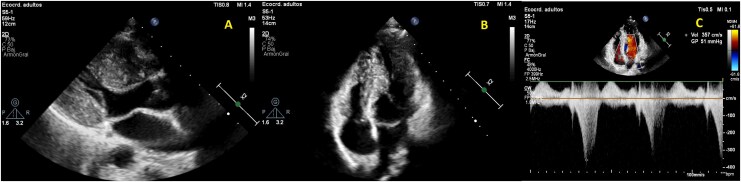
Recumbent resting echocardiography identifies only about half of patients with obstructive physiology. For a resting peak gradient below 50 mm Hg, TTE with provocative maneuvers is recommended (Class 1). For a symptomatic patient who does not reach 50 mm Hg at rest or with maneuvers, exercise echocardiography is recommended (Class 1); for an asymptomatic patient it is reasonable (Class 2a), because provocable obstruction changes advice on hydration and on drugs for comorbid conditions. Dobutamine is not used, because it produces intracavitary gradients in normal hearts and lacks specificity.
| Step | Method | Points of technique | Interpretation |
|---|---|---|---|
| 1. Rest | Color, pulsed-wave, and continuous-wave Doppler in sinus beats | The level of acceleration is localized, the MR envelope excluded, and blood pressure and rhythm recorded; post-extrasystolic beats are reported separately | Peak gradient ≥30 mm Hg = obstruction; ≥50 mm Hg = threshold for advanced therapy if symptomatic |
| 2. Valsalva strain | Continuous-wave Doppler during sustained strain | Coached effort; a smaller cavity and lower mitral E velocity confirm adequate strain | Rise to ≥30 mm Hg = provocable (latent) obstruction |
| 3. Standing or squat to stand | Continuous-wave Doppler through the first beats after standing | Preload is lowest before reflex compensation; delayed recording misses the peak | As for Valsalva |
| 4. Exercise echocardiography | Upright treadmill or bicycle, imaging immediately after exercise | Indicated when steps 1 to 3 remain below 50 mm Hg in a symptomatic patient; upright exercise lowers preload more than supine; postprandial testing when symptoms follow meals | Exercise gradient ≥50 mm Hg attributes exertional symptoms to obstruction |
| 5. Invasive hemodynamics | Simultaneous left ventricular and aortic pressure | End-hole or halo catheter to avoid entrapment; Valsalva, induced premature beat, or exercise | Indicated when noninvasive gradients are uncertain in a symptomatic patient (Class 1) |
Stepwise provocation of the outflow gradient, from the 2024 AHA/ACC guideline and the 2022 American Society of Echocardiography multimodality imaging recommendations. The report states the gradient with each maneuver, the blood pressure, and the rhythm.
Cardiac magnetic resonance: wall thickness, scar, and apical aneurysm
CMR provides sharp blood-myocardium contrast without acoustic window limits. It measures maximal wall thickness accurately in segments that echocardiography under- or overestimates, particularly the anterolateral wall, posterior septum, and apex, and it quantifies ventricular volumes, mass, and ejection fraction. CMR is indicated when echocardiography is inconclusive (Class 1), when an infiltrative or storage disease or athlete's heart is suspected (Class 1), and when the anatomic mechanism of obstruction is unclear before septal reduction (Class 1). It defines septal thickness at the point of mitral-septal contact, papillary muscle position, anomalous papillary insertion, accessory muscle bundles, and leaflet length.
LGE represents replacement fibrosis. In HCM it is typically patchy and midwall, located in the most hypertrophied segments and at the right ventricular insertion points, and it does not follow the subendocardial, coronary-territory pattern of infarction. LGE is present in about half of patients. Its extent, expressed as a percentage of left ventricular mass, relates continuously to sudden death risk: in 1,293 patients, risk rose by 46% for each 10% increase in LGE, and LGE involving 15% or more of the mass doubled the risk in patients otherwise judged to be at low risk. Absent or minimal LGE is associated with low risk. No quantification method has been standardized, and the extent can be estimated visually or by signal-intensity thresholds. Contrast CMR is recommended to assess maximal wall thickness, ejection fraction, apical aneurysm, and the extent of LGE in patients not otherwise identified as high risk or in whom the ICD decision remains uncertain after history, echocardiography, and ambulatory monitoring (Class 1). Repeat contrast CMR every 3 to 5 years for risk stratification may be considered (Class 2b).
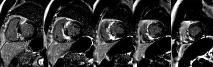
Parametric mapping adds tissue characterization. Native T1 and extracellular volume are mildly increased in sarcomeric HCM in proportion to diffuse fibrosis, markedly increased in amyloidosis, and low in Fabry disease before fibrosis develops. Values depend on scanner and sequence and are read against local reference ranges together with the LGE pattern. CMR also identifies apical aneurysms and thrombus missed by echocardiography, as well as crypts and elongated leaflets in genotype-positive relatives without hypertrophy, although the significance of these findings is uncertain.
Cardiac CT, invasive hemodynamics, and exercise testing
Cardiac CT may be considered for diagnosis when the echocardiogram is not diagnostic and CMR is unavailable (Class 2b). It defines wall thickness, subaortic membranes, thrombus, and coronary anatomy, at the cost of radiation and iodinated contrast. Coronary angiography by CT or invasive study is recommended in patients with symptoms or evidence of ischemia (Class 1) and before myectomy in those at risk of atherosclerosis (Class 1), and invasive angiography is part of every alcohol septal ablation.
Invasive hemodynamic assessment is recommended when the presence or severity of obstruction remains uncertain on noninvasive imaging in a symptomatic patient (Class 1). It distinguishes outflow from mitral signals, measures the gradient at rest and with Valsalva strain, an induced premature beat, or exercise, and quantifies filling pressures, cardiac output, and pulmonary pressures. Catheter entrapment in a small hyperdynamic cavity creates spurious gradients, so end-hole pigtail or halo catheters are used.
Exercise testing, preferably cardiopulmonary exercise testing (CPET), is reasonable in adults at the initial evaluation to measure functional capacity and provide prognostic information (Class 2a). It identifies provocable obstruction, an abnormal blood pressure response, chronotropic incompetence, arrhythmia, and ischemia. In more than 9,000 patients, reduced peak oxygen consumption, ventilatory inefficiency, and a low anaerobic threshold predicted ventricular arrhythmia, progression to advanced heart failure, and death. CPET is recommended in nonobstructive HCM with NYHA class III or IV symptoms to guide referral for transplantation or mechanical support (Class 1). Repeat testing every 2 to 3 years may be considered when it is unclear whether capacity has declined or when therapy depends on an ambiguous functional capacity (Class 2b).
Distinguishing HCM from athlete's heart and hypertensive hypertrophy
Athletic training, hypertension, aortic stenosis, and infiltrative disease all thicken the wall, and wall thickness in the range of 13 to 16 mm is the main gray zone. Among 947 elite Italian athletes, the thickest wall measured 16 mm, and only 16 (1.7%) had a wall of 13 mm or more; 15 of them were rowers or canoeists and one a cyclist, and all had enlarged cavities of 55 to 63 mm. No single measurement separates adaptation from disease, and the diagnosis rests on a combination of features.
| Feature | Favors HCM | Favors athlete's heart | Favors hypertensive heart disease |
|---|---|---|---|
| Distribution of hypertrophy | Asymmetric or focal; any segment; apical form | Mild and symmetric | Concentric, usually moderate; isolated basal septal bulge in older patients |
| Maximal thickness | Often ≥16 mm | Rarely ≥13 mm; 16 mm the reported upper limit | Rarely above 15 mm without another cause |
| Cavity size | Small, often below 45 mm | Enlarged in proportion to training (often above 55 mm) | Normal |
| SAM and dynamic obstruction | Common | Absent | Possible with a sigmoid septum and hypovolemia |
| Diastolic function | Reduced e′; raised filling-pressure markers | Normal or supranormal e′ | Impaired relaxation |
| Left atrium | Enlarged with dysfunction | Enlarged with normal reservoir function | Variable |
| ECG | Deep T inversion, pathological Q waves, ST depression | Isolated voltage criteria, early repolarization, sinus bradycardia | Voltage with strain pattern |
| CMR | Patchy midwall LGE; crypts; elongated leaflets | No LGE | Usually no or minimal LGE |
| Exercise capacity | Peak oxygen consumption reduced for age and sex | Supranormal | Normal or reduced |
| Family history and genetics | Affected relatives; sarcomere variant | Negative | Negative |
| Response to change in load | No regression | Regression with deconditioning | Regression with blood pressure control |
Differential diagnosis of left ventricular hypertrophy in the gray zone. Aortic stenosis is excluded by valve morphology and Doppler, and coexisting stenosis and dynamic obstruction are separated by pulsed-wave mapping along the outflow tract.
When findings remain equivocal, CMR, exercise testing with oxygen consumption, genetic testing, and a period of supervised detraining are complementary. Hypertension is present in 35% to 50% of adults with HCM, so the conditions frequently coexist; hypertension increases penetrance in variant carriers, and blood pressure control does not cause true HCM to regress.
Genetic testing, cascade testing, and family screening
Genetic counseling by an expert in cardiovascular genetics is recommended for every patient (Class 1), and genetic testing of the proband is beneficial to identify relatives at risk (Class 1). The first tier of testing includes the genes with strong evidence of causing HCM (Class 1); exome or genome sequencing is a second-tier option. Results are reported as pathogenic, likely pathogenic, variant of uncertain significance (VUS), likely benign, or benign. A pathogenic or likely pathogenic result confirms the diagnosis and enables cascade testing. A VUS is not clinically actionable: it does not guide testing of relatives or device decisions, although segregation analysis in affected relatives can help reclassify it. Variant classifications change; in one report, 11% of HCM variants were reclassified over 6 years in a way that altered family screening. Serial reevaluation of the variant is recommended (Class 1), and the guideline advises reconfirming pathogenicity every 2 to 3 years. In adults, the genotype does not by itself determine sudden death risk or ICD candidacy (Class 2b for its use in risk assessment). After a sudden unexplained death with a postmortem diagnosis of HCM, postmortem genetic testing is beneficial (Class 1).
First-degree relatives are offered both clinical screening with ECG and echocardiography and, when the proband carries a pathogenic or likely pathogenic variant, cascade genetic testing (Class 1). Relatives who test negative for the family variant are released from clinical surveillance (Class 3: No Benefit for continued screening), but they are notified and screening resumes if the variant is later downgraded. Relatives who carry the variant, and all relatives in families with no identified variant, undergo periodic clinical screening. Preconception and prenatal reproductive and genetic counseling is offered to affected families (Class 1); options include preimplantation genetic testing with in vitro fertilization, prenatal testing, and postnatal testing.
| Person screened | Start | ECG and echocardiogram |
|---|---|---|
| Adult first-degree relative, phenotype negative | When HCM is diagnosed in the family | Every 3 to 5 years, and with new symptoms |
| Adult carrier of the family variant (genotype positive, phenotype negative) | At cascade testing | Every 3 to 5 years with clinical assessment (Class 1) |
| Relative who does not carry the family pathogenic variant | — | No further screening unless the variant is downgraded |
| Families with malignant course or late-onset disease | Individualized | Interval shortened or extended by family history |
Family screening intervals from the 2024 AHA/ACC guideline, Table 7. Screening of children in genotype-positive or early-onset families begins when HCM is diagnosed in the family, and otherwise no later than puberty. Most programs continue clinical screening of adults to about 50 years of age, because new disease becomes less frequent but still occurs.
Genotype-positive, phenotype-negative individuals have preclinical HCM. Subtle findings such as reduced strain, impaired relaxation, crypts, elongated leaflets, ECG abnormalities, and raised natriuretic peptides can precede hypertrophy, but they do not drive treatment. Sudden death is rare in this group, and no risk model exists for it. An ICD is not recommended for primary prevention (Class 3: No Benefit), and preemptive drug therapy is not offered.
Sudden death risk stratification in adults
HCM has been one of the most common causes of sudden death in young people in North America, although it accounts for fewer than 10% of sudden deaths in young people and athletes in prospective registries. Risk is highest in adolescents and young adults and falls with age; event rates are low in stable patients older than 60 years. Most sudden deaths result from ventricular tachyarrhythmia, and in patients with ICDs, monomorphic VT and ventricular flutter are more common than previously thought. No sex or race difference in sudden death risk has been shown.
A comprehensive, systematic noninvasive risk assessment is recommended at the initial evaluation and every 1 to 2 years (Class 1). It includes the personal history of cardiac arrest, sustained ventricular arrhythmia, and syncope; the family history; maximal wall thickness, ejection fraction, and apical aneurysm on imaging; and nonsustained VT (NSVT) on 24- to 48-hour ambulatory monitoring, which is recommended at baseline and every 1 to 2 years (Class 1). CMR adds the extent of LGE when the decision remains uncertain.
| Risk factor | 2024 AHA/ACC definition | Role in the ICD decision |
|---|---|---|
| Prior cardiac arrest or sustained VT | Ventricular fibrillation or VT lasting more than 30 seconds or causing hemodynamic compromise | Secondary prevention: ICD recommended (Class 1) |
| Family history of HCM sudden death | Sudden death judged definitively or likely due to HCM in ≥1 first-degree or close relative aged ≤50 years; multiple deaths in more distant relatives are also relevant | Major risk factor |
| Massive hypertrophy | Wall thickness ≥30 mm in any segment by echocardiography or CMR; borderline values ≥28 mm may be considered | Major risk factor |
| Unexplained syncope | ≥1 episode judged unlikely to be vasovagal or due to LVOTO, especially within 6 months; events more than 5 years earlier have little relevance | Major risk factor |
| Apical aneurysm | Discrete thin-walled akinetic or dyskinetic distal segment with transmural scar or LGE, independent of size | Major risk factor |
| Systolic dysfunction | LVEF <50% by echocardiography or CMR | Major risk factor |
| Extensive LGE | Replacement fibrosis ≥15% of left ventricular mass, quantified or estimated visually | Modifier when no major risk factor is present |
| NSVT | ≥3 beats at ≥120 beats/min; greatest weight to runs that are frequent (≥3), long (≥10 beats), or fast (≥200 beats/min) | Modifier when no major risk factor is present |
Sudden death risk factors in adults, adapted from the 2024 AHA/ACC guideline, Table 8. Genotype status does not by itself guide ICD decisions in adults.
Each major risk factor rests on observational evidence. A wall thickness of 30 mm or more was associated with a cumulative 20-year sudden death risk near 40% in one cohort, with risk rising in proportion to thickness. Systolic dysfunction carries an annual sudden death event rate of about 2.5% in patients with an LVEF of 35% to 50%, and a particularly high arrhythmic burden below 35%. The 4.7% annual event rate with apical aneurysm and the doubling of risk with LGE of 15% or more are described above. NSVT is common, and short isolated runs add little in the absence of other markers, whereas NSVT in young patients and long, fast runs carry more weight.
The HCM Risk-SCD model, derived in 3,675 patients at six European centers, estimates 5-year sudden death risk from seven variables: age, maximal wall thickness, left atrial diameter, maximal left ventricular outflow tract (LVOT) gradient at rest or with Valsalva, family history of sudden death, NSVT, and unexplained syncope. Its discrimination is moderate. For patients 16 years and older, the 2024 AHA/ACC guideline finds it reasonable to obtain left atrial diameter and maximal gradient so that the 5-year estimate can inform shared decision-making (Class 2a). The writing committee did not assign recommendations to prespecified risk thresholds. The model does not include LVEF below 50%, apical aneurysm, or LGE, and the effect of these markers on an individual estimate is undetermined. The 2023 European Society of Cardiology (ESC) guideline places the HCM Risk-SCD estimate at the center of the decision. ICD implantation should be considered at an estimated 5-year risk of 6% or more (Class IIa) and may be considered at 4% to under 6% (Class IIb). Below 4%, extensive LGE (15% or more of left ventricular mass) or an LVEF below 50% may be weighed in shared decision-making (Class IIb), and an apical aneurysm is assessed through the Risk-SCD estimate rather than by its presence alone.
Primary-prevention ICD decisions and device selection
The 2024 guideline bases ICD decisions on a sensitive set of risk markers and on shared decision-making. For adults with one or more major risk factors, offering an ICD is reasonable (Class 2a), and discussing the 5-year sudden death risk and mortality estimates can be useful (Class 2a). When no major risk factor is present, an ICD may be considered in selected adults with extensive LGE or NSVT (Class 2b). An ICD should not be implanted in a patient without risk factors (Class 3: Harm) or solely to permit participation in competitive athletics (Class 3: Harm). Decisions involving a Class 2b recommendation, or an ambiguous profile, benefit from referral to a comprehensive HCM center. The major-marker strategy applies best to young and middle-aged adults.
2024 AHA/ACC ICD pathway
Adults with HCMAdapted from the 2024 AHA/ACC guideline, Section 7.2 and Figure 3. The class describes the strength of the recommendation to offer an ICD; the decision itself follows a shared discussion of risk, device complications, and patient preference.
HCM Risk-SCD estimate
Ages 16 years and olderFive-year risk = 1 − 0.998^exp(PI), where PI = 0.15939858 × MWT − 0.00294271 × MWT² + 0.0259082 × LAD + 0.00446131 × LVOTG + 0.4583082 × family history + 0.82639195 × NSVT + 0.71650361 × syncope − 0.01799934 × age (O'Mahony 2014). Left atrial diameter is measured by echocardiography in the parasternal long axis; the gradient is the maximal continuous-wave value at rest or with Valsalva, not with exercise. MWT, maximal wall thickness; LAD, left atrial diameter; LVOTG, outflow gradient.
Device selection. For patients receiving an ICD, either a single-chamber transvenous ICD or a subcutaneous ICD is recommended after shared decision-making that weighs age, lifestyle, and the potential need for pacing for bradycardia or for antitachycardia pacing (Class 1). Patients with HCM are younger than most ICD recipients, so lead failure, infection, and extraction over decades weigh heavily. The subcutaneous ICD avoids intravascular leads but cannot pace, is larger, and has a shorter battery life; candidates are screened for T-wave oversensing, including after exercise, because large T waves are common in HCM. With a transvenous system, single-coil leads are recommended for left-sided implants when the defibrillation threshold is adequate (Class 1), and threshold testing is considered with massive hypertrophy. A dual-chamber ICD is reasonable when atrial or atrioventricular sequential pacing is needed, or as an attempt to relieve obstructive symptoms, most often in patients older than 65 years (Class 2a). Cardiac resynchronization therapy (CRT) is reasonable in nonobstructive HCM with an LVEF below 50%, left bundle branch block, and NYHA class II to ambulatory class IV symptoms (Class 2a).
Ventricular arrhythmia after implantation. Antitachycardia pacing is recommended in patients with pacing-capable ICDs (Class 1), because a large share of appropriate therapies treat monomorphic VT or ventricular flutter that pacing terminates. For symptomatic ventricular arrhythmia or recurrent shocks despite a beta-blocker, antiarrhythmic therapy with amiodarone, dofetilide, mexiletine, or sotalol is recommended (Class 1). Catheter ablation, often combined epicardial and endocardial, is useful for recurrent monomorphic VT when drugs fail or are not preferred (Class 2a); apical aneurysms are a frequent substrate. Refractory life-threatening arrhythmia is an indication for transplant assessment (Class 1).
Drug therapy for symptomatic obstructive HCM
Drug therapy for obstruction relieves symptoms; no drug has been shown to alter the natural history. Because gradients vary throughout the day, success is judged by symptoms, not by a measured gradient. Asymptomatic patients with obstruction receive no gradient-directed drugs, but they are counseled on hydration and on avoiding vasodilators. Symptoms in the guideline's sense are exertional dyspnea or chest pain, and occasionally exertional syncope or presyncope, attributable to obstruction and interfering with daily life.
Nonvasodilating beta-blockers, titrated to effect or to the maximal tolerated dose, are first-line therapy (Class 1). Failure is not declared until physiologic beta-blockade, with suppression of the resting heart rate, is documented. When beta-blockers are ineffective or not tolerated, verapamil or diltiazem is substituted (Class 1). Their vasodilating action limits their use: verapamil is potentially harmful with severe dyspnea at rest, hypotension, or a very high resting gradient above 100 mm Hg (Class 3: Harm). Combining a beta-blocker with a nondihydropyridine calcium channel blocker as treatment for HCM is unsupported by evidence, although the combination may be used for hypertension.
When symptoms persist despite a beta-blocker or calcium channel blocker, three options carry Class 1 recommendations: adding a cardiac myosin inhibitor (adults), adding disopyramide in combination with an atrioventricular nodal blocker, or performing septal reduction therapy at an experienced center. The choice follows a discussion of success rates, risks, anatomy, comorbidities, local expertise, and patient preference. Disopyramide is a class IA antiarrhythmic with strong negative inotropic action. In a multicenter cohort of 118 patients followed for about 3 years, almost all also on a beta-blocker, two-thirds were managed without septal reduction, the resting gradient fell from 75 to 40 mm Hg, and no proarrhythmic excess was found. It is combined with a nodal blocker because it accelerates atrioventricular conduction in AF. Anticholinergic effects (dry mouth, urinary retention in older men, constipation, glaucoma) and QT prolongation limit its use, and the QT interval is monitored during initiation.
| Drug or intervention | Mechanism in obstruction | 2024 AHA/ACC class | Principal cautions |
|---|---|---|---|
| Nonvasodilating beta-blocker | Negative inotropy; longer diastolic filling | 1 (first line) | Titrate to resting heart rate suppression; chronotropic incompetence |
| Verapamil or diltiazem | Negative inotropy; improved relaxation | 1 (if beta-blocker fails) | Verapamil 3: Harm with rest dyspnea, hypotension, or gradient >100 mm Hg |
| Cardiac myosin inhibitor (mavacamten, aficamten) | Fewer actin-myosin cross-bridges; less hypercontractility | 1 (add-on, adults) | LVEF monitoring under the Risk Evaluation and Mitigation Strategy (REMS) program; cytochrome P450 (CYP) interactions; embryo-fetal toxicity in animal studies (mavacamten in pregnancy, Class 3: Harm) |
| Disopyramide | Negative inotropy | 1 (add-on, with a nodal blocker) | Anticholinergic effects; QT prolongation; accelerates atrioventricular conduction in AF |
| Septal reduction at an experienced center | Widens the outflow tract | 1 | See septal reduction |
| Intravenous phenylephrine for acute hypotension | Raises afterload without inotropy | 1 (if fluids fail) | May be combined with a beta-blocker |
| Low-dose oral diuretic | Relieves congestion | 2b | Aggressive diuresis worsens obstruction |
| Stopping vasodilators (angiotensin-converting enzyme [ACE] inhibitors, angiotensin receptor blockers [ARBs], dihydropyridines) and digoxin | Restores preload and afterload; removes inotropy | 2b | Alternative treatment of comorbid conditions |
Pharmacologic management of symptomatic obstructive HCM. Positive inotropes, pure vasodilators, and high-dose diuretics are relatively contraindicated when obstruction causes symptoms. The same principles apply to midventricular obstruction.
Acute hypotension in obstructive HCM is a medical urgency. Intravenous fluid restores preload, and if hypotension persists, intravenous phenylephrine or another vasoconstrictor without inotropic action is recommended, alone or with a beta-blocker that slows the rate and lengthens filling (Class 1). Inotropes such as dobutamine, dopamine, and milrinone, and vasodilators such as nitroglycerin, worsen obstruction and are avoided.
Cardiac myosin inhibitors: mavacamten and aficamten
Cardiac myosin inhibitors reduce the number of myosin heads available to bind actin, lowering hypercontractility, the outflow gradient, and filling pressures. Two are approved in the United States for symptomatic obstructive HCM to improve functional capacity and symptoms: mavacamten and aficamten. The 2024 guideline recommends adding a myosin inhibitor in adults whose symptoms persist despite a beta-blocker or calcium channel blocker (Class 1).
| Trial | Population | Comparison | Principal result |
|---|---|---|---|
| EXPLORER-HCM | 251 adults, NYHA II to III, LVOT gradient ≥50 mm Hg | Mavacamten vs placebo, 30 weeks | Composite of peak oxygen consumption and NYHA class met by 37% vs 17% |
| VALOR-HCM | 112 patients referred for septal reduction | Mavacamten vs placebo, 16 weeks | Decision to proceed with or continued eligibility for septal reduction in 18% vs 77% |
| SEQUOIA-HCM | 282 adults with symptomatic obstruction | Aficamten vs placebo, 24 weeks | Peak oxygen consumption +1.8 vs 0.0 mL/kg/min (difference 1.7); gradient reduction within 2 weeks |
| MAPLE-HCM | 175 adults with symptomatic obstruction, monotherapy | Aficamten vs metoprolol, 24 weeks | Peak oxygen consumption +1.1 vs −1.2 mL/kg/min (difference 2.3); secondary end points favored aficamten |
| ODYSSEY-HCM | Symptomatic nonobstructive HCM | Mavacamten vs placebo | No significant improvement in peak oxygen consumption or symptoms; LVEF <50% in 21.5% vs 1.7% |
Randomized trials of cardiac myosin inhibitors. In EXPLORER-HCM, a decrease in LVEF below 50% attributable to mavacamten occurred in 5.7% of patients.
Choosing between a myosin inhibitor and septal reduction. A myosin inhibitor suits a patient with symptomatic obstruction whose anatomy does not require surgery, who wishes to avoid or defer septal reduction, and who can attend serial echocardiography. Septal reduction is favored when the mitral apparatus, papillary muscles, or coexisting valve or coronary disease need surgical correction, when a single definitive procedure is preferred, when echocardiographic monitoring is impractical, when the patient is planning pregnancy, or when interacting drugs cannot be changed. Long-term outcome data for myosin inhibitors remain limited compared with the decades of follow-up after myectomy. Mavacamten has a long and variable half-life (6 to 9 days in normal CYP2C19 metabolizers and about 23 days in poor metabolizers), so steady state and recovery after interruption take weeks. Aficamten has a half-life of about 80 hours, and gradient reduction is evident within 2 weeks.
| Finding at scheduled echocardiogram | Mavacamten (adult label) | Aficamten (label) |
|---|---|---|
| Before starting | LVEF ≥55% required; start 5 mg daily | LVEF ≥55% required; start 5 mg daily |
| Initiation schedule | Echocardiogram at weeks 4, 8, and 12; at weeks 4 and 8, Valsalva gradient <20 mm Hg leads to dose reduction (5 to 2.5 mg) or, on 2.5 mg, withholding until week 12 | Echocardiogram 2 to 8 weeks after starting and after each dose change; increase by 5 mg every 2 to 8 weeks to a maximum of 20 mg |
| LVEF ≥55% and Valsalva gradient ≥30 mm Hg | From week 12, increase one level (2.5, 5, 10, 15 mg); recheck in 4 weeks; next increase no sooner than 12 weeks | Increase by 5 mg |
| LVEF ≥55% and Valsalva gradient <30 mm Hg | Maintain; clinical review at 3 months and echocardiogram at 6 months, then every 6 months | Maintain; echocardiogram every 6 months once the maintenance dose is set |
| LVEF 50% to <55% | Maintain; reassess in 3 months | Maintain; echocardiogram every 3 months |
| LVEF 40% to <50% | Interrupt; restart after 4 weeks at a lower dose if LVEF ≥50%; if interrupted on 2.5 mg, restart at 2.5 mg or discontinue | Decrease by 5 mg; on 5 mg, interrupt for at least 7 days |
| LVEF <40% or heart failure from systolic dysfunction | Interrupt | Interrupt for at least 7 days; restart at 5 mg when LVEF ≥55% |
| Intercurrent illness or new arrhythmia (infection, AF) | Delay dose increases; interruption is an option | Monitor LVEF; do not increase dose until resolved |
Echocardiographic monitoring decisions from the current US prescribing information (CAMZYOS revised 9/2026; MYQORZO revised 5/2026). Both drugs are dispensed only through the REMS program, with prescriber, pharmacy, and patient enrollment.
Drug interactions of mavacamten and aficamten. Mavacamten is metabolized mainly by CYP2C19 (74%) and CYP3A4 (18%). It is contraindicated with strong CYP2C19 inhibitors and with moderate or strong inducers of CYP2C19 or CYP3A4, because starting an inhibitor or stopping an inducer raises exposure and the risk of systolic dysfunction, and starting an inducer causes loss of effect. Starting a weak or moderate CYP2C19 inhibitor (the label names over-the-counter omeprazole, esomeprazole, and cimetidine) or a moderate or strong CYP3A4 inhibitor requires a one-level dose reduction and an echocardiogram 4 weeks later. Initiation of such inhibitors is avoided in patients on the lowest dose. The mavacamten label also advises against combining it with disopyramide, ranolazine, verapamil plus a beta-blocker, or diltiazem plus a beta-blocker, because of additive negative inotropy. Aficamten is metabolized mainly by CYP2C9, with smaller contributions from CYP3A, CYP2D6, and CYP2C19. It is contraindicated with rifampin; starting fluvoxamine or a strong CYP2C9 inhibitor, or stopping a moderate or strong CYP3A inducer, requires dose reduction and an echocardiogram 2 to 8 weeks later; starting fluconazole (for more than 3 days) or voriconazole requires reduction to 5 mg from 15 or 20 mg, and the combination is avoided in patients taking 5 or 10 mg.
Pregnancy and contraception with myosin inhibitors. Mavacamten is contraindicated in pregnancy (Class 3: Harm) because of teratogenicity in animal studies. Women of reproductive potential use effective contraception until 4 months after the last dose, and combined hormonal contraceptives containing a progestin other than norethindrone are avoided because mavacamten can reduce their effectiveness. Aficamten produced structural malformations in rats at exposures several times the human exposure, and no human pregnancy data exist.
Septal myectomy and alcohol septal ablation
Septal reduction therapy (SRT) is recommended for patients who remain symptomatic despite guideline-directed therapy (Class 1). Eligibility has three parts: clinical (severe dyspnea or chest pain, usually NYHA class III or IV, or exertional syncope or presyncope, attributable to obstruction and limiting daily life), hemodynamic (a dynamic gradient of about 50 mm Hg or more at rest or with physiologic provocation, with septal hypertrophy and SAM), and anatomic (a targeted septal thickness sufficient for a safe and effective procedure). SRT is performed at experienced centers whose outcomes meet defined targets (Class 1), because operative mortality of 5% to 10% has been observed at low-volume centers in the recent era. In a US nationwide database, both procedures had higher mortality and complication rates at low-volume hospitals.
Surgical septal myectomy. Transaortic extended septal myectomy resects a trough of basal and mid septum from below the aortic valve toward the papillary muscles, widening the outflow tract and redirecting flow so that SAM and SAM-mediated MR resolve. Extension of the resection, mobilization or resection of anomalous papillary muscles and accessory bundles, and mitral repair or plication address the mechanisms that septal thinning alone cannot. At experienced centers, operative mortality is below 1%, clinical success exceeds 90% to 95%, recurrent obstruction is rare, and long-term survival is similar to that of the age-matched general population. Intraoperative transesophageal echocardiography is recommended (Class 1). It defines the septal target, contact point, and mitral anatomy before bypass and confirms relief of obstruction and the absence of a ventricular septal defect or new aortic regurgitation afterward. Myectomy is the recommended procedure when associated cardiac disease requires surgery, such as an anomalous papillary muscle, a markedly elongated anterior leaflet, intrinsic mitral disease, multivessel coronary disease, or valvular aortic stenosis (Class 1). Surgical AF ablation or a maze procedure can be added (Class 2a). A transapical approach, alone or with a transaortic approach, treats midventricular obstruction.
Alcohol septal ablation. Ethanol injected into a septal perforator branch of the left anterior descending artery produces a localized basal septal infarction. Before injection, an ultrasound-enhancing agent injected into the candidate branch must opacify only the basal septum at the point of mitral-septal contact, with no opacification of papillary muscles, the right ventricle, or distant walls (Class 1 for contrast guidance). The gradient often falls immediately from stunning, then rises, and declines again as the scar matures over about 3 months, sometimes up to a year. Procedural mortality is below 1% at experienced centers. In adults who remain severely symptomatic and in whom surgery is contraindicated or the risk is unacceptable because of comorbidity or advanced age, alcohol septal ablation in eligible patients is recommended (Class 1). It carries a higher rate of permanent pacing and repeat intervention than myectomy. Survival is similar at 5 years, but survival at 10 years is lower after ablation than after myectomy.
| Feature | Surgical septal myectomy | Alcohol septal ablation |
|---|---|---|
| Access and recovery | Sternotomy and cardiopulmonary bypass; longer stay | Percutaneous; shorter stay |
| Preferred when | Mitral, papillary, valve, or coronary disease needing surgery; very thick septum; very high gradient; younger patients | Surgery contraindicated or high risk; frailty; suitable septal perforator; patient preference after full discussion |
| Unsuitable when | Prohibitive surgical risk | No suitable perforator; septum ≥30 mm or gradient ≥100 mm Hg (less effective); intrinsic mitral or papillary disease |
| Conduction effect | Left bundle branch block; complete block risk higher with preexisting right bundle branch block | Right bundle branch block; complete block risk higher with preexisting left bundle branch block |
| Target 30-day mortality | ≤1% | ≤1% |
| Target permanent pacemaker at 30 days | ≤5% | ≤10% |
| Target repeat procedure | ≤3% | ≤10% |
| Target mitral valve replacement within 1 year | ≤5% | — |
| Target success | >90% improve ≥1 NYHA class; >90% rest and provoked gradient <50 mm Hg | >90% improve ≥1 NYHA class; >90% rest and provoked gradient <50 mm Hg |
| Other complications | Ventricular septal defect, aortic regurgitation | Coronary dissection, remote infarction from ethanol leak, ventricular arrhythmia from the scar |
Comparison of septal reduction strategies, with outcome targets from the 2024 AHA/ACC guideline, Table 5. Targets for 30-day adverse complications (tamponade, coronary dissection, infection, major bleeding) and for more than moderate residual MR are ≤5% for both procedures.
Timing of septal reduction and role of mitral surgery. Earlier myectomy in NYHA class II may be reasonable at comprehensive centers when obstruction causes severe or progressive pulmonary hypertension, left atrial enlargement with symptomatic AF, poor functional capacity on treadmill testing, or very high resting gradients above 100 mm Hg in young adults (Class 2b). After shared decision-making, SRT may be considered as an alternative to escalating drug therapy in symptomatic patients (Class 2b). SRT is not recommended for asymptomatic patients with normal exercise capacity (Class 3: Harm). Mitral valve replacement solely to relieve obstruction should not be performed when SRT is an option (Class 3: Harm), because adding replacement increases hospital mortality more than 10-fold compared with isolated myectomy; when intrinsic mitral disease requires intervention, repair is preferred. Transcatheter edge-to-edge mitral repair has been used in selected patients at prohibitive surgical risk, but it is not a guideline-recommended treatment. Echocardiography 3 to 6 months after SRT is recommended to assess the result (Class 1).
Nonobstructive HCM with preserved ejection fraction
About one-third of patients have no gradient at rest or with provocation. Their symptoms arise from diastolic dysfunction, restrictive physiology, microvascular ischemia, chronotropic incompetence (including that caused by rate-slowing drugs), AF, and comorbidities such as obesity, hypertension, sleep apnea, and deconditioning. Overall HCM-related mortality is similar to that of obstructive disease. Coronary disease is excluded in patients with angina or coronary risk factors.
Beta-blockers or verapamil or diltiazem are recommended for exertional angina or dyspnea (Class 1); calcium channel blockers have the more direct trial evidence for chest pain and exercise capacity. Oral diuretics are reasonable when dyspnea persists (Class 2a), and the benefit of ACE inhibitors and angiotensin receptor blockers for symptoms is not established (Class 2b). In asymptomatic patients, the benefit of rate-slowing drugs is not established (Class 2b), and they can cause chronotropic incompetence; in patients with symptoms and no obstruction, chronotropic response is assessed by ambulatory monitoring or exercise testing. For younger patients (about 45 years or younger) with a sarcomere variant and a mild phenotype, valsartan may be beneficial to slow adverse remodeling (Class 2b). In the VANISH trial of 178 patients aged 45 years or younger, valsartan attenuated a composite of changes in wall thickness, mass, volumes, left atrial size, diastolic function, and biomarkers.
Disease-specific therapy for nonobstructive HCM remains an evidence gap. Mavacamten did not improve peak oxygen consumption or symptoms in ODYSSEY-HCM and reduced LVEF below 50% in about one in five treated patients. In ACACIA-HCM, 517 adults with symptomatic nonobstructive HCM were randomized to aficamten or placebo; at 36 weeks aficamten improved the KCCQ clinical summary score by 3.0 points and peak oxygen consumption by 0.67 mL/kg/min relative to placebo, and a reversible fall in LVEF below 50% occurred in 10.5% versus 0.8%. No myosin inhibitor carries a US indication for nonobstructive disease. Sodium-glucose cotransporter 2 (SGLT2) inhibitors and mineralocorticoid receptor antagonists reduce heart failure events in heart failure with preserved ejection fraction, but those trials did not address HCM, and the guideline lists their role in nonobstructive HCM as a question for investigation. A phase 3 trial of sotagliflozin in symptomatic obstructive and nonobstructive HCM has completed enrollment.
Atrial fibrillation and stroke prevention in HCM
AF is the most common sustained arrhythmia in HCM. In 1,558 patients followed for about 5 years, 20% had AF; with contemporary management, HCM-related mortality among them was 0.7% per year and mortality directly attributable to AF was 0.1% per year. Device monitoring detects subclinical AF in up to half of patients. Predictors include left atrial dilation, age, disease duration, and NYHA class III or IV symptoms. The HCM-AF score, derived in about 1,900 patients, combines these factors; high-risk patients developed AF at 2.7% to 3.4% per year. Extended ambulatory monitoring is recommended for patients at high risk of AF who are eligible for anticoagulation, at the initial evaluation and annually (Class 1), and may be considered every 1 to 2 years in adults without AF risk factors (Class 2b).
Thromboembolic risk is high and independent of the CHA₂DS₂-VASc score; many strokes occur in patients with a score of 0. A meta-analysis of 33 studies reported a thromboembolism prevalence of 27% among patients with HCM and AF. Anticoagulation is recommended for clinical AF regardless of the score, with a direct oral anticoagulant (DOAC) as first-line therapy and a vitamin K antagonist as second-line therapy (Class 1). For device-detected subclinical AF lasting more than 24 hours in a single episode, anticoagulation is recommended on the same terms (Class 1); for episodes of more than 5 minutes but less than 24 hours, anticoagulation can be beneficial after weighing episode duration, total burden, risk factors, and bleeding risk (Class 2a). Stored electrograms are reviewed to exclude artifact. Atrial flutter is treated in the same way. Left atrial appendage occlusion has not been tested in HCM.
Rapid AF is often poorly tolerated, because filling depends on atrial contraction and a short diastole. When rate control is chosen, beta-blockers, verapamil, or diltiazem are recommended (Class 1); the verapamil cautions in obstruction apply, and digoxin is avoided when obstruction is present. A rhythm-control strategy with cardioversion or antiarrhythmic drugs can be beneficial for poorly tolerated AF (Class 2a). Amiodarone, sotalol, and dofetilide are reasonable choices; disopyramide has modest antiarrhythmic efficacy; dronedarone has low efficacy; and flecainide and propafenone are generally avoided unless an ICD is present. Catheter ablation can be effective when drugs are ineffective, contraindicated, or not preferred (Class 2a), although recurrence is about twice as frequent as in patients without HCM and repeat procedures are common. In patients undergoing myectomy, concomitant surgical ablation can be beneficial (Class 2a). Anticoagulant selection and dosing follow the principles in atrial fibrillation.
HCM with systolic dysfunction and advanced heart failure
An LVEF below 50% defines HCM with systolic dysfunction, often called the end-stage or burned-out phase. Because the HCM ventricle is normally hyperdynamic, an LVEF in the low 50s already represents lost function. In three cohorts totaling 1,259 patients, 3.5% had end-stage disease, with a mortality of 11% per year; in SHaRe, 553 of 6,793 patients (8%) met criteria for systolic dysfunction, and 35% of them died or required transplantation or a ventricular assist device, at a median of 8.4 years after it was recognized. Remodeling is heterogeneous: some ventricles dilate and thin, while others retain thick walls and a small cavity with restrictive physiology. Contractility declines before cavity dilation and symptoms appear, so serial imaging is needed to detect the decline.
When LVEF falls below 50%, coexisting causes such as coronary disease, valve disease, and phenocopies are sought (Class 1). Guideline-directed therapy for heart failure with reduced ejection fraction is recommended (Class 1), myosin inhibitors are discontinued if the reduction persists (Class 1), and stopping verapamil, diltiazem, and disopyramide is reasonable (Class 2a), although they can be continued for AF rate or rhythm control case by case. An ICD can be beneficial for persistent LVEF below 50% (Class 2a), reflecting sudden death event rates of about 2.5% per year with an LVEF of 35% to 50%. CRT can be beneficial with an LVEF below 50%, NYHA class II to IV symptoms, and left bundle branch block (Class 2a).
Advanced heart failure develops in 3% to 8% of patients, and 20% to 50% of them have a preserved ejection fraction with restrictive physiology, so referral for transplantation does not require a reduced ejection fraction. CPET quantifies limitation and guides selection (Class 1), and transplant assessment is recommended for NYHA class III or IV symptoms despite therapy or for refractory life-threatening ventricular arrhythmia (Class 1). The 2018 United Network for Organ Sharing allocation policy added HCM-specific criteria that have increased transplant rates and shortened waiting times, and survival after transplantation is comparable to, or better than, that in other cardiomyopathies. A continuous-flow left ventricular assist device is reasonable as a bridge to transplantation (Class 2a); a small cavity limits device performance, and outcomes are better when the left ventricular end-diastolic dimension exceeds about 46 to 50 mm.
Exercise, sports participation, and occupation
Mild- to moderate-intensity recreational exercise is recommended to improve fitness, physical function, quality of life, and overall health, in keeping with general physical activity guidelines (Class 1). Intensity is graded as light below 3 metabolic equivalents (METs), moderate 3 to 6 METs, and vigorous above 6 METs, or by 40% to 50%, 50% to 70%, and above 70% of maximal heart rate. In the RESET-HCM randomized trial of 136 adults, 16 weeks of moderate-intensity training increased peak oxygen consumption by 1.35 mL/kg/min, against 0.08 mL/kg/min with usual activity, with no major adverse events. For most patients, universal restriction from vigorous activity or competitive sport is not indicated (Class 3: No Benefit).
Vigorous recreational activity is reasonable after an annual comprehensive evaluation and shared decision-making with an expert professional (Class 2a). In the LIVE-HCM prospective cohort of patients aged 8 to 60 years with mild or no symptoms, mostly followed at experienced centers, those who exercised vigorously had no more arrhythmic events than those who exercised moderately or were sedentary. For patients capable of a high level of performance, competitive sport may be considered after annual review by an expert experienced with athletes who have HCM (Class 2b). Prospective studies show a similar burden of ventricular arrhythmia in athletes who continue to compete and those who withdraw, and shock rates in athletes with ICDs resemble those of nonathletes, with most shocks occurring outside sport. The return-to-play discussion covers the patient's risk profile, the type of sport, the uncertainty of the data, emergency preparedness with an automated external defibrillator, hydration and avoidance of heat, and the role of team physicians and institutions, which often make the final eligibility decision. Genotype-positive, phenotype-negative individuals may compete at any intensity (Class 2a).
For commercial drivers, it is reasonable to follow Federal Motor Carrier Safety Administration guidance, which permits driving for patients without an ICD or major sudden death risk factors who are on guideline-directed therapy (Class 2a). For pilots, Federal Aviation Administration guidance permits consideration of multicrew duties for asymptomatic, low-risk patients who complete a maximal treadmill test to 85% of peak heart rate (Class 2a). Manual labor, heavy lifting, and high-performance occupations such as law enforcement and firefighting may be considered after evaluation and risk stratification (Class 2b).
Pregnancy in women with HCM
Most women with HCM tolerate pregnancy well. Maternal mortality is very low, with only three sudden deaths reported over 17 years, all in high-risk or undiagnosed patients. Symptoms or complications (dyspnea, chest pain, palpitations, heart failure, and arrhythmia) occur in about one-quarter of pregnancies, mostly in women symptomatic before conception, and outcomes do not differ between women with and without obstruction. It is reasonable to advise women with clinically stable HCM that pregnancy is generally safe (Class 2a). Women who are severely symptomatic before conception are offered options to reduce risk, such as SRT for refractory obstruction, advanced heart failure therapy, or an ICD for high-risk features.
Care is coordinated between the cardiologist and the obstetrician, with maternal-fetal medicine consultation for high-risk patients (Class 1). Selected beta-blockers are given for symptoms of obstruction or arrhythmia, with monitoring of fetal growth (Class 1); metoprolol, bisoprolol, labetalol, pindolol, and propranolol are generally considered safe, whereas atenolol carries evidence of fetal risk. Serial echocardiography is reasonable, particularly in the second and third trimesters, when hemodynamic load peaks (Class 2a). For AF or another indication for anticoagulation, low-molecular-weight heparin or a vitamin K antagonist at a dose below 5 mg daily is recommended (Class 1); DOACs lack safety data in pregnancy. Cardioversion of new or recurrent AF is reasonable (Class 2a). Mavacamten is contraindicated (Class 3: Harm).
Vaginal delivery is the recommended first choice (Class 1), because adverse cardiac outcomes are similar with vaginal and cesarean delivery and bleeding is greater with cesarean section; the Valsalva maneuver of labor is well tolerated. A delivery plan is set by the end of the second trimester. General or epidural anesthesia is reasonable with precautions to avoid hypotension (Class 2a). Preconception genetic counseling is offered (Class 1), and fetal echocardiography may be considered (Class 2b).
Comorbidities, anesthesia, and noncardiac surgery
Comorbid conditions shape symptoms and outcomes. More than 70% of adults with HCM have a body mass index above 25 kg/m² and more than 30% above 30 kg/m². Obesity increases hypertrophy, obstruction, and symptoms, reduces exercise capacity, and was independently associated with death, heart failure, AF, ventricular arrhythmia, and stroke in a large registry. Counseling and comprehensive lifestyle intervention for weight loss are recommended (Class 1), as is adherence to ACC/AHA primary prevention guidance (Class 1). Hypertension is treated to standard targets, with a preference for beta-blockers and nondihydropyridine calcium channel blockers when obstruction is present (Class 1); vasodilators and diuretics can be used in asymptomatic patients, and in younger patients with sarcomeric nonobstructive HCM and hypertension, valsartan is an option. Sleep-disordered breathing affects 55% to 70% of patients and is associated with AF, NSVT, and greater symptom burden; screening for symptoms and referral for evaluation and treatment are recommended (Class 1).
Anesthesia and noncardiac surgery require management of dynamic obstruction. The preoperative evaluation documents the resting and provoked gradient, ventricular function, rhythm, and device status. Beta-blockers and calcium channel blockers are continued, and myosin inhibitor therapy is reviewed with the treating HCM team because of the drugs' long or intermediate half-lives and interaction potential. Intraoperatively, hypovolemia, vasodilation from induction agents or neuraxial blockade, tachycardia, and catecholamine inotropes increase obstruction; adequate volume, a slower heart rate, and a phenylephrine-based vasopressor strategy counter it. Transesophageal echocardiography identifies SAM as the cause of intraoperative hypotension. ICD tachytherapy is suspended during electrocautery with continuous monitoring and restored afterward. Dynamic obstruction with SAM can also appear without HCM in hypovolemic or septic patients, after aortic valve replacement in a hypertrophied ventricle, after mitral repair, and in takotsubo syndrome, where inotropes worsen shock and volume and vasoconstriction relieve it.
Follow-up and surveillance schedule
Each review, scheduled or prompted by new symptoms or a clinical event, reassesses symptoms, functional capacity, obstruction, ventricular function, rhythm, and sudden death risk, because new risk markers such as systolic dysfunction, aneurysm, and progressive fibrosis appear over time.
| Test | Interval in stable adults | 2024 AHA/ACC class |
|---|---|---|
| Clinical evaluation and sudden death risk assessment | Every 1 to 2 years; individualized after age 60 when stable | 1 |
| 12-lead ECG | Every 1 to 2 years | 1 |
| Transthoracic echocardiography with provocation | Every 1 to 2 years; longer after repeated stability | 1 |
| 24- to 48-hour ambulatory ECG | Every 1 to 2 years | 1 |
| Extended monitoring for AF | Annually if at high AF risk and eligible for anticoagulation; every 1 to 2 years may be considered otherwise | 1 / 2b |
| Contrast CMR | Every 3 to 5 years for risk stratification | 2b |
| Exercise testing | Every 2 to 3 years when change in capacity is uncertain | 2b |
| Echocardiography on a myosin inhibitor | Per label: 2 to 12 weeks during titration, then every 3 to 6 months | Label |
| Echocardiography after septal reduction | 3 to 6 months after the procedure | 1 |
| Reevaluation of genetic variant classification | Every 2 to 3 years | 1 |
Surveillance in adults with HCM, from the 2024 AHA/ACC guideline and current myosin inhibitor labeling. Patients with ICDs undergo routine device follow-up, which replaces ambulatory monitoring for arrhythmia detection.
Self-assessment
A 34-year-old man with hypertrophic cardiomyopathy has a maximal septal thickness of 24 mm, a left atrial diameter of 44 mm, a resting LVOT gradient of 18 mm Hg rising to 72 mm Hg with Valsalva strain, no syncope, and no family history of sudden death. Ambulatory monitoring shows one 4-beat run of NSVT at 150 beats/min, LVEF is 68%, and CMR shows no apical aneurysm with late gadolinium enhancement involving 19% of left ventricular mass. How is his primary-prevention ICD recommendation classified under the 2024 AHA/ACC guideline?
Cardiovascular Medicine Board ReviewReferences
- Ommen SR, Ho CY, Asif IM, et al. 2024 AHA/ACC/AMSSM/HRS/PACES/SCMR guideline for the management of hypertrophic cardiomyopathy: a report of the American Heart Association/American College of Cardiology Joint Committee on Clinical Practice Guidelines. Circulation. 2024;149:e1239–e1311. doi:10.1161/CIR.0000000000001250 (also published in J Am Coll Cardiol. 2024;83:2324–2405. doi:10.1016/j.jacc.2024.02.014)
- Arbelo E, Protonotarios A, Gimeno JR, et al. 2023 ESC guidelines for the management of cardiomyopathies. Eur Heart J. 2023;44:3503–3626. doi:10.1093/eurheartj/ehad194
- Nagueh SF, Phelan D, Abraham T, et al. Recommendations for multimodality cardiovascular imaging of patients with hypertrophic cardiomyopathy: an update from the American Society of Echocardiography, in collaboration with the American Society of Nuclear Cardiology, the Society for Cardiovascular Magnetic Resonance, and the Society of Cardiovascular Computed Tomography. J Am Soc Echocardiogr. 2022;35:533–569. doi:10.1016/j.echo.2022.03.012
- Maron BJ, Gardin JM, Flack JM, et al. Prevalence of hypertrophic cardiomyopathy in a general population of young adults: echocardiographic analysis of 4111 subjects in the CARDIA Study. Circulation. 1995;92:785–789. doi:10.1161/01.cir.92.4.785
- Semsarian C, Ingles J, Maron MS, Maron BJ. New perspectives on the prevalence of hypertrophic cardiomyopathy. J Am Coll Cardiol. 2015;65:1249–1254. doi:10.1016/j.jacc.2015.01.019
- Ho CY, Day SM, Ashley EA, et al. Genotype and lifetime burden of disease in hypertrophic cardiomyopathy: insights from the Sarcomeric Human Cardiomyopathy Registry (SHaRe). Circulation. 2018;138:1387–1398. doi:10.1161/CIRCULATIONAHA.117.033200
- Maron BJ, Rowin EJ, Casey SA, et al. Hypertrophic cardiomyopathy in adulthood associated with low cardiovascular mortality with contemporary management strategies. J Am Coll Cardiol. 2015;65:1915–1928. doi:10.1016/j.jacc.2015.02.061
- Maron BJ. Clinical course and management of hypertrophic cardiomyopathy. N Engl J Med. 2018;379:655–668. doi:10.1056/NEJMra1710575
- Maron MS, Olivotto I, Betocchi S, et al. Effect of left ventricular outflow tract obstruction on clinical outcome in hypertrophic cardiomyopathy. N Engl J Med. 2003;348:295–303. doi:10.1056/NEJMoa021332
- Maron MS, Olivotto I, Zenovich AG, et al. Hypertrophic cardiomyopathy is predominantly a disease of left ventricular outflow tract obstruction. Circulation. 2006;114:2232–2239. doi:10.1161/CIRCULATIONAHA.106.644682
- Sherrid MV, Gunsburg DZ, Moldenhauer S, Pearle G. Systolic anterior motion begins at low left ventricular outflow tract velocity in obstructive hypertrophic cardiomyopathy. J Am Coll Cardiol. 2000;36:1344–1354. doi:10.1016/S0735-1097(00)00830-5
- Kitaoka H, Doi Y, Casey SA, et al. Comparison of prevalence of apical hypertrophic cardiomyopathy in Japan and the United States. Am J Cardiol. 2003;92:1183–1186. doi:10.1016/j.amjcard.2003.07.027
- Eriksson MJ, Sonnenberg B, Woo A, et al. Long-term outcome in patients with apical hypertrophic cardiomyopathy. J Am Coll Cardiol. 2002;39:638–645. doi:10.1016/S0735-1097(01)01778-8
- Rowin EJ, Maron BJ, Haas TS, et al. Hypertrophic cardiomyopathy with left ventricular apical aneurysm: implications for risk stratification and management. J Am Coll Cardiol. 2017;69:761–773. doi:10.1016/j.jacc.2016.11.063
- Pelliccia A, Maron BJ, Spataro A, Proschan MA, Spirito P. The upper limit of physiologic cardiac hypertrophy in highly trained elite athletes. N Engl J Med. 1991;324:295–301. doi:10.1056/NEJM199101313240504
- Maron BJ, Pelliccia A, Spirito P. Cardiac disease in young trained athletes: insights into methods for distinguishing athlete's heart from structural heart disease, with particular emphasis on hypertrophic cardiomyopathy. Circulation. 1995;91:1596–1601. doi:10.1161/01.cir.91.5.1596
- Spirito P, Bellone P, Harris KM, et al. Magnitude of left ventricular hypertrophy and risk of sudden death in hypertrophic cardiomyopathy. N Engl J Med. 2000;342:1778–1785. doi:10.1056/NEJM200006153422403
- O'Mahony C, Jichi F, Pavlou M, et al. A novel clinical risk prediction model for sudden cardiac death in hypertrophic cardiomyopathy (HCM Risk-SCD). Eur Heart J. 2014;35:2010–2020. doi:10.1093/eurheartj/eht439
- Chan RH, Maron BJ, Olivotto I, et al. Prognostic value of quantitative contrast-enhanced cardiovascular magnetic resonance for the evaluation of sudden death risk in patients with hypertrophic cardiomyopathy. Circulation. 2014;130:484–495. doi:10.1161/CIRCULATIONAHA.113.007094
- Sherrid MV, Barac I, McKenna WJ, et al. Multicenter study of the efficacy and safety of disopyramide in obstructive hypertrophic cardiomyopathy. J Am Coll Cardiol. 2005;45:1251–1258. doi:10.1016/j.jacc.2005.01.012
- Olivotto I, Oreziak A, Barriales-Villa R, et al. Mavacamten for treatment of symptomatic obstructive hypertrophic cardiomyopathy (EXPLORER-HCM): a randomised, double-blind, placebo-controlled, phase 3 trial. Lancet. 2020;396:759–769. doi:10.1016/S0140-6736(20)31792-X
- Desai MY, Owens A, Geske JB, et al. Myosin inhibition in patients with obstructive hypertrophic cardiomyopathy referred for septal reduction therapy. J Am Coll Cardiol. 2022;80:95–108. doi:10.1016/j.jacc.2022.04.048
- Maron MS, Masri A, Nassif ME, et al. Aficamten for symptomatic obstructive hypertrophic cardiomyopathy. N Engl J Med. 2024;390:1849–1861. doi:10.1056/NEJMoa2401424
- Garcia-Pavia P, Maron MS, Masri A, et al. Aficamten or metoprolol monotherapy for obstructive hypertrophic cardiomyopathy. N Engl J Med. 2025;393:949–960. doi:10.1056/NEJMoa2504654
- Desai MY, Owens AT, Abraham T, et al. Mavacamten in symptomatic nonobstructive hypertrophic cardiomyopathy. N Engl J Med. 2025;393:961–972. doi:10.1056/NEJMoa2505927
- CAMZYOS (mavacamten) capsules, prescribing information. Bristol-Myers Squibb; revised 9/2026. packageinserts.bms.com/pi/pi_camzyos.pdf
- MYQORZO (aficamten) tablets, prescribing information. Cytokinetics; revised 5/2026. cytokinetics.com/pi_myqorzo
- Ho CY, Day SM, Axelsson A, et al. Valsartan in early-stage hypertrophic cardiomyopathy: a randomized phase 2 trial. Nat Med. 2021;27:1818–1824. doi:10.1038/s41591-021-01505-4
- Ommen SR, Maron BJ, Olivotto I, et al. Long-term effects of surgical septal myectomy on survival in patients with obstructive hypertrophic cardiomyopathy. J Am Coll Cardiol. 2005;46:470–476. doi:10.1016/j.jacc.2005.02.090
- Kim LK, Swaminathan RV, Looser P, et al. Hospital volume outcomes after septal myectomy and alcohol septal ablation for treatment of obstructive hypertrophic cardiomyopathy: US nationwide inpatient database, 2003–2011. JAMA Cardiol. 2016;1:324–332. doi:10.1001/jamacardio.2016.0252
- Liebregts M, Vriesendorp PA, Mahmoodi BK, et al. A systematic review and meta-analysis of long-term outcomes after septal reduction therapy in patients with hypertrophic cardiomyopathy. JACC Heart Fail. 2015;3:896–905. doi:10.1016/j.jchf.2015.06.011
- Nishimura RA, Seggewiss H, Schaff HV. Hypertrophic obstructive cardiomyopathy: surgical myectomy and septal ablation. Circ Res. 2017;121:771–783. doi:10.1161/CIRCRESAHA.116.309348
- Rowin EJ, Hausvater A, Link MS, et al. Clinical profile and consequences of atrial fibrillation in hypertrophic cardiomyopathy. Circulation. 2017;136:2420–2436. doi:10.1161/CIRCULATIONAHA.117.029267
- Carrick RT, Maron MS, Adler A, et al. Development and validation of a clinical predictive model for identifying hypertrophic cardiomyopathy patients at risk for atrial fibrillation: the HCM-AF score. Circ Arrhythm Electrophysiol. 2021;14:e009796. doi:10.1161/CIRCEP.120.009796
- Harris KM, Spirito P, Maron MS, et al. Prevalence, clinical profile, and significance of left ventricular remodeling in the end-stage phase of hypertrophic cardiomyopathy. Circulation. 2006;114:216–225. doi:10.1161/CIRCULATIONAHA.105.583500
- Marstrand P, Han L, Day SM, et al. Hypertrophic cardiomyopathy with left ventricular systolic dysfunction: insights from the SHaRe Registry. Circulation. 2020;141:1371–1383. doi:10.1161/CIRCULATIONAHA.119.044366
- Saberi S, Wheeler M, Bragg-Gresham J, et al. Effect of moderate-intensity exercise training on peak oxygen consumption in patients with hypertrophic cardiomyopathy: a randomized clinical trial. JAMA. 2017;317:1349–1357. doi:10.1001/jama.2017.2503
- Lampert R, Ackerman MJ, Marino BS, et al. Vigorous exercise in patients with hypertrophic cardiomyopathy. JAMA Cardiol. 2023;8:595–605. doi:10.1001/jamacardio.2023.1042
- Addis DR, Townsley MM. Perioperative implications of the 2024 AHA/ACC/AMSSM/HRS/PACES/SCMR guideline for the management of hypertrophic cardiomyopathy. J Cardiothorac Vasc Anesth. 2024;38:2510–2512. doi:10.1053/j.jvca.2024.07.039
- Masri A, Maron MS, Bhatia A, et al. Aficamten for symptomatic nonobstructive hypertrophic cardiomyopathy. N Engl J Med. 2026. Published online August 28, 2026. doi:10.1056/NEJMoa2603021