Library › Heart failure › HFrEF
Heart failure with reduced ejection fraction
Heart failure with reduced ejection fraction in adults from definition and cause through guideline-directed drug therapy, devices, acute decompensation, improved ejection fraction, and referral for advanced therapies.
Heart failure with reduced ejection fraction (HFrEF) is the clinical syndrome of heart failure with a left ventricular ejection fraction (LVEF) of 40% or less. Four drug classes reduce death and hospitalization: an angiotensin receptor-neprilysin inhibitor (ARNI; or an angiotensin-converting enzyme [ACE] inhibitor or angiotensin receptor blocker [ARB]), an evidence-based beta blocker, a mineralocorticoid receptor antagonist (MRA), and a sodium-glucose cotransporter 2 (SGLT2) inhibitor. All four are started early at tolerated doses and titrated within weeks. Ejection fraction is remeasured after optimization before an implantable cardioverter-defibrillator (ICD) or cardiac resynchronization therapy (CRT) device is chosen, and therapy continues after the ejection fraction improves.
Definition and ejection-fraction categories of heart failure
Heart failure is a clinical syndrome of symptoms and signs caused by a structural or functional cardiac abnormality and corroborated by elevated natriuretic peptides or by objective evidence of pulmonary or systemic congestion. This is the 2021 universal definition endorsed by the Heart Failure Society of America (HFSA), the Heart Failure Association of the European Society of Cardiology, and the Japanese Heart Failure Society, and it was adopted by the 2022 American Heart Association/American College of Cardiology/HFSA (AHA/ACC/HFSA) guideline. The definition makes heart failure a diagnosis of syndrome plus mechanism: a reduced ejection fraction without symptoms is pre-heart failure (stage B), and dyspnea without a cardiac abnormality is not heart failure.
The ejection fraction divides the syndrome into categories because the randomized trials enrolled patients by ejection fraction, and therapies proven in HFrEF are prescribed to patients who resemble the trial populations. The categories in the 2022 guideline remain the standard in US practice.
| Category | LVEF | Additional requirement | Therapeutic implication |
|---|---|---|---|
| HFrEF (reduced) | ≤40% | Clinical syndrome of heart failure | Four foundational drug classes; ICD and CRT by criteria |
| HFimpEF (improved) | Previous LVEF ≤40%, follow-up LVEF >40% | A documented trajectory, not a new diagnosis | HFrEF therapy continued (Class 1) |
| HFmrEF (mildly reduced) | 41–49% | Evidence of spontaneous or provokable increased filling pressure | SGLT2 inhibitor (Class 2a); other HFrEF classes may be considered (Class 2b) |
| HFpEF (preserved) | ≥50% | Evidence of spontaneous or provokable increased filling pressure | Managed as a separate syndrome |
Ejection-fraction categories in the 2022 AHA/ACC/HFSA guideline. The 2021 universal definition defines improved ejection fraction more strictly: baseline LVEF 40% or less, an increase of at least 10 percentage points, and a second measurement above 40%. HFrEF, heart failure with reduced ejection fraction; HFimpEF, heart failure with improved ejection fraction; HFmrEF, heart failure with mildly reduced ejection fraction; HFpEF, heart failure with preserved ejection fraction.
Ejection fraction is a load-dependent, method-dependent measurement with a reproducibility of roughly 5 to 10 percentage points between studies and readers. A value near a threshold is therefore confirmed with a second technique or a repeat study before it decides a device or a drug. Three-dimensional echocardiography or cardiac magnetic resonance (CMR) gives the most reproducible serial values, and the same method is used for the measurements that are compared over time.
The AHA, ACC, European Society of Cardiology (ESC), and World Heart Federation published a second universal definition of heart failure in 2026. It keeps the syndrome-based definition and names the ejection fraction categories reduced, preserved, and improved. It interprets ejection fraction in light of age, sex, and ethnicity in place of fixed cutoffs, describes heart failure as a dynamic condition that can improve, remit, or progress, and adds a standard classification of causes for trials and registries. Treatment recommendations on this page follow the 2022 guideline and the 2024 ACC Expert Consensus Decision Pathway (ECDP), whose eligibility criteria use the numerical thresholds tested in the trials.
Epidemiology, outcomes, and burden of HFrEF
About 7.7 million US adults have heart failure according to the AHA 2026 statistical update, up from 6.7 million in the previous survey cycle, and prevalence is projected to rise as the population ages and survival after myocardial infarction improves. Roughly half of patients have a reduced ejection fraction. HFrEF is more common in men, in patients with prior myocardial infarction, and in Black adults, in whom heart failure begins at a younger age and is more often related to hypertension. Incidence rises steeply after 65 years of age.
Heart failure is the leading cause of hospitalization among US adults older than 65 years. After a first hospitalization for heart failure, mortality and readmission rates are high: roughly 1 patient in 5 is readmitted within 30 days, and 5-year mortality after a heart failure hospitalization was about 75% among Medicare beneficiaries in the Get With The Guidelines-Heart Failure registry, similar across ejection-fraction categories. The period immediately after discharge carries the highest risk of death and readmission, and this vulnerable phase is the target of rapid in-hospital and post-discharge optimization of therapy.
Treatment has changed the outlook. Each foundational drug class reduces mortality independently, and modeling from the trial results suggests that comprehensive therapy with all four classes extends event-free survival by years for a middle-aged patient compared with an ACE inhibitor and beta blocker alone. The gap between that potential and practice is large: in US registries most eligible outpatients do not receive all four classes, and fewer still receive target doses. Underuse is greater in women, older adults, and patients with kidney disease, low income, or limited insurance coverage.
Causes of HFrEF and the search for a reversible cause
Coronary artery disease causes about half of HFrEF in the United States. The remainder is nonischemic: genetic, toxic, inflammatory, infiltrative, valvular, tachycardia-mediated, metabolic, peripartum, or idiopathic dilated cardiomyopathy. The cause changes management, because some causes respond to specific treatment and some reverse completely. The 2022 guideline recommends a thorough history and physical examination for cardiac and noncardiac causes (Class 1), a three-generation family history in nonischemic cardiomyopathy (Class 1), and specific testing when a cause is suspected.
| Cause | Clues | Confirming test | Specific action |
|---|---|---|---|
| Ischemic cardiomyopathy | Prior infarction, angina, regional wall motion abnormality, Q waves | Coronary angiography or coronary CT angiography; CMR scar in a coronary distribution | Revascularization when anatomy is suitable; secondary prevention |
| Tachycardia-mediated cardiomyopathy | Persistent atrial fibrillation or flutter with rapid rate, incessant atrial tachycardia, premature ventricular complex burden above about 10% to 15% | Ambulatory monitoring | Rate or rhythm control, ablation; recovery over weeks to months |
| Alcohol | Sustained heavy intake | History | Abstinence; recovery is common |
| Cardiotoxic cancer therapy | Anthracycline, HER2-targeted therapy, immune checkpoint inhibitor, radiation | Serial LVEF and global longitudinal strain, troponin | Cardio-oncology care, GDMT, review of the cancer regimen |
| Myocarditis and sarcoidosis | Recent viral illness, chest pain, conduction block, ventricular arrhythmia, troponin rise | CMR; FDG PET; endomyocardial biopsy for fulminant or giant cell myocarditis | Immunosuppression for giant cell myocarditis and sarcoidosis |
| Genetic dilated cardiomyopathy | Family history, early conduction disease, skeletal myopathy, arrhythmias out of proportion to LVEF | Genetic testing and counseling | Cascade screening of relatives; earlier ICD in high-risk genotypes such as LMNA |
| Peripartum cardiomyopathy | LVEF <45% in late pregnancy or the months after delivery | Echocardiography; exclusion of other causes | Pregnancy-compatible therapy, anticoagulation when LVEF is very low |
| Infiltrative disease | Increased wall thickness with low voltage, carpal tunnel syndrome, neuropathy | Bone scintigraphy with serum and urine light-chain testing; CMR | Disease-specific therapy for transthyretin and light-chain amyloidosis |
| Chagas cardiomyopathy | Birth or residence in endemic Latin America, right bundle branch block with left anterior fascicular block, apical aneurysm | Serologic testing for Trypanosoma cruzi | Arrhythmia and thromboembolism prevention; antiparasitic therapy in selected patients |
| Valvular disease | Severe primary regurgitation or stenosis | Echocardiography | Valve intervention by valve guideline criteria |
| Endocrine, metabolic, and nutritional | Thyroid disease, pheochromocytoma, thiamine deficiency, hemochromatosis, hypocalcemia | Thyroid function, iron studies, targeted tests | Treatment of the disorder |
| Stimulants | Cocaine or methamphetamine use | History, toxicology | Abstinence |
Causes of HFrEF and the tests that establish them. GDMT, guideline-directed medical therapy; FDG PET, fluorodeoxyglucose positron emission tomography.
Tachycardia-mediated cardiomyopathy. Rapid ventricular rates of any origin depress contractility and dilate the ventricle, and the dysfunction reverses when the rate is controlled. The diagnosis is often made in retrospect, after rhythm or rate control restores function, because atrial fibrillation is both a cause and a consequence of HFrEF. Recurrence of the arrhythmia can cause rapid and severe relapse, so patients remain under surveillance after recovery.
Cancer therapy-related cardiac dysfunction. Anthracycline toxicity is dose-dependent and is often irreversible once it causes heart failure; HER2-targeted therapy causes dysfunction that often recovers after interruption. Immune checkpoint inhibitors cause myocarditis that is uncommon but frequently fulminant. Baseline and serial LVEF with global longitudinal strain and troponin identify dysfunction before symptoms, and patients with established HFrEF from cancer therapy receive the same GDMT as other patients.
Genetic dilated cardiomyopathy. A pathogenic or likely pathogenic variant is found in about 10% to 30% of patients with idiopathic dilated cardiomyopathy (about 25% of patients of European ancestry and 8% of patients of African ancestry in the DCM Precision Medicine Study), and more often with a family history, most often truncating variants in TTN, followed by LMNA, FLNC, DSP, RBM20, MYH7, and others. Genetic testing is reasonable in nonischemic cardiomyopathy when a genetic cause is suspected (Class 2a), and genetic counseling is part of the process. A result changes management in two ways: first-degree relatives are offered cascade screening, and some genotypes carry a high risk of sudden death even when the ejection fraction is above 35%. LMNA, FLNC, DSP, and RBM20 variants are the leading examples, and risk calculators specific to LMNA inform the timing of a defibrillator.
Peripartum, alcohol, and myocarditis. These three causes share a high rate of recovery with therapy, which shapes the timing of a defibrillator decision and the counseling about prognosis. Fulminant myocarditis with shock is managed as cardiogenic shock and may require temporary mechanical support.
Amyloidosis and Chagas disease. Cardiac amyloidosis classically produces a thick-walled ventricle with preserved or mildly reduced ejection fraction. Advanced transthyretin and light-chain disease lower the ejection fraction, and amyloidosis is excluded in older patients whose wall thickness exceeds what their blood pressure history explains (covered on the cardiac amyloidosis page). Chagas cardiomyopathy is the leading cause of nonischemic cardiomyopathy in endemic regions of Latin America and is present in immigrant populations in the United States. It carries a higher risk of sudden death, conduction disease, and apical aneurysm with thromboembolism than other dilated cardiomyopathies.
Neurohormonal activation and remodeling: why the four drug classes work
A fall in stroke volume, whether from infarction, a genetic sarcomere defect, toxic injury, or chronic overload, is sensed as reduced effective arterial filling. The sympathetic nervous system, the renin-angiotensin-aldosterone system, and arginine vasopressin are activated. In the short term these responses support blood pressure and perfusion by raising heart rate, contractility, vascular tone, and intravascular volume. Sustained, they become the disease: norepinephrine is directly toxic to myocytes and arrhythmogenic, angiotensin II and aldosterone drive myocyte hypertrophy, apoptosis, and interstitial fibrosis, and sodium retention raises filling pressures.
The ventricle remodels. Chamber dilation raises wall stress by the law of Laplace, which further increases oxygen demand and promotes more dilation; the ventricle becomes spherical, the mitral annulus dilates, and the papillary muscles are displaced, which produces secondary mitral regurgitation and adds a volume load. The Frank-Starling relationship is depressed and flat, so additional preload raises filling pressure with little gain in stroke volume. Congestion, and not low output, causes most symptoms in most patients.
The natriuretic peptide system is the counter-regulatory response. B-type natriuretic peptide (BNP) is released from ventricular myocytes in response to wall stress and promotes natriuresis, vasodilation, and inhibition of renin and aldosterone. It is overwhelmed in established HFrEF, and it is the system that neprilysin inhibition amplifies.
The four foundational drug classes act on these pathways in parallel. Renin-angiotensin inhibition reduces afterload, aldosterone release, and fibrosis; neprilysin inhibition adds augmentation of natriuretic peptides and other vasoactive peptides. Beta blockers counter adrenergic toxicity, slow the heart rate, reduce arrhythmic and pump-failure death, and produce the largest reverse remodeling of any drug class, typically a rise in LVEF over 3 to 12 months. Mineralocorticoid receptor antagonists block the effects of aldosterone that persist despite ACE inhibition (aldosterone escape), including fibrosis and potassium and magnesium loss. SGLT2 inhibitors act through mechanisms that are not fully defined: proximal natriuresis without reflex neurohormonal activation, reduced preload and afterload, improved myocardial energetics, and renal protection. The mechanisms are complementary, which is the physiologic basis for combining all four early.
ACC/AHA stages of heart failure and New York Heart Association functional class
Two classifications are used together. The ACC/AHA stages describe the development and progression of disease and move in one direction; New York Heart Association (NYHA) class describes current symptoms and changes with treatment. A patient who has been symptomatic remains in stage C after symptoms resolve on therapy, because the structural disease persists, while the NYHA class can improve from III to I.
| ACC/AHA stage | Definition | NYHA class | Definition |
|---|---|---|---|
| A: at risk | Hypertension, diabetes, obesity, coronary disease, cardiotoxin exposure, genetic variant, or family history of cardiomyopathy; no structural disease, no symptoms | — | — |
| B: pre-heart failure | No current or prior symptoms, with structural heart disease (reduced LVEF, hypertrophy, chamber enlargement, wall motion abnormality, valve disease), evidence of increased filling pressure, or persistently elevated natriuretic peptide or troponin | I | No limitation of ordinary physical activity |
| C: symptomatic heart failure | Structural heart disease with current or prior symptoms | I to IV | II: slight limitation, comfortable at rest. III: marked limitation, comfortable at rest, less than ordinary activity causes symptoms. IV: symptoms at rest or with any activity |
| D: advanced heart failure | Marked symptoms that interfere with daily life and recurrent hospitalizations despite attempts to optimize GDMT | Usually III to IV | Ambulatory class IV denotes symptoms with minimal exertion without continuous intravenous therapy |
ACC/AHA stages from the 2022 guideline and NYHA functional class. Stage B was renamed pre-heart failure in the 2021 universal definition and the 2022 guideline.
Asymptomatic left ventricular dysfunction (stage B with LVEF 40% or less) is treated to prevent symptoms and death. ACE inhibition (Class 1, from the SOLVD prevention trial), an evidence-based beta blocker (Class 1), and an ARB when an ACE inhibitor is not tolerated are recommended. After myocardial infarction with LVEF 40% or less, the same therapy applies, and an ICD is indicated by the criteria below. When LVEF is 30% or less at least 40 days after infarction in a patient with NYHA class I symptoms on GDMT, a primary-prevention ICD is recommended (Class 1).
NYHA class is subjective and poorly reproducible between observers, especially in separating class II from III. Objective measures supplement it: the 6-minute walk distance, peak oxygen consumption on cardiopulmonary exercise testing, and patient-reported health status measured by the Kansas City Cardiomyopathy Questionnaire, which most modern trials use and which predicts outcome.
Symptoms, examination, electrocardiogram, and chest radiograph
Exertional dyspnea and fatigue are the cardinal symptoms. Orthopnea, paroxysmal nocturnal dyspnea, and bendopnea (dyspnea on bending forward) reflect elevated left-sided filling pressure. Right-sided congestion produces edema, abdominal fullness, early satiety, and right upper quadrant discomfort from hepatic congestion. Low output produces fatigue, cool extremities, cognitive slowing, oliguria, and cachexia. Many patients with elevated filling pressures have few signs, and dyspnea in older adults has many causes.
On examination, an elevated jugular venous pressure is the most useful sign of elevated right-sided and, in most patients with chronic HFrEF, left-sided filling pressure. The abdominojugular reflux test and a jugular venous pressure that fails to fall with inspiration (Kussmaul sign) support it. A third heart sound, a displaced and diffuse apical impulse, and the holosystolic murmur of secondary mitral regurgitation are typical of a dilated ventricle. Rales are often absent in chronic heart failure because lymphatic drainage increases. A narrow proportional pulse pressure (pulse pressure divided by systolic pressure below 25%), pulsus alternans, cool extremities, and hypotension suggest a low cardiac index.
The electrocardiogram is almost never normal in HFrEF. It shows Q waves of prior infarction, left bundle branch block (LBBB) or other conduction disease, atrial fibrillation, left ventricular hypertrophy, or low voltage. QRS duration and morphology are recorded at diagnosis and after optimization because they decide resynchronization. Right bundle branch block with left anterior fascicular block in a patient from an endemic region raises Chagas disease; conduction block in a young patient raises sarcoidosis, laminopathy, or other genetic disease.
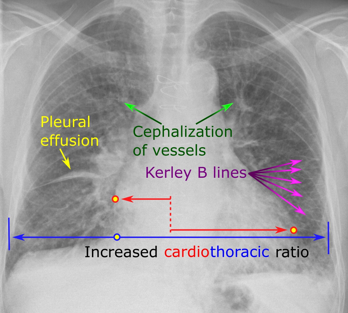

The chest radiograph shows cardiomegaly and the progression of pulmonary venous hypertension: redistribution of flow to the upper lobes, then interstitial edema with Kerley B lines and peribronchial cuffing, then alveolar edema and pleural effusions. A normal radiograph does not exclude elevated filling pressure in chronic heart failure. Lung ultrasound, with three or more B-lines in two or more zones on each side, detects interstitial edema with higher sensitivity and is used at the bedside to follow decongestion.
Initial laboratory testing comprises a complete blood count, urinalysis, electrolytes, urea nitrogen, creatinine, glucose, lipid profile, liver function tests, iron studies (ferritin and transferrin saturation), and thyroid-stimulating hormone (Class 1), with a natriuretic peptide and testing directed at specific causes. The 2022 guideline recommends a 12-lead electrocardiogram in all patients with heart failure (Class 1).
Natriuretic peptides: diagnostic thresholds and confounders
BNP and the inactive N-terminal fragment of its prohormone (NT-proBNP) are released in proportion to ventricular wall stress. Natriuretic peptide measurement supports or excludes heart failure in patients with dyspnea (Class 1). In established heart failure, a baseline value establishes prognosis (Class 1), and a predischarge value is reasonable to establish post-discharge prognosis (Class 2a). In patients at risk (stage A), natriuretic peptide screening followed by team-based care can prevent the development of left ventricular dysfunction or heart failure (Class 2a).
| Setting | BNP | NT-proBNP | Use |
|---|---|---|---|
| Ambulatory, nonacute symptoms | ≥35 pg/mL | ≥125 pg/mL | Supports heart failure; below these values heart failure is unlikely |
| Acute dyspnea, rule-out | <100 pg/mL | <300 pg/mL | Acute heart failure unlikely |
| Acute dyspnea, rule-in, age <50 years | — | >450 pg/mL | Supports acute heart failure |
| Acute dyspnea, rule-in, age 50–75 years | — | >900 pg/mL | Supports acute heart failure |
| Acute dyspnea, rule-in, age >75 years | — | >1,800 pg/mL | Supports acute heart failure |
Thresholds from the 2021 universal definition (ambulatory and hospital thresholds) and the ICON-RELOADED emergency department study (age-stratified NT-proBNP cutpoints and the 300 pg/mL rule-out value). Values between the rule-out and rule-in cutpoints are indeterminate.
Natriuretic peptides are confounded in both directions. Obesity lowers concentrations, and a body mass index above 30 kg/m² is associated with values about 50% lower for the same filling pressure, so lower thresholds apply in obese patients. Higher values occur with older age, atrial fibrillation, chronic kidney disease, pulmonary embolism, pulmonary hypertension and right ventricular strain, sepsis, critical illness, acute coronary syndrome, and anemia. Flash pulmonary edema can occur before BNP has risen, and constrictive pericarditis and mitral stenosis produce lower values than equivalent congestion from myocardial disease.
Sacubitril/valsartan changes interpretation. BNP is a substrate of neprilysin, so its concentration rises or falls less than expected during the first weeks of sacubitril/valsartan; NT-proBNP is not a substrate and falls as wall stress falls. NT-proBNP is therefore the marker used to follow patients receiving an ARNI. A fall in NT-proBNP after starting therapy predicts reverse remodeling and better outcomes. Titration of therapy to a natriuretic peptide target did not improve outcomes compared with usual care in GUIDE-IT, and titration to a natriuretic peptide target is not an established strategy.
Imaging and testing to establish etiology: echo, CMR, and coronary evaluation
Transthoracic echocardiography is recommended at initial evaluation (Class 1). It measures LVEF and volumes, wall thickness and regional wall motion, diastolic function and estimated filling pressure, right ventricular size and function, valve disease (including the severity and mechanism of mitral regurgitation), pulmonary artery systolic pressure, and the pericardium. Biplane Simpson volumes are the standard two-dimensional method; three-dimensional volumes are more accurate and reproducible where image quality allows, and contrast opacification improves endocardial definition when two or more segments are not seen. Global longitudinal strain detects early dysfunction, especially in cardio-oncology. Repeat echocardiography is recommended when clinical status changes, after a major event, and after 3 to 6 months of optimized therapy when the result will change a device decision (Class 1).
Regional wall motion abnormalities in a coronary distribution suggest ischemic disease. Multivessel disease can produce global hypokinesis, and some nonischemic diseases produce regional abnormalities (sarcoidosis classically in the basal septum, Chagas disease at the apex and inferolateral base). Left bundle branch block causes early septal activation with a septal flash and delayed lateral contraction, which is a feature of dyssynchrony, not of ischemia. Dyssynchrony measures by echocardiography do not predict response to resynchronization and are not used to select patients (Class 3: No Benefit in the 2023 Heart Rhythm Society/Asia Pacific Heart Rhythm Society/Latin American Heart Rhythm Society (HRS/APHRS/LAHRS) guideline).
Cardiac magnetic resonance. Alternative imaging, most often CMR, is recommended when echocardiography is inadequate (Class 1), and CMR is the next test when an infiltrative, inflammatory, or other specific cause is suspected. It is the reference method for volumes and ejection fraction, and late gadolinium enhancement defines the pattern of scar. Subendocardial or transmural enhancement in a coronary distribution indicates infarction. Midwall septal enhancement occurs in nonischemic dilated cardiomyopathy and predicts sudden death and lower likelihood of recovery independently of LVEF. Subepicardial enhancement suggests myocarditis; patchy enhancement with focal edema suggests sarcoidosis; diffuse subendocardial enhancement with abnormal gadolinium kinetics and elevated extracellular volume suggests amyloidosis; and T2* below 20 ms indicates myocardial iron overload. Parametric T1, T2, and extracellular volume mapping quantify edema and diffuse fibrosis.
Coronary evaluation. Coronary artery disease is excluded or defined in every patient with new HFrEF of uncertain cause. Invasive coronary angiography is reasonable in patients who may be candidates for revascularization (Class 2a), and coronary CT angiography is an alternative in patients with low to intermediate probability of coronary disease. Stress testing with myocardial perfusion imaging, positron emission tomography, stress CMR, or dobutamine echocardiography identifies ischemia and viability. Viability testing informs revascularization decisions but did not identify patients who benefit from surgical or percutaneous revascularization in STICH or REVIVED-BCIS2.
Endomyocardial biopsy, nuclear imaging, and invasive hemodynamics. Endomyocardial biopsy is reasonable when the result changes treatment, chiefly suspected giant cell myocarditis, fulminant myocarditis, eosinophilic myocarditis, and infiltrative disease not established by noninvasive tests (Class 2a). Technetium-99m pyrophosphate or other bone-tracer scintigraphy, with exclusion of a monoclonal protein, diagnoses transthyretin amyloidosis without biopsy. FDG PET identifies active cardiac sarcoidosis. Right heart catheterization is reasonable when filling pressures or cardiac output are uncertain, when the cause of hypotension or worsening renal function is unclear, and in the evaluation for advanced therapies (covered on the right heart catheterization page). Routine invasive hemodynamic monitoring is not recommended in acute heart failure without these indications.
The four foundational drug classes and their trial evidence
Guideline-directed medical therapy (GDMT) for symptomatic HFrEF rests on four drug classes, each supported by randomized trials that reduced death, hospitalization, or both on top of the therapy that preceded it. The trials enrolled patients with LVEF of 35% to 40% or less and NYHA class II to IV symptoms. The 2022 guideline gives each class a Class 1 recommendation with level of evidence A, and the 2024 ACC ECDP recommends that all four be started early, ideally within weeks, without waiting for any one drug to reach its target dose.
| Drug class and trial | Population | Comparison | Principal result |
|---|---|---|---|
| ACE inhibitor: SOLVD-Treatment (1991) | LVEF ≤35%, mostly NYHA II–III | Enalapril vs placebo | All-cause death 35.2% vs 39.7%; 16% relative reduction |
| ARNI: PARADIGM-HF (2014) | LVEF ≤40% (later ≤35%), NYHA II–IV, elevated natriuretic peptide | Sacubitril/valsartan 97/103 mg twice daily vs enalapril 10 mg twice daily | Cardiovascular death or HF hospitalization 21.8% vs 26.5%; HR 0.80 |
| Beta blocker: CIBIS-II (1999) | LVEF ≤35%, NYHA III–IV | Bisoprolol vs placebo | All-cause death 11.8% vs 17.3%; HR 0.66 |
| Beta blocker: MERIT-HF (1999) | LVEF ≤40%, NYHA II–IV | Metoprolol succinate vs placebo | All-cause death reduced by 34% |
| Beta blocker: COPERNICUS (2001) | LVEF <25%, symptoms at rest or minimal exertion, euvolemic | Carvedilol vs placebo | All-cause death reduced by 35% |
| MRA: RALES (1999) | LVEF ≤35%, NYHA III–IV | Spironolactone 25 mg vs placebo | All-cause death 35% vs 46%; relative risk 0.70 |
| MRA: EMPHASIS-HF (2011) | LVEF ≤35%, NYHA II | Eplerenone up to 50 mg vs placebo | Cardiovascular death or HF hospitalization 18.3% vs 25.9%; HR 0.63 |
| SGLT2 inhibitor: DAPA-HF (2019) | LVEF ≤40%, NYHA II–IV, with or without diabetes | Dapagliflozin 10 mg vs placebo | Worsening HF or cardiovascular death 16.3% vs 21.2%; HR 0.74 |
| SGLT2 inhibitor: EMPEROR-Reduced (2020) | LVEF ≤40%, NYHA II–IV, with or without diabetes | Empagliflozin 10 mg vs placebo | Cardiovascular death or HF hospitalization 19.4% vs 24.7%; HR 0.75 |
Landmark randomized trials of the four foundational drug classes in HFrEF. HR, hazard ratio; HF, heart failure; MRA, mineralocorticoid receptor antagonist.
Renin-angiotensin inhibition and the ARNI. In patients with chronic symptomatic HFrEF and NYHA class II to III symptoms, an ARNI is recommended to reduce morbidity and mortality (Class 1). In patients who tolerate an ACE inhibitor or ARB, replacement by an ARNI is recommended (Class 1). When an ARNI is not feasible because of cost, access, or intolerance, an ACE inhibitor is recommended (Class 1), and an ARB when an ACE inhibitor causes cough or angioedema (Class 1). De novo initiation of sacubitril/valsartan without prior ACE inhibitor exposure is supported by PIONEER-HF, in which sacubitril/valsartan started in hospitalized, stabilized patients reduced NT-proBNP more than enalapril without excess hypotension, hyperkalemia, or worsening renal function. ARNIs and ACE inhibitors are contraindicated with a history of angioedema (Class 3: Harm), an ARNI is not given within 36 hours of the last ACE inhibitor dose (Class 3: Harm), and all renin-angiotensin inhibitors are contraindicated in pregnancy.
Beta blockers. Only three beta blockers reduced mortality in HFrEF: bisoprolol, carvedilol, and sustained-release metoprolol succinate (Class 1). Metoprolol tartrate, atenolol, and nebivolol are not substitutes, and in COMET carvedilol reduced mortality compared with metoprolol tartrate. Mortality benefit is established for these three agents and is not assumed for the beta blocker class as a whole. Beta blockers are started in euvolemic or nearly euvolemic patients and are continued during most admissions for decompensation unless shock or marked hypoperfusion is present.
Mineralocorticoid receptor antagonists. Spironolactone or eplerenone is recommended in NYHA class II to IV HFrEF when the estimated glomerular filtration rate (eGFR) is above 30 mL/min/1.73 m² and potassium is below 5.0 mEq/L, with careful monitoring of potassium, renal function, and diuretic dosing (Class 1). When potassium cannot be kept below 5.5 mEq/L, the MRA is discontinued (Class 3: Harm). Gynecomastia with spironolactone results from antiandrogenic effects and resolves on switching to the selective agent eplerenone. Finerenone, a nonsteroidal MRA, reduced worsening heart failure events in heart failure with LVEF 40% or above in FINEARTS-HF and is not a substitute for spironolactone or eplerenone in HFrEF.
SGLT2 inhibitors. In symptomatic chronic HFrEF, an SGLT2 inhibitor is recommended to reduce hospitalization for heart failure and cardiovascular mortality, irrespective of diabetes (Class 1). The benefit appears within weeks, requires no titration, and is accompanied by slower decline in kidney function. An initial fall in eGFR of a few mL/min/1.73 m² is hemodynamic and expected. Dapagliflozin is not started below an eGFR of 25 mL/min/1.73 m² and empagliflozin was studied to an eGFR of 20. Both are withheld for type 1 diabetes, dialysis, and acute illness with poor oral intake, and they are stopped at least 3 days before elective surgery because of the risk of euglycemic ketoacidosis. Genital mycotic infections are the most common adverse effect. Sotagliflozin, a combined SGLT1 and SGLT2 inhibitor, reduced cardiovascular death and heart failure events in patients with diabetes recently hospitalized for heart failure (SOLOIST-WHF) and is approved for heart failure.
The 2022 guideline attached value statements to these recommendations: GDMT including ACE inhibitors or ARBs, beta blockers, MRAs, and ARNIs provides high economic value, and SGLT2 inhibitors provide intermediate economic value at their prices at the time.
Starting and titrating GDMT: doses, sequence, and laboratory checks
The traditional sequence, in which an ACE inhibitor was titrated to target before a beta blocker was added and an MRA came months later, reflected the order in which the trials were published. It is not supported by evidence. The benefit of each class appears within 30 days of starting it, most of the benefit is achieved at low and intermediate doses, and the drugs offset one another's adverse effects: SGLT2 inhibitors lower the risk of hyperkalemia from MRAs and soften the eGFR decline with renin-angiotensin inhibition, and sacubitril/valsartan lowers potassium relative to enalapril. The 2024 ACC ECDP therefore recommends initiating all four classes, in any order the clinical situation allows, and titrating to target or maximally tolerated doses with the aim of completing optimization within about 3 months. Titration as frequently as every 1 to 2 weeks, guided by symptoms, vital signs, and laboratory values, is reasonable (Class 2a in the 2022 guideline).
STRONG-HF tested this strategy after hospitalization for acute heart failure. Patients randomized to high-intensity care had renin-angiotensin inhibitors, beta blockers, and MRAs titrated to full recommended doses within 2 weeks of discharge, with scheduled visits at 1, 2, 3, and 6 weeks monitoring symptoms, blood pressure, potassium, creatinine, and NT-proBNP. Readmission for heart failure or death at 180 days fell from 23.3% to 15.2%. STRONG-HF did not include SGLT2 inhibitors, so it supports rapid titration with close follow-up but is not a trial of the complete four-class regimen.
| Drug | Starting dose | Target dose | Titration step |
|---|---|---|---|
| Sacubitril/valsartan | 49/51 mg twice daily; 24/26 mg twice daily when not taking or taking low doses of an ACE inhibitor or ARB, eGFR <30, moderate hepatic impairment, or older age | 97/103 mg twice daily | Double every 2–4 weeks |
| Enalapril | 2.5 mg twice daily | 10–20 mg twice daily | Double every 1–2 weeks |
| Lisinopril | 2.5–5 mg once daily | 20–40 mg once daily | Double every 1–2 weeks |
| Ramipril | 1.25–2.5 mg once daily | 10 mg once daily | Double every 1–2 weeks |
| Captopril | 6.25 mg three times daily | 50 mg three times daily | Double every 1–2 weeks |
| Candesartan | 4–8 mg once daily | 32 mg once daily | Double every 1–2 weeks |
| Losartan | 25–50 mg once daily | 50–150 mg once daily | Double every 1–2 weeks |
| Valsartan | 20–40 mg twice daily | 160 mg twice daily | Double every 1–2 weeks |
| Bisoprolol | 1.25 mg once daily | 10 mg once daily | Double every 2 weeks |
| Carvedilol | 3.125 mg twice daily | 25 mg twice daily; 50 mg twice daily if weight >85 kg | Double every 2 weeks |
| Carvedilol controlled release | 10 mg once daily | 80 mg once daily | Double every 2 weeks |
| Metoprolol succinate | 12.5–25 mg once daily | 200 mg once daily | Double every 2 weeks |
| Spironolactone | 12.5–25 mg once daily | 25–50 mg once daily | Increase after about 4 weeks if potassium and creatinine allow |
| Eplerenone | 25 mg once daily | 50 mg once daily | Increase after about 4 weeks if potassium and creatinine allow |
| Dapagliflozin | 10 mg once daily | 10 mg once daily | None |
| Empagliflozin | 10 mg once daily | 10 mg once daily | None |
| Sotagliflozin | 200 mg once daily | 400 mg once daily | After 2 weeks |
Starting and target doses adapted from the 2022 AHA/ACC/HFSA guideline (drugs commonly used for HFrEF) and the 2024 ACC ECDP. Target doses are those used in the outcome trials; the maximally tolerated dose is the goal when a target is not reached. With eGFR 31 to 49 mL/min/1.73 m², MRAs start at spironolactone 12.5 mg daily or eplerenone 25 mg every other day.
| Clinical profile | Sequence that fits the physiology |
|---|---|
| Congested, adequate blood pressure | Diuretic plus SGLT2 inhibitor and ARNI first (both contribute natriuresis); beta blocker once near-euvolemic; MRA early |
| Euvolemic, heart rate above 70/min, adequate blood pressure | Beta blocker and SGLT2 inhibitor first, then ARNI and MRA |
| Systolic pressure 90–100 mm Hg | SGLT2 inhibitor and MRA first (small effect on blood pressure); low-dose ARNI and beta blocker in sequence; nonprognostic antihypertensives stopped |
| Heart rate below 60/min | ARNI, MRA, and SGLT2 inhibitor first; beta blocker at low dose after review of nodal drugs and conduction disease |
| eGFR 20–30 mL/min/1.73 m² | SGLT2 inhibitor and beta blocker first; ARNI at 24/26 mg; MRA withheld at eGFR 30 or less |
| Potassium 5.0–5.5 mEq/L | SGLT2 inhibitor and beta blocker first; ARNI preferred over an ACE inhibitor; MRA deferred until potassium is below 5.0 or a binder is in place |
| Hospitalized and stabilized | All tolerated classes started before discharge; follow-up within 7 days with laboratory review |
Sequencing of foundational therapy by clinical profile, after the 2024 ACC ECDP. No single order is required; the order is chosen to start the classes that the current physiology tolerates.
Each step is followed by measurement of blood pressure (including orthostatic pressure in older patients), heart rate, symptoms, weight, potassium, and creatinine. After starting or increasing an ARNI, ACE inhibitor, ARB, or MRA, potassium and creatinine are checked within 1 to 2 weeks; after starting or increasing an MRA they are checked at about 1 week and 4 weeks, then every 6 months, with more frequent testing during clinical instability (2022 guideline). Beta-blocker titration is guided by heart rate, blood pressure, and congestion. Clinical pharmacists, nurse-led titration clinics, and telehealth visits increase the proportion of patients who reach target doses, and team-based multidisciplinary care is recommended (Class 1).
GDMT titration and device eligibility checker
The checker applies the eligibility thresholds of the 2022 AHA/ACC/HFSA guideline, the 2024 ACC ECDP, and the 2023 HRS/APHRS/LAHRS guideline to one set of values. For each foundational drug class, it returns whether initiation or titration fits current blood pressure, heart rate, potassium, and kidney function, and it classifies ICD and CRT eligibility. Volume status, symptoms, drug interactions, and patient preferences are outside its inputs.
Thresholds: ARNI, ACE inhibitor, or ARB not started at systolic pressure below 90 mm Hg or potassium above 5.0 mEq/L; MRA started only with eGFR above 30 and potassium below 5.0 and held above 5.5; SGLT2 inhibitors studied to eGFR 20 (empagliflozin) and 25 (dapagliflozin); ivabradine for sinus rhythm at 70/min or more with LVEF 35% or less. Device classes from the 2022 AHA/ACC/HFSA, 2017 AHA/ACC/HRS, and 2023 HRS/APHRS/LAHRS guidelines. Expected survival with good functional status beyond 1 year is assumed.
Managing hyperkalemia, hypotension, bradycardia, and worsening renal function during titration
Most apparent intolerance of GDMT is managed without stopping a prognostic drug. The physiology that limits titration is identified first, and nonprognostic drugs are reduced before prognostic ones.
| Problem | Threshold | Management |
|---|---|---|
| Asymptomatic low blood pressure | Systolic pressure 85–100 mm Hg without dizziness or hypoperfusion | No dose change; systolic pressure in the 90s is common in HFrEF trials and is not by itself a reason to withhold therapy |
| Symptomatic hypotension | Orthostatic symptoms, presyncope | Nitrates, alpha blockers, calcium channel blockers, and other nonprognostic drugs stopped first; diuretic reduced in a euvolemic patient; doses staggered through the day; then ARNI or beta-blocker dose reduced, not stopped |
| Bradycardia | Heart rate <50–55/min with symptoms, or advanced atrioventricular block | Digoxin, amiodarone, and other nodal agents reduced or stopped first; beta-blocker dose then reduced; conduction disease and pacing indication evaluated |
| Potassium 5.1–5.5 mEq/L | Mild hyperkalemia | Supplements, salt substitutes, NSAIDs, and trimethoprim reviewed; dietary counseling; therapy continued with repeat testing |
| Potassium >5.5 mEq/L | Moderate or severe hyperkalemia | MRA held; renin-angiotensin inhibitor reduced or held until potassium is controlled; both reintroduced at a lower dose; a potassium binder to facilitate continuation of renin-angiotensin inhibition has uncertain effect on outcomes (Class 2b) |
| Creatinine rise up to about 30% (ESC: up to 50%, or to 3.0 mg/dL, or eGFR to 25) | Expected hemodynamic effect after ARNI, ACE inhibitor, ARB, MRA, or SGLT2 inhibitor | Therapy continued; creatinine and potassium repeated in 1–2 weeks |
| Larger creatinine rise or eGFR <20–25 | Rise or fall beyond the limits in the row above | Volume depletion, NSAIDs, obstruction, and renal artery stenosis excluded; diuretic reduced if the patient is dry; renin-angiotensin inhibitor or MRA dose halved and laboratory values repeated; drug stopped only if the decline persists |
Management of common obstacles during titration, after the 2024 ACC ECDP, the 2022 AHA/ACC/HFSA guideline, and the 2021 ESC guideline. NSAIDs, nonsteroidal anti-inflammatory drugs.
Hyperkalemia. Patiromer and sodium zirconium cyclosilicate bind potassium in the gut and allow continuation or reintroduction of renin-angiotensin inhibitors and MRAs. In DIAMOND, patiromer allowed more patients with prior hyperkalemia to remain on spironolactone 50 mg and target renin-angiotensin inhibitor doses, with fewer hyperkalemia events and fewer MRA dose reductions; the trial was not designed to test clinical outcomes. The 2022 guideline gives binders a Class 2b recommendation in patients with hyperkalemia (potassium 5.5 mEq/L or more) while taking renin-angiotensin inhibitors, to allow continuation of therapy. Sodium zirconium cyclosilicate contains sodium, which is relevant in a congested patient.
Worsening renal function. A modest rise in creatinine after renin-angiotensin inhibition or SGLT2 inhibition reflects reduced intraglomerular pressure and is associated with long-term preservation of kidney function. In PARADIGM-HF and the SGLT2 inhibitor trials, patients with a small early fall in eGFR derived the same benefit as other patients. Discontinuation for a modest rise is a common cause of undertreatment. Hyperkalemia and renal decline are more frequent with an ACE inhibitor than with sacubitril/valsartan, and SGLT2 inhibitors reduce the risk of hyperkalemia during MRA therapy.
Chronic kidney disease. The trials of ACE inhibitors, ARNIs, beta blockers, and MRAs excluded patients with eGFR below about 30 mL/min/1.73 m², and evidence below that level is limited. Beta blockers are continued in advanced kidney disease and dialysis. Renin-angiotensin inhibitors are continued in stable advanced kidney disease when potassium allows, with close monitoring. MRAs are not started at eGFR 30 or less.
Diuretic strategy and diuretic resistance
Loop diuretics relieve congestion and are recommended in patients with HFrEF who have fluid retention (Class 1); they have not been shown to reduce mortality. The goal is the lowest dose that maintains euvolemia, because overdiuresis causes hypotension and renal hypoperfusion that limit GDMT. As GDMT is titrated, and particularly after SGLT2 inhibitors and ARNIs are added, diuretic needs often fall.
| Diuretic | Initial oral dose | Maximum total daily dose | Duration of action | Approximate oral equivalence |
|---|---|---|---|---|
| Furosemide | 20–40 mg once or twice daily | 600 mg | 6–8 h | 40 mg (oral bioavailability 10%–90%, mean about 50%) |
| Bumetanide | 0.5–1.0 mg once or twice daily | 10 mg | 4–6 h | 1 mg |
| Torsemide | 10–20 mg once daily | 200 mg | 12–16 h | 20 mg (bioavailability above 80%) |
| Metolazone | 2.5 mg once daily | 20 mg | 12–24 h | Added to a loop diuretic |
| Chlorthalidone | 12.5–25 mg once daily | 100 mg | 24–72 h | Added to a loop diuretic |
| Hydrochlorothiazide | 25 mg once or twice daily | 200 mg | 6–12 h | Added to a loop diuretic |
| Chlorothiazide | 250–500 mg once or twice daily | 1,000 mg | 6–12 h | Added to a loop diuretic; intravenous form used in hospital |
| Acetazolamide (intravenous) | 500 mg once daily | — | — | Added to intravenous loop diuretic (ADVOR) |
Diuretics in heart failure, adapted from the 2022 AHA/ACC/HFSA guideline. Intravenous furosemide is about twice as potent as the same oral dose.
Loop diuretics have a threshold and a ceiling: a dose below the natriuretic threshold has almost no effect, and once the ceiling is reached higher doses add little. The effective single dose is found first, and frequency is then increased. Between doses, sodium is reabsorbed avidly (postdiuretic sodium retention), so a once-daily short-acting loop diuretic with unrestricted sodium intake can produce a neutral sodium balance. TRANSFORM-HF showed no difference in all-cause mortality between torsemide and furosemide after a heart failure hospitalization (26.1% vs 26.2%), so the agent is chosen for absorption, prior response, dosing convenience, and cost.
Diuretic resistance is persistent congestion despite an adequate loop dose. Its causes are reduced delivery to the tubule (low renal perfusion, hypoalbuminemia, reduced absorption with gut edema), distal tubular hypertrophy with avid distal reabsorption after chronic loop exposure, high sodium intake, and NSAIDs. The response is assessed early. In hospitalized patients, a spot urine sodium below 50 to 70 mEq/L about 2 hours after a dose, or a urine output below about 100 to 150 mL/h in the first 6 hours, identifies an inadequate natriuretic response, and the loop dose is then doubled. Persistent resistance is treated with sequential nephron blockade, using a thiazide-type diuretic (metolazone or intravenous chlorothiazide) added to the loop diuretic (Class 2a in hospitalized patients) or intravenous acetazolamide. In ADVOR, acetazolamide increased successful decongestion at 3 days from 30.5% to 42.2%. Sequential blockade causes hypokalemia, hyponatremia, hypomagnesemia, and contraction alkalosis, and electrolytes are measured daily. Ultrafiltration is reserved for congestion refractory to stepped pharmacologic therapy; it did not improve outcomes over stepped diuretic therapy in CARRESS-HF.
Additional drugs: ivabradine, vericiguat, hydralazine-isosorbide dinitrate, and digoxin
After the four foundational classes are established, additional drugs are selected by phenotype: persistent sinus tachycardia, a recent worsening event, self-identified Black race with persistent symptoms, intolerance of renin-angiotensin inhibitors, or atrial fibrillation with poor rate control.
| Drug | Eligible patient | Dose | Trial result | 2022 class |
|---|---|---|---|---|
| Ivabradine | NYHA II–III, stable, LVEF ≤35%, sinus rhythm, resting heart rate ≥70/min on maximally tolerated beta blocker | 5 mg twice daily (2.5 mg in older patients or conduction disease), titrated to heart rate 50–60/min, maximum 7.5 mg twice daily | SHIFT: cardiovascular death or HF hospitalization reduced (HR 0.82), driven by fewer HF hospitalizations | 2a |
| Vericiguat | LVEF <45%, recent worsening (HF hospitalization or outpatient intravenous diuretic), elevated natriuretic peptide, on GDMT | 5 mg once daily with food (2.5 mg if at risk of symptomatic hypotension), doubled about every 2 weeks to 10 mg once daily | VICTORIA: cardiovascular death or HF hospitalization 35.5% vs 38.5% (HR 0.90) | 2b |
| Hydralazine-isosorbide dinitrate | Self-identified Black patients with NYHA III–IV on optimized GDMT | Fixed dose hydralazine 37.5 mg with isosorbide dinitrate 20 mg three times daily, increased to 75 mg/40 mg three times daily | A-HeFT: all-cause death 6.2% vs 10.2% (43% relative reduction) | 1 |
| Hydralazine-isosorbide dinitrate | Any patient unable to take an ARNI, ACE inhibitor, or ARB | As above, or hydralazine up to 300 mg/day with isosorbide dinitrate up to 120 mg/day in divided doses | V-HeFT I: trend toward lower mortality than placebo in the era before ACE inhibitors | 2b |
| Digoxin | Persistent symptoms on GDMT, or atrial fibrillation needing rate control | 0.125–0.25 mg daily, lower with renal impairment, older age, and amiodarone; serum level 0.5 to <0.9 ng/mL | DIG: no mortality effect (34.8% vs 35.1%); HF hospitalization 26.8% vs 34.7% | 2b |
| Omega-3 polyunsaturated fatty acids | NYHA II–IV | 1 g daily | GISSI-HF: small reduction in death or cardiovascular hospitalization | 2b |
Additional pharmacologic therapy in HFrEF and its class of recommendation in the 2022 AHA/ACC/HFSA guideline. HR, hazard ratio.
Ivabradine inhibits the sinoatrial node funny current (If) and slows the sinus rate without negative inotropy. It has no effect in atrial fibrillation, causes luminous visual phenomena (phosphenes) in a minority of patients, increases the risk of atrial fibrillation, and causes fetal toxicity in animal studies, so females of reproductive potential are advised to use effective contraception. The beta blocker is titrated first; ivabradine is not a substitute for it.
Vericiguat stimulates soluble guanylate cyclase directly and independently of nitric oxide, restoring cyclic guanosine monophosphate signaling. Its effect in VICTORIA was modest and came from fewer heart failure hospitalizations. It is contraindicated with riociguat, and its use with phosphodiesterase-5 inhibitors is not recommended. Patients taking long-acting nitrates were excluded from VICTORIA, and experience with that combination in heart failure is limited.
Hydralazine-isosorbide dinitrate. Hydralazine reduces afterload and limits nitrate tolerance; isosorbide dinitrate supplies nitric oxide. Three-times-daily dosing, headache, and dizziness limit adherence, and the fixed-dose combination is expensive.
Digoxin inhibits the sodium-potassium ATPase. At serum concentrations of 0.5 to 0.9 ng/mL it has a neutral or favorable association with outcome, and higher levels are associated with excess mortality. Toxicity (nausea, visual changes, atrial tachycardia with block, bidirectional ventricular tachycardia) is promoted by hypokalemia, renal dysfunction, and interacting drugs such as amiodarone, verapamil, and some macrolides.
Agents without established benefit. Omecamtiv mecarbil, a cardiac myosin activator, modestly reduced heart failure events in GALACTIC-HF (HR 0.92) without reducing cardiovascular death and is not approved in the United States. Statins in patients without another indication, nutritional supplements, hormonal therapy other than for deficiency, and erythropoiesis-stimulating agents for anemia (Class 3: No Benefit) do not improve outcomes.
Iron deficiency and anemia in HFrEF
Iron deficiency affects up to half of ambulatory patients with HFrEF and impairs exercise capacity and quality of life independently of hemoglobin. In heart failure it is defined as ferritin below 100 ng/mL, or ferritin 100 to 299 ng/mL with transferrin saturation below 20%. The threshold is higher than in general medicine because inflammation raises ferritin independently of iron stores.
In patients with HFrEF and iron deficiency, with or without anemia, intravenous iron replacement is reasonable to improve functional status and quality of life (Class 2a). Oral iron is poorly absorbed in heart failure and did not improve exercise capacity in IRONOUT-HF. Ferric carboxymaltose (FAIR-HF, CONFIRM-HF) and ferric derisomaltose improved symptoms, 6-minute walk distance, and quality of life. The hard-outcome evidence is less consistent. IRONMAN (ferric derisomaltose, LVEF 45% or less) showed a nonsignificant reduction in recurrent heart failure hospitalization and cardiovascular death (rate ratio 0.82). HEART-FID (ferric carboxymaltose, LVEF 40% or less) did not meet its hierarchical primary end point. FAIR-HF2 (ferric carboxymaltose, LVEF 45% or less) found a hazard ratio of 0.79 for cardiovascular death or first heart failure hospitalization (P=0.04) but did not meet the trial's prespecified multiplicity criteria for its three primary end points. Pooled analyses suggest a reduction in heart failure hospitalization without an effect on mortality.
Ferric carboxymaltose causes hypophosphatemia, which can be symptomatic and cause osteomalacia with repeated doses; ferric derisomaltose causes less. Iron indices are rechecked about 3 months after repletion and every 6 to 12 months, and a gastrointestinal source of loss is sought when deficiency recurs or anemia is present.
Anemia is present in about one-third of patients with HFrEF and is associated with worse symptoms and survival. Its causes include iron deficiency, chronic kidney disease, hemodilution, inflammation, and drug effects. Darbepoetin alfa did not reduce death or heart failure hospitalization in RED-HF and increased thromboembolic events, and erythropoiesis-stimulating agents are not recommended (Class 3: No Benefit).
Drugs that worsen HFrEF
Medication review is part of every visit, because several commonly prescribed drugs worsen heart failure by negative inotropy, sodium retention, or proarrhythmia. The 2022 guideline lists many of them as Class 3: Harm.
| Drug or class | Mechanism of harm | 2022 recommendation |
|---|---|---|
| Nondihydropyridine calcium channel blockers (diltiazem, verapamil) | Negative inotropy | 3: Harm in HFrEF |
| Class IC antiarrhythmics (flecainide, propafenone) and dronedarone | Proarrhythmia and increased mortality in structural heart disease and HFrEF | 3: Harm in HFrEF; dronedarone is also harmful after recent decompensation and in NYHA IV (2023 atrial fibrillation guideline) |
| Thiazolidinediones (pioglitazone, rosiglitazone) | Sodium and fluid retention | 3: Harm in HFrEF |
| Saxagliptin and alogliptin | Increased heart failure hospitalization in outcome trials | 3: Harm in patients with heart failure |
| NSAIDs, including COX-2 inhibitors | Sodium retention, diuretic resistance, renal dysfunction, hyperkalemia | 3: Harm in HFrEF; avoided or withdrawn whenever possible |
| ACE inhibitor within 36 hours of an ARNI | Angioedema from combined bradykinin and neprilysin substrate accumulation | 3: Harm |
| Combination of ACE inhibitor, ARB, and MRA | Hyperkalemia and renal dysfunction | Avoided |
| Other drugs | Cilostazol (contraindicated in heart failure), itraconazole (negative inotropy), anthracyclines and other cardiotoxic chemotherapy, pseudoephedrine, high-dose sodium-containing preparations, tumor necrosis factor inhibitors in NYHA III–IV | Avoided or used with monitoring |
Drugs that cause or worsen heart failure. Amiodarone and dofetilide are the antiarrhythmic drugs used for rhythm control in HFrEF; sotalol is generally avoided at low LVEF.
Amlodipine and felodipine are neutral in HFrEF and are used when hypertension or angina persists after GDMT is optimized. Alpha blockers increase heart failure events in hypertension trials and are second-line choices for blood pressure in heart failure.
Comorbidities: atrial fibrillation, diabetes, hypertension, and sleep apnea
Atrial fibrillation is present in about one-third of patients with HFrEF and is both cause and consequence. Anticoagulation follows the CHA₂DS₂-VASc score (heart failure contributes one point), with a direct oral anticoagulant preferred over warfarin except with moderate or severe mitral stenosis or a mechanical valve. For rate control, beta blockers are used, digoxin is added when rate remains uncontrolled, and diltiazem and verapamil are avoided. Rhythm control is favored in HFrEF. In CASTLE-AF, catheter ablation in patients with LVEF 35% or less and an implanted device reduced the composite of death or heart failure hospitalization from 44.6% to 28.5% and all-cause death from 25.0% to 13.4%. The 2023 ACC/AHA/ACCP/HRS atrial fibrillation guideline recommends catheter ablation in appropriate patients with atrial fibrillation and HFrEF receiving GDMT to improve symptoms, quality of life, ventricular function, and cardiovascular outcomes (Class 1). When atrial fibrillation is suspected of causing cardiomyopathy, rhythm control is pursued and recovery of LVEF confirms the diagnosis (covered on the atrial fibrillation page).
Diabetes. SGLT2 inhibitors are recommended in patients with type 2 diabetes and HFrEF (Class 1) and are also the preferred glucose-lowering class. Metformin is safe when eGFR is 30 mL/min/1.73 m² or more. GLP-1 receptor agonists have not improved heart failure outcomes in HFrEF (FIGHT). Saxagliptin, alogliptin, and thiazolidinediones are avoided. Hypoglycemia is minimized because it is arrhythmogenic.
Hypertension. In HFrEF with hypertension, GDMT is titrated to target doses before other antihypertensive drugs are added (Class 1), because the foundational drugs also lower blood pressure. Amlodipine or felodipine is added when hypertension persists.
Sleep-disordered breathing. Obstructive and central sleep apnea are each present in a substantial proportion of patients with HFrEF. Formal sleep assessment is reasonable when sleep apnea is suspected (Class 2a), and continuous positive airway pressure is reasonable in obstructive sleep apnea to improve sleep quality and daytime sleepiness (Class 2b). Central sleep apnea with Cheyne-Stokes respiration often improves with optimization of heart failure therapy. SERVE-HF found higher all-cause and cardiovascular mortality with adaptive servo-ventilation in patients with LVEF 45% or less and predominantly central sleep apnea, and the 2022 guideline classifies adaptive servo-ventilation for central sleep apnea in NYHA II–IV HFrEF as Class 3: Harm. ADVENT-HF, which used a different device and algorithm, did not reproduce the mortality signal. The 2025 American Academy of Sleep Medicine guideline limits adaptive servo-ventilation in HFrEF to experienced centers with close monitoring and does not prohibit it outright.
Chronic lung disease, depression, frailty, and cognitive impairment are common, worsen self-care, and increase readmission. Cardioselective beta blockers are tolerated in chronic obstructive pulmonary disease and are continued. Depression, frailty, and cognitive function are assessed as part of team-based care, and screening for social determinants of health that affect self-care is reasonable (Class 2a).
Self-care, vaccination, exercise training, and cardiac rehabilitation
Patients with heart failure receive specific education and support for self-care, including recognition of worsening symptoms, daily weights, adherence, and diet (Class 1). Multidisciplinary disease management programs reduce hospitalization (Class 1). A rise in weight of about 1 kg in a day or 2 kg in a week prompts contact with the care team or a prespecified diuretic adjustment.
Sodium and fluid. Avoidance of excessive sodium intake is reasonable to reduce congestive symptoms (Class 2a). SODIUM-HF, which compared a target below 1,500 mg/day with usual care in ambulatory patients, did not reduce clinical events, and severe restriction can worsen nutrition. In advanced heart failure with hyponatremia, the benefit of fluid restriction to reduce congestive symptoms is uncertain (Class 2b), and routine fluid restriction is not supported.
Vaccination. Vaccination against respiratory illnesses is reasonable to reduce mortality (Class 2a). Influenza vaccination is given annually, and pneumococcal and COVID-19 vaccination follow current US recommendations.
Exercise training and cardiac rehabilitation. Exercise training, or regular physical activity, is recommended to improve functional status, exercise performance, and quality of life (Class 1). In HF-ACTION, aerobic training in 2,331 patients with HFrEF was safe and produced a modest reduction in the composite of death or hospitalization after adjustment for prognostic factors, with improved health status. Cardiac rehabilitation is reasonable to improve functional capacity, exercise tolerance, and health-related quality of life (Class 2a), and Medicare covers it for stable patients with LVEF 35% or less and NYHA II–IV symptoms on optimal therapy for 6 weeks.
ICD for prevention of sudden death
Sudden death accounts for a large share of mortality in mild HFrEF, and the implantable cardioverter-defibrillator (ICD) prevents arrhythmic death. Modern GDMT has reduced the absolute rate of sudden death, and the gap between drug and device benefit has narrowed, which makes reassessment of LVEF after optimization essential.
| Indication | Criteria | Class | Evidence |
|---|---|---|---|
| Primary prevention, ischemic or nonischemic | LVEF ≤35%, NYHA II–III on GDMT; at least 40 days after myocardial infarction (and, by convention and US coverage policy, 90 days after revascularization); meaningful survival >1 year expected | 1 | SCD-HeFT, MADIT-II |
| Primary prevention, ischemic, NYHA I | LVEF ≤30%, at least 40 days after myocardial infarction, on GDMT | 1 | MADIT-II |
| Secondary prevention | Survivor of cardiac arrest from ventricular fibrillation or hemodynamically unstable ventricular tachycardia, or sustained ventricular tachycardia, not from a completely reversible cause | 1 | AVID, CIDS, CASH |
| Nonischemic, genetic high-risk substrate | For example LMNA variant with risk factors; LVEF may exceed 35% | 2a | Observational data |
| Wearable cardioverter-defibrillator | Increased risk before ICD eligibility (within 40 days after myocardial infarction, new nonischemic cardiomyopathy, recent revascularization, myocarditis) or ICD explanted for infection | 2b; 2a after explant | VEST |
| Not indicated | NYHA IV refractory to therapy and not a candidate for CRT, a left ventricular assist device (LVAD), or transplantation; life expectancy <1 year; incessant ventricular arrhythmia until controlled | 3: No Benefit | — |
ICD indications in HFrEF from the 2022 AHA/ACC/HFSA and 2017 AHA/ACC/HRS guidelines. AVID, CIDS, and CASH are the secondary-prevention trials.
SCD-HeFT randomized 2,521 patients with LVEF 35% or less and NYHA II–III symptoms, half with nonischemic disease, to a single-chamber shock-only ICD, amiodarone, or placebo. The ICD reduced all-cause mortality by 23% (HR 0.77) with an absolute reduction of 7.2 percentage points at 5 years, and amiodarone had no effect. MADIT-II showed a 31% reduction in mortality in patients with prior myocardial infarction and LVEF 30% or less.
In nonischemic cardiomyopathy, DANISH randomized 1,116 patients, 58% of whom received CRT, to an ICD or usual care. All-cause death did not differ (21.6% vs 23.4%; HR 0.87; P=0.28), although sudden death was halved (4.3% vs 8.2%). In a prespecified analysis, mortality was lower with an ICD in patients 68 years or younger. The guideline recommendation remains Class 1 for nonischemic cardiomyopathy with LVEF 35% or less, and the decision weighs age, comorbidity, and the patient's preferences. CMR midwall fibrosis and high-risk genotypes identify patients with nonischemic disease at higher arrhythmic risk.
Timing. The 40-day waiting period after myocardial infarction comes from DINAMIT and IRIS, in which early ICD implantation reduced arrhythmic death but increased nonarrhythmic death with no net benefit. The waiting periods after revascularization and after a new diagnosis of nonischemic cardiomyopathy allow time for recovery. The wearable cardioverter-defibrillator bridges these periods in selected patients; in VEST, arrhythmic death at 90 days after infarction was not significantly reduced (1.6% vs 2.4%), although total mortality was lower in the device group (3.1% vs 4.9%), and adherence was a major limitation.
Device selection. A transvenous ICD provides bradycardia pacing, antitachycardia pacing, and the option of resynchronization. A subcutaneous ICD avoids intravascular leads and is reasonable in patients without a need for bradycardia pacing, antitachycardia pacing, or CRT. It is preferred when venous access is inadequate or infection risk is high. In PRAETORIAN, the subcutaneous ICD was noninferior to the transvenous ICD for the composite of device complications and inappropriate shocks. Programming with high rate cutoffs and long detection times reduces inappropriate and unnecessary therapy (MADIT-RIT). At the time of generator replacement, the indication is reassessed with the patient, especially after LVEF recovery or in advanced illness.
Detailed management of ventricular arrhythmias is on the ventricular tachycardia and sudden cardiac death page.
Cardiac resynchronization therapy and conduction system pacing
Left bundle branch block delays activation of the lateral left ventricular wall by 100 ms or more relative to the septum. The walls then contract at different times, the septum moves paradoxically, stroke volume falls, and functional mitral regurgitation increases. Cardiac resynchronization therapy (CRT) paces both ventricles to restore synchronous contraction. In well-selected patients it improves symptoms, exercise capacity, and LVEF, reduces mitral regurgitation, and reduces heart failure hospitalization and death. In CARE-HF, CRT without a defibrillator reduced death or unplanned cardiovascular hospitalization from 55% to 39% (HR 0.63) in NYHA III–IV patients. In MADIT-CRT, CRT with a defibrillator reduced death or heart failure events (17.2% vs 25.3%; HR 0.66) in NYHA I–II patients with QRS 130 ms or more, with the benefit concentrated in patients with left bundle branch block and QRS 150 ms or more. RAFT extended the mortality benefit to NYHA II–III patients.
| LVEF, QRS, and symptoms (on GDMT) | Recommendation | Class |
|---|---|---|
| LVEF ≤35%, sinus rhythm, LBBB, QRS ≥150 ms, NYHA II–ambulatory IV | Biventricular CRT | 1 |
| Same, when effective biventricular CRT cannot be achieved | His bundle pacing with LBBB correction or left bundle branch area pacing | 2a |
| Same | Conduction system pacing as an alternative to biventricular CRT | 2b |
| LVEF ≤35%, sinus rhythm, LBBB, QRS 120–149 ms, NYHA II–IV | Biventricular CRT (Class 1 in women and other selected patients in the 2023 HRS guideline) | 2a |
| LVEF ≤35%, sinus rhythm, non-LBBB, QRS ≥150 ms, NYHA III–ambulatory IV | Biventricular CRT | 2a |
| LVEF ≤35%, sinus rhythm, non-LBBB, QRS ≥150 ms, NYHA II | Physiologic pacing (biventricular or conduction system) in the 2023 HRS guideline; biventricular CRT is Class 2a for NYHA II to ambulatory IV in the 2022 AHA/ACC/HFSA guideline | 2b (HRS); 2a (AHA/ACC/HFSA) |
| LVEF ≤35%, sinus rhythm, non-LBBB, QRS 120–149 ms, NYHA III–ambulatory IV | Physiologic pacing; usefulness not well established | 2b |
| LVEF ≤30%, sinus rhythm, LBBB, QRS ≥150 ms, NYHA I | Biventricular CRT (ischemic cause in the 2022 guideline) | 2b |
| LVEF ≤35% with atrial fibrillation meeting the criteria above | CRT when near-100% biventricular pacing is achievable with rate control or atrioventricular node ablation | 2a |
| LVEF 36%–50% with a pacing indication and substantial anticipated ventricular pacing (about 20%–40% or more) | Physiologic pacing (CRT or conduction system pacing) to prevent pacing-induced cardiomyopathy | 2a |
| LVEF 36%–50%, LBBB, QRS ≥150 ms, NYHA II–IV | Physiologic pacing to maintain or improve LVEF | 2b |
| QRS <120 ms | CRT not recommended | 3: No Benefit |
| Non-LBBB, QRS <150 ms, NYHA I–II | CRT not recommended | 3: No Benefit |
Indications for cardiac resynchronization and conduction system pacing in the 2022 AHA/ACC/HFSA guideline and the 2023 HRS/APHRS/LAHRS guideline on cardiac physiologic pacing. LBBB, left bundle branch block.
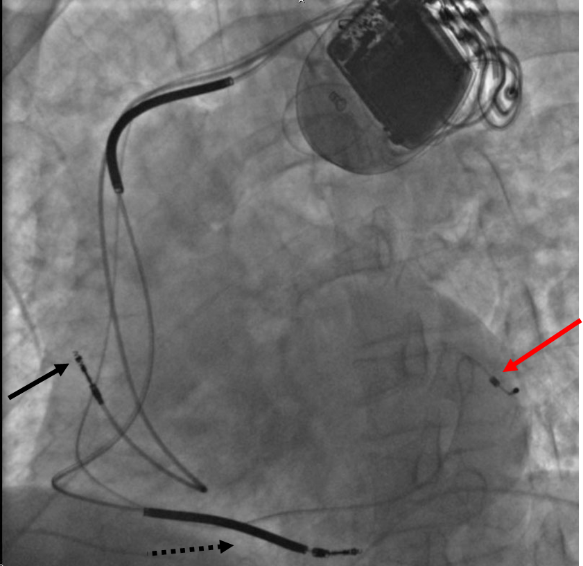

Choice of CRT with or without a defibrillator. Patients who meet both CRT and ICD criteria receive CRT with a defibrillator (CRT-D). CRT with pacing only (CRT-P) is chosen for older patients, those with major comorbidity or limited life expectancy, and many patients with nonischemic cardiomyopathy after shared decision-making, an approach supported by the DANISH results and by CARE-HF, which showed a mortality benefit with CRT-P alone.
Response and nonresponse. About one-third of patients do not improve after CRT. Response is more likely with LBBB, QRS 150 ms or more, female sex, nonischemic cause, smaller ventricles, and absence of extensive scar at the lead site. The left ventricular lead is placed in a lateral or posterolateral vein, away from the apex and from scar. Effective therapy requires biventricular pacing in at least 98% of beats; atrial fibrillation, premature ventricular complexes, and long programmed atrioventricular delays reduce it, and catheter ablation of the atrioventricular node or of premature ventricular complexes restores it. Electrocardiographic confirmation of biventricular or conduction system capture is required at implantation and follow-up.
Conduction system pacing. His bundle pacing and left bundle branch area pacing activate the ventricles through the His-Purkinje system and can correct left bundle branch block. Left bundle branch area pacing has lower and more stable thresholds than His bundle pacing and is widely used. Randomized comparisons with biventricular CRT are small. The 2023 HRS guideline recommends biventricular CRT as the standard, conduction system pacing when biventricular pacing fails (Class 2a), and conduction system pacing as an alternative in selected patients (Class 2b).
Transcatheter repair of secondary mitral regurgitation
Secondary (functional) mitral regurgitation in HFrEF arises from a remodeled ventricle with apical and lateral displacement of the papillary muscles, leaflet tethering, annular dilation, and dyssynchrony; the leaflets themselves are normal. Its severity falls with GDMT and CRT, so the 2020 ACC/AHA valve guideline requires optimization of GDMT, including CRT when indicated, before secondary mitral regurgitation is graded for intervention. Severe secondary mitral regurgitation in the 2020 guideline is defined by an effective regurgitant orifice area of 0.40 cm² or more and a regurgitant volume of 60 mL or more, the same thresholds as primary disease.
| Trial | Population | Result | Interpretation |
|---|---|---|---|
| MITRA-FR (2018) | 304 patients; LVEF 15%–40%; severe secondary MR by ERO >20 mm² or RVol >30 mL; larger ventricles; background therapy adjusted at investigator discretion | Death or unplanned HF hospitalization at 12 months 54.6% vs 51.3%; no difference | Regurgitation proportionate to ventricular dilation; the ventricle drives outcome |
| COAPT (2018) | 614 patients; LVEF 20%–50%; LV end-systolic diameter ≤70 mm; moderate-to-severe or severe MR; symptomatic despite maximally tolerated GDMT confirmed by a central committee | HF hospitalization 35.8% vs 67.9% per patient-year (HR 0.53); all-cause death at 24 months 29.1% vs 46.1% | Regurgitation disproportionate to ventricular size; correcting the valve changes outcome |
| RESHAPE-HF2 (2024) | 505 patients; moderate to severe secondary MR; symptomatic on GDMT | HF hospitalization or cardiovascular death 37.0 vs 58.9 events per 100 patient-years (rate ratio 0.64); improved health status | Benefit extends to less than severe regurgitation in selected patients |
Randomized trials of mitral transcatheter edge-to-edge repair (TEER) in secondary mitral regurgitation. MR, mitral regurgitation; LV, left ventricular; ERO, effective regurgitant orifice area; RVol, regurgitant volume.
In patients with chronic severe secondary mitral regurgitation, LVEF 20% to 50%, left ventricular end-systolic diameter 70 mm or less, and pulmonary artery systolic pressure 70 mm Hg or less, who remain NYHA II–IV despite optimal GDMT, TEER is reasonable (Class 2a in the 2020 ACC/AHA valve guideline). The concept of proportionate and disproportionate regurgitation reconciles MITRA-FR and COAPT in part. The trials also differed in the rigor of background therapy, the definition of severity, and procedural results, and no single ratio selects patients. A heart team including a heart failure specialist makes the decision. Mitral valve surgery is reasonable in patients with severe secondary mitral regurgitation undergoing coronary bypass surgery, and chordal-sparing replacement may be chosen over downsized annuloplasty in severe ischemic mitral regurgitation. Detailed grading is on the mitral regurgitation page.
Coronary revascularization in ischemic cardiomyopathy
In patients with HFrEF, LVEF 35% or less, and coronary disease suitable for surgery, coronary artery bypass grafting is recommended to improve symptoms, cardiovascular hospitalization, and long-term all-cause mortality (Class 1 in the 2022 guideline). The recommendation rests on STICH and its extension STICHES: at a median of 9.8 years, all-cause death occurred in 58.9% of the surgical group and 66.1% of the medical group (HR 0.84), a benefit that emerged only after several years and came at the price of early surgical mortality. Viability testing did not identify which patients benefited.
Percutaneous revascularization did not improve outcomes in REVIVED-BCIS2. In 700 patients with LVEF 35% or less, extensive coronary disease suitable for percutaneous coronary intervention (PCI), and demonstrated viability, death or heart failure hospitalization occurred in 37.2% after PCI and 38.0% with optimal medical therapy (HR 0.99), with no difference in LVEF at 6 or 12 months. PCI in ischemic cardiomyopathy is therefore performed for angina or acute coronary syndromes, not to improve ventricular function or survival. Revascularization decisions in diabetes and multivessel disease involve the heart team and the patient's surgical risk.
Remote hemodynamic monitoring
Rising filling pressures precede symptoms and weight gain by days to weeks. Implantable pulmonary artery pressure sensors allow diuretics and vasodilators to be adjusted before congestion becomes clinical. In CHAMPION, sensor-guided management in NYHA III patients with a heart failure hospitalization in the prior year reduced heart failure hospitalization by 28% at 6 months, regardless of ejection fraction. GUIDE-HF did not meet its primary end point overall, although a prespecified analysis before the COVID-19 pandemic suggested fewer heart failure events. In selected patients with NYHA III symptoms despite GDMT and a heart failure hospitalization within the past year, or elevated natriuretic peptides, remote pulmonary artery pressure monitoring may be considered to reduce hospitalization (Class 2b). Device-based multiparameter algorithms (thoracic impedance, heart sounds, activity, and heart rate variability) from ICDs and CRT devices predict decompensation, and remote device monitoring is standard care. Telemonitoring of weight and symptoms alone has not reduced hospitalization consistently.
Acute decompensated heart failure: hemodynamic profiles and initial treatment
Acute decompensated heart failure is new or worsening heart failure requiring urgent therapy, most often hospitalization. Every admission requires two assessments: the hemodynamic profile and the precipitant. Common precipitants are nonadherence to drugs or diet, atrial fibrillation or other arrhythmia, acute coronary syndrome, uncontrolled hypertension, infection, worsening renal function, NSAIDs and other harmful drugs, pulmonary embolism, thyroid disease, anemia, and progression of the underlying disease.
| Profile | Congestion | Perfusion | Typical findings | Initial treatment |
|---|---|---|---|---|
| A: warm and dry | Absent | Adequate | Compensated | Oral GDMT optimized; another cause of symptoms sought |
| B: warm and wet | Present | Adequate | Orthopnea, elevated jugular venous pressure, edema, rales; warm extremities | Intravenous loop diuretic; vasodilator when hypertensive; GDMT continued |
| C: cold and wet | Present | Reduced | Congestion with cool extremities, narrow pulse pressure, oliguria, confusion, rising lactate | Diuretic with inotropic support when hypoperfusion is present; vasodilator if blood pressure allows; evaluation for cardiogenic shock and mechanical support |
| L: cold and dry | Absent | Reduced | Low output without congestion; often overdiuresed or advanced disease | Assessment of filling pressures; cautious volume when underfilled; inotrope when output is low despite adequate filling |
Bedside hemodynamic profiles. Profile B accounts for most admissions; profile C carries the highest mortality. Treatment decisions use blood pressure, end-organ function, and, when uncertain, invasive hemodynamics.
Diuretics. Patients admitted with congestion receive intravenous loop diuretics promptly (Class 1). For patients taking a loop diuretic before admission, the initial intravenous dose equals or exceeds the total daily oral dose and is titrated by response. In DOSE, bolus and continuous infusion produced similar symptom relief and renal function. The high-dose strategy (2.5 times the oral dose) produced greater weight and fluid loss, a nonsignificant trend toward better symptom relief, and transient creatinine rises that did not affect 60-day outcomes. Diuretic response is assessed within hours by urine output or spot urine sodium, and the dose is doubled when response is inadequate. Decongestion is the goal of hospitalization; residual congestion at discharge predicts readmission and death, and modest creatinine rises during effective decongestion do not justify stopping diuresis.
Vasodilators and inotropes. Intravenous nitroglycerin or nitroprusside may be considered as an adjunct to diuretics for dyspnea in patients without hypotension (Class 2b). Inotropes are reserved for low output with hypoperfusion, and routine inotrope use in patients without hypoperfusion is harmful. Dobutamine is a beta-1 agonist whose effect is blunted by beta blockade; milrinone is a phosphodiesterase-3 inhibitor that acts downstream of the beta receptor, causes more vasodilation and hypotension, accumulates in renal failure, and is favored when pulmonary vascular resistance is high and blood pressure permits. Neither has been shown to improve survival. Norepinephrine is the preferred vasopressor when hypotension persists. Cardiogenic shock, temporary mechanical circulatory support, and shock-team management are covered on the cardiogenic shock page.
Other inpatient care. Venous thromboembolism prophylaxis is recommended (Class 1). Noninvasive positive-pressure ventilation is used for respiratory distress with pulmonary edema. Hyponatremia is a marker of severity; fluid restriction and vasopressin antagonists may be used for symptomatic or severe hyponatremia, with tolvaptan limited to 30 days and avoided in liver disease. Invasive hemodynamic monitoring is reasonable when volume status, perfusion, or vascular resistance is uncertain, or when symptoms persist despite empiric therapy.
In-hospital GDMT, discharge, and the vulnerable post-discharge period
GDMT is continued during hospitalization unless the patient is hemodynamically unstable or has a contraindication (Class 1). Discontinuation of beta blockers during admission is associated with higher mortality; the dose is reduced or held only for cardiogenic shock, marked hypoperfusion, symptomatic bradycardia, or high-grade block. In patients with HFrEF, GDMT is started during hospitalization after clinical stability is achieved (Class 1), because drugs not started before discharge are often never started. Sacubitril/valsartan (PIONEER-HF) and SGLT2 inhibitors (EMPULSE) are safe to start in stabilized hospitalized patients. In EMPULSE, empagliflozin started at a median of 3 days after admission improved a hierarchical composite of death, heart failure events, and symptoms.
Discharge planning includes confirmation of euvolemia, an oral diuretic dose tested for at least 24 hours, medication reconciliation, a documented plan for titration, education in self-care, and an early follow-up visit. An early follow-up visit, generally within 7 days of discharge, is reasonable (Class 2a), and the STRONG-HF strategy of laboratory and clinical review at 1, 2, 3, and 6 weeks shows what rapid titration requires. Referral to a heart failure program or disease management program at discharge is recommended for patients at high risk of readmission.
Readmission within 30 days is the basis of the Centers for Medicare & Medicaid Services Hospital Readmissions Reduction Program, and heart failure performance measures from the ACC/AHA include prescription of each GDMT class at discharge, ICD counseling, and timely follow-up. Readmission rates are influenced by social risk, access to care, and medication cost as well as clinical care.
Heart failure with improved ejection fraction: continue or withdraw therapy
Heart failure with improved ejection fraction (HFimpEF) describes a patient with a previous LVEF of 40% or less whose follow-up LVEF is above 40%. Improvement occurs in a large minority of patients receiving modern GDMT and is more common in women, younger patients, nonischemic disease, shorter duration of disease, absence of CMR scar, and reversible causes such as tachycardia, alcohol, and peripartum cardiomyopathy. Patients with HFimpEF have fewer events than patients with persistent HFrEF but more than the general population, and the underlying myocardial disease usually persists at the molecular and structural level: abnormalities of strain, volumes, and biomarkers often remain.
In patients with HFimpEF after treatment, GDMT is continued to prevent relapse of heart failure and left ventricular dysfunction, even in patients who become asymptomatic (Class 1). The recommendation rests on TRED-HF, in which 51 patients with recovered dilated cardiomyopathy (LVEF from below 40% to 50% or more, normal left ventricular end-diastolic volume, normal NT-proBNP, and no symptoms) were randomized to phased withdrawal of therapy or continuation. Within 6 months, 44% of patients in the withdrawal group relapsed, defined by a fall in LVEF, a rise in end-diastolic volume, a doubling of NT-proBNP, or clinical heart failure, compared with no relapses in the continuation group. The 2026 universal definition describes remission and recovery as possible states, but no trial has identified patients in whom withdrawal of therapy is safe.
A defibrillator implanted for primary prevention is usually left in place. Replacement of the generator is individualized in a patient whose LVEF has risen above 35% and who has never had appropriate therapy. A patient whose LVEF improved with CRT continues biventricular pacing at device replacement (2023 HRS guideline). Diuretic doses can often be reduced. Surveillance echocardiography is repeated when symptoms change and periodically to detect relapse.
Pregnancy and peripartum cardiomyopathy
Peripartum cardiomyopathy is heart failure with LVEF below 45%, usually with a dilated ventricle, presenting toward the end of pregnancy or in the months after delivery, without another identifiable cause. Risk factors include Black race, older maternal age, multiparity, multiple gestation, preeclampsia, and hypertension. A cleaved 16-kDa fragment of prolactin with antiangiogenic effects is implicated, and truncating TTN variants are overrepresented. About half of patients recover LVEF, usually within 6 to 12 months; recovery is less likely when LVEF is below 30% at diagnosis or the ventricle is markedly dilated.
During pregnancy, ACE inhibitors, ARBs, ARNIs, MRAs, SGLT2 inhibitors, and ivabradine are avoided. Beta blockers (metoprolol succinate preferred), hydralazine with nitrates, loop diuretics for congestion, and digoxin are used. After delivery, standard GDMT is given; enalapril and captopril are compatible with breastfeeding, and SGLT2 inhibitors and ARNIs lack lactation data. Anticoagulation is used when LVEF is very low because thrombus risk is high in the peripartum state. Bromocriptine, which suppresses prolactin, is used with anticoagulation in European practice in selected patients; it is not approved for this indication in the United States and is used there mainly in research or specialist settings. A wearable defibrillator may bridge the period of potential recovery.
A subsequent pregnancy carries a risk of relapse and death that is highest when LVEF has not normalized. Women with persistent LVEF below 50% are counseled against a further pregnancy, and contraception is part of the discharge plan. Women with HFrEF from other causes who are considering pregnancy receive preconception counseling, and pregnancy is managed by a cardio-obstetric team.
Recognizing advanced heart failure and when to refer
Advanced (stage D) heart failure is persistent severe symptoms despite optimized GDMT. Its recognition matters because the therapies that change its course, heart transplantation and durable left ventricular assist devices (LVADs), require adequate renal, hepatic, and pulmonary function and reversible pulmonary vascular resistance. Late referral is the common failure, and progressive intolerance of GDMT, often attributed to drug side effects, is one of its earliest markers.
| Letter | Trigger for referral |
|---|---|
| I | Intravenous inotropes required |
| N | NYHA class IIIB to IV, or persistently elevated natriuretic peptides |
| E | End-organ dysfunction: worsening renal or hepatic function |
| E | Ejection fraction ≤35% |
| D | Defibrillator shocks, recurrent appropriate therapy |
| H | Hospitalizations, more than one in the past 12 months |
| E | Edema despite escalating diuretics |
| L | Low blood pressure (systolic ≤90 mm Hg), high heart rate |
| P | Prognostic medication: progressive intolerance or down-titration of GDMT |
The "I NEED HELP" mnemonic for timely referral to an advanced heart failure center.
The 2022 guideline recommends referral to a heart failure specialty team for patients with advanced heart failure when consistent with the patient's goals (Class 1). Other markers include diuretic needs above about 160 mg/day of furosemide or need for metolazone, persistent hyponatremia below 134 mEq/L, cardiac cachexia, and a peak oxygen consumption below 14 mL/kg/min (or 12 mL/kg/min with beta blockade) or below 50% of predicted on cardiopulmonary exercise testing. The Interagency Registry for Mechanically Assisted Circulatory Support (INTERMACS) profiles stratify patients considered for durable support from profile 1 (critical cardiogenic shock) to profile 7 (advanced NYHA III).
Durable LVAD implantation, as a bridge to transplantation or as destination therapy, improves functional status, quality of life, and survival in selected patients with advanced HFrEF and NYHA IV symptoms who depend on intravenous inotropes or temporary mechanical support (Class 1). It is reasonable in selected NYHA IV patients despite GDMT (Class 2a). Transplantation is indicated in selected patients to improve survival and quality of life (Class 1). Palliative care, including symptom management and goals-of-care discussions, is recommended throughout the course of advanced heart failure (Class 1), and continuous intravenous inotropic support is reasonable as palliative therapy in patients who are not candidates for advanced therapies (Class 2b). Device deactivation, including defibrillator therapy, is discussed as goals change.
Prognosis and risk models
Validated multivariable risk scores estimate mortality and inform decisions about devices and advanced therapy (Class 2a for their use in ambulatory or hospitalized patients). The Seattle Heart Failure Model uses age, sex, NYHA class, weight, LVEF, systolic pressure, ischemic cause, medications, devices, and laboratory values and estimates the effect of adding therapies. The Meta-Analysis Global Group in Chronic Heart Failure (MAGGIC) score uses 13 readily available variables and was derived in more than 39,000 patients. In hospitalized patients, the ADHERE risk tree (blood urea nitrogen, systolic pressure, creatinine) and the Get With The Guidelines-Heart Failure score estimate in-hospital mortality.
Markers of worse prognosis include NYHA III–IV symptoms, low peak oxygen consumption and steep ventilatory efficiency slope, high and rising natriuretic peptide and troponin, hyponatremia, worsening renal function, anemia, low systolic pressure, wide QRS, right ventricular dysfunction, pulmonary hypertension, frailty, cachexia, and repeated hospitalization. Risk models underestimate risk in patients with advanced disease and overestimate it in patients receiving contemporary four-drug therapy, and they are used together with clinical judgment.
Cost, access, and adherence
Out-of-pocket cost limits use of sacubitril/valsartan, SGLT2 inhibitors, vericiguat, and ivabradine in the United States. The 2022 guideline identifies out-of-pocket cost as a barrier to GDMT and considers screening for social determinants of health reasonable (Class 2a). Generic formulations as they reach the market, manufacturer assistance programs, Medicare Part D coverage, and 90-day prescriptions reduce cost and improve adherence. When an ARNI is unaffordable, an ACE inhibitor or ARB at target dose is used; when an SGLT2 inhibitor is unaffordable, the other three classes remain fully effective.
Adherence improves with once-daily regimens, pill organizers, combination of tasks with clinic visits, pharmacist-led reconciliation, and simplification of nonprognostic drugs. Disparities in prescription of GDMT, device implantation, and advanced therapies by race, sex, and income are documented, and quality programs track prescription rates as performance measures.
How European and Canadian guidance differs
The 2021 ESC heart failure guideline uses the same ejection-fraction categories. It recommends an ACE inhibitor or ARNI as initial renin-angiotensin therapy (with ARNI recommended as a replacement for an ACE inhibitor). The 2023 ESC focused update recommends an intensive strategy of early initiation and rapid titration with close follow-up after a heart failure hospitalization (Class 1, based on STRONG-HF) and intravenous iron to reduce heart failure hospitalization in symptomatic HFrEF and HFmrEF with iron deficiency (Class 2a). The ESC gives a primary-prevention ICD in nonischemic dilated cardiomyopathy a Class 2a recommendation, where the US guideline gives Class 1. The 2021 Canadian Cardiovascular Society and Canadian Heart Failure Society update recommends combination therapy with one drug from each of the four classes, started as early as possible, with an ARNI preferred over an ACE inhibitor or ARB. It endorses rapid titration over sequential addition of each class after the previous one reaches target.
Sources of error in the management of HFrEF
- Titration of one drug to target before the next class is started: patients remain without proven therapy during the months of highest risk.
- Withholding GDMT for asymptomatic systolic pressure in the 90s or for a creatinine rise of less than 30%: both are expected effects, and trial patients with them gained the same benefit.
- Prescription of metoprolol tartrate, atenolol, or nebivolol as the heart failure beta blocker: mortality benefit is established only for bisoprolol, carvedilol, and metoprolol succinate.
- Interpretation of BNP after sacubitril/valsartan as if neprilysin were not inhibited: BNP can rise as filling pressures fall; NT-proBNP is the marker used during ARNI therapy.
- Implantation of a primary-prevention ICD at the time of diagnosis: LVEF is remeasured after at least 3 months of optimized therapy, 40 days after myocardial infarction, and 90 days after revascularization.
- Selection of CRT candidates by echocardiographic dyssynchrony: QRS morphology and duration select patients, and dyssynchrony indices do not predict response.
- Withdrawal of GDMT after LVEF recovers: relapse occurred in 44% within 6 months in TRED-HF.
- Prescription of ivabradine in atrial fibrillation or before the beta blocker is titrated: it acts only on the sinus node and does not replace beta blockade.
- Grading of secondary mitral regurgitation for TEER before GDMT and CRT are optimized: regurgitation often falls with medical therapy and resynchronization.
- Referral for advanced therapies only after continuous inotrope dependence: end-organ dysfunction may by then exclude transplantation or LVAD.
Self-assessment
A 64-year-old man with nonischemic dilated cardiomyopathy has taken sacubitril/valsartan 97/103 mg twice daily, metoprolol succinate 200 mg daily, spironolactone 25 mg daily, and dapagliflozin 10 mg daily for 4 months. He is NYHA class II, in sinus rhythm at 64/min, with LVEF 30% on repeat echocardiography and left bundle branch block with a QRS duration of 162 ms. Which device is indicated, and what is the class of recommendation for each component?
Cardiovascular Medicine Board ReviewReferences
- Heidenreich PA, Bozkurt B, Aguilar D, et al. 2022 AHA/ACC/HFSA guideline for the management of heart failure: a report of the American College of Cardiology/American Heart Association Joint Committee on Clinical Practice Guidelines. Circulation. 2022;145:e895–e1032. doi:10.1161/CIR.0000000000001063
- Maddox TM, Januzzi JL Jr, Allen LA, et al. 2024 ACC expert consensus decision pathway for treatment of heart failure with reduced ejection fraction: a report of the American College of Cardiology Solution Set Oversight Committee. J Am Coll Cardiol. 2024;83:1444–1488. doi:10.1016/j.jacc.2023.12.024
- Bozkurt B, Coats AJS, Tsutsui H, et al. Universal definition and classification of heart failure: a report of the Heart Failure Society of America, Heart Failure Association of the European Society of Cardiology, Japanese Heart Failure Society and Writing Committee of the Universal Definition of Heart Failure. J Card Fail. 2021;27:387–413. doi:10.1016/j.cardfail.2021.01.022
- Walsh MN, Køber L, Sliwa K, et al. AHA/ACC/ESC/WHF expert consensus document: second universal definition of heart failure (2026). J Am Coll Cardiol. 2026;88:1797–1812. doi:10.1016/j.jacc.2026.05.036
- Martin SS, Aday AW, Allen NB, et al. 2025 heart disease and stroke statistics: a report of US and global data from the American Heart Association. Circulation. 2025;151:e41–e660. doi:10.1161/CIR.0000000000001303
- McDonagh TA, Metra M, Adamo M, et al. 2021 ESC guidelines for the diagnosis and treatment of acute and chronic heart failure. Eur Heart J. 2021;42:3599–3726. doi:10.1093/eurheartj/ehab368
- McDonagh TA, Metra M, Adamo M, et al. 2023 focused update of the 2021 ESC guidelines for the diagnosis and treatment of acute and chronic heart failure. Eur Heart J. 2023;44:3627–3639. doi:10.1093/eurheartj/ehad195
- McDonald M, Virani S, Chan M, et al. CCS/CHFS heart failure guidelines update: defining a new pharmacologic standard of care for heart failure with reduced ejection fraction. Can J Cardiol. 2021;37:531–546. doi:10.1016/j.cjca.2021.01.017
- Januzzi JL Jr, Chen-Tournoux AA, Christenson RH, et al. N-terminal pro-B-type natriuretic peptide in the emergency department: the ICON-RELOADED study. J Am Coll Cardiol. 2018;71:1191–1200. doi:10.1016/j.jacc.2018.01.021
- SOLVD Investigators; Yusuf S, Pitt B, et al. Effect of enalapril on survival in patients with reduced left ventricular ejection fractions and congestive heart failure. N Engl J Med. 1991;325:293–302. doi:10.1056/NEJM199108013250501
- McMurray JJV, Packer M, Desai AS, et al. Angiotensin-neprilysin inhibition versus enalapril in heart failure (PARADIGM-HF). N Engl J Med. 2014;371:993–1004. doi:10.1056/NEJMoa1409077
- CIBIS-II Investigators and Committees. The Cardiac Insufficiency Bisoprolol Study II (CIBIS-II): a randomised trial. Lancet. 1999;353:9–13. doi:10.1016/S0140-6736(98)11181-9
- MERIT-HF Study Group. Effect of metoprolol CR/XL in chronic heart failure: Metoprolol CR/XL Randomised Intervention Trial in Congestive Heart Failure (MERIT-HF). Lancet. 1999;353:2001–2007. doi:10.1016/S0140-6736(99)04440-2
- Packer M, Coats AJS, Fowler MB, et al. Effect of carvedilol on survival in severe chronic heart failure (COPERNICUS). N Engl J Med. 2001;344:1651–1658. doi:10.1056/NEJM200105313442201
- Pitt B, Zannad F, Remme WJ, et al. The effect of spironolactone on morbidity and mortality in patients with severe heart failure (RALES). N Engl J Med. 1999;341:709–717. doi:10.1056/NEJM199909023411001
- Zannad F, McMurray JJV, Krum H, et al. Eplerenone in patients with systolic heart failure and mild symptoms (EMPHASIS-HF). N Engl J Med. 2011;364:11–21. doi:10.1056/NEJMoa1009492
- McMurray JJV, Solomon SD, Inzucchi SE, et al. Dapagliflozin in patients with heart failure and reduced ejection fraction (DAPA-HF). N Engl J Med. 2019;381:1995–2008. doi:10.1056/NEJMoa1911303
- Packer M, Anker SD, Butler J, et al. Cardiovascular and renal outcomes with empagliflozin in heart failure (EMPEROR-Reduced). N Engl J Med. 2020;383:1413–1424. doi:10.1056/NEJMoa2022190
- Mebazaa A, Davison B, Chioncel O, et al. Safety, tolerability and efficacy of up-titration of guideline-directed medical therapies for acute heart failure (STRONG-HF): a multinational, open-label, randomised trial. Lancet. 2022;400:1938–1952. doi:10.1016/S0140-6736(22)02076-1
- Swedberg K, Komajda M, Böhm M, et al. Ivabradine and outcomes in chronic heart failure (SHIFT): a randomised placebo-controlled study. Lancet. 2010;376:875–885. doi:10.1016/S0140-6736(10)61198-1
- Armstrong PW, Pieske B, Anstrom KJ, et al. Vericiguat in patients with heart failure and reduced ejection fraction (VICTORIA). N Engl J Med. 2020;382:1883–1893. doi:10.1056/NEJMoa1915928
- Taylor AL, Ziesche S, Yancy C, et al. Combination of isosorbide dinitrate and hydralazine in blacks with heart failure (A-HeFT). N Engl J Med. 2004;351:2049–2057. doi:10.1056/NEJMoa042934
- Digitalis Investigation Group. The effect of digoxin on mortality and morbidity in patients with heart failure. N Engl J Med. 1997;336:525–533. doi:10.1056/NEJM199702203360801
- Kalra PR, Cleland JGF, Petrie MC, et al. Intravenous ferric derisomaltose in patients with heart failure and iron deficiency in the UK (IRONMAN): an investigator-initiated, prospective, randomised, open-label, blinded-endpoint trial. Lancet. 2022;400:2199–2209. doi:10.1016/S0140-6736(22)02083-9
- Anker SD, Friede T, Butler J, et al. Intravenous ferric carboxymaltose in heart failure with iron deficiency: the FAIR-HF2 DZHK05 randomized clinical trial. JAMA. 2025;333:1965–1976. doi:10.1001/jama.2025.3833
- Felker GM, Lee KL, Bull DA, et al. Diuretic strategies in patients with acute decompensated heart failure (DOSE). N Engl J Med. 2011;364:797–805. doi:10.1056/NEJMoa1005419
- Mullens W, Dauw J, Martens P, et al. Acetazolamide in acute decompensated heart failure with volume overload (ADVOR). N Engl J Med. 2022;387:1185–1195. doi:10.1056/NEJMoa2203094
- Mentz RJ, Anstrom KJ, Eisenstein EL, et al. Effect of torsemide vs furosemide after discharge on all-cause mortality in patients hospitalized with heart failure: the TRANSFORM-HF randomized clinical trial. JAMA. 2023;329:214–223. doi:10.1001/jama.2022.23924
- Al-Khatib SM, Stevenson WG, Ackerman MJ, et al. 2017 AHA/ACC/HRS guideline for management of patients with ventricular arrhythmias and the prevention of sudden cardiac death. Circulation. 2018;138:e272–e391. doi:10.1161/CIR.0000000000000549
- Bardy GH, Lee KL, Mark DB, et al. Amiodarone or an implantable cardioverter-defibrillator for congestive heart failure (SCD-HeFT). N Engl J Med. 2005;352:225–237. doi:10.1056/NEJMoa043399
- Køber L, Thune JJ, Nielsen JC, et al. Defibrillator implantation in patients with nonischemic systolic heart failure (DANISH). N Engl J Med. 2016;375:1221–1230. doi:10.1056/NEJMoa1608029
- Olgin JE, Pletcher MJ, Vittinghoff E, et al. Wearable cardioverter-defibrillator after myocardial infarction (VEST). N Engl J Med. 2018;379:1205–1215. doi:10.1056/NEJMoa1800781
- Chung MK, Patton KK, Lau CP, et al. 2023 HRS/APHRS/LAHRS guideline on cardiac physiologic pacing for the avoidance and mitigation of heart failure. Heart Rhythm. 2023;20:e17–e91. doi:10.1016/j.hrthm.2023.03.1538
- Cleland JGF, Daubert JC, Erdmann E, et al. The effect of cardiac resynchronization on morbidity and mortality in heart failure (CARE-HF). N Engl J Med. 2005;352:1539–1549. doi:10.1056/NEJMoa050496
- Moss AJ, Hall WJ, Cannom DS, et al. Cardiac-resynchronization therapy for the prevention of heart-failure events (MADIT-CRT). N Engl J Med. 2009;361:1329–1338. doi:10.1056/NEJMoa0906431
- Otto CM, Nishimura RA, Bonow RO, et al. 2020 ACC/AHA guideline for the management of patients with valvular heart disease: a report of the American College of Cardiology/American Heart Association Joint Committee on Clinical Practice Guidelines. Circulation. 2021;143:e72–e227. doi:10.1161/CIR.0000000000000923
- Stone GW, Lindenfeld J, Abraham WT, et al. Transcatheter mitral-valve repair in patients with heart failure (COAPT). N Engl J Med. 2018;379:2307–2318. doi:10.1056/NEJMoa1806640
- Obadia JF, Messika-Zeitoun D, Leurent G, et al. Percutaneous repair or medical treatment for secondary mitral regurgitation (MITRA-FR). N Engl J Med. 2018;379:2297–2306. doi:10.1056/NEJMoa1805374
- Anker SD, Friede T, von Bardeleben RS, et al. Transcatheter valve repair in heart failure with moderate to severe mitral regurgitation (RESHAPE-HF2). N Engl J Med. 2024;391:1799–1809. doi:10.1056/NEJMoa2314328
- Velazquez EJ, Lee KL, Jones RH, et al. Coronary-artery bypass surgery in patients with ischemic cardiomyopathy (STICHES). N Engl J Med. 2016;374:1511–1520. doi:10.1056/NEJMoa1602001
- Perera D, Clayton T, O'Kane PD, et al. Percutaneous revascularization for ischemic left ventricular dysfunction (REVIVED-BCIS2). N Engl J Med. 2022;387:1351–1360. doi:10.1056/NEJMoa2206606
- Halliday BP, Wassall R, Lota AS, et al. Withdrawal of pharmacological treatment for heart failure in patients with recovered dilated cardiomyopathy (TRED-HF): an open-label, pilot, randomised trial. Lancet. 2019;393:61–73. doi:10.1016/S0140-6736(18)32484-X
- Marrouche NF, Brachmann J, Andresen D, et al. Catheter ablation for atrial fibrillation with heart failure (CASTLE-AF). N Engl J Med. 2018;378:417–427. doi:10.1056/NEJMoa1707855
- Joglar JA, Chung MK, Armbruster AL, et al. 2023 ACC/AHA/ACCP/HRS guideline for the diagnosis and management of atrial fibrillation. J Am Coll Cardiol. 2024;83:109–279. doi:10.1016/j.jacc.2023.08.017
- Crespo-Leiro MG, Metra M, Lund LH, et al. Advanced heart failure: a position statement of the Heart Failure Association of the European Society of Cardiology. Eur J Heart Fail. 2018;20:1505–1535. doi:10.1002/ejhf.1236
- Palaniappan LP, Allen NB, Almarzooq ZI, et al. 2026 heart disease and stroke statistics: a report of US and global data from the American Heart Association. Circulation. 2026;153:e275–e906. doi:10.1161/CIR.0000000000001412
- Shah KS, Xu H, Matsouaka RA, et al. Heart failure with preserved, borderline, and reduced ejection fraction: 5-year outcomes. J Am Coll Cardiol. 2017;70:2476–2486. doi:10.1016/j.jacc.2017.08.074
- Jordan E, Kinnamon DD, Haas GJ, et al. Genetic architecture of dilated cardiomyopathy in individuals of African and European ancestry. JAMA. 2023;330:432–441. doi:10.1001/jama.2023.11970
- Baumwol J. "I Need Help"—a mnemonic to aid timely referral in advanced heart failure. J Heart Lung Transplant. 2017;36:593–594. doi:10.1016/j.healun.2017.02.010