Library › Valvular heart disease › Aortic stenosis
Aortic stenosis
Aortic stenosis in adults from cause and natural history through multimodality grading, timing and choice of valve replacement, special populations, and surveillance after intervention.
Severe aortic stenosis is defined by a peak aortic velocity of 4.0 m/s or more or a mean gradient of 40 mm Hg or more, with a valve area usually 1.0 cm² or less. Symptomatic severe aortic stenosis is a Class 1 indication for aortic valve replacement. When the valve area is in the severe range but velocity and gradient are not, transvalvular flow, ejection fraction, blood pressure, and valve calcium decide the grade.
Definition and ACC/AHA stages of aortic stenosis
Aortic stenosis is fixed obstruction to left ventricular outflow at the level of the aortic valve. Obstruction below the valve (a discrete subaortic membrane, a fibromuscular tunnel, or dynamic septal obstruction) or above it (supravalvular stenosis) is a different disease with different treatment. The level of obstruction is therefore established before the valve is graded.
The 2020 American College of Cardiology/American Heart Association (ACC/AHA) valve guideline classifies aortic stenosis in four stages that combine valve anatomy, hemodynamics, ventricular consequences, and symptoms. Stage A denotes patients at risk, stage B progressive (mild or moderate) disease, stage C asymptomatic severe disease, and stage D symptomatic severe disease. Stage D is divided by flow and ejection fraction because low-flow states lower the gradient across a severely stenotic valve. Valve area is supportive in high-gradient disease and is not required to define it.
| Stage | Definition | Valve hemodynamics | Ventricle and symptoms |
|---|---|---|---|
| A | At risk: bicuspid or other congenital valve anomaly, or aortic sclerosis | Vmax <2.0 m/s | None |
| B | Progressive: mild to moderate calcification or fibrosis of a bicuspid or trileaflet valve, or rheumatic commissural fusion | Mild: Vmax 2.0–2.9 m/s or mean gradient <20 mm Hg. Moderate: Vmax 3.0–3.9 m/s or mean gradient 20–39 mm Hg | Early diastolic dysfunction possible; normal LVEF; no symptoms |
| C1 | Asymptomatic severe | Vmax ≥4.0 m/s or mean gradient ≥40 mm Hg | Normal LVEF; mild hypertrophy; exercise testing reasonable to confirm symptom status |
| C2 | Asymptomatic severe with left ventricular systolic dysfunction | Vmax ≥4.0 m/s or mean gradient ≥40 mm Hg | LVEF <50% |
| D1 | Symptomatic severe high-gradient | Vmax ≥4.0 m/s or mean gradient ≥40 mm Hg; AVA typically ≤1.0 cm², larger with mixed stenosis and regurgitation | Exertional dyspnea or reduced exercise tolerance, angina, syncope or presyncope, heart failure |
| D2 | Symptomatic severe low-flow, low-gradient with reduced LVEF | AVA ≤1.0 cm² with resting Vmax <4.0 m/s or mean gradient <40 mm Hg; dobutamine stress shows AVA ≤1.0 cm² with Vmax ≥4.0 m/s at any flow rate | LVEF <50% |
| D3 | Symptomatic severe low-gradient with normal LVEF (paradoxical low flow) | AVA ≤1.0 cm² (indexed ≤0.6 cm²/m²) with Vmax <4.0 m/s or mean gradient <40 mm Hg, stroke volume index <35 mL/m², measured with systolic pressure <140 mm Hg | LVEF ≥50%; small, thick-walled ventricle with restrictive filling |
Adapted from the 2020 ACC/AHA valve guideline, Table 13. Very severe aortic stenosis is a Vmax of 5.0 m/s or more or a mean gradient of 60 mm Hg or more. Vmax, peak aortic jet velocity; AVA, aortic valve area; LVEF, left ventricular ejection fraction.
Epidemiology of aortic stenosis
Calcific aortic stenosis is the most common valve lesion requiring intervention in North America and Europe, and its prevalence rises steeply with age. The 2020 ACC/AHA guideline cites a prevalence of 1% to 2% in people older than 65 years and 3% to 8% in those older than 75 years. A pooled analysis of population studies in adults 75 years and older found aortic stenosis of any severity in about 12% and severe aortic stenosis in about 3%, with roughly three-quarters of severe cases symptomatic. In a US population-based echocardiographic study, moderate or severe valve disease of any kind was uncommon before age 65 and reached 13.3% at 75 years and older; mitral regurgitation was the most common lesion, and aortic stenosis was present in about 2.8% of those 75 years and older.
Age at presentation depends on valve anatomy. A congenitally bicuspid valve is present in 1% to 2% of the population, and these patients reach severe stenosis one to two decades earlier than patients with a trileaflet valve. In a large surgical pathology series of adults undergoing isolated aortic valve replacement for stenosis, congenitally malformed (unicuspid or bicuspid) valves predominated in patients younger than 70 years, and trileaflet valves predominated in older patients. Rheumatic aortic stenosis remains common where rheumatic fever is endemic and almost always coexists with rheumatic mitral disease.
Risk factors for calcific disease overlap with those for atherosclerosis: age, male sex, hypertension, smoking, diabetes, chronic kidney disease, elevated low-density lipoprotein cholesterol, and lipoprotein(a). A genome-wide association study identified a variant in the LPA locus associated with aortic valve calcium and incident aortic stenosis, an association mediated by lipoprotein(a) levels and replicated across ethnic groups, which supports a causal role for lipoprotein(a). End-stage kidney disease and disordered calcium-phosphate metabolism accelerate leaflet calcification.
Causes and valve morphology of aortic stenosis
Calcific disease of a trileaflet valve. Calcific aortic stenosis is an active cellular process. Endothelial injury from mechanical stress allows lipid and lipoprotein(a) to enter the leaflet, where oxidized lipids provoke inflammation. Valvular interstitial cells then differentiate toward an osteoblast-like phenotype, and leaflet fibrosis is followed by nodular calcification. Calcium begins at the base of the cusps on the aortic side and spreads toward the free edges. The commissures remain unfused, which distinguishes calcific from rheumatic disease. Aortic sclerosis, the earliest visible stage, is leaflet thickening or calcification without significant obstruction (peak velocity 2.5 m/s or less in the American Society of Echocardiography and European Association of Cardiovascular Imaging [ASE/EACVI] document, below 2.0 m/s in stage A of the ACC/AHA guideline). In a large echocardiographic cohort, about 16% of patients with aortic sclerosis developed aortic stenosis during follow-up, and 2.5% developed severe stenosis.
Bicuspid aortic valve. Fusion of two cusps produces a functional two-leaflet valve, most often with a raphe; fusion of the right and left coronary cusps is the most common pattern. Turbulent flow and abnormal leaflet stress accelerate calcification. A bicuspid valve is a disease of the valve and the aorta: dilation of the aortic sinuses or ascending aorta is common and persists after valve replacement. Systolic doming of the leaflets in the long-axis view and an elliptical ("fish-mouth") orifice in the short-axis view are the echocardiographic hallmarks. Severe calcification can obscure the morphology, and CT or transesophageal echocardiography then defines the number of cusps and the raphe. A unicuspid valve, with one commissure or none, obstructs early and often presents in adolescence or young adulthood.
Rheumatic aortic stenosis. Rheumatic disease fuses the commissures and thickens the leaflet edges, producing a small, triangular central orifice. Isolated rheumatic aortic stenosis without mitral involvement is unusual.
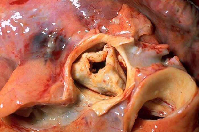

Radiation-associated valve disease. Mediastinal radiation, most often for Hodgkin lymphoma or breast cancer, causes fibrosis and calcification of the aortic and mitral valves that typically appears 10 to 20 years after treatment. The aorto-mitral curtain is often heavily calcified, and coronary disease, conduction disease, pericardial constriction, and a scarred mediastinum frequently coexist. These features raise the risk of sternotomy, and the 2020 ACC/AHA guideline lists prior chest irradiation as a factor favoring transcatheter replacement.
Less common causes and nonvalvular obstruction. Fabry disease, alkaptonuria (ochronosis), systemic lupus erythematosus, severe familial hypercholesterolemia, Paget disease of bone, and end-stage kidney disease can cause valvular stenosis. Supravalvular aortic stenosis is an elastin arteriopathy, characteristic of Williams syndrome, with an hourglass narrowing at the sinotubular junction. Discrete subaortic stenosis is a fibromuscular membrane below the valve whose jet damages the aortic leaflets and often causes regurgitation. Because a membrane is resected and a valve is replaced, pulsed-wave and color Doppler localize the obstruction before the gradient is attributed to the valve.
Congenital aortic stenosis in adolescents and young adults. A noncalcified, pliable congenital valve can be treated with balloon valvotomy, a procedure that has a role in children, adolescents, and young adults but a very limited role in older adults with calcific disease. Restenosis and regurgitation after valvotomy lead to surgical replacement or the Ross procedure in early adulthood, and these patients are followed lifelong in adult congenital heart disease programs.
Pathophysiology: pressure overload, hypertrophy, and myocardial fibrosis
A stenotic valve forces the left ventricle to generate a systolic pressure well above aortic pressure. By the law of Laplace, wall stress rises with pressure and radius and falls with wall thickness. The ventricle adds sarcomeres in parallel, thickening its walls while the cavity stays normal or small, and concentric hypertrophy normalizes systolic wall stress. Compensation preserves the ejection fraction for years but carries costs that explain the symptoms.
The thick ventricle is stiff. Relaxation is slow and compliance falls, so filling requires a higher left atrial pressure, and atrial contraction contributes a large share of preload. Loss of atrial systole with atrial fibrillation can therefore precipitate heart failure in a previously compensated patient.
Coronary flow reserve falls even when the epicardial arteries are normal. Myocardial oxygen demand rises with mass and systolic pressure, while supply is limited by capillary rarefaction, compression of the subendocardium by high intramyocardial pressure, a shortened diastolic perfusion time, and a raised left ventricular diastolic pressure that narrows the coronary perfusion gradient. Repeated subendocardial ischemia leads to myocyte death and replacement fibrosis. Diffuse interstitial fibrosis develops first and can regress after valve replacement. Focal replacement fibrosis, seen as midwall late gadolinium enhancement on cardiac magnetic resonance, does not regress.
Afterload mismatch occurs when hypertrophy can no longer normalize wall stress. The ejection fraction then falls because of excessive afterload, and contractility may still be largely intact. This form of dysfunction improves after valve replacement. When systolic dysfunction is caused by infarction or a primary cardiomyopathy, improvement after replacement is limited.
The ventricle faces the valve and the systemic circulation in series. Hypertension and reduced arterial compliance add a second load. The valvuloarterial impedance (Zva) expresses this global afterload: Zva = (systolic arterial pressure + mean transvalvular gradient) ÷ stroke volume index. In asymptomatic patients with at least moderate aortic stenosis, a Zva of 4.5 mm Hg·mL⁻¹·m² or more was associated with about a 2.8-fold adjusted increase in mortality, and the ASE/EACVI document lists 5 mm Hg·mL⁻¹·m² as the cutoff for a high global load. Zva is a research and adjudication index and is not a guideline trigger for intervention.
Sex modifies the response. Women develop less valve calcium for the same hemodynamic severity, with a larger fibrotic component, which is the basis for lower CT calcium thresholds in women. Women more often have a small, concentric ventricle with low stroke volume and a paradoxical low-flow pattern, whereas men more often show eccentric remodeling and focal fibrosis.
Natural history and prognosis of aortic stenosis
Calcific aortic stenosis progresses in nearly every patient once the peak velocity reaches 2 m/s. The average annual change in a trileaflet calcific valve is an increase in velocity of about 0.3 m/s, an increase in mean gradient of 7 to 8 mm Hg, and a decrease in valve area of 0.1 to 0.15 cm², with wide individual variation. Older age, more severe leaflet calcification, chronic kidney disease, and a rapid rate of change on serial studies predict faster progression. In asymptomatic severe aortic stenosis, the combination of moderate or severe calcification and an increase in velocity of 0.3 m/s or more in 1 year identified patients at high risk of events in a prospective cohort.
The asymptomatic phase of severe aortic stenosis carries a low risk of sudden death, less than 1% per year when patients are followed closely and report symptoms promptly. Symptoms, however, develop in most patients within a few years. In a prospective cohort, survival without valve replacement at 2 years was 84% with a velocity below 3.0 m/s, 66% with a velocity of 3.0 to 4.0 m/s, and 21% with a velocity above 4.0 m/s, and the 2020 ACC/AHA guideline cites an event-free survival of only 30% to 50% at 2 years in severe stenosis. In very severe aortic stenosis, symptoms develop in about 50% of patients within 2 years. In a large asymptomatic cohort, a velocity of 5 m/s or more was associated with a more than 6-fold increase in cardiovascular mortality, and an LVEF below 60% with a more than 4-fold higher risk of cardiovascular death.
After symptoms begin, prognosis without valve replacement is poor. In untreated historical cohorts, average survival after symptom onset was about 5 years with angina, 3 years with syncope, and 2 years with heart failure. These figures predate widespread valve replacement. The principle still holds: symptom onset marks the transition to a high-risk phase, and no medical therapy alters the course. Exertional dyspnea and reduced exercise tolerance, not the classic triad, are now the most common first symptoms.
Complications of untreated severe disease include heart failure, atrial fibrillation (poorly tolerated by the stiff ventricle), sudden death (usually in symptomatic patients), infective endocarditis, and gastrointestinal bleeding from angiodysplasia. The last, Heyde syndrome, results from acquired von Willebrand syndrome: high shear stress across the stenotic valve unfolds large von Willebrand factor multimers and exposes them to cleavage by ADAMTS13. The degree of multimer loss correlates with stenosis severity, and the abnormality corrects after successful valve replacement.
Symptoms and physical examination in aortic stenosis
The classic symptoms are exertional angina, exertional syncope or presyncope, and dyspnea or heart failure. Angina reflects subendocardial ischemia in a hypertrophied ventricle and occurs with or without obstructive coronary disease. Exertional syncope results from a fixed cardiac output during exercise-induced vasodilation, sometimes compounded by a vasodepressor reflex from left ventricular baroreceptors; arrhythmia is a less common cause. Dyspnea reflects elevated filling pressure from diastolic dysfunction and, later, systolic failure.
Patients often reduce their activity without recognizing it, attributing limitation to age. A history that records specific activities stopped, slowed, or delegated (stairs, walking pace, yard work) identifies symptomatic patients who describe themselves as asymptomatic.
The murmur of aortic stenosis is a harsh, crescendo-decrescendo systolic ejection murmur, loudest at the right upper sternal border and radiating to both carotid arteries. It begins after S1, often after an ejection click in a pliable bicuspid valve, and ends before A2. Intensity does not grade severity: a severe stenosis with low flow, a large body habitus, or emphysema can produce a soft murmur. Features that indicate severe obstruction are a murmur that peaks late in systole, a soft or absent A2 producing a single S2, and a delayed, small-volume carotid upstroke (pulsus parvus et tardus). Paradoxical splitting of S2 occurs when ejection is markedly prolonged. A sustained, nondisplaced apical impulse and an S4 reflect concentric hypertrophy. In older patients with stiff arteries, the carotid upstroke can feel normal despite severe stenosis, so a brisk pulse does not exclude it.
In the Gallavardin phenomenon, the high-frequency components of the aortic stenosis murmur radiate to the apex, where the murmur can sound musical and holosystolic and mimic mitral regurgitation. Bedside maneuvers separate fixed valvular obstruction from dynamic obstruction. The murmur of aortic stenosis softens with standing and the strain phase of the Valsalva maneuver and becomes louder in the beat after a premature contraction, whereas the murmur of obstructive hypertrophic cardiomyopathy becomes louder with reduced preload. After a premature beat, the arterial pulse pressure rises in valvular stenosis and falls in obstructive hypertrophic cardiomyopathy (the Brockenbrough-Braunwald-Morrow sign).
| Condition | Distinguishing bedside findings | Distinguishing echocardiographic findings |
|---|---|---|
| Valvular aortic stenosis | Late-peaking murmur radiating to the carotids; soft or single S2; pulsus parvus et tardus | Calcified or doming leaflets with restricted opening; acceleration begins at the cusps |
| Aortic sclerosis | Early-peaking soft murmur; normal S2 and carotid upstroke | Thickened leaflets that open well; Vmax ≤2.5 m/s |
| Obstructive hypertrophic cardiomyopathy | Murmur increases with standing and Valsalva strain; brisk, bifid carotid pulse | Septal hypertrophy, systolic anterior motion; late-peaking, dagger-shaped continuous-wave envelope; normal valve |
| Discrete subaortic stenosis | Often an early diastolic murmur of associated regurgitation; no ejection click | Membrane or ridge in the outflow tract; acceleration begins below the valve |
| Supravalvular stenosis | Thrill and murmur maximal higher in the right second interspace; right arm pressure higher than left | Narrowing at the sinotubular junction |
| Mitral regurgitation | Holosystolic apical murmur radiating to the axilla; unchanged after a premature beat | Regurgitant jet begins at mitral closure and continues through isovolumic relaxation |
Differential diagnosis of a systolic ejection murmur in adults.
Electrocardiogram and chest radiograph in aortic stenosis
The electrocardiogram most often shows left ventricular hypertrophy with secondary repolarization abnormalities (a strain pattern) and left atrial enlargement. Hypertrophy voltage correlates poorly with severity, and its absence does not exclude severe disease. Calcium can extend from the valve into the conduction system, producing first-degree atrioventricular block, left bundle branch block, or higher-grade block, most often in older patients and those with heavy annular calcium. A pre-existing right bundle branch block identifies a high risk of complete heart block after transcatheter valve replacement. Atrial fibrillation in severe stenosis usually indicates advanced disease or coexisting mitral disease. Low limb-lead voltage despite increased wall thickness, or a pseudo-infarct pattern, raises the question of coexisting cardiac amyloidosis.
Deep-learning models applied to the 12-lead electrocardiogram can identify patients with moderate or severe aortic stenosis in retrospective cohorts, and similar models are being developed for automated echocardiographic grading. These tools detect disease for confirmation by Doppler echocardiography and do not replace it in any guideline.
The chest radiograph is often normal in compensated disease because concentric hypertrophy does not enlarge the cardiac silhouette. Typical findings are rounding of the left ventricular apex, poststenotic dilation of the ascending aorta, and valve calcium, best seen on the lateral view. Cardiomegaly and pulmonary venous congestion indicate decompensation or coexisting regurgitation.
Echocardiographic grading of aortic stenosis
Transthoracic echocardiography is the Class 1 initial test in patients with signs or symptoms of aortic stenosis or a bicuspid valve. It establishes the cause, the hemodynamic severity, left ventricular size, wall thickness and function, coexisting valve lesions, pulmonary pressure, and the dimensions of the aorta. Severity is graded by integrating three primary measurements (peak velocity, mean gradient, and valve area) with the dimensionless index, the flow state, and valve morphology. Values are never averaged across methods, and discordance is explained before a grade is assigned.
| Parameter | At risk (A) | Mild (B) | Moderate (B) | Severe (C, D) |
|---|---|---|---|---|
| Peak velocity | <2.0 m/s | 2.0–2.9 m/s | 3.0–3.9 m/s | ≥4.0 m/s |
| Mean gradient | — | <20 mm Hg | 20–39 mm Hg | ≥40 mm Hg |
| Valve area | — | >1.5 cm²* | 1.0–1.5 cm²* | ≤1.0 cm² |
| Indexed valve area | — | >0.85 cm²/m²* | 0.60–0.85 cm²/m²* | ≤0.6 cm²/m² |
| Dimensionless index | — | >0.50* | 0.25–0.50* | ≤0.25 |
Velocity and gradient thresholds and the severe valve area from the 2020 ACC/AHA guideline. *Mild and moderate valve area, indexed area, and dimensionless index ranges from the 2017 ASE/EACVI recommendations, which also grade aortic sclerosis as Vmax ≤2.5 m/s and mild stenosis from 2.6 m/s. The ASE/EACVI severe valve area is <1.0 cm², so an area of exactly 1.0 cm² is severe by ACC/AHA criteria and moderate by ASE/EACVI criteria.
Peak velocity is the most reproducible measurement and the strongest predictor of outcome. The peak instantaneous gradient (4v²) is larger than the peak-to-peak gradient reported at catheterization, which compares two pressures that do not occur at the same instant. The two are not interchangeable, and the peak gradient is not used for grading. The mean gradient is the average of the instantaneous gradients across the traced envelope and is the gradient used for grading and for comparison with catheter data. When the outflow tract velocity exceeds 1.5 m/s (anemia, septal hypertrophy, aortic regurgitation), the simplified Bernoulli equation overestimates the valvular gradient, and the proximal velocity is included (expanded Bernoulli equation).
The dimensionless index (velocity ratio) divides the outflow tract velocity or velocity-time integral by the aortic velocity or velocity-time integral. It removes the outflow tract diameter from the calculation, is independent of body size, and equals the valve area as a fraction of the outflow tract area. A ratio of 0.25 or less is consistent with severe stenosis and predicts symptom onset and adverse outcome. The 2020 ACC/AHA guideline rates it as reasonable (Class 2a) to further define severity in suspected low-flow, low-gradient disease.
Indexing valve area to body surface area matters in small patients, in whom a valve area just above 1.0 cm² can still represent severe obstruction. In obesity, indexing overstates severity because body surface area grows faster than the normal valve area, and the unindexed area is preferred.
Aortic valve area by the continuity equation
The continuity equation states that the stroke volume passing through the left ventricular outflow tract (LVOT) equals the stroke volume passing through the stenotic orifice. Valve area is the LVOT cross-sectional area multiplied by the ratio of the LVOT velocity-time integral (VTI) to the aortic valve VTI. The result is the effective orifice area at the vena contracta, which is smaller than the anatomic orifice and is the area that predicts outcome.
Valve area = π × (LVOT diameter ÷ 2)² × LVOT VTI ÷ aortic valve VTI. The diameter is squared, so a 1-mm error in a 2.0-cm outflow tract changes the calculated area by about 10%. Stroke volume = LVOT area × LVOT VTI; stroke volume index = stroke volume ÷ body surface area.
Doppler measurement technique in aortic stenosis
- Outflow tract diameter: measured in mid-systole on a zoomed parasternal long-axis view, inner edge to inner edge, at the annulus or at the level of the pulsed-wave sample. The same site is used on every study of the same patient.
- Outflow tract velocity: pulsed-wave Doppler from the apical five-chamber or three-chamber view, with the sample volume just proximal to the zone of flow acceleration, where the envelope is smooth with a narrow velocity band. If flow at the annulus is not laminar, the sample is moved apically by 0.5 to 1.0 cm.
- Aortic jet velocity: continuous-wave Doppler from the apical, right parasternal, and suprasternal windows, and the subcostal window when feasible, with a dedicated nonimaging (pencil) transducer as well as the imaging transducer. The highest velocity from a dense, well-defined envelope is reported, and the window is named.
- Envelope tracing: the dense outer edge of the spectral signal is traced, excluding fine linear signals from the transit-time effect. In sinus rhythm, representative beats are measured; in atrial fibrillation, at least five consecutive beats are averaged, and beats after a long pause or a premature contraction are excluded.
- Blood pressure and heart rhythm: recorded with every study.
In severe valvular stenosis, the velocity peaks in mid-systole and the envelope is rounded; in mild stenosis, the velocity peaks early and the envelope is triangular. Dynamic outflow obstruction produces a late-peaking, concave (dagger-shaped) envelope. Acceleration time and the ratio of acceleration time to ejection time lengthen with severity and are supportive measurements.
Planimetry of the anatomic orifice by two-dimensional or three-dimensional transthoracic or transesophageal echocardiography is an alternative when Doppler data are unavailable or inconsistent. A planimetered area of 1.0 cm² or less supports severe stenosis. Planimetry measures the anatomic and not the effective orifice, so it is systematically larger than the continuity area, and acoustic shadowing from calcium makes the orifice edge difficult to trace. When planimetry is used, transesophageal imaging is preferred, and the smallest orifice is measured at the leaflet tips, which in a doming congenital valve lie above the leaflet bodies.
The outflow tract is elliptical in most patients, so a sagittal diameter assumes a circle smaller than the true area. Three-dimensional echocardiographic or CT outflow tract areas are larger and raise the calculated valve area. They are used only with thresholds validated for that method and are not compared serially with areas derived from a two-dimensional diameter.
Discordant grading and low-flow, low-gradient aortic stenosis
Discordance between valve area and gradient is common. At normal flow, a valve area of 1.0 cm² corresponds more closely to a mean gradient of about 30 to 35 mm Hg than to 40 mm Hg, so an area of 0.8 to 1.0 cm² with a moderate gradient frequently reflects inconsistency in the cutoffs themselves. The first step in every discordant study is a search for measurement error: an underestimated outflow tract diameter, a misplaced pulsed-wave sample, an off-axis continuous-wave recording, unmatched beats, and hypertension at the time of the study. The dimensionless index provides a check independent of the outflow tract diameter. Stroke volume index separates the remaining patterns into low-flow and normal-flow phenotypes.
| Pattern | Defining findings | Adjudication |
|---|---|---|
| Concordant severe (D1) | Vmax ≥4.0 m/s or mean gradient ≥40 mm Hg; AVA ≤1.0 cm² | Severe; no adjudication needed |
| High gradient, area >1.0 cm², normal flow | Vmax ≥4.0 m/s or mean gradient ≥40 mm Hg; AVA >1.0 cm²; SVi ≥35 mL/m² | Diameter rechecked; usually severe, because the gradient is the more reproducible measurement |
| High-flow state | Severe gradient with moderate AVA and DVI; anemia, fever, thyrotoxicosis, arteriovenous fistula, or severe aortic regurgitation | Cause corrected and study repeated; AVA and DVI reflect anatomic severity |
| Classical low-flow, low-gradient (D2) | LVEF <50%; SVi <35 mL/m²; AVA ≤1.0 cm²; low gradient | Low-dose dobutamine stress echocardiography; CT calcium score if flow reserve is absent or the result is indeterminate |
| Paradoxical low-flow, low-gradient (D3) | LVEF ≥50%; SVi <35 mL/m²; AVA ≤1.0 cm²; low gradient; systolic pressure <140 mm Hg | Low flow confirmed by a second method; indexed valve area; CT calcium score |
| Normal-flow, low-gradient | LVEF ≥50%; SVi ≥35 mL/m²; AVA ≤1.0 cm²; low gradient | Usually measurement error or moderate stenosis near the cutoff; CT calcium score when uncertainty persists |
Discordant grading of aortic stenosis, after the 2020 ACC/AHA guideline and the 2017 ASE/EACVI recommendations. SVi, stroke volume index; DVI, dimensionless index.
Classical low-flow, low-gradient aortic stenosis with reduced ejection fraction (stage D2). A weak ventricle may not open a moderately stenotic valve fully, so a small calculated area can reflect either a truly severe, fixed orifice or moderate stenosis with a primary cardiomyopathy (pseudosevere stenosis). Low-dose dobutamine stress echocardiography is reasonable (Class 2a) in this setting. The infusion begins at 2.5 or 5 µg/kg/min and increases every 3 to 5 minutes to a maximum of 20 µg/kg/min, with valve area, velocity, mean gradient, and stroke volume measured at each stage. True severe stenosis is confirmed when the velocity reaches 4.0 m/s or more (mean gradient 40 mm Hg or more) while the valve area remains 1.0 cm² or less at any flow rate. Pseudosevere stenosis is present when the valve area increases above 1.0 cm² and the gradient remains low. The test ends at the maximum dose, a positive result, a fall in blood pressure, a significant arrhythmia, or a marked rise in heart rate.
An increase in stroke volume of 20% or more defines flow reserve, previously called contractile reserve. Its absence predicts high operative mortality and poor long-term outcome but neither excludes severe stenosis nor establishes futility; patients without flow reserve can still benefit from valve replacement. When flow rises but the response fits neither pattern, the projected valve area at a normal flow rate of 250 mL/s is calculated from the slope of valve area against mean flow rate (stroke volume ÷ ejection time). A projected area below 1.0 cm² supports severe stenosis. When dobutamine fails to raise flow, the CT calcium score adjudicates severity.
Paradoxical low-flow, low-gradient aortic stenosis with preserved ejection fraction (stage D3). A small, concentrically hypertrophied ventricle with restrictive filling can eject a low stroke volume despite a normal ejection fraction. The pattern is common in older women and often accompanies hypertension, atrial fibrillation, and mitral disease. The 2020 ACC/AHA guideline recommends optimizing blood pressure before the measurement of severity (Class 1). The diagnostic sequence comprises confirmation of accurate measurement, blood pressure control, indexing of the valve area (0.6 cm²/m² or less suggests severe stenosis), confirmation of low flow by a second method such as left ventricular volumes, and CT calcium scoring. Symptoms are attributed to the valve only after other causes, including amyloidosis, are excluded. Exercise or dobutamine stress is rarely informative in these small ventricles.
Normal-flow, low-gradient aortic stenosis. A small area with a normal stroke volume index and a low gradient is physically inconsistent with severe stenosis and most often results from an underestimated outflow tract diameter or a missed peak velocity. The ASE/EACVI document classifies the pattern as consistent with moderate stenosis. These patients are followed closely, with CT calcium scoring when the grade remains uncertain.
Sources of error in grading aortic stenosis
- An underestimated outflow tract diameter: the error is squared in the area and stroke volume calculations and produces an apparently small valve area with apparently low flow. A 3-mm underestimate of a 2.1-cm diameter lowers the calculated area by about one-quarter.
- A pulsed-wave sample within the zone of prestenotic acceleration: the LVOT VTI and the valve area are overestimated. A sample placed too far apically has the opposite effect.
- Continuous-wave misalignment: any angle between beam and jet lowers the recorded velocity, the gradient error is about twice the velocity error, and severity is underestimated. A study limited to the apical window is incomplete.
- A mitral regurgitant jet mistaken for the aortic jet: the mitral signal begins at mitral closure, continues through isovolumic relaxation, and usually has a higher velocity. Timing against the electrocardiogram separates the two.
- Severe hypertension at the time of the study: added afterload lowers stroke volume and gradient. A low-gradient study recorded during marked hypertension is repeated after blood pressure is controlled.
- High-flow states and aortic regurgitation: velocity and gradient rise across a moderately narrowed valve, while the area and dimensionless index remain in the moderate range.
- Atrial fibrillation and premature beats: unmatched outflow and aortic beats change the ratio of velocity-time integrals; beats are matched or averaged.
- Pressure recovery with a small ascending aorta: the Doppler gradient exceeds the net pressure loss measured farther downstream.
Pressure recovery and energy loss
Doppler measures the pressure drop from the left ventricle to the vena contracta. Downstream of the vena contracta, the jet decelerates and part of its kinetic energy converts back to pressure, so the net pressure loss across the valve is smaller than the Doppler gradient. Recovery is greatest when the ascending aorta is small relative to the orifice, and it becomes clinically relevant mainly when the ascending aorta at the sinotubular junction is less than 30 mm in diameter. Pressure recovery explains why a catheter placed several centimeters beyond the valve records a lower gradient than Doppler when both measurements are accurate. The effect is magnified across bileaflet mechanical prostheses.
The energy loss coefficient corrects the valve area for recovery: ELCo = (AVA × AA) ÷ (AA − AVA), where AA is the cross-sectional area of the ascending aorta at the sinotubular junction. Indexed to body surface area, an energy loss index of 0.5 cm²/m² or less indicates severe stenosis in the ASE/EACVI document. The index was a better predictor of outcome than the continuity area in the original validation study. It is calculated selectively, most often in patients with a small aorta and borderline severity.
Myocardial strain and left ventricular function
Ejection fraction is a late and insensitive marker of myocardial damage in aortic stenosis because concentric hypertrophy maintains it while longitudinal function declines. An LVEF below 50% defines stage C2 and is a Class 1 indication for valve replacement. An LVEF below 60% is already associated with higher mortality, and a progressive decline to below 60% on at least three serial studies is a Class 2b indication in the 2020 ACC/AHA guideline. The requirement for three studies ensures that the change exceeds measurement and physiological variability.
Global longitudinal strain detects subclinical dysfunction while ejection fraction is preserved. In an individual participant data meta-analysis of asymptomatic patients with significant aortic stenosis and preserved ejection fraction, impaired global longitudinal strain was associated with a higher risk of death. Strain is afterload dependent, differs among vendors, and has no guideline threshold for intervention. It is used as supporting evidence when the decision is balanced. A pattern of relative apical sparing in a hypertrophied ventricle raises the question of coexisting amyloidosis.
Other markers of a ventricle near decompensation are a left ventricular mass index or wall thickness out of proportion to the gradient, a high E/e′ ratio, left atrial enlargement, and pulmonary hypertension at rest.
Exercise testing in asymptomatic severe aortic stenosis
In apparently asymptomatic severe aortic stenosis (stage C1), exercise testing is reasonable (Class 2a) to confirm the absence of symptoms and to assess the blood pressure response. Under direct supervision with continuous blood pressure and electrocardiographic monitoring, testing in asymptomatic patients is relatively safe. Exercise testing is harmful (Class 3: Harm) in symptomatic severe aortic stenosis.
Symptoms provoked during testing (angina, early or excessive dyspnea, dizziness, or syncope) reclassify the patient as symptomatic, which is a Class 1 indication for valve replacement. In patients without overt symptoms, a fall in systolic pressure of 10 mm Hg or more from baseline to peak exercise, or exercise tolerance below the normal range for age and sex, is associated with symptom onset in about 60% to 80% within 1 to 2 years. In a low-risk patient, valve replacement is reasonable (Class 2a) with either finding. The 2025 European Society of Cardiology/European Association for Cardio-Thoracic Surgery (ESC/EACTS) guideline uses a sustained fall in systolic pressure of more than 20 mm Hg as its adverse exercise finding. Exercise stress echocardiography adds the change in mean gradient with exercise. An increase of more than 20 mm Hg has been associated with a worse prognosis, but exercise hemodynamics add little to baseline severity and functional status in most patients and are not a guideline trigger.
Natriuretic peptides in aortic stenosis
B-type natriuretic peptide (BNP) and N-terminal pro-BNP rise with left ventricular wall stress, hypertrophy, fibrosis, and filling pressure. In asymptomatic severe aortic stenosis, an elevated level predicts symptom onset, death, and persistent symptoms after valve replacement. The ratio of measured BNP to the age- and sex-specific upper normal value is more informative than the absolute value. In a cohort of 387 asymptomatic adults with severe stenosis cited by the ACC/AHA guideline, a BNP above 300 pg/mL (3 times normal) carried a hazard ratio of 7.38 for aortic stenosis–related events at 5 years. Valve replacement is reasonable (Class 2a) in apparently asymptomatic severe aortic stenosis with low surgical risk when BNP is more than 3 times normal. The level is interpreted in light of renal function, atrial fibrillation, age, sex, and obesity, all of which affect it. Repeated elevation without another cause carries the most weight.
Computed tomography: valve calcium and TAVR planning
Aortic valve calcium score. Noncontrast ECG-gated CT measures valve calcium with the Agatston method. Calcium load is independent of flow, which makes it the preferred adjudicator in low-flow and discordant stenosis. Measurement of the calcium score is reasonable (Class 2a) in suspected stage D2 or D3 disease. The 2020 ACC/AHA guideline gives sex-specific thresholds for severe stenosis of 2,000 Agatston units in men and 1,300 in women. The ASE/EACVI recommendations and the 2025 ESC/EACTS guideline grade the likelihood of severe stenosis in three bands.
| Interpretation | Men | Women |
|---|---|---|
| Severe stenosis very likely | ≥3,000 AU | ≥1,600 AU |
| Severe stenosis likely | ≥2,000 AU | ≥1,200 AU |
| Severe stenosis unlikely | <1,600 AU | <800 AU |
| 2020 ACC/AHA threshold for severe stenosis | 2,000 AU | 1,300 AU |
Aortic valve calcium score thresholds (Agatston units, AU) from the 2017 ASE/EACVI recommendations and the 2020 ACC/AHA guideline.
The lower thresholds in women reflect a larger contribution of leaflet fibrosis to stiffness. The score is less reliable in bicuspid valves, rheumatic and radiation-associated disease, and other predominantly fibrotic stenoses, which obstruct with less calcium. Contrast-enhanced images cannot be scored with the Agatston method. A high score also identifies patients with faster progression and worse outcome.
CT for transcatheter valve planning. Contrast-enhanced, ECG-gated CT of the aortic root with a noncontrast or contrast run-off of the aorta, iliac, and femoral arteries is the standard planning study for transcatheter aortic valve replacement (TAVR). The annulus is measured in systole as a virtual ring at the hinge points of the leaflets, with area, perimeter, and minimum and maximum diameters; area- and perimeter-derived diameters determine prosthesis size. The report includes:
- Heights of the coronary ostia above the annulus
- Sinus of Valsalva dimensions and sinotubular junction height, which predict coronary obstruction and future coronary access
- Number and fusion pattern of the leaflets
- Amount and distribution of leaflet, annular, and outflow tract calcium, which predict annular injury and paravalvular regurgitation
- Membranous septal length, which relates to conduction injury
- Angle of the aortic root
- Minimal luminal diameters, calcification, and tortuosity of the access vessels
In patients with a low pretest probability of coronary disease, the same acquisition can include coronary CT angiography, which is an accepted alternative to invasive angiography before transcatheter replacement.
Cardiac magnetic resonance: myocardial fibrosis and flow
Cardiac magnetic resonance measures left ventricular volumes, mass, and ejection fraction with high reproducibility and characterizes the myocardium. Midwall late gadolinium enhancement identifies replacement fibrosis and is associated with higher mortality before and after valve replacement; because it does not regress, it marks irreversible damage. Native T1 and extracellular volume fraction quantify diffuse interstitial fibrosis, which can regress after replacement. Late enhancement in a subendocardial or transmural coronary distribution indicates prior infarction, and a diffuse subendocardial pattern with abnormal gadolinium kinetics and a markedly raised extracellular volume suggests cardiac amyloidosis.
Phase-contrast velocity mapping measures stroke volume and aortic flow independently of the outflow tract diameter. Planimetry of the valve orifice on cine images is possible, though the spatial resolution limits it in heavily calcified valves. Cardiac magnetic resonance is useful when echocardiographic windows are poor, when stroke volume must be confirmed in low-flow disease, and in the evaluation of the aorta in bicuspid valve disease.
In the EVOLVED trial, early valve replacement in asymptomatic severe aortic stenosis with midwall fibrosis did not significantly reduce the composite of death or unplanned aortic stenosis–related hospitalization (18% versus 23%; hazard ratio 0.79; 95% confidence interval [CI], 0.44 to 1.43). The trial enrolled 224 of a planned 356 patients, so its confidence interval was wide.
Invasive hemodynamics and coronary assessment
Doppler echocardiography provides diagnostic data in nearly all patients, and invasive hemodynamic measurement is reserved for patients in whom noninvasive testing is nondiagnostic or discordant with the clinical findings. The mean gradient is measured from simultaneous left ventricular and ascending aortic pressures, preferably with a dual-lumen catheter or two transducers, and a pullback from ventricle to aorta is a less accurate substitute. Femoral sheath pressure is not used as the aortic pressure because peripheral amplification raises it. Retrograde crossing of a stenotic valve carries a small risk of cerebral embolism.
Valve area is calculated with the Gorlin formula from cardiac output by thermodilution or Fick:
Gorlin: AVA (cm²) = cardiac output (mL/min) ÷ [heart rate × systolic ejection period (s/beat) × 44.3 × √mean gradient (mm Hg)]. Hakki simplification: AVA ≈ cardiac output (L/min) ÷ √peak-to-peak or mean gradient (mm Hg).
The Gorlin constant was derived empirically, and the formula underestimates valve area at low flow, which exaggerates severity in low-output states. The Hakki simplification agreed closely with the Gorlin formula in the original series (r = 0.96); it assumes a normal heart rate and loses accuracy in marked bradycardia or tachycardia. Thermodilution is unreliable in significant tricuspid regurgitation and low output, and Fick output depends on an assumed oxygen consumption. Pressure recovery and catheter placement explain much of the residual disagreement between the Gorlin and continuity areas. Low-dose dobutamine infusion during catheterization serves the same purpose as dobutamine echocardiography in stage D2 disease.
Invasive coronary angiography is indicated before valve intervention in patients with angina, objective ischemia, reduced left ventricular systolic function, a history of coronary disease, or coronary risk factors, including men older than 40 years and postmenopausal women. Coronary CT angiography is reasonable in selected patients with a low to intermediate pretest probability.
Coronary disease coexists with severe aortic stenosis in 15% to 80% of patients depending on definition and population. Coronary bypass is performed at the time of surgical replacement when significant disease is present. Before transcatheter replacement, percutaneous revascularization of significant left main or proximal disease is reasonable (Class 2a). In complex multivessel or left main disease with a SYNTAX score above 33, surgical valve replacement with bypass grafting is reasonable and preferred over transcatheter replacement with percutaneous intervention.
Aortic stenosis with cardiac amyloidosis
Wild-type transthyretin cardiac amyloidosis and calcific aortic stenosis share an elderly population, and the two coexist more often than chance would predict. Scintigraphic screening found transthyretin amyloidosis in 16% of older patients undergoing transcatheter replacement, and myocardial biopsy at surgical replacement found it in 6% of patients older than 65 years. Amyloid infiltration produces a small, thick-walled ventricle with low stroke volume, so coexisting amyloidosis is a frequent explanation for paradoxical low-flow, low-gradient stenosis.
Findings that suggest coexisting amyloidosis include wall thickness out of proportion to the gradient, low or normal electrocardiographic voltage despite thick walls, conduction disease, relative apical sparing of longitudinal strain, a restrictive filling pattern, a very low stroke volume index, carpal tunnel syndrome, lumbar spinal stenosis, and spontaneous biceps tendon rupture. Bone-avid scintigraphy (technetium-99m pyrophosphate, 3,3-diphosphono-1,2-propanodicarboxylic acid [DPD], or hydroxymethylene diphosphonate [HMDP]) with grade 2 or 3 myocardial uptake, together with exclusion of a monoclonal protein by serum free light chains and serum and urine immunofixation, establishes transthyretin amyloidosis without biopsy.
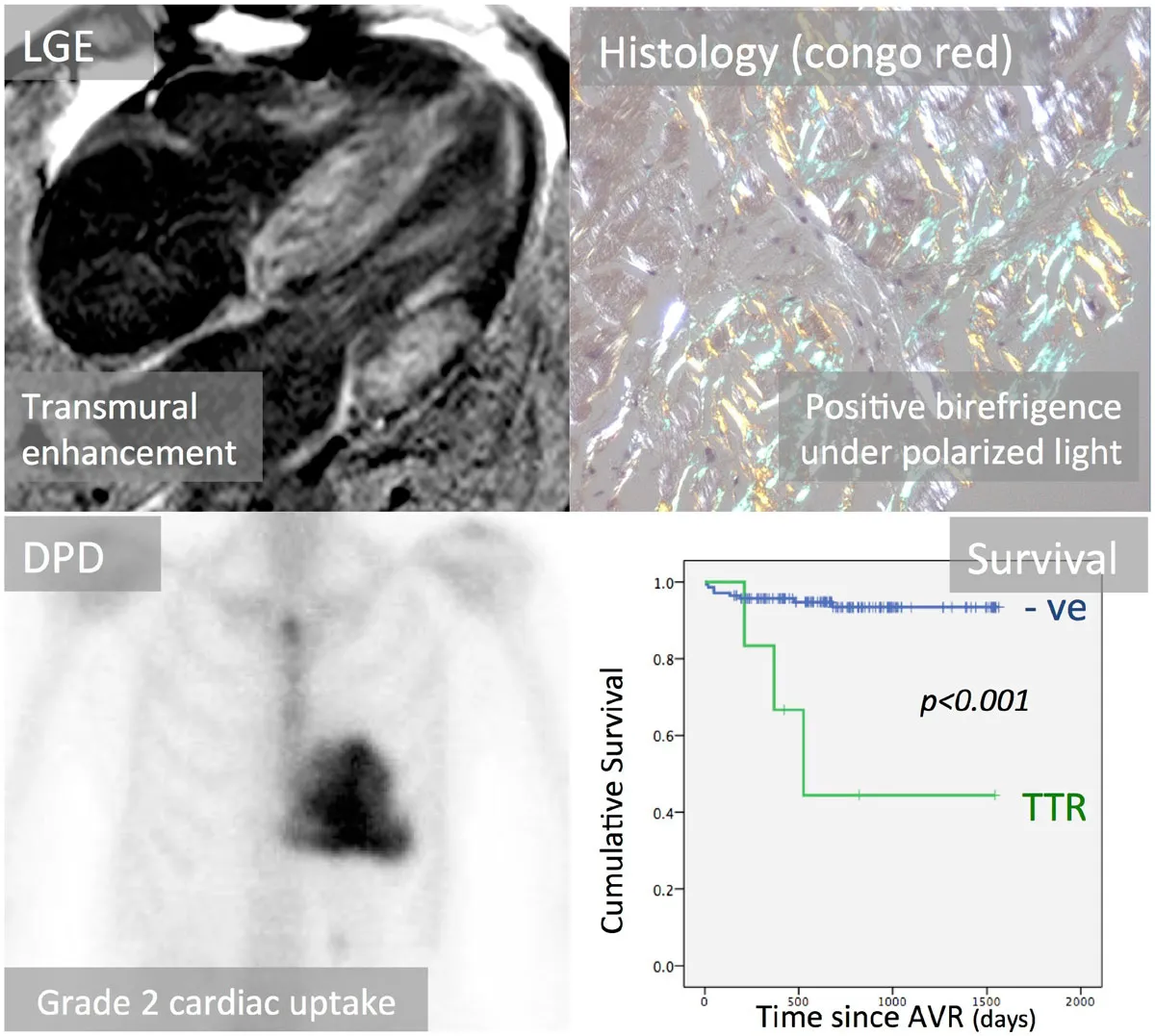

Dual pathology carries a worse prognosis than aortic stenosis alone. Valve replacement relieves the obstruction and is not withheld because of amyloidosis; in a multicenter observational cohort, transcatheter replacement was associated with lower mortality than medical therapy in patients with both conditions, and transthyretin-directed therapy is added for the cardiomyopathy. The amyloid diagnosis also changes the interpretation of low-flow physiology, the expected recovery of ventricular function, and the high risk of conduction disease after transcatheter replacement.
Medical therapy in aortic stenosis
No drug slows the progression of calcific aortic stenosis, and medical therapy addresses comorbid conditions and symptoms until the valve is replaced.
Hypertension. In patients at risk (stage A) and with asymptomatic aortic stenosis (stages B and C), hypertension is treated according to standard guidelines, starting at a low dose and titrating gradually (Class 1). Vasodilator-induced hypotension has not been a problem in clinical studies of patients with aortic stenosis. Diuretics can reduce stroke volume in a small ventricle, and the 2025 ESC/EACTS guideline prefers renin-angiotensin system blockers. After transcatheter replacement, an angiotensin-converting enzyme inhibitor or angiotensin receptor blocker may be considered (Class 2b) to reduce long-term mortality, on the basis of observational data.
Statins and lipid lowering. Statin therapy is indicated in all patients with calcific aortic stenosis for the primary and secondary prevention of atherosclerosis according to standard risk scores (Class 1). Statins are not indicated to slow hemodynamic progression (Class 3: No Benefit). Three randomized trials established this: SALTIRE (atorvastatin), SEAS (simvastatin with ezetimibe), and ASTRONOMER (rosuvastatin) each showed no reduction in hemodynamic progression or valve events in mild to moderate stenosis. In SEAS, lipid lowering reduced ischemic cardiovascular events, the basis for continued statin therapy. Lipoprotein(a) lowering is under investigation in early-stage calcific valve disease.
Heart failure and arrhythmia. In decompensated patients awaiting valve replacement, diuretics relieve congestion and are used cautiously because a small, stiff ventricle depends on preload. In critically ill patients with severe aortic stenosis and an LVEF of 35% or less, nitroprusside under invasive hemodynamic monitoring increased cardiac index and served as a bridge to valve replacement. When valve replacement is declined or not feasible, guideline-directed therapy for systolic heart failure is continued. Atrial fibrillation is poorly tolerated; restoration of sinus rhythm or rate control preserves filling, and anticoagulation follows standard atrial fibrillation criteria (aortic stenosis is not a contraindication to direct oral anticoagulants).
Endocarditis prophylaxis. Native aortic stenosis is not an indication for antibiotic prophylaxis. After valve replacement, prophylaxis before dental procedures that manipulate the gingiva or periapical region or perforate the oral mucosa is reasonable (Class 2a) in patients with any prosthetic valve, including transcatheter prostheses, and in patients with previous endocarditis. Prophylaxis is not recommended for nondental procedures such as transesophageal echocardiography, endoscopy, or colonoscopy.
Indications and timing of aortic valve replacement
The 2020 ACC/AHA recommendations for timing apply to both surgical aortic valve replacement (SAVR) and TAVR. Symptoms remain the dominant trigger, and the asymptomatic indications identify patients in whom waiting carries more risk than intervention.
| Patient group | Recommendation | Class (LOE) |
|---|---|---|
| Symptomatic severe high-gradient AS (D1), symptoms by history or on exercise testing | AVR is indicated | 1 (A) |
| Asymptomatic severe AS with LVEF <50% (C2) | AVR is indicated | 1 (B-NR) |
| Asymptomatic severe AS (C1) undergoing other cardiac surgery | AVR is indicated | 1 (B-NR) |
| Symptomatic low-flow, low-gradient severe AS with reduced LVEF (D2) | AVR is recommended | 1 (B-NR) |
| Symptomatic low-flow, low-gradient severe AS with normal LVEF (D3) | AVR is recommended if AS is the most likely cause of symptoms | 1 (B-NR) |
| Apparently asymptomatic severe AS (C1), low surgical risk, abnormal exercise test (reduced exercise tolerance or systolic fall ≥10 mm Hg) | AVR is reasonable | 2a (B-NR) |
| Asymptomatic very severe AS (Vmax ≥5.0 m/s), low surgical risk | AVR is reasonable | 2a (B-R) |
| Apparently asymptomatic severe AS (C1), low surgical risk, BNP >3 times normal | AVR is reasonable | 2a (B-NR) |
| Asymptomatic severe high-gradient AS (C1), low surgical risk, Vmax increase ≥0.3 m/s per year | AVR is reasonable | 2a (B-NR) |
| Asymptomatic severe high-gradient AS (C1), LVEF falling to <60% on ≥3 serial studies | AVR may be considered | 2b (B-NR) |
| Moderate AS (B) undergoing cardiac surgery for other indications | AVR may be considered | 2b (C-EO) |
Indications for aortic valve replacement (AVR) in the 2020 ACC/AHA guideline. AS, aortic stenosis; LOE, level of evidence.
Symptoms carry the same weight whether reported spontaneously or provoked on exercise testing. Symptomatic patients benefit from replacement in survival, symptoms, and ventricular function, and outcomes are excellent when procedural risk is not high. In stage C2, the reduced ejection fraction is often caused by afterload mismatch and improves after replacement. In stage D2, outcomes are worse than in high-gradient disease but still better with replacement than with medical therapy, particularly when flow reserve is present. The additive risk of replacing a severely stenotic valve during other cardiac surgery is lower than the risk of reoperation within 5 years, which is the basis for concomitant replacement.
In the 2025 ESC/EACTS guideline, intervention is recommended in asymptomatic severe aortic stenosis with an LVEF below 50% without another cause. Adverse prognostic features that support intervention in a low-risk patient include very high velocity, natriuretic peptide levels more than three times the age- and sex-corrected normal range, severe valve calcification with rapid progression, an LVEF below 55%, and a sustained fall in systolic pressure of more than 20 mm Hg during exercise.
Early intervention trials in asymptomatic severe aortic stenosis
Four randomized trials have compared early valve replacement with clinical surveillance in asymptomatic severe aortic stenosis. In the surveillance groups of all four, most patients ultimately required replacement, so the trials largely compare early with deferred intervention.
| Trial | Population | Intervention | Primary result |
|---|---|---|---|
| RECOVERY (2020) | 145 patients with very severe AS: AVA ≤0.75 cm² with Vmax ≥4.5 m/s or mean gradient ≥50 mm Hg | Early SAVR | Operative or cardiovascular death 1% versus 15% (HR 0.09); all-cause death 7% versus 21% |
| AVATAR (2022) | 157 patients with severe AS, LVEF >50%, and a normal exercise test; STS score <8% | Early SAVR | Death, MI, stroke, or unplanned heart failure hospitalization 15.2% versus 34.7% (HR 0.46) over a median of 32 months |
| EVOLVED (2025) | 224 patients with severe AS and midwall fibrosis on cardiac magnetic resonance | Early SAVR or TAVR | Death or unplanned AS-related hospitalization 18% versus 23% (HR 0.79; not significant) |
| EARLY TAVR (2025) | 901 patients with asymptomatic severe AS and LVEF ≥50%, anatomy suitable for transfemoral balloon-expandable TAVR | Early transfemoral TAVR | Death, stroke, or unplanned cardiovascular hospitalization 26.8% versus 45.3% (HR 0.50) over a median of 3.8 years |
Randomized trials of early aortic valve replacement versus clinical surveillance. HR, hazard ratio; MI, myocardial infarction; STS, Society of Thoracic Surgeons.
RECOVERY and AVATAR showed that early surgery in selected low-risk patients reduced hard events, with no operative deaths in RECOVERY and one in AVATAR. In EARLY TAVR, the benefit was driven mainly by fewer unplanned cardiovascular hospitalizations; death and stroke did not differ significantly. In its surveillance group, 87% underwent valve replacement during follow-up, at a median of 11.1 months, and patients who converted after presenting with symptoms or heart failure had worse outcomes than those who converted electively. A meta-analysis of the four trials, cited in the 2025 ESC/EACTS guideline, showed fewer unplanned hospitalizations with early intervention.
The 2020 ACC/AHA guideline predates EARLY TAVR and EVOLVED. Its asymptomatic recommendations are limited to the specific triggers listed among the indications for replacement, and for patients with a Class 2a asymptomatic indication it recommends surgical over transcatheter replacement (Class 1), because the comparative trials then available enrolled only symptomatic patients. The 2025 ESC/EACTS guideline states that early intervention should be considered (Class 2a, level A) in asymptomatic severe high-gradient aortic stenosis with an LVEF of 50% or more and low procedural risk, as an alternative to close active surveillance.
Moderate aortic stenosis with heart failure. Moderate aortic stenosis with reduced ejection fraction carries a poor prognosis, but early replacement has not been shown to change it. In TAVR UNLOAD, 178 patients with heart failure, reduced ejection fraction, and moderate aortic stenosis were randomized to transfemoral TAVR or surveillance with replacement on progression. TAVR was not superior for the primary hierarchical composite (win ratio 1.31, 95% CI 0.91 to 1.88) at a median of 23 months, although health status was better at 1 year. In the surveillance group, 43% underwent TAVR. Management remains optimized heart failure therapy, confirmation that the stenosis is truly moderate and not low-flow severe, and replacement when a standard indication develops.
Choice of prosthesis: mechanical, bioprosthetic, and pulmonary autograft
The choice of prosthesis is a shared decision that weighs durability and the risk of reintervention against the burden and bleeding risk of lifelong vitamin K antagonist therapy (Class 1). A bioprosthesis is recommended at any age when anticoagulation is contraindicated, cannot be managed, or is not desired (Class 1). A mechanical prosthesis is reasonable in patients younger than 50 years without a contraindication to anticoagulation (Class 2a), the choice is individualized between 50 and 65 years (Class 2a), and a bioprosthesis is reasonable in patients older than 65 years (Class 2a). The Ross procedure (pulmonary autograft) may be considered in patients younger than 50 years who prefer a biological valve and have suitable anatomy, when performed at a Comprehensive Valve Center (Class 2b).
Bioprosthetic durability depends strongly on age at implantation. The predicted 15-year risk of reoperation for structural deterioration is about 22% at age 50, 30% at age 40, and 50% at age 20, whereas in patients older than 65 years the likelihood of structural deterioration at 15 to 20 years is about 10%. Transcatheter valve-in-valve replacement has changed the calculation in younger patients, because a failed surgical bioprosthesis can often be treated without reoperation. Valve-in-valve feasibility depends on the original prosthesis size and design. A small surgical valve leaves a high residual gradient after valve-in-valve implantation unless it is fractured, which is one reason to avoid small prostheses and patient-prosthesis mismatch at the first operation. Lifetime management also considers whether a first transcatheter valve will allow a second transcatheter valve-in-transcatheter valve (TAV-in-TAV) procedure without sequestering the coronary sinuses.
Surgical versus transcatheter aortic valve replacement
For patients in whom a bioprosthesis is appropriate, the 2020 ACC/AHA guideline frames the choice by age as a surrogate for life expectancy, set against the known durability of each valve type:
- Younger than 65 years or life expectancy more than 20 years: SAVR is recommended (Class 1, A).
- 65 to 80 years with no anatomic contraindication to transfemoral TAVR: either SAVR or transfemoral TAVR is recommended after shared decision-making about expected longevity and valve durability (Class 1, A).
- Older than 80 years, or younger with life expectancy less than 10 years, and no anatomic contraindication: transfemoral TAVR is recommended in preference to SAVR (Class 1, A).
- High or prohibitive surgical risk at any age: TAVR is recommended if predicted post-TAVR survival exceeds 12 months with acceptable quality of life (Class 1, A).
- Valve or vascular anatomy unsuitable for transfemoral TAVR: SAVR is recommended (Class 1, A).
The age breakpoints derive from US actuarial data. Average additional life expectancy is 25, 17, and 10 years for a woman at ages 60, 70, and 80, and 22, 14, and 8 years for a man. These figures are starting points: comorbidity, frailty, and patient values move individual patients across the breakpoints. The 2025 ESC/EACTS guideline uses a single age threshold of 70 years: transfemoral TAVR is recommended in patients 70 years or older with a trileaflet valve and suitable anatomy (Class 1, A), and SAVR in patients younger than 70 years at low surgical risk.
| Factor | Favors SAVR | Favors TAVR | Favors palliation |
|---|---|---|---|
| Age and life expectancy | Younger age, longer life expectancy | Older age, fewer expected years of life | Limited life expectancy |
| Valve anatomy | Bicuspid valve; rheumatic disease; subaortic (outflow tract) calcification; small or large annulus | Calcific stenosis of a trileaflet valve | — |
| Prosthesis preference | Mechanical or surgical bioprosthetic valve preferred; concern for patient-prosthesis mismatch (annular enlargement possible) | Bioprosthesis preferred; favorable ratio of life expectancy to durability; larger effective orifice area than a surgical valve of the same size | — |
| Concurrent cardiac conditions | Aortic dilation; severe primary MR; severe CAD requiring bypass; septal hypertrophy requiring myectomy; AF (surgical ablation and appendage closure) | Porcelain aorta; severe MR due to annular calcification | Irreversible severe LV systolic dysfunction |
| Noncardiac conditions | — | Severe lung, liver, or kidney disease; mobility limits that raise sternotomy risk | Symptoms likely due to noncardiac disease; severe dementia; moderate to severe involvement of two or more other organ systems |
| Frailty | Not frail or few frailty measures | Frailty likely to improve after TAVR | Severe frailty unlikely to improve |
| Procedural risk | Low SAVR risk; high TAVR risk | Low to medium TAVR risk; high to prohibitive SAVR risk | Prohibitive SAVR risk (>15%) or post-TAVR life expectancy <1 year |
| Procedure-specific impediments | Annular size, valve anatomy, or low coronary ostial height precludes TAVR; no transfemoral access | Previous cardiac surgery with at-risk grafts; previous chest irradiation | — |
| Goals of care | Less uncertainty about durability; avoiding reintervention; lower risk of permanent pacemaker | Faster recovery; less pain; shorter hospital stay | Life prolongation not a goal; symptom relief |
Adapted from the 2020 ACC/AHA guideline, Table 14. AF, atrial fibrillation; CAD, coronary artery disease; MR, mitral regurgitation.
Surgical risk and frailty. The Society of Thoracic Surgeons predicted risk of mortality (STS-PROM) is combined with frailty, major organ compromise, and procedure-specific impediments. Low surgical risk requires an STS-PROM below 3%, no frailty, no major organ compromise, and no procedure-specific impediment. High risk is any of an STS-PROM above 8%, one moderate to severe frailty index, compromise of one or two major organ systems, or a possible procedure-specific impediment. Prohibitive risk is any of a predicted 30-day risk of death or major morbidity above 50%, two or more moderate to severe frailty indices, compromise of three or more major organ systems, or a severe procedure-specific impediment. Frailty is measured objectively (gait speed, grip strength, activities of daily living, cognition, and nutrition), and the decision is made by a multidisciplinary heart team with the patient.
Transcatheter replacement in bicuspid valves. Bicuspid valves were excluded from the pivotal randomized trials. Asymmetric and bulky leaflet calcium, calcified raphes, an elliptical annulus, and associated aortopathy raise the risk of paravalvular regurgitation, incomplete expansion, annular injury, and an aorta left untreated. Observational registries report acceptable results in selected patients with favorable anatomy. The 2020 ACC/AHA guideline lists bicuspid anatomy as a factor favoring SAVR. The 2025 ESC/EACTS guideline keeps surgery as the primary treatment of stenotic bicuspid valves, particularly in young patients or those with aortopathy, and lists TAVR as an option (Class 2b) at increased surgical risk with suitable anatomy. In a bicuspid valve with indications for SAVR, replacement of the aortic sinuses or ascending aorta is reasonable when the diameter is 4.5 cm or more and the operation is performed at a Comprehensive Valve Center (Class 2a).
Randomized trials of TAVR versus surgery
Transcatheter replacement was tested first against medical therapy in inoperable patients, then against surgery in progressively lower-risk populations. Most low-risk trial patients were in their 70s with trileaflet valves.
| Trial | Population | Key result |
|---|---|---|
| PARTNER 1B (2010) | Inoperable patients; balloon-expandable TAVR versus standard therapy (84% had balloon valvuloplasty) | All-cause death at 1 year 30.7% versus 50.7%; more strokes and major vascular complications with TAVR |
| PARTNER 1A, CoreValve High Risk | High surgical risk | TAVR noninferior or superior to SAVR for mortality |
| PARTNER 2A, SURTAVI | Intermediate surgical risk | TAVR noninferior to SAVR for death or disabling stroke |
| PARTNER 3 (2019; 5-year 2023) | 1,000 low-risk patients, mean age about 73; balloon-expandable valve | Death, stroke, or rehospitalization at 1 year 8.5% versus 15.1%; at 5 years 22.8% versus 27.2% (not significant); bioprosthetic valve failure 3.3% versus 3.8% |
| Evolut Low Risk (2019; 5-year 2025) | 1,414 low-risk patients; self-expanding valve | Death or disabling stroke at 2 years 5.3% versus 6.7% (noninferior); at 5 years 15.5% versus 16.4% (not significant); reintervention 3.3% versus 2.5% |
| NOTION (10-year 2024) | 280 patients aged 70 years or older at low risk; self-expanding valve | Death, stroke, or MI at 10 years 65.5% in both arms; severe structural deterioration 1.5% versus 10.0%; bioprosthetic valve failure 9.7% versus 13.8% (not significant); permanent pacemaker 44.7% versus 14.0% |
Selected randomized trials of transcatheter aortic valve replacement (TAVR). Rates are TAVR versus comparator.
Across the low-risk trials, TAVR produced shorter hospital stays, less pain, faster recovery, less atrial fibrillation, less major bleeding, and less acute kidney injury. SAVR produced less paravalvular regurgitation, fewer permanent pacemakers, and fewer vascular complications. The early advantages of TAVR in the composite end points narrowed over 5 years, and long-term mortality and stroke were similar. In a meta-analysis cited by the 2020 ACC/AHA guideline, transfemoral TAVR was associated with lower mortality (hazard ratio 0.88) and stroke (hazard ratio 0.81) than surgery.
Durability. Durability is now defined by Valve Academic Research Consortium 3 (VARC-3) criteria, which separate hemodynamic structural deterioration from bioprosthetic valve failure (death, reintervention, or severe hemodynamic deterioration attributable to the valve). Five-year data from the low-risk trials and 10-year data from NOTION show no excess structural deterioration or valve failure with transcatheter valves. Transcatheter valves also have larger effective orifice areas and lower gradients than surgical valves of the same size. Data beyond 10 years and data in patients younger than 65 years remain limited, which is why age still governs the choice in US guidance. Failed transcatheter valves are treated with a second transcatheter valve or with surgical explantation, which carries higher risk than a first operation.
Procedural complications of TAVR and SAVR
Conduction disturbance. The prosthesis seats against the membranous septum near the His bundle. New left bundle branch block and high-grade atrioventricular block are more frequent after TAVR than after SAVR and with self-expanding than with balloon-expandable valves. Pre-existing right bundle branch block, a short membranous septum, deep implantation, and oversizing increase the risk. Pacemaker rates have fallen with higher implant positions and cusp-overlap techniques, but permanent pacemaker implantation remains the most common complication.
Paravalvular regurgitation. Leaks result from calcium nodules, an elliptical annulus, undersizing, or malposition. Moderate or greater paravalvular regurgitation is associated with higher mortality, and even mild regurgitation has been associated with worse outcome in some series. It is graded after deployment by transthoracic or transesophageal echocardiography, aortography, and hemodynamics, and treated by postdilation, a second valve, or later plug closure.
Stroke. Most periprocedural strokes are embolic, occur early, and arise from manipulation of calcified valve and aortic tissue. Rates are low and similar to surgery in contemporary trials. Cerebral embolic protection devices capture debris, but randomized data have not shown a significant reduction in stroke.
Coronary obstruction, annular rupture, and vascular injury. Coronary obstruction occurs when a displaced native leaflet covers a low coronary ostium in a shallow sinus and is more likely in valve-in-valve procedures; CT predicts it. Annular rupture is rare and associated with heavy outflow tract calcium and balloon-expandable oversizing. Vascular complications reflect sheath size and iliofemoral disease.
Patient-prosthesis mismatch. Mismatch exists when the effective orifice area of a normally functioning prosthesis is too small for the patient's body size. VARC-3 defines it by the indexed effective orifice area: moderate mismatch at 0.85 cm²/m² or less and severe mismatch at 0.65 cm²/m² or less, with lower thresholds (0.70 and 0.55 cm²/m²) when the body mass index is 30 kg/m² or more. Mismatch is associated with persistent hypertrophy, high gradients, less symptom improvement, and higher mortality, most strongly when severe or when ventricular function is reduced. It is more frequent after SAVR than TAVR, particularly in small annuli, and is prevented by prosthesis selection, including supra-annular designs, or by annular enlargement. Mismatch is distinguished from prosthetic stenosis by comparison with the early postoperative study and by normal leaflet appearance and motion.
Balloon aortic valvuloplasty and palliative care
Balloon valvuloplasty in calcific aortic stenosis fractures calcium within the leaflets and stretches the annulus. The immediate result is a modest increase in valve area, usually well short of normal, and restenosis occurs within months in most patients. The procedure does not improve long-term survival. In critically ill patients with severe aortic stenosis, balloon dilation may be considered as a bridge to SAVR or TAVR (Class 2b). It also serves as a bridge to urgent noncardiac surgery in unstable patients, as a test of whether symptoms are caused by the valve when the cause is uncertain, and as palliation for patients who are not candidates for replacement.
When predicted survival after TAVR or SAVR is less than 12 months, or minimal improvement in quality of life is expected, palliative care is recommended after shared decision-making about the patient's values and preferences (Class 1). Factors associated with futility include severe frailty unlikely to improve; advanced dementia; severe lung, liver, or kidney disease; and multiorgan compromise. TAVR is not recommended when life expectancy is less than 1 year even with a successful procedure, or when the chance of survival with benefit at 2 years is less than 25%. Palliative management relieves congestion with cautious diuresis, treats atrial fibrillation, and includes advance care planning.
Noncardiac surgery in aortic stenosis
Undiagnosed severe aortic stenosis is associated with a 10% to 30% rate of cardiac complications during noncardiac surgery. Anesthesia, vasodilation, blood loss, and tachycardia lower preload and coronary perfusion pressure in a hypertrophied ventricle that tolerates neither. Preoperative echocardiography is recommended in patients with clinically suspected moderate or greater valvular stenosis (Class 1); an echocardiogram from the previous 12 months suffices when the clinical course has not changed.
In patients who meet standard criteria for valve intervention on the basis of symptoms and severity, valve replacement is performed before elective noncardiac surgery when possible (Class 1). TAVR is a reasonable way to avoid delaying semi-urgent noncardiac surgery, and balloon valvuloplasty is an option as a bridge in unstable patients at high or prohibitive risk. In asymptomatic patients with moderate or greater aortic stenosis and normal ventricular function, elective noncardiac surgery is reasonable (Class 2a), with anesthetic management directed at maintaining sinus rhythm, a normal heart rate, preload, and systemic vascular resistance. Coexisting mitral regurgitation, pulmonary hypertension, and coronary disease predict adverse outcome. Elevated-risk procedures in patients with severe aortic stenosis call for heart team review and a decision about invasive hemodynamic or transesophageal monitoring.
Pregnancy in women with aortic stenosis
Pregnancy raises cardiac output by 30% to 50%, which raises the transvalvular gradient across a fixed orifice, and labor and delivery add abrupt changes in preload. Women of reproductive age with aortic stenosis usually have a bicuspid or other congenital valve, often with dilation of the aortic sinuses or ascending aorta, so the aorta is imaged before pregnancy because of the risk of aortic enlargement and dissection.
The 2020 ACC/AHA guideline recommends valve intervention before pregnancy in symptomatic women with severe valve disease (Class 1) and finds intervention before pregnancy reasonable in asymptomatic women with severe aortic stenosis (Class 2a). For asymptomatic women with severe stenosis who do not meet a Class 1 criterion and have a preconception evaluation confirming the absence of symptoms, including a normal exercise test and natriuretic peptide level, medical management during pregnancy may be considered to avoid valve replacement (Class 2b). Options before pregnancy include balloon dilation of a noncalcified congenital valve, the Ross procedure, and surgical replacement; bioprosthetic valves are preferred over mechanical valves in women of childbearing age because of the maternal and fetal risks of anticoagulation (Class 2a). Transcatheter valves have not been studied in young women.
During pregnancy, women with severe stenosis are followed at a tertiary center by a team experienced in cardiac disease in pregnancy, with serial clinical and echocardiographic assessment. Valve intervention during pregnancy is reasonable in severe aortic stenosis (mean gradient 40 mm Hg or more) with hemodynamic deterioration or New York Heart Association (NYHA) class III or IV heart failure (Class 2a). Balloon valvuloplasty and TAVR are the options when intervention cannot wait, because cardiopulmonary bypass carries a high fetal risk. Delivery is usually vaginal with regional anesthesia titrated slowly and an assisted second stage, and cesarean delivery is reserved for obstetric indications or decompensation.
Aortic stenosis with mixed and multivalve disease
Aortic stenosis with aortic regurgitation. Most adults with aortic stenosis have some regurgitation. Regurgitation raises forward systolic flow, so velocity and gradient are higher for a given valve area, whereas the continuity area and dimensionless index remain valid because both terms measure the same systolic flow. The ventricle faces pressure and volume overload together, and the combination can be hemodynamically severe when neither lesion is severe alone. In the 2020 ACC/AHA guideline, valve replacement is recommended in symptomatic patients with combined stenosis and regurgitation and a peak velocity of 4.0 m/s or more or a mean gradient of 40 mm Hg or more (Class 1). Surgical replacement is recommended in asymptomatic patients with a velocity of 4.0 m/s or more and an LVEF below 50% (Class 1). The indications are therefore the same as for severe isolated stenosis, even when neither lesion alone meets severe criteria by area or regurgitant measures.
Aortic stenosis with mitral disease. Significant mitral regurgitation lowers forward stroke volume and therefore the aortic gradient, creating a low-flow state. Mitral stenosis does the same by limiting filling. Mitral annular calcification with mitral stenosis, common in older patients and those with kidney disease, complicates both surgery and the assessment of transmitral gradients. Functional mitral regurgitation from pressure overload often improves after aortic valve replacement, whereas primary mitral disease does not. Severe primary mitral regurgitation favors surgery that addresses both valves; when the mitral disease is secondary or the patient is at high surgical risk, TAVR followed by reassessment is common practice.
Significant tricuspid regurgitation and pulmonary hypertension at rest indicate advanced disease and predict worse outcome after either form of replacement.
Echocardiographic surveillance before valve replacement
Periodic monitoring is required for all patients in whom valve replacement is not yet indicated, because symptoms develop insidiously and patients may not recognize them. The 2020 ACC/AHA guideline sets echocardiographic intervals for asymptomatic patients with normal ventricular function and normal stroke volume. Repeat imaging is also performed whenever symptoms or examination findings change (a louder or later-peaking murmur, a soft A2), and when hemodynamic demands increase, such as pregnancy, noncardiac surgery, infection, anemia, or gastrointestinal bleeding.
| Status | Echocardiographic interval |
|---|---|
| Mild AS (stage B, Vmax 2.0–2.9 m/s) | Every 3 to 5 years |
| Moderate AS (stage B, Vmax 3.0–3.9 m/s) | Every 1 to 2 years |
| Asymptomatic severe AS (stage C1, Vmax ≥4.0 m/s) | Every 6 to 12 months |
| Bicuspid valve with aortic diameter ≥4.0 cm at the time of AVR | Continued aortic imaging after replacement |
| After surgical bioprosthetic AVR | Baseline 1 to 3 months after surgery, then at 5 and 10 years, then annually |
| After transcatheter AVR | Baseline 1 to 3 months after implantation, then annually |
| After mechanical AVR | Baseline study; repeat for symptoms, new murmur, or embolic events |
Imaging intervals from the 2020 ACC/AHA guideline (Tables 5 and 12). Clinical review is annual after any valve intervention; mixed valve disease, rapid progression, and severe disease with risk features warrant shorter intervals.
Serial studies are most useful when acquired with the same windows, the same nonimaging transducer, and the same outflow tract measurement site, with current and prior spectra compared side by side. The 2025 ESC/EACTS guideline recommends reassessment of asymptomatic severe aortic stenosis at least every 6 months, with patient education to report symptoms promptly.
Surveillance after aortic valve replacement
The baseline postprocedural echocardiogram, ideally 1 to 3 months after implantation once loading conditions have normalized, records the prosthesis gradients, effective orifice area, dimensionless index, paravalvular and central regurgitation, and ventricular function. It is the reference against which later studies are judged.
Antithrombotic therapy. After TAVR, aspirin 75 to 100 mg daily is reasonable in the absence of another indication for oral anticoagulation (Class 2a). Dual antiplatelet therapy with aspirin and clopidogrel for 3 to 6 months (Class 2b) or a vitamin K antagonist for at least 3 months (Class 2b) may be reasonable in patients at low bleeding risk. Low-dose rivaroxaban with aspirin is harmful in patients without another indication for anticoagulation (Class 3: Harm). Patients with atrial fibrillation continue oral anticoagulation, generally without routine added antiplatelet therapy. After surgical bioprosthetic replacement, aspirin is reasonable (Class 2a), and a vitamin K antagonist for 3 to 6 months is reasonable in patients at low bleeding risk (Class 2a). After mechanical aortic replacement, vitamin K antagonist therapy is recommended with an international normalized ratio (INR) target of 2.5, or 3.0 with additional thromboembolic risk factors or an older-generation prosthesis.
Subclinical leaflet thrombosis. Hypoattenuated leaflet thickening (HALT) on contrast CT, with or without reduced leaflet motion, represents thin leaflet thrombus. It was first reported in patients after TAVR and surgical bioprosthetic replacement and is more frequent with transcatheter valves and in patients not receiving anticoagulation. Most cases cause no symptoms and little change in gradient, and some resolve spontaneously. HALT is suspected when the mean gradient rises on routine echocardiography, and three-dimensional transesophageal echocardiography or four-dimensional CT confirms leaflet thrombosis (Class 2a). In stable patients with clinically significant bioprosthetic valve thrombosis, a vitamin K antagonist is reasonable initial treatment (Class 2a) and often restores leaflet motion and gradients. Routine anticoagulation after TAVR in patients without another indication reduces subclinical leaflet thrombosis but has not improved clinical outcomes; in GALILEO, low-dose rivaroxaban with aspirin increased death, thromboembolic events, and bleeding (Class 3: Harm).
Structural valve deterioration. Bioprosthetic stenosis or regurgitation from leaflet calcification, thickening, or tear develops progressively, most often after 10 years for surgical valves and earlier in younger patients and those with kidney failure, diabetes, or hypercalcemia. VARC-3 grades hemodynamic deterioration by the increase in mean gradient with a decrease in effective orifice area and dimensionless index, or by new or worsening intraprosthetic regurgitation, compared with the baseline study. Nonstructural dysfunction (patient-prosthesis mismatch, paravalvular regurgitation), thrombosis, and endocarditis are separate categories. A rising gradient is evaluated for thrombosis before it is attributed to degeneration, because thrombosis responds to anticoagulation. Failed bioprostheses are treated with transcatheter valve-in-valve implantation or surgical reoperation according to anatomy, prosthesis size, coronary risk, and surgical risk.
Prosthetic valve endocarditis after TAVR and SAVR carries high mortality. Fever, new regurgitation, a rising gradient, or new conduction disease in a patient with a prosthetic valve leads to blood cultures and transesophageal echocardiography, with CT or positron emission tomography when echocardiography is inconclusive.
Self-assessment
A 79-year-old woman has exertional dyspnea, LVEF 64%, blood pressure 126/72 mm Hg, aortic valve area 0.78 cm², indexed area 0.48 cm²/m², mean gradient 29 mm Hg, stroke volume index 30 mL/m², and an aortic valve calcium score of 1,680 Agatston units. What ACC/AHA stage is this, and what does the calcium score indicate?
Echocardiography Board ReviewReferences
- Otto CM, Nishimura RA, Bonow RO, et al. 2020 ACC/AHA guideline for the management of patients with valvular heart disease: a report of the American College of Cardiology/American Heart Association Joint Committee on Clinical Practice Guidelines. Circulation. 2021;143:e72–e227. doi:10.1161/CIR.0000000000000923
- Praz F, Borger MA, Lanz J, et al. 2025 ESC/EACTS guidelines for the management of valvular heart disease. Eur Heart J. 2025;46:4635–4736. doi:10.1093/eurheartj/ehaf194
- Baumgartner H, Hung J, Bermejo J, et al. Recommendations on the echocardiographic assessment of aortic valve stenosis: a focused update from the European Association of Cardiovascular Imaging and the American Society of Echocardiography. J Am Soc Echocardiogr. 2017;30:372–392. doi:10.1016/j.echo.2017.02.009
- Généreux P, Piazza N, Alu MC, et al. Valve Academic Research Consortium 3: updated endpoint definitions for aortic valve clinical research. J Am Coll Cardiol. 2021;77:2717–2746. doi:10.1016/j.jacc.2021.02.038
- Ross J Jr, Braunwald E. Aortic stenosis. Circulation. 1968;38(1 Suppl):61–67. doi:10.1161/01.cir.38.1s5.v-61
- Otto CM, Burwash IG, Legget ME, et al. Prospective study of asymptomatic valvular aortic stenosis: clinical, echocardiographic, and exercise predictors of outcome. Circulation. 1997;95:2262–2270. doi:10.1161/01.cir.95.9.2262
- Rosenhek R, Binder T, Porenta G, et al. Predictors of outcome in severe, asymptomatic aortic stenosis. N Engl J Med. 2000;343:611–617. doi:10.1056/NEJM200008313430903
- Osnabrugge RL, Mylotte D, Head SJ, et al. Aortic stenosis in the elderly: disease prevalence and number of candidates for transcatheter aortic valve replacement: a meta-analysis and modeling study. J Am Coll Cardiol. 2013;62:1002–1012. doi:10.1016/j.jacc.2013.05.015
- Nkomo VT, Gardin JM, Skelton TN, et al. Burden of valvular heart diseases: a population-based study. Lancet. 2006;368:1005–1011. doi:10.1016/S0140-6736(06)69208-8
- Lindman BR, Clavel MA, Mathieu P, et al. Calcific aortic stenosis. Nat Rev Dis Primers. 2016;2:16006. doi:10.1038/nrdp.2016.6
- Thanassoulis G, Campbell CY, Owens DS, et al. Genetic associations with valvular calcification and aortic stenosis. N Engl J Med. 2013;368:503–512. doi:10.1056/NEJMoa1109034
- Roberts WC, Ko JM. Frequency by decades of unicuspid, bicuspid, and tricuspid aortic valves in adults having isolated aortic valve replacement for aortic stenosis, with or without associated aortic regurgitation. Circulation. 2005;111:920–925. doi:10.1161/01.CIR.0000155623.48408.C5
- Pibarot P, Dumesnil JG. Low-flow, low-gradient aortic stenosis with normal and depressed left ventricular ejection fraction. J Am Coll Cardiol. 2012;60:1845–1853. doi:10.1016/j.jacc.2012.06.051
- Monin JL, Quéré JP, Monchi M, et al. Low-gradient aortic stenosis: operative risk stratification and predictors for long-term outcome: a multicenter study using dobutamine stress hemodynamics. Circulation. 2003;108:319–324. doi:10.1161/01.CIR.0000079171.43055.46
- Clavel MA, Messika-Zeitoun D, Pibarot P, et al. The complex nature of discordant severe calcified aortic valve disease grading: new insights from combined Doppler echocardiographic and computed tomographic study. J Am Coll Cardiol. 2013;62:2329–2338. doi:10.1016/j.jacc.2013.08.1621
- Hachicha Z, Dumesnil JG, Pibarot P. Usefulness of the valvuloarterial impedance to predict adverse outcome in asymptomatic aortic stenosis. J Am Coll Cardiol. 2009;54:1003–1011. doi:10.1016/j.jacc.2009.04.079
- Garcia D, Pibarot P, Dumesnil JG, et al. Assessment of aortic valve stenosis severity: a new index based on the energy loss concept. Circulation. 2000;101:765–771. doi:10.1161/01.cir.101.7.765
- Magne J, Cosyns B, Popescu BA, et al. Distribution and prognostic significance of left ventricular global longitudinal strain in asymptomatic significant aortic stenosis: an individual participant data meta-analysis. JACC Cardiovasc Imaging. 2019;12:84–92. doi:10.1016/j.jcmg.2018.11.005
- Clavel MA, Malouf J, Michelena HI, et al. B-type natriuretic peptide clinical activation in aortic stenosis: impact on long-term survival. J Am Coll Cardiol. 2014;63:2016–2025. doi:10.1016/j.jacc.2014.02.581
- Cohen-Shelly M, Attia ZI, Friedman PA, et al. Electrocardiogram screening for aortic valve stenosis using artificial intelligence. Eur Heart J. 2021;42:2885–2896. doi:10.1093/eurheartj/ehab153
- Gorlin R, Gorlin SG. Hydraulic formula for calculation of the area of the stenotic mitral valve, other cardiac valves, and central circulatory shunts. Am Heart J. 1951;41:1–29. doi:10.1016/0002-8703(51)90002-6
- Hakki AH, Iskandrian AS, Bemis CE, et al. A simplified valve formula for the calculation of stenotic cardiac valve areas. Circulation. 1981;63:1050–1055. doi:10.1161/01.cir.63.5.1050
- Castaño A, Narotsky DL, Hamid N, et al. Unveiling transthyretin cardiac amyloidosis and its predictors among elderly patients with severe aortic stenosis undergoing transcatheter aortic valve replacement. Eur Heart J. 2017;38:2879–2887. doi:10.1093/eurheartj/ehx350
- Treibel TA, Fontana M, Gilbertson JA, et al. Occult transthyretin cardiac amyloid in severe calcific aortic stenosis: prevalence and prognosis in patients undergoing surgical aortic valve replacement. Circ Cardiovasc Imaging. 2016;9:e005066. doi:10.1161/CIRCIMAGING.116.005066
- Vincentelli A, Susen S, Le Tourneau T, et al. Acquired von Willebrand syndrome in aortic stenosis. N Engl J Med. 2003;349:343–349. doi:10.1056/NEJMoa022831
- Cowell SJ, Newby DE, Prescott RJ, et al. A randomized trial of intensive lipid-lowering therapy in calcific aortic stenosis. N Engl J Med. 2005;352:2389–2397. doi:10.1056/NEJMoa043876
- Rossebø AB, Pedersen TR, Boman K, et al. Intensive lipid lowering with simvastatin and ezetimibe in aortic stenosis. N Engl J Med. 2008;359:1343–1356. doi:10.1056/NEJMoa0804602
- Chan KL, Teo K, Dumesnil JG, et al. Effect of lipid lowering with rosuvastatin on progression of aortic stenosis: results of the Aortic Stenosis Progression Observation: Measuring Effects of Rosuvastatin (ASTRONOMER) trial. Circulation. 2010;121:306–314. doi:10.1161/CIRCULATIONAHA.109.900027
- Kang DH, Park SJ, Lee SA, et al. Early surgery or conservative care for asymptomatic aortic stenosis. N Engl J Med. 2020;382:111–119. doi:10.1056/NEJMoa1912846
- Banovic M, Putnik S, Penicka M, et al. Aortic valve replacement versus conservative treatment in asymptomatic severe aortic stenosis: the AVATAR trial. Circulation. 2022;145:648–658. doi:10.1161/CIRCULATIONAHA.121.057639
- Loganath K, Craig NJ, Everett RJ, et al. Early intervention in patients with asymptomatic severe aortic stenosis and myocardial fibrosis: the EVOLVED randomized clinical trial. JAMA. 2025;333:213–221. doi:10.1001/jama.2024.22730
- Généreux P, Schwartz A, Oldemeyer JB, et al. Transcatheter aortic-valve replacement for asymptomatic severe aortic stenosis. N Engl J Med. 2025;392:217–227. doi:10.1056/NEJMoa2405880
- Van Mieghem NM, Elmariah S, Spitzer E, et al. Transcatheter aortic valve replacement in patients with systolic heart failure and moderate aortic stenosis: TAVR UNLOAD. J Am Coll Cardiol. 2025;85:878–890. doi:10.1016/j.jacc.2024.10.070
- Leon MB, Smith CR, Mack M, et al. Transcatheter aortic-valve implantation for aortic stenosis in patients who cannot undergo surgery. N Engl J Med. 2010;363:1597–1607. doi:10.1056/NEJMoa1008232
- Mack MJ, Leon MB, Thourani VH, et al. Transcatheter aortic-valve replacement with a balloon-expandable valve in low-risk patients. N Engl J Med. 2019;380:1695–1705. doi:10.1056/NEJMoa1814052
- Mack MJ, Leon MB, Thourani VH, et al. Transcatheter aortic-valve replacement in low-risk patients at five years. N Engl J Med. 2023;389:1949–1960. doi:10.1056/NEJMoa2307447
- Popma JJ, Deeb GM, Yakubov SJ, et al. Transcatheter aortic-valve replacement with a self-expanding valve in low-risk patients. N Engl J Med. 2019;380:1706–1715. doi:10.1056/NEJMoa1816885
- Forrest JK, Yakubov SJ, Deeb GM, et al. 5-year outcomes after transcatheter or surgical aortic valve replacement in low-risk patients with aortic stenosis. J Am Coll Cardiol. 2025;85:1523–1532. doi:10.1016/j.jacc.2025.03.004
- Thyregod HGH, Jørgensen TH, Ihlemann N, et al. Transcatheter or surgical aortic valve implantation: 10-year outcomes of the NOTION trial. Eur Heart J. 2024;45:1116–1124. doi:10.1093/eurheartj/ehae043
- Makkar RR, Fontana G, Jilaihawi H, et al. Possible subclinical leaflet thrombosis in bioprosthetic aortic valves. N Engl J Med. 2015;373:2015–2024. doi:10.1056/NEJMoa1509233
- Cosmi JE, Kort S, Tunick PA, et al. The risk of the development of aortic stenosis in patients with “benign” aortic valve thickening. Arch Intern Med. 2002;162:2345–2347.
- Khot UN, Novaro GM, Popović ZB, et al. Nitroprusside in critically ill patients with left ventricular dysfunction and aortic stenosis. N Engl J Med. 2003;348:1756–1763.
- Dangas GD, Tijssen JGP, Wöhrle J, et al. A controlled trial of rivaroxaban after transcatheter aortic-valve replacement (GALILEO). N Engl J Med. 2020;382:120–129.
- Nitsche C, Scully PR, Patel KP, et al. Prevalence and outcomes of concomitant aortic stenosis and cardiac amyloidosis. J Am Coll Cardiol. 2021;77:128–139.