Library › Vascular disease › Pulmonary embolism
Pulmonary embolism
Acute pulmonary embolism in adults from venous thrombosis and clinical probability through imaging, the 2026 AHA/ACC clinical categories, anticoagulation and reperfusion, special populations, and follow-up for chronic thromboembolic disease.
Acute pulmonary embolism is graded by its physiologic consequences: the 2026 American Heart Association/American College of Cardiology (AHA/ACC) guideline assigns clinical categories A to E from symptoms, a clinical severity score, right ventricular imaging, cardiac biomarkers, and perfusion, with a respiratory modifier. Anticoagulation treats every category. Systemic thrombolysis is harmful in categories A to C2 (Class 3: Harm), uncertain in C3 and D, and reasonable in cardiopulmonary failure (category E). Clot burden on CT does not decide treatment.
Definition, venous sources, and risk factor categories of pulmonary embolism
Pulmonary embolism (PE) is obstruction of the pulmonary arterial tree by material carried in the venous circulation, almost always thrombus from the deep veins of the legs or pelvis. Deep vein thrombosis (DVT) and PE are one disease, venous thromboembolism (VTE). Proximal DVT is present in many patients with PE at the time of diagnosis, and silent PE is common in patients with proximal DVT. Thrombi originating in the calf veins embolize less often than thrombi that propagate into the popliteal, femoral, and iliac veins. Upper-extremity thrombosis, usually catheter- or device-related, is a less frequent source. Right atrial or ventricular thrombus, including thrombus in transit, and thrombus on pacing leads or central catheters account for the remainder.
Virchow's triad describes the mechanisms. Stasis results from immobility, paralysis, long travel, heart failure, and venous obstruction. Endothelial injury follows surgery, trauma, central catheters, and prior thrombosis. Hypercoagulability accompanies cancer, pregnancy and the postpartum period, estrogen therapy, inflammatory disease, nephrotic syndrome, antiphospholipid syndrome, and inherited thrombophilia. Thrombus begins in the valve pockets of the deep veins, where flow is slow and hypoxia activates the endothelium. Most patients have more than one factor.
Provoked and unprovoked embolism. The 2026 AHA/ACC guideline, in keeping with the International Society on Thrombosis and Haemostasis, replaces the binary "provoked/unprovoked" label with the strength and persistence of the associated risk factor. The distinction decides the duration of anticoagulation, because recurrence after stopping treatment is low when a major transient factor caused the event and high when no factor is identified or the factor persists.
| Risk factor category | Examples | Implication for duration |
|---|---|---|
| Major transient (reversible) | Surgery under general anesthesia for 30 minutes or more; hospitalization of 72 hours or more confined to bed for acute medical illness; cesarean section; lower-limb fracture | Stop at 3 to 6 months (Class 1) |
| Minor transient (reversible) | Surgery under general anesthesia for less than 30 minutes; hospitalization of less than 72 hours; illness outside hospital with 72 hours or more confined to bed; estrogen therapy; peripartum period; trauma with reduced mobility for 72 hours or more | Shared decision on stopping or extending (Class 2a) |
| Persistent | Active cancer; autoimmune disease; inflammatory bowel disease; chronic immobility | Extended therapy reasonable while the factor persists |
| No identifiable factor | First event with no transient or persistent factor (formerly "unprovoked") | Extended therapy beyond 3 to 6 months (Class 1) |
Risk factor categories and examples from Table 9 of the 2026 AHA/ACC acute PE guideline, with the corresponding duration recommendations.
Epidemiology and risk factors for pulmonary embolism
VTE is the third most common acute cardiovascular syndrome after myocardial infarction and stroke. Incidence rises steeply with age, and hospitalization rates have increased over two decades, partly because CT detects smaller and incidental emboli. Case fatality has fallen over the same period, yet roughly one in five patients with high-risk presentations dies. PE is a leading preventable cause of death in hospitalized patients.
The 2026 guideline lists the major acquired risk factors as surgery, trauma, hospitalization, prolonged immobility, pregnancy and the 6 weeks after delivery, oral contraceptive or estrogen therapy, obesity (body mass index 30 kg/m² or more), smoking, atherosclerotic cardiovascular disease, cancer, and thrombophilia, with risk increasing after age 40. Cancer raises VTE risk several-fold, and the risk is highest with pancreatic, gastric, brain, lung, ovarian, and hematologic malignancy, with metastatic disease, and in the first months after diagnosis. Systemic chemotherapy, immunomodulatory drugs, antiangiogenic agents, and central venous catheters add risk. Other acquired conditions include heart failure, chronic kidney disease and nephrotic syndrome, inflammatory bowel disease, myeloproliferative neoplasms, paroxysmal nocturnal hemoglobinuria, and heparin-induced thrombocytopenia.
Inherited and acquired thrombophilia. Factor V Leiden and the prothrombin G20210A variant are the most common inherited thrombophilias in populations of European ancestry. Deficiencies of antithrombin, protein C, and protein S are rarer and confer higher risk. Antiphospholipid syndrome is the acquired thrombophilia with the most direct effect on treatment, because it changes the choice of anticoagulant: warfarin is recommended over a direct oral anticoagulant (DOAC) in thrombotic antiphospholipid syndrome (Class 1). Testing for inherited thrombophilia does not change management for most patients and is not performed during the acute event.
Pathophysiology: right ventricular failure and impaired gas exchange
Acute obstruction of the pulmonary arteries raises pulmonary vascular resistance mechanically and through vasoconstriction mediated by thromboxane A2, serotonin, and hypoxia. A previously normal right ventricle (RV) is thin-walled and cannot generate a mean pulmonary artery pressure much above 40 mm Hg. A tricuspid regurgitant velocity in the range of severe pulmonary hypertension therefore suggests chronic pressure overload, either chronic thromboembolic pulmonary disease (CTEPD) or another cause on which the acute event is superimposed.
The RV responds to the sudden afterload by dilating. Wall tension rises and RV oxygen demand increases while coronary perfusion of the RV falls, because systemic pressure declines and RV intramural pressure increases. RV ischemia and myocyte injury explain the troponin release, and wall stretch explains the natriuretic peptide rise. Ventricular interdependence completes the spiral: the dilated RV shifts the septum leftward, the left ventricle (LV) underfills, cardiac output and systemic pressure fall, and RV perfusion falls further. Obstructive shock and cardiac arrest, usually pulseless electrical activity, are the end of this sequence. Systemic hypotension in PE is therefore a late sign, and normotensive patients can already have reduced cardiac output and tissue hypoperfusion, the basis of the "normotensive shock" category.
Hypoxemia results from ventilation-perfusion mismatch, with blood redistributed to underventilated lung, from reduced mixed venous oxygen saturation when cardiac output falls, and from right-to-left shunting through a patent foramen ovale when right atrial pressure exceeds left. Increased alveolar dead space widens the alveolar–arterial oxygen gradient and, in severe obstruction, the arterial–end-tidal carbon dioxide gradient. Hyperventilation produces hypocapnia and respiratory alkalosis. Pulmonary infarction occurs in a minority, usually with smaller distal emboli, because the lung has a dual blood supply. Infarction causes pleuritic pain, hemoptysis, and pleural-based opacities.
Most thrombus resolves over weeks to months through endogenous fibrinolysis, and perfusion recovers in most patients. Incomplete resolution with organized fibrotic material and a secondary small-vessel arteriopathy is the substrate of CTEPD.
Symptoms, signs, and differential diagnosis
Dyspnea, often abrupt, is the most common symptom. Pleuritic chest pain reflects pleural irritation from distal emboli and infarction, and substernal pressure can reflect RV ischemia. Cough, hemoptysis, and leg swelling or pain from coexisting DVT are less frequent. Syncope or presyncope signals a transient fall in cardiac output and is associated with larger emboli and RV dysfunction, although syncope alone does not mandate advanced therapy. Some patients present with shock or cardiac arrest, and others have no symptoms, with emboli found on CT performed for another reason (category A).
Tachypnea and tachycardia are the most common signs. Hypoxemia is frequent but not universal, and a normal oxygen saturation does not exclude PE. Signs of RV pressure overload include elevated jugular venous pressure, a parasternal heave, a loud pulmonic component of S2, a right-sided S3, and tricuspid regurgitation. Low-grade fever can accompany pulmonary infarction. Examination of the legs identifies DVT in a minority. The shock index (heart rate divided by systolic pressure) is a continuous marker of instability; values of 1 or more are associated with adverse outcome, and the 2026 guideline notes that no consensus cut-point exists.
The differential diagnosis includes acute coronary syndrome, aortic dissection, pneumothorax, pneumonia, pericarditis and tamponade, acute decompensated heart failure, exacerbation of chronic lung disease, and musculoskeletal pain. Several of these produce troponin elevation and RV dilation, so the diagnosis of PE rests on pulmonary arterial imaging.
Paradoxical embolism. In patients with a patent foramen ovale or atrial septal defect, a raised right atrial pressure in acute PE can cause right-to-left shunting with refractory hypoxemia and paradoxical systemic embolism, including stroke. Thrombus straddling the foramen ovale is a recognized finding on echocardiography and a high-risk presentation.
Nonthrombotic embolism. Fat embolism follows long-bone fracture or orthopedic surgery and combines respiratory failure, neurologic dysfunction, and petechiae 24 to 72 hours after injury. Venous air embolism follows central line insertion or removal, surgery, or trauma; the patient is placed in the left lateral decubitus Trendelenburg position and given high-flow oxygen, and air can be aspirated from a central catheter. Amniotic fluid embolism presents during labor or soon after delivery with hypoxemia, cardiovascular collapse, and disseminated intravascular coagulation. Cement embolism after vertebroplasty, septic embolism from tricuspid endocarditis or infected thrombophlebitis, and tumor embolism are other causes. Anticoagulation has no role in nonthrombotic embolism except septic thrombophlebitis in selected cases.
Clinical probability: Wells, revised Geneva, PERC, and YEARS
Diagnosis begins with an estimate of pretest probability from a targeted history and full examination (Class 1), combined with a validated rule or clinician gestalt. The 2026 guideline names the Wells and revised Geneva scores and gestalt as acceptable tools and divides patients into a low or intermediate group (probability below 50%) and a high group (above 50%). In the low or intermediate group, a D-dimer is measured, and a result below the threshold excludes PE. Imaging is recommended when probability is high or the D-dimer is elevated (Class 1).
The Wells score was derived in 2000 from outpatients and inpatients with suspected PE. The two-tier version (PE unlikely at 4 points or less, PE likely above 4) is used with D-dimer testing. The item "an alternative diagnosis is less likely than PE" carries 3 points and is subjective, which makes the score partly a structured gestalt. The revised Geneva score is fully standardized and uses no subjective item. In its validation cohort, the prevalence of PE was 8% in the low (0 to 3 points), 28% in the intermediate (4 to 10 points), and 74% in the high (11 or more points) category.
Wells score for pulmonary embolism
Three-tier and two-tier interpretationThree-tier: low below 2, moderate 2 to 6, high above 6. Two-tier: PE unlikely at 4 or less, PE likely above 4. Clinical signs of DVT and a high heart rate (4.5 points) make PE likely; adding "alternative diagnosis less likely" gives 7.5 points, high probability.
Revised Geneva score
Standardized items without clinician judgmentHeart rate scores 3 points at 75 to 94/min and 5 points at 95/min or more. The example (age above 65, unilateral leg pain, heart rate 100/min) scores 9, intermediate probability.
PERC rule. The pulmonary embolism rule-out criteria (PERC) apply only when the clinician's gestalt probability is low, below about 15%. A patient who meets all eight criteria (age under 50 years, heart rate under 100/min, oxygen saturation 95% or more, no hemoptysis, no estrogen use, no prior VTE, no unilateral leg swelling, and no surgery or trauma requiring hospitalization within 4 weeks) needs no D-dimer. In the PROPER cluster-randomized trial in French emergency departments, a PERC-based strategy was noninferior to conventional testing for 3-month thromboembolic events and reduced CT use and length of stay. PERC appears in the 2026 AHA/ACC table of clinical decision rules, limited to a gestalt probability below 15%, but carries no class-graded recommendation. It is endorsed by emergency medicine practice guidance and is used in the United States as an emergency department rule-out step.
YEARS algorithm. YEARS asks three questions: clinical signs of DVT, hemoptysis, and whether PE is the most likely diagnosis. With none present, PE is excluded by a D-dimer below 1,000 ng/mL; with one or more, the threshold is 500 ng/mL. In the YEARS cohort of 3,465 patients, the 3-month VTE rate after PE was excluded was 0.61%, and CT pulmonary angiography was not indicated in 48% of patients, compared with 34% had the Wells rule with a fixed 500 ng/mL threshold been applied, an absolute difference of 14 percentage points. The 2026 guideline states that YEARS can identify patients who do not need imaging (Class 2a).
PERC rule and age-adjusted D-dimer threshold
For low gestalt probabilityAge-adjusted threshold: age × 10 µg/L (fibrinogen-equivalent units) above age 50, and 500 µg/L at 50 or younger. A 72-year-old has a threshold of 720 µg/L. The rule is not applied in pregnancy or when gestalt probability exceeds 15%.
D-dimer testing and age-adjusted thresholds
D-dimer is a degradation product of cross-linked fibrin. High-sensitivity quantitative assays (enzyme-linked immunosorbent or immunoturbidimetric) have a sensitivity above 95% for acute VTE, so a normal result in a patient with low or intermediate probability excludes PE. Specificity is poor: D-dimer rises with age, cancer, pregnancy, infection, inflammation, surgery, trauma, and hospitalization, and in older or hospitalized patients a "positive" result is the rule. A D-dimer is not measured when probability is high, because a negative result does not lower the probability enough to withhold imaging. A positive D-dimer does not diagnose PE. Assays report in fibrinogen-equivalent units or D-dimer units (roughly half the value), and the threshold is applied in the units of the assay used.
The age-adjusted threshold (age × 10 µg/L above age 50) is recommended in patients with low or intermediate probability (Class 2a). In the ADJUST-PE management study, the 3-month thromboembolic failure rate was 0.3% (1 of 331) in patients whose D-dimer lay between 500 µg/L and the age-adjusted cutoff and who were left untreated. Among patients 75 years or older, the age-adjusted threshold raised the proportion in whom PE could be excluded without imaging from 6.4% to 29.7% without additional false-negative results. Clinical probability–adjusted strategies (YEARS, and the PEGeD strategy, which uses 1,000 ng/mL with low Wells probability) apply the same principle.
CT pulmonary angiography, ventilation-perfusion scanning, and leg ultrasound
CT pulmonary angiography (CTPA) is the reference test and is preferred over ventilation-perfusion (V/Q) scanning for confirmation (Class 1). A positive CTPA or a high-probability V/Q scan is sufficient to diagnose PE (Class 1). In PIOPED II, multidetector CTPA had a sensitivity of 83% and a specificity of 96%; the positive predictive value was 96% with high clinical probability and fell to 58% with low probability, and a negative CTPA with high clinical probability had a negative predictive value of only 60%. Current scanners have better sensitivity for segmental and subsegmental clots, and a technically adequate negative CTPA excludes PE in patients without high probability. Discordance between probability and the scan result is resolved with further testing.
The direct sign of acute PE is an intraluminal filling defect: a central defect surrounded by contrast (the "polo mint" sign in cross-section and "railway track" sign longitudinally), an occlusive defect that expands the artery, or a saddle embolus straddling the bifurcation of the main pulmonary artery. Chronic thrombus is eccentric and adherent to the wall, forms webs and bands, narrows or abruptly cuts off vessels, and calcifies, and the bronchial arteries enlarge and lung attenuation becomes mosaic. Reporting chronic features may help identify patients at risk of CTEPD (Class 2b). Flow artifact from poor contrast timing, respiratory motion, mixing of unopacified blood from the inferior vena cava, and partial-volume averaging mimic emboli.
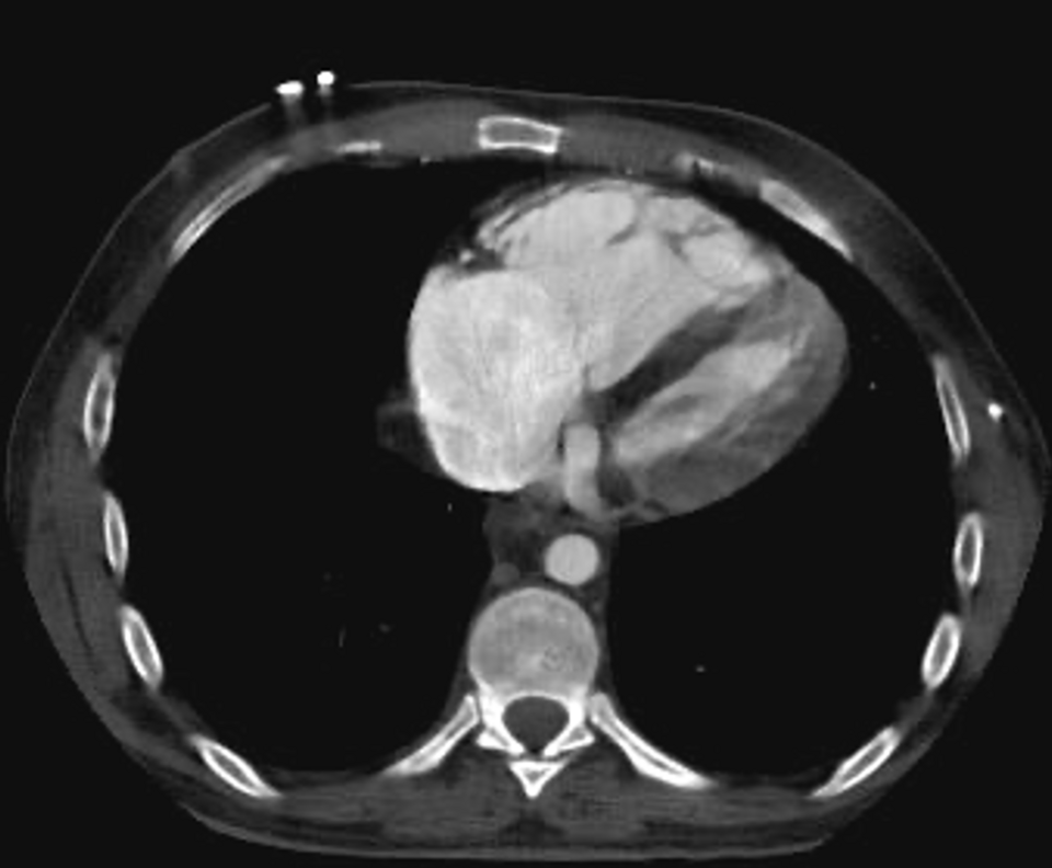

RV assessment on CT. The RV/LV diameter ratio is measured at the widest internal diameters, from endocardium to septum, on axial images or reformatted four-chamber views. The 2026 guideline recommends reporting the numerical ratio over subjective assessment (Class 1). In a meta-analysis of 49 studies, an increased RV/LV ratio was associated with about a 2.5-fold risk of all-cause death and a 5-fold risk of PE-related death, whereas thrombus load and central location did not predict all-cause mortality. Reflux of contrast into the inferior vena cava and hepatic veins and septal flattening or bowing are supportive signs. Quantification of thrombus burden is not used for risk stratification in categories A to C (Class 3: No Benefit).
V/Q scanning. A normal perfusion scan excludes PE, and a high-probability scan (two or more large mismatched segmental perfusion defects) confirms it. Intermediate and low-probability readings are nondiagnostic in patients with discordant clinical probability. Single-photon emission computed tomography (SPECT) V/Q is reasonable over planar imaging (Class 2a), because it reduces nondiagnostic results. V/Q performs best with a normal chest radiograph and no chronic lung disease. It is the alternative when CTPA cannot be performed (Class 2a, over contrast magnetic resonance angiography), most often with iodinated contrast allergy or severe kidney disease, and it is the screening test for CTEPD. Magnetic resonance pulmonary angiography has low sensitivity for distal emboli and a high rate of inadequate studies.
Compression ultrasound. Venous duplex of the legs is reasonable when finding DVT would change management or prognosis (Class 2b). A proximal DVT in a patient with suspected PE justifies anticoagulation without chest imaging when CTPA is contraindicated or the patient is unstable, and DVT in a patient with PE marks a higher risk of early death and recurrence. Duplex adds nothing after a negative CTPA or a normal SPECT V/Q (Class 3: No Benefit), and CT venography is not added routinely to CTPA (Class 3: No Benefit).
Subsegmental pulmonary embolism. Isolated subsegmental emboli are detected more often with modern CT and include false-positive readings. The CHEST 2021 guideline suggests clinical surveillance over anticoagulation for isolated subsegmental PE without proximal DVT in patients at low risk of recurrence, and anticoagulation for patients at high risk of recurrence (hospitalization, reduced mobility, active cancer, no reversible risk factor) or with poor cardiopulmonary reserve. In a prospective cohort managed without anticoagulation after negative bilateral leg ultrasound, recurrent VTE at 90 days was 3.1% overall: 2.1% with a single and 5.7% with multiple subsegmental emboli. The rate exceeded the predefined safety threshold, and the study stopped early.
| Test | Strengths | Limitations | Preferred setting |
|---|---|---|---|
| CT pulmonary angiography | Direct visualization to subsegmental level; RV/LV ratio; alternative diagnoses | Iodinated contrast; radiation, highest to the breast; artifacts | Default test (Class 1 over V/Q) |
| SPECT V/Q | No iodinated contrast; low radiation to breast; fewer nondiagnostic results than planar | No alternative diagnoses; less available after hours; abnormal with lung disease | Contrast allergy, severe kidney disease, normal chest radiograph; CTEPD screening |
| Planar V/Q | Normal and high-probability scans are definitive | Many intermediate or low-probability readings | When SPECT is unavailable |
| Leg compression ultrasound | Bedside; no radiation; proximal DVT justifies treatment | A negative study does not exclude PE | Unstable patients, pregnancy with leg symptoms, contraindication to CTPA |
| Magnetic resonance pulmonary angiography | No radiation or iodinated contrast | Low sensitivity for distal emboli; many inadequate studies | Experienced centers only |
| Echocardiography | Bedside RV assessment; thrombus in transit; alternative causes of shock | Normal RV does not exclude PE; RV dilation is not specific | Risk stratification; shock when CTPA is unsafe (Class 3: No Benefit for diagnosis) |
Diagnostic tests for acute PE and their place in the 2026 AHA/ACC recommendations.
Electrocardiogram, chest radiograph, and laboratory findings
The electrocardiogram (ECG) neither confirms nor excludes PE. Sinus tachycardia is the most common abnormality. Signs of RV strain include T-wave inversion in V1 to V4, often with inferior T-wave inversion, complete or incomplete right bundle branch block, right axis deviation, P pulmonale, and the S1Q3T3 pattern (a prominent S wave in lead I, a Q wave and inverted T wave in lead III). S1Q3T3 is insensitive and occurs in other causes of acute cor pulmonale. Atrial fibrillation or flutter can be new. ST elevation in V1 and aVR or diffuse ST depression appears with severe RV ischemia. RV strain on the ECG is associated with RV dysfunction and adverse short-term outcome.
The chest radiograph is often normal or shows nonspecific atelectasis, small effusion, or an elevated hemidiaphragm. Its role is to identify alternative diagnoses and to interpret V/Q scans. Classic signs are uncommon: the Westermark sign (regional oligemia distal to an occluded artery), the Hampton hump (a peripheral wedge-shaped opacity from infarction), and the Fleischner sign (an enlarged central pulmonary artery).
Arterial blood gases typically show hypoxemia, hypocapnia, respiratory alkalosis, and a widened alveolar–arterial gradient; a normal gradient does not exclude PE. Hypercapnia indicates respiratory failure or massive obstruction with exhaustion.
Biomarkers. Cardiac troponin rises with RV myocyte injury, and B-type natriuretic peptide (BNP) or N-terminal proBNP (NT-proBNP) rises with RV wall stretch. Both are prognostic markers. Their negative predictive value for adverse early outcome is high, and their positive predictive value is low. At least one cardiac biomarker is measured in category C (Class 1), and the result, with RV imaging, assigns C1, C2, or C3. Lactate is measured in categories C to E at acute care facilities (Class 1): a level above 2 mmol/L is a marker of hypoperfusion that defines normotensive shock in category D2 and is associated with death independent of blood pressure.
Echocardiographic signs of right ventricular strain
Echocardiography is not used to confirm or exclude PE (Class 3: No Benefit), because a normal RV does not exclude PE and RV dilation has many causes. Its roles are risk stratification in categories C and D, for which RV imaging is recommended (Class 1) and echocardiography is preferred over CT (Class 2a), and bedside evaluation of shock when CT cannot be performed safely. In a patient in shock with high clinical probability, absence of RV dilation or dysfunction makes PE an unlikely cause of the shock.
| Parameter | Abnormal value | Comment |
|---|---|---|
| RV end-diastolic diameter | ≥30 mm; basal diameter ≥41 mm | End-diastole in the RV-focused parasternal long-axis view; basal diameter in the RV-focused apical view with the free wall visible throughout the cycle |
| RV/LV end-diastolic diameter ratio | >0.9 | Apical four-chamber or subcostal view |
| TAPSE | <17 mm | M-mode along the lateral tricuspid annulus; load-dependent and falls with acute afterload |
| Tricuspid annular systolic velocity (TDI S′) | <9.5 cm/s | Pulsed tissue Doppler at the lateral tricuspid annulus |
| Peak tricuspid regurgitant velocity | ≥2.9 m/s | Suggests pulmonary hypertension; underestimates pressure when RV function is poor |
Echocardiographic RV parameters in Table 4 of the 2026 AHA/ACC guideline, which recommends reporting them (Class 1). TAPSE, tricuspid annular plane systolic excursion.
McConnell sign. Akinesis of the mid RV free wall with normal or hyperdynamic apical motion was described in 1996 as a specific sign of acute PE. Later studies found the same pattern in RV infarction, which is distinguished by inferior LV wall motion abnormality from proximal right coronary occlusion. The sign supports acute RV pressure overload; it does not establish the cause.
60/60 sign. A pulmonary acceleration time of 60 ms or less with a tricuspid regurgitant pressure gradient of 60 mm Hg or less indicates an acutely pressure-loaded RV that has not had time to hypertrophy. In chronic pulmonary hypertension, the gradient is usually higher. The sign has good specificity and low sensitivity.
Other findings. A dilated, noncollapsing inferior vena cava indicates raised right atrial pressure. Septal flattening in systole and diastole (a D-shaped LV in the parasternal short axis) reflects RV pressure overload and an underfilled LV. RV free-wall thickness above 5 mm suggests chronic disease. TAPSE and RV free-wall strain fall with acute afterload and recover after reperfusion.
Thrombus in transit. A mobile, serpiginous thrombus in the right atrium or ventricle, sometimes prolapsing through the tricuspid valve or straddling a patent foramen ovale, is a venous cast in transit to the lungs. It is associated with RV dysfunction and high early mortality. Advanced therapy over anticoagulation alone is reasonable for free-floating clot in transit in categories C3 to E2 (Class 2a). Options include systemic thrombolysis, catheter aspiration, and surgical removal, chosen by bleeding risk, the size and location of the thrombus, the presence of a patent foramen ovale, and local expertise.
AHA/ACC acute pulmonary embolism clinical categories
The 2026 guideline replaces the older terms "massive," "submassive," and "low risk," and the European high, intermediate-high, intermediate-low, and low-risk classes, with five clinical categories. They combine symptoms, a clinical severity score, RV imaging, cardiac biomarkers, perfusion, and respiratory status. A patient is placed in the highest category any feature qualifies for and can move between categories as the illness evolves.
| Category | Definition | Criteria | Disposition and reperfusion |
|---|---|---|---|
| A | Subclinical: incidental, asymptomatic PE | No PE-attributable symptoms | Outpatient care in most; no reperfusion |
| B | Symptomatic PE with low clinical severity | PESI class I–II (≤85), sPESI 0, Bova ≤4, or Hestia negative. B1 subsegmental PE; B2 more proximal PE | Early discharge for selected patients (Class 2a); systemic thrombolysis harmful |
| C1 | Symptomatic PE with elevated clinical severity | PESI class III–V, sPESI ≥1, Bova >4, or Hestia positive; normal RV and normal biomarkers | Hospitalization; anticoagulation alone; catheter and surgical therapy not recommended (Class 3: No Benefit) |
| C2 | Elevated severity with RV or biomarker abnormality | Abnormal RV on imaging or at least one abnormal biomarker (troponin or natriuretic peptide) | Systemic thrombolysis harmful (Class 3: Harm); catheter therapy of uncertain benefit (Class 2b) |
| C3 | Elevated severity with RV and biomarker abnormality | Abnormal RV and at least one abnormal biomarker; MAP below 80 mm Hg may flag escalation (Class 2a) | Systemic thrombolysis, catheter-directed thrombolysis, or mechanical thrombectomy of uncertain benefit (Class 2b) |
| D1 | Incipient cardiopulmonary failure: transient hypotension | Systolic pressure below 90 mm Hg or a fall of more than 40 mm Hg lasting less than 15 minutes, or responding to fluids; no hypoperfusion | Advanced therapy may be considered (Class 2b) |
| D2 | Incipient cardiopulmonary failure: normotensive shock | Any of: lactate above 2 mmol/L, acute kidney injury or urine output below 0.5 mL/kg/h, altered mental status, cardiac index below 2.2 L/min/m², MAP below 60 mm Hg, or a higher shock stage | Advanced therapy may be considered (Class 2b); vasopressors or inotropes for shock (Class 1) |
| E1 | Cardiopulmonary failure | Persistent or recurrent hypotension with cardiogenic shock | Systemic thrombolysis, catheter-directed thrombolysis, mechanical thrombectomy, or surgical embolectomy reasonable (Class 2a) |
| E2 | Refractory cardiogenic shock or cardiac arrest | Refractory cardiogenic shock (SCAI stage D–E), or cardiac arrest without return of spontaneous circulation after 30 minutes of resuscitation | Systemic thrombolysis reasonable (Class 2a); VA-ECMO reasonable (Class 2a) |
2026 AHA/ACC acute PE clinical categories. A respiratory modifier (R, for example C3R) is added in categories C to E for clinically important hypoxemia, rising oxygen requirement, tachypnea, or ventilatory failure. Abnormal RV is defined by the echocardiographic criteria in the preceding table or by the CT RV/LV ratio. PESI, Pulmonary Embolism Severity Index; sPESI, simplified PESI; MAP, mean arterial pressure; VA-ECMO, venoarterial extracorporeal membrane oxygenation.
The categories separate questions that the older classes merged. Category C contains most hospitalized patients. Its three subcategories reflect the observation that RV dysfunction and biomarker elevation each add risk, and that the combination (C3, roughly the European intermediate-high group) carries the highest risk of decompensation in a normotensive patient. Category D recognizes two states that precede overt shock: transient hypotension (D1), often syncope or a fluid-responsive fall in pressure, and normotensive shock (D2), in which pressure is maintained by vasoconstriction while perfusion fails.
| AHA/ACC 2026 category | European Society of Cardiology (ESC) 2019 risk class | AHA 2011 term |
|---|---|---|
| A, B | Low risk (PESI I–II or sPESI 0) | Low risk |
| C1 | Intermediate-low (elevated score, no RV or troponin abnormality) | Low risk |
| C2 | Intermediate-low (RV dysfunction or elevated troponin) | Submassive (RV dysfunction or myocardial necrosis) |
| C3 | Intermediate-high (RV dysfunction and elevated troponin) | Submassive |
| D1, D2 | Intermediate-high, or high risk when hypotension persists | Submassive or massive |
| E1, E2 | High risk (cardiac arrest, obstructive shock, or persistent hypotension) | Massive (systolic pressure below 90 mm Hg for at least 15 minutes or requiring inotropic support) |
Approximate correspondence between the three classification systems. The mapping is not exact, because the systems use different variables, and trials enrolled patients by the older definitions.
The European high-risk definition uses cardiac arrest, obstructive shock (systolic pressure below 90 mm Hg or vasopressors to maintain 90 mm Hg despite adequate filling, with end-organ hypoperfusion), or persistent hypotension (systolic pressure below 90 mm Hg or a fall of 40 mm Hg or more for more than 15 minutes, not caused by arrhythmia, hypovolemia, or sepsis). The older AHA term "submassive" combined normotension with RV dysfunction or myocardial necrosis and grouped patients whose risks differ several-fold.
Clinical severity scores: PESI, sPESI, Bova, and Hestia
A PE-specific score is applied after diagnosis. Hestia, PESI, or sPESI is recommended to identify low-severity patients in categories A and B (Class 1), and a PE-specific score is reasonable in hemodynamically stable patients in categories C and D (Class 2a). The National Early Warning Score (NEWS) and NEWS2 are alternatives (Class 2b).
PESI uses 11 variables, with age entered in years. Thirty-day mortality in the derivation and validation samples was 0% to 1.6% in class I, 1.7% to 3.5% in class II, 3.2% to 7.1% in class III, 4.0% to 11.4% in class IV, and 10.0% to 24.5% in class V. The simplified PESI (sPESI) keeps six variables with one point each. Patients with sPESI 0 had a 30-day mortality of 1.0%, and those with 1 or more had a mortality of 10.9%. The scores capture comorbidity and physiology, so an older patient with cancer can have an elevated score from a small embolism, and a younger patient with a large embolism and RV dilation can score low. The scores define clinical severity; RV imaging and biomarkers define the RV component.
PESI and simplified PESI
30-day mortality classesPESI points: age in years; male sex 10; cancer 30; chronic heart failure 10; chronic lung disease 10; heart rate ≥110/min 20; systolic pressure <100 mm Hg 30; respiratory rate ≥30/min 20; temperature <36 °C 20; altered mental status 60; oxygen saturation <90% 20. Classes: I ≤65, II 66–85, III 86–105, IV 106–125, V >125. sPESI: one point each for age >80 years, cancer, chronic cardiopulmonary disease, heart rate ≥110/min, systolic pressure <100 mm Hg, and saturation <90%. The example (72-year-old man with chronic lung disease and heart rate 112/min) scores PESI 112, class IV, and sPESI 2.
Bova score. The Bova score was derived in normotensive patients and combines systolic pressure 90 to 100 mm Hg (2 points), elevated troponin (2), RV dysfunction (2), and heart rate 110/min or more (1). Thirty-day rates of PE-related death, hemodynamic collapse, or recurrence were 4.2% in stage I (0 to 2 points), 10.8% in stage II (3 to 4), and 29.2% in stage III (more than 4). Because it includes RV and troponin data, it overlaps with the C2 and C3 subcategories.
Bova score
Normotensive patients with confirmed PEStages: I, 0 to 2 points; II, 3 to 4; III, more than 4. The example (elevated troponin, RV dysfunction, heart rate 110/min or more) scores 5, stage III.
Hestia criteria. Hestia is a set of 11 yes-or-no questions. They address hemodynamic instability, the need for thrombolysis or embolectomy, active bleeding or high bleeding risk, oxygen for more than 24 hours to keep saturation above 90%, PE diagnosed during anticoagulation, severe pain needing intravenous analgesia for more than 24 hours, a medical or social reason for admission longer than 24 hours, creatinine clearance below 30 mL/min, severe liver impairment, pregnancy, and a history of heparin-induced thrombocytopenia. Any "yes" makes the rule positive. In the HOME-PE trial of 1,975 normotensive patients, triage by Hestia and by sPESI (with physician override) produced similar 30-day rates of recurrence, major bleeding, or death (3.82% and 3.57%) and similar rates of home treatment (38.4% and 36.6%). Neither home-treatment group had a recurrent or fatal PE.
Outpatient treatment of low-risk pulmonary embolism
A decision tool (Hestia, PESI, or sPESI) is reasonable to assess suitability for outpatient care (Class 2a), and outpatient treatment is reasonable in selected patients in categories A and B with a low risk of 90-day adverse events, when anticoagulant access and follow-up are assured (Class 2a). DOACs that need no parenteral lead-in (apixaban and rivaroxaban) make same-day discharge practical.
Discharge from the emergency department requires more than a low score. The patient is hemodynamically stable with a normal or near-normal heart rate, needs no supplemental oxygen, has adequate pain control and no active bleeding or high bleeding risk, has no condition needing admission, can obtain and take the drug the same day, understands warning symptoms, and has follow-up arranged within a week. Social circumstances (housing, support, transport, and the ability to return) decide as often as physiology. Patients with sPESI 0 who have RV dilation on CT, cancer, or an elevated biomarker are evaluated individually, because these findings carry risk not captured by the score.
Anticoagulation: agents, dosing, and special circumstances
Anticoagulation is started in every patient with confirmed PE who has no absolute contraindication (Class 1). When suspicion is high, imaging is delayed, and bleeding risk is low, therapeutic anticoagulation before confirmation is reasonable in patients likely to be in category C2 or higher (Class 2a). Low-molecular-weight heparin (LMWH) is recommended over unfractionated heparin (UFH) for initial parenteral therapy in categories C1 to E1 (Class 1). It has a more predictable dose response and lower rates of recurrence, bleeding, and heparin-induced thrombocytopenia. UFH is chosen when an immediate procedure, systemic thrombolysis, or rapid reversal is likely, and in severe kidney failure. After thrombolysis or an endovascular procedure, LMWH is preferred over UFH (Class 1), and LMWH is reasonable even when a procedure is likely (Class 2a).
When oral therapy is appropriate, a DOAC is recommended over a vitamin K antagonist (Class 1). In the phase 3 trials (EINSTEIN-PE, AMPLIFY, Hokusai-VTE, RE-COVER), DOACs were noninferior to warfarin for recurrent VTE and caused less major or clinically relevant bleeding, most clearly less intracranial hemorrhage. Apixaban and rivaroxaban are started at a higher loading dose without parenteral therapy. Dabigatran and edoxaban follow at least 5 days of parenteral anticoagulation. Warfarin is overlapped with parenteral therapy for at least 5 days and until the international normalized ratio (INR) is 2.0 or more for 24 hours, with a target of 2.5 (range 2.0 to 3.0).
| Agent | Treatment dose | Dose adjustment and cautions |
|---|---|---|
| Unfractionated heparin | 80 U/kg bolus, then 18 U/kg/h, adjusted to aPTT or anti-Xa level | Preferred with planned systemic lysis, imminent procedure, or creatinine clearance <30 mL/min with bleeding concern; short half-life; protamine reverses fully |
| Enoxaparin | 1 mg/kg every 12 h (or 1.5 mg/kg once daily) | 1 mg/kg once daily when creatinine clearance <30 mL/min; anti-Xa monitoring reasonable in severe kidney disease |
| Dalteparin | 200 IU/kg once daily | Cancer regimen: 200 IU/kg daily for 1 month, then 150 IU/kg daily |
| Fondaparinux | 5 mg (<50 kg), 7.5 mg (50–100 kg), 10 mg (>100 kg) once daily | Contraindicated with creatinine clearance <30 mL/min; used in heparin-induced thrombocytopenia |
| Apixaban | 10 mg twice daily for 7 days, then 5 mg twice daily | Extended phase: 2.5 mg twice daily after 6 months; no parenteral lead-in |
| Rivaroxaban | 15 mg twice daily for 21 days, then 20 mg once daily with food | Extended phase: 10 mg once daily after 6 months; avoid with creatinine clearance <15 mL/min |
| Edoxaban | 60 mg once daily after ≥5 days of parenteral therapy | 30 mg with creatinine clearance 15–50 mL/min, weight ≤60 kg, or certain P-glycoprotein inhibitors |
| Dabigatran | 150 mg twice daily after 5–10 days of parenteral therapy | Avoid with creatinine clearance <30 mL/min; idarucizumab reverses |
| Warfarin | Overlap with parenteral therapy ≥5 days and until INR ≥2.0 for 24 h; target INR 2.5 | Preferred in thrombotic antiphospholipid syndrome, Child-Pugh C liver disease, and some valve indications |
Doses from US prescribing information and the CHEST 2021 guideline. aPTT, activated partial thromboplastin time; anti-Xa, anti–factor Xa activity.
Kidney disease, body weight, and liver disease. In chronic kidney disease stages 2 and 3, a DOAC is recommended over a vitamin K antagonist (Class 1). In stages 4 and 5 or on dialysis, the benefit of apixaban over warfarin is uncertain (Class 2b), and dosing follows labeling. With a body mass index above 30 kg/m², a DOAC is reasonable over a vitamin K antagonist (Class 2a), including severe obesity, with apixaban and rivaroxaban having the most data. With a body mass index above 40 kg/m², reducing LMWH dose may be reasonable (Class 2b). Routine anti-Xa monitoring is not indicated for most patients on weight-based LMWH (Class 3: No Benefit); when it is used, a peak level is drawn 3 to 5 hours after a dose at steady state. A DOAC is reasonable in Child-Pugh A liver disease (Class 2a), may be reasonable in Child-Pugh B (Class 2b), and is not recommended in Child-Pugh C (Class 3: Harm).
Antiphospholipid syndrome and other drug choices. In TRAPS, which enrolled triple-positive patients, thromboembolic events occurred in 12% of patients on rivaroxaban and in none on warfarin, and the trial stopped early. A DOAC may be an alternative when only a single anticardiolipin or anti-β2-glycoprotein I antibody is present (Class 2b). In patients with a primary or metastatic brain tumor, a DOAC may be considered over LMWH to reduce intracranial hemorrhage (Class 2b). Drug interactions with strong inhibitors or inducers of cytochrome P450 3A4 and P-glycoprotein (some antiretrovirals, azole antifungals, rifampin, carbamazepine, phenytoin) exclude or alter DOAC use.
Bleeding and reversal. Major bleeding on anticoagulation is managed by stopping the drug, local hemostasis, transfusion, and reversal for life-threatening or critical-site bleeding. Protamine fully reverses UFH and partially reverses LMWH. Idarucizumab reverses dabigatran. In the United States, bleeding on apixaban, rivaroxaban, or edoxaban is treated with four-factor prothrombin complex concentrate. Andexanet alfa was withdrawn from the US market after the US Food and Drug Administration concluded that its risks outweigh its benefits, but it remains available in some other countries. Warfarin is reversed with four-factor prothrombin complex concentrate and intravenous vitamin K. The decision to restart anticoagulation balances recurrent VTE risk, highest in the first month after diagnosis, against rebleeding. An inferior vena cava filter bridges the interval when anticoagulation must be withheld during the first weeks.
Pulmonary embolism response teams and the reperfusion decision
A pulmonary embolism response team (PERT) is a multidisciplinary group, typically cardiology, pulmonary and critical care, emergency medicine, interventional radiology or cardiology, cardiac surgery, hematology, and vascular medicine, that can be activated at any hour. Assessment by a PERT is recommended for categories C to E (Class 1). Its tasks are to confirm the category, judge bleeding risk, choose and time therapy, and plan transfer when needed. Transfer of stable C3 and D patients to a center offering advanced therapy may be considered (Class 2b). Patients in category E are not transferred before stabilization (Class 3: Harm). Systemic or catheter-directed thrombolysis requires intensive or intermediate care monitoring (Class 1).
The reperfusion decision weighs the physiologic trajectory more than clot burden. Advanced therapy reduces RV afterload faster than anticoagulation alone; the risks are bleeding, including intracranial hemorrhage with thrombolytics, and procedural complications. In categories A to C1, anticoagulation alone is the standard, and catheter-based and surgical therapies are not recommended (Class 3: No Benefit). In C2, systemic thrombolysis remains harmful (Class 3: Harm), and catheter-directed thrombolysis and mechanical thrombectomy have unclear benefit (Class 2b). In C3, systemic thrombolysis, catheter-directed thrombolysis, and mechanical thrombectomy all have unclear benefit (Class 2b). Surgical embolectomy is not recommended over anticoagulation alone in categories A to C3 (Class 3: No Benefit). The randomized trials published since the guideline bear on categories C2 and C3 most. In categories D and E, the risk of death outweighs bleeding risk for most patients.
| Therapy | A–C1 | C2 | C3 | D1–D2 | E1 | E2 |
|---|---|---|---|---|---|---|
| Anticoagulation | 1 | 1 | 1 | 1 | 1 | 1 |
| Systemic thrombolysis | 3: Harm | 3: Harm | 2b | 2b | 2a | 2a |
| Catheter-directed thrombolysis | 3: No Benefit | 2b | 2b | 2b | 2a | — |
| Mechanical thrombectomy | 3: No Benefit | 2b | 2b | 2b | 2a | — |
| Surgical embolectomy | 3: No Benefit | 3: No Benefit | 3: No Benefit | 2b | 2a | 3: No Benefit over other advanced therapy without MCS |
| VA-ECMO | — | — | — | — | — | 2a |
| IVC filter added to anticoagulation | 3: Harm | 3: Harm | 3: Harm | 2b (with advanced therapy) | 2b (with advanced therapy) | 2b (with advanced therapy) |
Class of recommendation by clinical category in the 2026 AHA/ACC guideline. A dash indicates no category-specific recommendation. MCS, mechanical circulatory support; IVC, inferior vena cava.
Systemic thrombolysis: regimens, evidence, and contraindications
Systemic thrombolysis is given with anticoagulation when bleeding risk is acceptable. Lower-dose regimens may be considered to reduce bleeding (Class 2b).
| Regimen | Dose | Notes |
|---|---|---|
| Alteplase, standard (FDA-approved) | 100 mg intravenously over 2 hours | UFH is held during the infusion and resumed without a bolus when the aPTT is below twice the upper limit of normal |
| Alteplase, accelerated | 0.6 mg/kg (maximum 50 mg) over 15 minutes | Used in cardiac arrest and imminent arrest |
| Alteplase, reduced dose | 50 mg over 2 hours (MOPETT used 0.5 mg/kg below 50 kg) | Class 2b; smaller trials only |
| Tenecteplase (off label in the United States) | Weight-based single bolus, 30–50 mg | PEITHO regimen; higher intracranial hemorrhage in older patients |
| Catheter-directed alteplase | Standard 5–10 mg per pulmonary artery; SEATTLE II 24 mg total; ULTIMA 10–20 mg over 15 hours | Doses below 5 mg per artery are not recommended over 5–10 mg (Class 3: No Benefit) |
Thrombolytic regimens for acute PE. Thrombolysis is most effective within 48 hours of symptom onset and has benefit up to 14 days.
Evidence. PEITHO randomized 1,005 normotensive patients with RV dysfunction and a positive troponin (European intermediate-high risk, roughly AHA/ACC category C3) to weight-based tenecteplase or placebo with heparin. Death or hemodynamic decompensation within 7 days occurred in 2.6% with tenecteplase and 5.6% with placebo, a benefit driven by fewer decompensations. Mortality did not differ at 7 days (1.2% and 1.8%) or 30 days (2.4% and 3.2%). Major extracranial bleeding occurred in 6.3% and 1.2%, and stroke in 2.4% (10 of 12 hemorrhagic) and 0.2%. In a meta-analysis of 16 trials, thrombolysis lowered all-cause mortality (odds ratio 0.53) at the cost of more major bleeding (9.24% vs 3.42%) and intracranial hemorrhage (1.46% vs 0.19%), with the bleeding excess concentrated in patients older than 65 years. These data are the basis for withholding systemic thrombolysis in normotensive patients until they deteriorate. In MOPETT, half-dose alteplase in "moderate" PE reduced pulmonary hypertension at follow-up without major bleeding in a small, open-label trial.
| Absolute contraindications | Relative contraindications |
|---|---|
| Any prior intracranial hemorrhage | Age above 75 years |
| Known structural cerebral vascular lesion or malignant intracranial neoplasm | Current therapeutic anticoagulation |
| Ischemic stroke within 3 months | Pregnancy or first postpartum week |
| Suspected aortic dissection | Noncompressible vascular puncture |
| Active bleeding or bleeding diathesis | Traumatic or prolonged (more than 10 minutes) cardiopulmonary resuscitation |
| Recent brain or spinal surgery | Internal bleeding within 2 to 4 weeks |
| Significant closed-head or facial trauma within 3 months | Uncontrolled hypertension (systolic above 180 or diastolic above 110 mm Hg) |
| Major surgery within 3 weeks; remote ischemic stroke; dementia |
Contraindications to systemic thrombolysis, adapted from the 2011 AHA scientific statement and the CHEST guideline. In cardiac arrest from PE, contraindications are weighed against certain death.
Catheter-directed thrombolysis and mechanical thrombectomy
Catheter-directed thrombolysis (CDL) delivers a small dose of alteplase through multi–side-hole infusion catheters in one or both pulmonary arteries, with or without ultrasound to disperse fibrin. Mechanical thrombectomy (MT) removes thrombus with large-bore aspiration or extraction devices without a thrombolytic. Both reduce the RV/LV ratio and pulmonary pressure within hours to days.
CDL and MT carry identical class ratings in every category. Whether CDL or MT is better than systemic thrombolysis in D1 to E1 is uncertain, though both may cause less bleeding (Class 2b). The relative benefit of CDL and MT in C2 to D2 is uncertain (Class 2b). During CDL, therapeutic LMWH or UFH, or subtherapeutic UFH, is recommended over no anticoagulation (Class 1).
| Trial | Design and population | Intervention | Main result |
|---|---|---|---|
| PEITHO (2014) | RCT, 1,005 normotensive patients with RV dysfunction and positive troponin | Tenecteplase vs placebo with heparin | Death or decompensation at 7 days 2.6% vs 5.6%; stroke 2.4% vs 0.2% |
| ULTIMA (2014) | RCT, 59 patients, echocardiographic RV/LV ≥1.0 | Ultrasound-assisted CDL, 10–20 mg alteplase over 15 h, vs heparin | RV/LV reduction at 24 h 0.30 vs 0.03; no major bleeding |
| SEATTLE II (2015) | Single-arm, 150 patients, massive or submassive PE | Ultrasound-facilitated CDL, 24 mg alteplase | RV/LV 1.55 to 1.13 at 48 h; PA systolic pressure 51 to 37 mm Hg; 10% moderate or severe bleeding; no intracranial hemorrhage |
| FLARE (2019) | Single-arm, 106 patients, intermediate-risk PE | Large-bore aspiration thrombectomy (FlowTriever) | RV/LV reduction 0.38 at 48 h; major bleeding 1.0%; 41% needed no intensive care stay |
| PEERLESS (2025) | RCT, 550 intermediate-risk patients | Large-bore MT vs CDL | Win ratio 5.01 favoring MT, driven by less deterioration or bailout (1.8% vs 5.4%) and less intensive care use; no difference in mortality, intracranial hemorrhage, or major bleeding |
| HI-PEITHO (2026) | RCT, 544 patients with RV/LV ≥1.0, elevated troponin, and at least two markers of cardiorespiratory distress | Ultrasound-facilitated CDL plus anticoagulation vs anticoagulation alone | PE-related death, decompensation, or recurrence at 7 days 4.0% vs 10.3% (relative risk 0.39); major bleeding at 30 days 4.1% vs 3.0%; no intracranial hemorrhage |
Key trials of thrombolytic and catheter-based therapy in acute PE. RCT, randomized controlled trial; PA, pulmonary artery.
HI-PEITHO, published after the 2026 guideline, is the first randomized trial to show a clinical benefit of a catheter-based therapy over anticoagulation alone in normotensive patients. Its population resembles category C3 with at least two markers of cardiorespiratory distress (heart rate 100/min or more, systolic pressure 110 mm Hg or less, or respiratory rate above 20/min). The benefit came from fewer cases of cardiorespiratory decompensation; PE-related death and recurrence did not differ, and mortality was low in both groups. The result applies to the ultrasound-facilitated system and alteplase protocol tested. PEERLESS compared two catheter strategies and did not include an anticoagulation-alone group. In STORM-PE, 100 normotensive patients with an RV/LV ratio of 1.0 or more and elevated biomarkers were randomized to computer-assisted vacuum thrombectomy plus anticoagulation or anticoagulation alone; the RV/LV ratio fell more at 48 hours with thrombectomy (0.52 vs 0.24), and major adverse events at 7 days did not differ (4.3% vs 7.5%), in a trial not powered for clinical outcomes. PEERLESS II (large-bore thrombectomy vs anticoagulation alone, NCT06055920) and PEITHO-3 (reduced-dose alteplase vs placebo, NCT04430569) test clinical outcomes in the same population.
Procedural complications include access-site bleeding, pulmonary artery injury and hemoptysis, arrhythmia during passage through the right heart, and blood loss with large-bore aspiration. After MT, stable patients are monitored on telemetry with nursing trained in the device (Class 2a).
Surgical embolectomy, mechanical circulatory support, and resuscitation
Surgical embolectomy. Surgical pulmonary embolectomy is performed through median sternotomy on cardiopulmonary bypass, usually normothermic and on the beating heart, with arteriotomy of the main and proximal branch pulmonary arteries and removal of central thrombus. It is suited to patients with central thrombus and a contraindication to thrombolysis, failed thrombolysis or catheter therapy, thrombus in transit or straddling a patent foramen ovale, and to centers with surgical expertise. In E2 without mechanical circulatory support, it is not recommended over other advanced therapies (Class 3: No Benefit), because outcomes in patients operated on during arrest are poor.
Hemodynamic support of the failing RV. Vasopressors or inotropes are recommended for cardiogenic shock in categories D2 to E2 (Class 1). Norepinephrine restores systemic pressure and RV coronary perfusion and is the vasopressor of choice; vasopressin can be added because it raises systemic resistance with little effect on pulmonary resistance. Dobutamine increases RV contractility but can cause vasodilation and hypotension and is used with a vasopressor. A cautious volume challenge may be considered when preload is low (Class 2b), and large volumes are avoided, because RV dilation worsens septal shift, tricuspid regurgitation, and LV filling. Inhaled pulmonary vasodilators (nitric oxide or epoprostenol) may be considered to reduce RV afterload in categories C2 to E (Class 2b).
Intubation and ventilation. Induction of anesthesia and positive-pressure ventilation can precipitate arrest in a patient with an afterload-sensitive RV, by vasodilation, loss of sympathetic tone, and increased intrathoracic pressure that reduces venous return and raises pulmonary resistance. In two surgical embolectomy series cited by the guideline, cardiopulmonary resuscitation was needed after anesthesia induction in 19% and 28% of patients, including patients who were hemodynamically stable at induction. Deep sedation and mechanical ventilation are avoided unless clinically required (Class 3: Harm). Heated high-flow nasal cannula is beneficial over standard nasal oxygen for moderate to severe hypoxemia (Class 2a). When intubation is unavoidable, vasopressors, inotropes, and venoarterial ECMO are prepared beforehand (Class 1), hemodynamically neutral induction drugs are used, and low tidal volumes and plateau pressures are targeted.
Venoarterial ECMO. VA-ECMO is reasonable in category E2 refractory cardiogenic shock (Class 2a). It unloads the RV, restores systemic perfusion and oxygenation, and provides time for endogenous or therapeutic clot resolution. Parenteral anticoagulation is continued on ECMO unless bleeding prevents it (Class 1). The benefit of adding thrombolysis, CDL, MT, or surgery on ECMO has not been established (Class 2b). Other RV support devices, including percutaneous RV assist pumps, have been used in selected patients.
Cardiac arrest. PE causes a substantial share of in-hospital arrests with pulseless electrical activity. When PE is suspected or confirmed during cardiopulmonary resuscitation, thrombolysis is reasonable, and resuscitation is continued for at least 60 to 90 minutes after thrombolytic administration because return of circulation can be delayed. Extracorporeal cardiopulmonary resuscitation is an option in experienced systems. Point-of-care echocardiography during pulse checks can show a dilated RV, which supports, though it does not prove, PE as the cause.
Inferior vena cava filters
A filter is indicated for acute PE or proximal DVT when anticoagulation cannot be given (Class 2a), and a retrievable filter is preferred over a permanent one (Class 1). Routine filter placement in patients who are therapeutically anticoagulated is harmful (Class 3: Harm). In PREPIC2, a retrievable filter added to anticoagulation in 399 patients with PE and DVT did not reduce recurrent PE at 3 months (3.0% vs 1.5%). In the earlier PREPIC trial, permanent filters reduced early PE but increased DVT at 2 years. A filter may be considered for recurrent PE despite optimal anticoagulation (Class 2b), and its benefit in patients in categories D and E receiving advanced therapy is uncertain (Class 2b).
Retrieval is attempted once anticoagulation is tolerated and the acute risk has fallen (Class 1). The US Food and Drug Administration recommends considering removal between 29 and 54 days after implantation, when the risk-benefit balance favors removal. Structured follow-up through a filter registry or clinic is reasonable (Class 2a), because many filters are never retrieved and complications (fracture, migration, caval perforation, caval thrombosis, and recurrent DVT) accumulate with dwell time.
Duration of anticoagulation and extended therapy
The 2026 AHA/ACC guideline names two phases: the initial treatment phase (the first 3 to 6 months, which includes the higher-intensity lead-in of the first 5 to 21 days) and the extended treatment phase (beyond 6 months, without a planned stop date and with periodic review). Every patient receives at least 3 months of treatment. The decision at 3 to 6 months is based on the risk factor category, bleeding risk, and patient preference.
| Clinical situation | Recommendation | Class |
|---|---|---|
| First PE with a major reversible risk factor | Stop at 3 to 6 months | 1 |
| First PE without a major reversible risk factor | Extend beyond 3 to 6 months | 1 |
| Persistent risk factor | Extend beyond 3 to 6 months | 1 |
| Minor reversible risk factor | Shared decision on stopping or extending | 2a |
| Agent for extended therapy | DOAC over vitamin K antagonist | 1 |
| Dose for extended therapy | Apixaban 2.5 mg twice daily or rivaroxaban 10 mg daily over full dose to reduce bleeding | 1 |
| Cancer, extended phase | DOAC or LMWH over vitamin K antagonist | 1 |
| DOAC contraindicated, no cancer | Vitamin K antagonist over aspirin or no therapy | 1 |
| Anticoagulation declined or contraindicated | Low-dose aspirin over no therapy | 2a |
| All patients on extended therapy | Periodic reassessment of risks and benefits | 1 |
Duration recommendations from Section 5.2 of the 2026 AHA/ACC guideline.
After a first event without a major reversible factor, stopping anticoagulation leads to recurrence in about 10% of patients in the first year and about 25% over 5 years, and recurrence presents as PE more often after an index PE than after DVT. Extended therapy prevents most of these events. In AMPLIFY-EXT, recurrent VTE or VTE-related death over 12 months occurred in 8.8% of patients on placebo and in 1.7% on apixaban 2.5 mg or 5 mg twice daily, with no increase in major bleeding. In EINSTEIN CHOICE, recurrent VTE occurred in 1.5% with rivaroxaban 20 mg, 1.2% with 10 mg, and 4.4% with aspirin 100 mg, with similar major bleeding. Aspirin reduces recurrence by about one-third and is the option only when anticoagulation is declined or contraindicated.
Recurrence risk scores. Scores estimate recurrence after stopping treatment in patients without a major transient factor. HERDOO2 applies to women: post-thrombotic signs (hyperpigmentation, edema, or redness), D-dimer of 250 µg/L or more on anticoagulation, body mass index of 30 kg/m² or more, and age 65 years or older. In REVERSE II, women with 0 or 1 point who stopped anticoagulation had a recurrence rate of about 3% per patient-year. The Vienna prediction model (sex, proximal DVT or PE, and D-dimer after stopping) and the DASH score (D-dimer after stopping, age under 50, male sex, and hormone use in women) estimate recurrence in men and women. These scores inform shared decisions; none is incorporated as a class-graded recommendation in the 2026 AHA/ACC guideline.
Bleeding risk. Bleeding risk on extended therapy rises with age, prior bleeding, cancer, kidney or liver disease, thrombocytopenia, antiplatelet therapy, poor anticoagulant control, and frailty. Validated tools include VTE-BLEED. A high bleeding risk favors a reduced DOAC dose or stopping after the primary phase; no fixed threshold dictates the choice.
Recurrent pulmonary embolism. Suspected recurrence is confirmed with CTPA or V/Q (Class 1) and compared with the baseline study, so a baseline image is valuable when residual defects are likely. Causes are evaluated (Class 1): nonadherence, subtherapeutic dosing, drug interactions, cancer, antiphospholipid syndrome, and heparin-induced thrombocytopenia. For true failure in an adherent patient, switching drug class is reasonable (Class 2a). Recurrence on a reduced-dose DOAC is treated with the full dose (Class 2a), and recurrence on LMWH in cancer is treated with a 20% to 25% dose increase (Class 2a).
Pulmonary embolism in pregnancy and the postpartum period
Pregnancy raises VTE risk several-fold, and the risk is highest in the 6 weeks after delivery. PE is a leading cause of maternal death in the United States. D-dimer rises through pregnancy, and dyspnea, tachycardia, and leg swelling are common in normal pregnancy, so both clinical assessment and testing are less specific.
The pregnancy-adapted YEARS algorithm may be reasonable (Class 2b). It begins with compression ultrasound when leg symptoms are present, then applies the three YEARS items with D-dimer thresholds of 1,000 ng/mL (no items) and 500 ng/mL (any item). In the Artemis study of 498 women, it excluded PE without CTPA in 39% (65% in the first trimester and 32% in the third), and the 3-month VTE rate was 0.21%. In pregnancy with a normal chest radiograph, low-dose CTPA is reasonable over perfusion scintigraphy (Class 2a). Fetal radiation exposure with either test is far below the threshold for harm. CTPA delivers more radiation to maternal breast tissue, and perfusion scanning delivers slightly more to the fetus. The American Society of Hematology 2018 pregnancy guideline accepts either test after a normal chest radiograph.
LMWH or UFH is recommended in pregnancy (Class 1), and DOACs and warfarin are potentially harmful (Class 3: Harm). Warfarin crosses the placenta and causes embryopathy, and DOACs cross the placenta with insufficient safety data. LMWH is given at therapeutic weight-based doses, once or twice daily, through pregnancy and for at least 6 weeks after delivery, with a total duration of at least 3 months. The value of anti-Xa monitoring in each trimester is not established (Class 2b). Planned delivery allows LMWH to be stopped 24 hours before neuraxial anesthesia or induction, sometimes with a switch to intravenous UFH near term. During breastfeeding, LMWH, UFH, or warfarin is recommended over DOACs (Class 1).
Systemic thrombolysis is used for life-threatening PE in pregnancy; bleeding risk is highest around delivery. Catheter-based therapy and surgical embolectomy are alternatives when thrombolysis is contraindicated. A multidisciplinary pregnancy heart team, including maternal-fetal medicine and obstetric anesthesia, plans care. For a future pregnancy after PE, care by a specialized thrombosis team is useful (Class 2a), with prophylactic LMWH antepartum and postpartum in most women. Estrogen-containing contraception is avoided after VTE; it may be considered in a patient who remains anticoagulated (Class 2b). Contraception options are discussed with every patient of childbearing potential (Class 1).
Cancer-associated pulmonary embolism
Cancer raises both recurrence and bleeding risk on anticoagulation. LMWH replaced warfarin as standard therapy after the CLOT trial, and DOACs have since been compared with dalteparin. In Hokusai VTE Cancer, edoxaban after 5 days of LMWH was noninferior to dalteparin for the composite of recurrent VTE and major bleeding. Recurrent VTE was 7.9% vs 11.3%, and major bleeding was 6.9% vs 4.0%, with the excess in gastrointestinal cancer. In SELECT-D, rivaroxaban reduced recurrence with more clinically relevant nonmajor bleeding. In Caravaggio, apixaban was noninferior to dalteparin for recurrent VTE (5.6% vs 7.9%) with similar major bleeding (3.8% vs 4.0%), including in gastrointestinal cancer.
In extended therapy for cancer, the choice between a DOAC and LMWH depends on the tumor site (luminal gastrointestinal and genitourinary cancers carry more bleeding with edoxaban and rivaroxaban), drug interactions with chemotherapy, thrombocytopenia, kidney function, nausea and oral intake, planned procedures, and patient preference. Apixaban is often selected in gastrointestinal cancer. Anticoagulation continues while cancer is active or treatment is ongoing. Reduced-dose apixaban in extended therapy for cancer was evaluated in API-CAT, which supported 2.5 mg twice daily after 6 months. Platelet counts below 50,000/µL lead to dose reduction or temporary interruption, guided by the recency of the thrombosis.
Incidental PE found on staging CT is treated like symptomatic PE in most patients with cancer, because outcomes are similar. Routine CT or positron emission tomography–CT to search for occult cancer after PE without an identifiable risk factor is not recommended (Class 3: No Benefit), and a thorough history, examination, and age-appropriate cancer screening are recommended (Class 1).
Follow-up after pulmonary embolism and screening for CTEPH
Follow-up consists of clinical contact within 1 week of discharge, a clinic visit at or before 3 months, and review of symptoms and functional limitation at every visit for at least 1 year (all Class 1). The 3-month visit decides the duration of anticoagulation, reviews bleeding and adherence, determines whether thrombophilia or cancer evaluation is needed, and screens for persistent dyspnea. A specialized PE clinic is reasonable for complex patients (Class 2a): complex anticoagulation, recurrent VTE, unresolved symptoms suggesting CTEPD, pregnancy-related PE, PE with cancer or severe chronic illness, and requests for a second opinion.
Post-PE syndrome. A substantial share of survivors report persistent dyspnea, reduced exercise capacity, or reduced quality of life months after PE. Most have deconditioning, comorbid cardiopulmonary disease, or obesity, and a minority have CTEPD. Early ambulation is encouraged over bed rest (Class 2a). A performance test (6-minute walk or shuttle walk) may be reasonable in patients with symptoms at 3 to 6 months (Class 2b). Pulmonary rehabilitation is recommended when CTEPD has been excluded and symptoms persist. Anxiety, depression, and post-traumatic stress are common, and screening questionnaires are reasonable (Class 2a).
Thrombophilia testing. Testing may be reasonable after PE without a major reversible risk factor when there is a family history of VTE or the patient is younger than 55 years (Class 2b). It is performed when the result would change management (antiphospholipid syndrome changes the drug, and some results inform family counseling) and is not performed during the acute event or on anticoagulants that interfere with the assays.
Travel and activity. Graduated compression stockings for travel of 5 hours or more are useful (Class 2a). A single prophylactic dose of an oral anticoagulant or LMWH for long travel may be reasonable in patients no longer anticoagulated (Class 2b). Restricting long-haul travel for 4 weeks may be reasonable after PE in categories C2 to E (Class 2b).
Chronic thromboembolic pulmonary disease. CTEPD is persistent symptoms with mismatched perfusion defects and organized thromboembolic obstruction after at least 3 months of therapeutic anticoagulation. Chronic thromboembolic pulmonary hypertension (CTEPH) is the subset with pulmonary hypertension at rest, defined by a mean pulmonary artery pressure above 20 mm Hg and pulmonary vascular resistance above 2 Wood units. CTEPH develops in about 3% of PE survivors. Risk is higher with unprovoked or recurrent PE, large perfusion defects, signs of chronic disease or pulmonary hypertension on the index CT or echocardiogram, and antiphospholipid syndrome. Many patients with CTEPH have no recognized prior PE.
Patients with dyspnea or functional impairment that persists after 3 months of anticoagulation are evaluated for CTEPD (Class 1). Echocardiography combined with a perfusion scan is reasonable over echocardiography alone (Class 2a), and cardiopulmonary exercise testing is reasonable to exclude CTEPD (Class 2a). A normal perfusion scan essentially excludes it. Routine follow-up CTPA or perfusion imaging to document resolution is not useful when suspicion is low (Class 3: No Benefit). Anticoagulation continues during the evaluation unless bleeding risk is high (Class 1). Patients with CTEPD and pulmonary hypertension are referred to an expert CTEPH center (Class 1), and referral can be beneficial for CTEPD without pulmonary hypertension (Class 2a).
Confirmation requires right heart catheterization and pulmonary angiography (CT, conventional, or both). Pulmonary endarterectomy under deep hypothermic circulatory arrest is the treatment of choice for surgically accessible disease and can be curative. Balloon pulmonary angioplasty treats inoperable distal disease and residual disease after surgery. Riociguat, a soluble guanylate cyclase stimulator, is approved for inoperable or persistent CTEPH; in CHEST-1, it increased 6-minute walk distance by a mean of 46 m over placebo and reduced pulmonary vascular resistance. Lifelong anticoagulation is recommended in CTEPH. CTEPH is group 4 in the pulmonary hypertension classification.
| Time point | Content | Class |
|---|---|---|
| Within 1 week of discharge | Treatment plan, adherence, drug access, bleeding | 1 |
| At or before 3 months | Duration decision; thrombophilia and cancer evaluation where indicated; symptom screen | 1 |
| Every visit for at least 1 year | Dyspnea and functional limitation screen for CTEPD | 1 |
| Persistent symptoms at 3 to 6 months | Echocardiography with perfusion scan, or cardiopulmonary exercise testing; walk test may be reasonable | 1 (evaluation); 2a (tests); 2b (walk test) |
| Extended therapy | Periodic reassessment of bleeding and recurrence risk | 1 |
| Indwelling IVC filter | Retrieval once anticoagulation is tolerated; structured filter follow-up | 1; 2a |
Follow-up schedule after acute PE in the 2026 AHA/ACC guideline.
Prognosis and prevention of venous thromboembolism
Short-term mortality ranges from below 1% in category B to more than one-third with refractory shock or arrest. Early deaths in normotensive patients result from RV failure and recurrent emboli in the first days, most within 7 days. Later deaths are more often caused by cancer, other comorbidity, or bleeding than by PE. Predictors of early death include hypotension and shock, an elevated clinical severity score, RV dilation on CT or echocardiography, elevated troponin and natriuretic peptides, elevated lactate, coexisting DVT, and thrombus in transit. Long-term consequences are recurrence, functional limitation, and CTEPD.
Prevention of VTE in hospitalized patients is the most effective way to reduce PE deaths. Medical inpatients are assessed with a risk model (Padua or IMPROVE), and those at increased risk without high bleeding risk receive pharmacologic prophylaxis with LMWH, UFH, or fondaparinux. Surgical patients receive prophylaxis according to procedure and patient risk, with extended prophylaxis after major orthopedic and cancer surgery. Mechanical prophylaxis with intermittent pneumatic compression is used when bleeding risk precludes drugs.
Differences between the 2026 AHA/ACC and 2019 ESC acute pulmonary embolism guidelines
| Topic | 2026 AHA/ACC | 2019 ESC/ERS |
|---|---|---|
| Classification | Clinical categories A–E with subcategories and respiratory modifier | High, intermediate-high, intermediate-low, and low risk |
| Normotensive shock | Recognized as category D2 (lactate, kidney injury, mental status, cardiac index) | Not a separate class |
| Systemic thrombolysis in intermediate-high risk | C3: uncertain benefit (Class 2b) | Not routinely recommended; rescue thrombolysis for hemodynamic deterioration (Class I) |
| Catheter-based therapy | Reasonable in E1 (Class 2a); may be considered in D (Class 2b) | Alternative to rescue thrombolysis in deteriorating intermediate-high risk (Class IIa); in high risk when thrombolysis is contraindicated or failed (Class IIa) |
| Echocardiography for RV assessment | Preferred over CT in categories C–D (Class 2a) | Echocardiography or CT |
| Extended-phase dose | Reduced-dose apixaban or rivaroxaban recommended (Class 1) | Reduced dose to be considered after 6 months (Class IIa) |
Main differences between the 2026 AHA/ACC and 2019 ESC acute PE guidelines.
Sources of error in the diagnosis and management of pulmonary embolism
- D-dimer in a patient with high clinical probability: a negative result does not exclude PE, and imaging is required.
- Fixed D-dimer threshold in older patients: the age-adjusted threshold excludes PE in many more patients older than 75 years with the same safety.
- Echocardiography used to diagnose or exclude PE: a normal RV does not exclude PE, and RV dilation, including the McConnell pattern, occurs in RV infarction and chronic pulmonary hypertension.
- Clot burden taken as severity: a saddle embolus in a patient with a normal RV and normal biomarkers is category B or C1, and systemic thrombolysis is harmful.
- Normal blood pressure taken as stability: lactate above 2 mmol/L, oliguria, confusion, or a cardiac index below 2.2 L/min/m² defines normotensive shock (category D2).
- Aggressive fluid loading in RV failure: volume dilates the RV further and worsens LV filling; norepinephrine restores RV perfusion pressure.
- Routine intubation: induction and positive-pressure ventilation can precipitate arrest; high-flow nasal oxygen is preferred, and support is prepared if intubation is unavoidable.
- Precordial T-wave inversion with tachycardia read as anterior ischemia: RV strain from PE produces the same pattern.
- An IVC filter added to anticoagulation: routine use is harmful, and a filter placed for a temporary contraindication is retrieved.
- Stopping anticoagulation at 3 months after PE without a major reversible factor: recurrence is high, and extended reduced-dose DOAC therapy is recommended.
- Persistent dyspnea attributed to deconditioning without a perfusion scan: CTEPH is treatable and often curable.
Self-assessment
A 61-year-old man has acute pulmonary embolism on CT with an RV/LV diameter ratio of 1.3, troponin elevated above the upper reference limit, heart rate 112/min, blood pressure 108/70 mm Hg, lactate 1.4 mmol/L, and oxygen saturation 93% on room air. His sPESI is 1. Which AHA/ACC clinical category applies, and what is the class of recommendation for systemic thrombolysis?
Cardiovascular Medicine Board ReviewReferences
- Creager MA, Barnes GD, Giri J, et al. 2026 AHA/ACC/ACCP/ACEP/CHEST/SCAI/SHM/SIR/SVM/SVN guideline for the evaluation and management of acute pulmonary embolism in adults: a report of the American College of Cardiology/American Heart Association Joint Committee on Clinical Practice Guidelines. Circulation. 2026;153:e977–e1051. doi:10.1161/CIR.0000000000001415
- Konstantinides SV, Meyer G, Becattini C, et al. 2019 ESC guidelines for the diagnosis and management of acute pulmonary embolism developed in collaboration with the European Respiratory Society (ERS). Eur Heart J. 2020;41:543–603. doi:10.1093/eurheartj/ehz405
- Stevens SM, Woller SC, Baumann Kreuziger L, et al. Antithrombotic therapy for VTE disease: second update of the CHEST guideline and expert panel report. Chest. 2021;160:e545–e608. doi:10.1016/j.chest.2021.07.055
- Jaff MR, McMurtry MS, Archer SL, et al. Management of massive and submassive pulmonary embolism, iliofemoral deep vein thrombosis, and chronic thromboembolic pulmonary hypertension: a scientific statement from the American Heart Association. Circulation. 2011;123:1788–1830. doi:10.1161/CIR.0b013e318214914f
- Bates SM, Rajasekhar A, Middeldorp S, et al. American Society of Hematology 2018 guidelines for management of venous thromboembolism: venous thromboembolism in the context of pregnancy. Blood Adv. 2018;2:3317–3359. doi:10.1182/bloodadvances.2018024802
- Humbert M, Kovacs G, Hoeper MM, et al. 2022 ESC/ERS guidelines for the diagnosis and treatment of pulmonary hypertension. Eur Heart J. 2022;43:3618–3731. doi:10.1093/eurheartj/ehac237
- Wells PS, Anderson DR, Rodger M, et al. Derivation of a simple clinical model to categorize patients' probability of pulmonary embolism: increasing the model's utility with the SimpliRED D-dimer. Thromb Haemost. 2000;83:416–420.
- Le Gal G, Righini M, Roy PM, et al. Prediction of pulmonary embolism in the emergency department: the revised Geneva score. Ann Intern Med. 2006;144:165–171. doi:10.7326/0003-4819-144-3-200602070-00004
- Kline JA, Mitchell AM, Kabrhel C, et al. Clinical criteria to prevent unnecessary diagnostic testing in emergency department patients with suspected pulmonary embolism. J Thromb Haemost. 2004;2:1247–1255. doi:10.1111/j.1538-7836.2004.00790.x
- Freund Y, Cachanado M, Aubry A, et al. Effect of the pulmonary embolism rule-out criteria on subsequent thromboembolic events among low-risk emergency department patients: the PROPER randomized clinical trial. JAMA. 2018;319:559–566. doi:10.1001/jama.2017.21904
- Righini M, Van Es J, Den Exter PL, et al. Age-adjusted D-dimer cutoff levels to rule out pulmonary embolism: the ADJUST-PE study. JAMA. 2014;311:1117–1124. doi:10.1001/jama.2014.2135
- van der Hulle T, Cheung WY, Kooij S, et al. Simplified diagnostic management of suspected pulmonary embolism (the YEARS study): a prospective, multicentre, cohort study. Lancet. 2017;390:289–297. doi:10.1016/S0140-6736(17)30885-1
- van der Pol LM, Tromeur C, Bistervels IM, et al. Pregnancy-adapted YEARS algorithm for diagnosis of suspected pulmonary embolism. N Engl J Med. 2019;380:1139–1149. doi:10.1056/NEJMoa1813865
- Stein PD, Fowler SE, Goodman LR, et al. Multidetector computed tomography for acute pulmonary embolism. N Engl J Med. 2006;354:2317–2327. doi:10.1056/NEJMoa052367
- Le Gal G, Kovacs MJ, Bertoletti L, et al. Risk for recurrent venous thromboembolism in patients with subsegmental pulmonary embolism managed without anticoagulation: a multicenter prospective cohort study. Ann Intern Med. 2022;175:29–35. doi:10.7326/M21-2981
- Meinel FG, Nance JW Jr, Schoepf UJ, et al. Predictive value of computed tomography in acute pulmonary embolism: systematic review and meta-analysis. Am J Med. 2015;128:747–759.e2. doi:10.1016/j.amjmed.2015.01.023
- McConnell MV, Solomon SD, Rayan ME, et al. Regional right ventricular dysfunction detected by echocardiography in acute pulmonary embolism. Am J Cardiol. 1996;78:469–473. doi:10.1016/s0002-9149(96)00339-6
- Kurzyna M, Torbicki A, Pruszczyk P, et al. Disturbed right ventricular ejection pattern as a new Doppler echocardiographic sign of acute pulmonary embolism. Am J Cardiol. 2002;90:507–511. doi:10.1016/s0002-9149(02)02523-7
- Aujesky D, Obrosky DS, Stone RA, et al. Derivation and validation of a prognostic model for pulmonary embolism. Am J Respir Crit Care Med. 2005;172:1041–1046. doi:10.1164/rccm.200506-862OC
- Jiménez D, Aujesky D, Moores L, et al. Simplification of the pulmonary embolism severity index for prognostication in patients with acute symptomatic pulmonary embolism. Arch Intern Med. 2010;170:1383–1389. doi:10.1001/archinternmed.2010.199
- Bova C, Sanchez O, Prandoni P, et al. Identification of intermediate-risk patients with acute symptomatic pulmonary embolism. Eur Respir J. 2014;44:694–703. doi:10.1183/09031936.00006114
- Zondag W, Mos IC, Creemers-Schild D, et al. Outpatient treatment in patients with acute pulmonary embolism: the Hestia Study. J Thromb Haemost. 2011;9:1500–1507. doi:10.1111/j.1538-7836.2011.04388.x
- Roy PM, Penaloza A, Hugli O, et al. Triaging acute pulmonary embolism for home treatment by Hestia or simplified PESI criteria: the HOME-PE randomized trial. Eur Heart J. 2021;42:3146–3157. doi:10.1093/eurheartj/ehab373
- EINSTEIN-PE Investigators; Büller HR, Prins MH, Lensing AW, et al. Oral rivaroxaban for the treatment of symptomatic pulmonary embolism. N Engl J Med. 2012;366:1287–1297. doi:10.1056/NEJMoa1113572
- Agnelli G, Buller HR, Cohen A, et al. Oral apixaban for the treatment of acute venous thromboembolism. N Engl J Med. 2013;369:799–808. doi:10.1056/NEJMoa1302507
- Hokusai-VTE Investigators; Büller HR, Décousus H, Grosso MA, et al. Edoxaban versus warfarin for the treatment of symptomatic venous thromboembolism. N Engl J Med. 2013;369:1406–1415. doi:10.1056/NEJMoa1306638
- Schulman S, Kearon C, Kakkar AK, et al. Dabigatran versus warfarin in the treatment of acute venous thromboembolism. N Engl J Med. 2009;361:2342–2352. doi:10.1056/NEJMoa0906598
- Pengo V, Denas G, Zoppellaro G, et al. Rivaroxaban vs warfarin in high-risk patients with antiphospholipid syndrome. Blood. 2018;132:1365–1371. doi:10.1182/blood-2018-04-848333
- Meyer G, Vicaut E, Danays T, et al. Fibrinolysis for patients with intermediate-risk pulmonary embolism. N Engl J Med. 2014;370:1402–1411. doi:10.1056/NEJMoa1302097
- Chatterjee S, Chakraborty A, Weinberg I, et al. Thrombolysis for pulmonary embolism and risk of all-cause mortality, major bleeding, and intracranial hemorrhage: a meta-analysis. JAMA. 2014;311:2414–2421. doi:10.1001/jama.2014.5990
- Sharifi M, Bay C, Skrocki L, et al. Moderate pulmonary embolism treated with thrombolysis (from the "MOPETT" Trial). Am J Cardiol. 2013;111:273–277. doi:10.1016/j.amjcard.2012.09.027
- Kucher N, Boekstegers P, Müller OJ, et al. Randomized, controlled trial of ultrasound-assisted catheter-directed thrombolysis for acute intermediate-risk pulmonary embolism. Circulation. 2014;129:479–486. doi:10.1161/CIRCULATIONAHA.113.005544
- Piazza G, Hohlfelder B, Jaff MR, et al. A prospective, single-arm, multicenter trial of ultrasound-facilitated, catheter-directed, low-dose fibrinolysis for acute massive and submassive pulmonary embolism: the SEATTLE II study. JACC Cardiovasc Interv. 2015;8:1382–1392. doi:10.1016/j.jcin.2015.04.020
- Rosenfield K, Klok FA, Piazza G, et al. Ultrasound-facilitated, catheter-directed fibrinolysis for acute pulmonary embolism. N Engl J Med. 2026;394:1979–1990. doi:10.1056/NEJMoa2516567
- Tu T, Toma C, Tapson VF, et al. A prospective, single-arm, multicenter trial of catheter-directed mechanical thrombectomy for intermediate-risk acute pulmonary embolism: the FLARE study. JACC Cardiovasc Interv. 2019;12:859–869. doi:10.1016/j.jcin.2018.12.022
- Jaber WA, Gonsalves CF, Stortecky S, et al. Large-bore mechanical thrombectomy versus catheter-directed thrombolysis in the management of intermediate-risk pulmonary embolism: primary results of the PEERLESS randomized controlled trial. Circulation. 2025;151:260–273. doi:10.1161/CIRCULATIONAHA.124.072364
- Mismetti P, Laporte S, Pellerin O, et al. Effect of a retrievable inferior vena cava filter plus anticoagulation vs anticoagulation alone on risk of recurrent pulmonary embolism: a randomized clinical trial. JAMA. 2015;313:1627–1635. doi:10.1001/jama.2015.3780
- Weitz JI, Lensing AWA, Prins MH, et al. Rivaroxaban or aspirin for extended treatment of venous thromboembolism. N Engl J Med. 2017;376:1211–1222. doi:10.1056/NEJMoa1700518
- Agnelli G, Buller HR, Cohen A, et al. Apixaban for extended treatment of venous thromboembolism. N Engl J Med. 2013;368:699–708. doi:10.1056/NEJMoa1207541
- Rodger MA, Le Gal G, Anderson DR, et al. Validating the HERDOO2 rule to guide treatment duration for women with unprovoked venous thrombosis: multinational prospective cohort management study. BMJ. 2017;356:j1065. doi:10.1136/bmj.j1065
- Raskob GE, van Es N, Verhamme P, et al. Edoxaban for the treatment of cancer-associated venous thromboembolism. N Engl J Med. 2018;378:615–624. doi:10.1056/NEJMoa1711948
- Agnelli G, Becattini C, Meyer G, et al. Apixaban for the treatment of venous thromboembolism associated with cancer. N Engl J Med. 2020;382:1599–1607. doi:10.1056/NEJMoa1915103
- Ende-Verhaar YM, Cannegieter SC, Vonk Noordegraaf A, et al. Incidence of chronic thromboembolic pulmonary hypertension after acute pulmonary embolism: a contemporary view of the published literature. Eur Respir J. 2017;49:1601792. doi:10.1183/13993003.01792-2016
- Ghofrani HA, D'Armini AM, Grimminger F, et al. Riociguat for the treatment of chronic thromboembolic pulmonary hypertension. N Engl J Med. 2013;369:319–329. doi:10.1056/NEJMoa1209657
- Khan F, Rahman A, Carrier M, et al. Long term risk of symptomatic recurrent venous thromboembolism after discontinuation of anticoagulant treatment for first unprovoked venous thromboembolism event: systematic review and meta-analysis. BMJ. 2019;366:l4363. doi:10.1136/bmj.l4363
- Lookstein RA, Konstantinides SV, Weinberg I, et al. Randomized controlled trial of mechanical thrombectomy with anticoagulation versus anticoagulation alone for acute intermediate-high risk pulmonary embolism: primary outcomes from the STORM-PE trial. Circulation. 2026;153:21–34. doi:10.1161/CIRCULATIONAHA.125.077232
- Lee AY, Levine MN, Baker RI, et al. Low-molecular-weight heparin versus a coumarin for the prevention of recurrent venous thromboembolism in patients with cancer. N Engl J Med. 2003;349:146–153. doi:10.1056/NEJMoa025313
- Young AM, Marshall A, Thirlwall J, et al. Comparison of an oral factor Xa inhibitor with low molecular weight heparin in patients with cancer with venous thromboembolism: results of a randomized trial (SELECT-D). J Clin Oncol. 2018;36:2017–2023. doi:10.1200/JCO.2018.78.8034
- Mahé I, Carrier M, Mayeur D, et al. Extended reduced-dose apixaban for cancer-associated venous thromboembolism. N Engl J Med. 2025;392:1363–1373. doi:10.1056/NEJMoa2416112
- Kearon C, de Wit K, Parpia S, et al. Diagnosis of pulmonary embolism with d-dimer adjusted to clinical probability. N Engl J Med. 2019;381:2125–2134. doi:10.1056/NEJMoa1909159
- Klok FA, Hösel V, Clemens A, et al. Prediction of bleeding events in patients with venous thromboembolism on stable anticoagulation treatment. Eur Respir J. 2016;48:1369–1376. doi:10.1183/13993003.00280-2016
- Barbar S, Noventa F, Rossetto V, et al. A risk assessment model for the identification of hospitalized medical patients at risk for venous thromboembolism: the Padua Prediction Score. J Thromb Haemost. 2010;8:2450–2457. doi:10.1111/j.1538-7836.2010.04044.x
- Spyropoulos AC, Anderson FA Jr, FitzGerald G, et al. Predictive and associative models to identify hospitalized medical patients at risk for VTE. Chest. 2011;140:706–714. doi:10.1378/chest.10-1944
- US Food and Drug Administration. Update on the safety of Andexxa by AstraZeneca: FDA safety communication. 2025. fda.gov