Library › Arrhythmia › Atrial fibrillation
Atrial fibrillation
Atrial fibrillation and atrial flutter in adults, from mechanisms and diagnosis through stroke prevention, rate and rhythm control, ablation, appendage occlusion, and special populations.
Atrial fibrillation (AF) is managed as four parallel tasks: risk-factor and comorbidity treatment, stroke prevention, rate control, and rhythm control. The 2023 ACC/AHA/ACCP/HRS guideline recommends anticoagulation at an estimated annual thromboembolic risk of 2% or more (CHA₂DS₂-VASc ≥2 in men or ≥3 in women), prefers a direct oral anticoagulant (DOAC) to warfarin except with a mechanical valve or moderate to severe rheumatic mitral stenosis, and moves rhythm control earlier: catheter ablation is first-line therapy in selected patients with symptomatic paroxysmal AF and a Class 1 treatment for AF with heart failure with reduced ejection fraction (HFrEF).
Definition, patterns, and the 2023 ACC/AHA stages of atrial fibrillation
AF is a supraventricular arrhythmia with uncoordinated atrial electrical activation and consequently ineffective atrial contraction. On the surface electrocardiogram it appears as irregularly irregular R-R intervals without consistent P waves. Atrial activity runs at 300 to 500 per minute, and the ventricular response is set by the refractoriness of the atrioventricular (AV) node. A diagnosis requires a recording interpreted by a clinician. Automated algorithms, smartwatch notifications, and device counters identify candidate episodes, and the 2023 guideline recommends that an initial AF diagnosis be made by visual review of the electrocardiographic signal, whatever the recording device (Class 1).
Temporal patterns guide treatment decisions. Paroxysmal AF terminates spontaneously or with intervention within 7 days. Persistent AF is continuous for more than 7 days, including episodes terminated by cardioversion after 7 days. Long-standing persistent AF is continuous for more than 12 months while a rhythm-control strategy is still pursued. Permanent AF is a therapeutic designation: patient and clinician have agreed not to pursue further attempts to restore sinus rhythm. The pattern does not determine stroke risk; anticoagulation is decided by the risk of thromboembolism whether AF is paroxysmal, persistent, long-standing persistent, or permanent (Class 1).
The 2023 guideline added a staging system that treats AF as a progressive disease of the atria, from risk factors to permanent arrhythmia, so that prevention, screening, and early rhythm control have defined places. The stages are cumulative: risk-factor treatment continues through every stage.
| Stage | Definition | Management emphasis |
|---|---|---|
| 1. At risk for AF | Modifiable risk factors (obesity, physical inactivity, hypertension, sleep apnea, alcohol, diabetes) or nonmodifiable factors (genetics, male sex, age) | Primary prevention through risk-factor treatment |
| 2. Pre-AF | Structural or electrical findings that predispose to AF: atrial enlargement, frequent atrial ectopy, short runs of atrial tachycardia, atrial flutter, other markers of atrial myopathy | Risk-factor treatment; monitoring when the result would change therapy |
| 3A. Paroxysmal AF | Intermittent AF that terminates within 7 days | Stroke prevention by thromboembolic risk; symptom and burden reduction; early rhythm control in appropriate patients; continued risk-factor treatment |
| 3B. Persistent AF | Continuous AF for more than 7 days that requires intervention | |
| 3C. Long-standing persistent AF | Continuous AF for more than 12 months | |
| 3D. Successful AF ablation | Freedom from AF after percutaneous or surgical intervention | Surveillance for recurrence; anticoagulation by underlying stroke risk |
| 4. Permanent AF | No further attempts at rhythm control after shared decision-making | Rate control, stroke prevention, risk-factor treatment |
Adapted from the 2023 ACC/AHA/ACCP/HRS AF guideline, Figure 4. Progression between stages is not inevitable, and treatment can move a patient back from persistent to paroxysmal AF or to stage 3D.
AF burden, the proportion of monitored time spent in AF, is a continuous measure that implantable and wearable monitors quantify. Higher burden and persistent patterns are associated with higher stroke risk and more atrial remodeling, and the guideline lists AF burden among the factors that modify stroke risk at intermediate scores.
Epidemiology, lifetime risk, and prognosis of atrial fibrillation
AF is the most common sustained arrhythmia. The 2023 guideline cites a US prevalence estimated at 5.2 million in 2010 and projected to reach 12.1 million in 2030, with incidence rising from 1.2 million to 2.6 million new cases a year over the same period. Lifetime risk is about 30% to 40% in White individuals, about 20% in African American individuals, and about 15% in Chinese individuals. Prevalence rises steeply with age, and AF is common in people older than 80 years.
AF is associated with a 1.5- to 2-fold increase in mortality, and the guideline notes that the excess risk may be higher in women. It raises the risk of ischemic stroke about fivefold, and AF-related strokes are larger and more often fatal or disabling than strokes from other mechanisms. AF is also associated with heart failure, myocardial infarction, chronic kidney disease, dementia, and reduced quality of life. In anticoagulated trial cohorts, cardiac deaths, mainly from heart failure and sudden death, outnumber deaths from stroke, so stroke prevention alone does not remove the excess mortality.
The arrhythmia tends to progress. In observational cohorts, a substantial minority of patients with paroxysmal AF progress to persistent forms within a decade, at rates that increase with age, hypertension, heart failure, obesity, and atrial size. AF lasting only hours remodels the atrium electrically, and months of AF add structural remodeling.
Mechanisms: pulmonary vein triggers, atrial substrate, and remodeling
AF requires a trigger and a substrate that sustains it. The observation that ectopic beats arising in the muscular sleeves of the pulmonary veins initiate most episodes of paroxysmal AF established pulmonary vein isolation as the foundation of catheter ablation. Pulmonary vein myocytes have a less negative resting membrane potential, stretch-activated channels, and abrupt changes in fiber orientation that favor triggered activity and local reentry. Non-pulmonary-vein triggers arise from the superior vena cava, the posterior left atrial wall, the coronary sinus, the ligament of Marshall, and the crista terminalis, and they account for a larger share of recurrences after ablation.
Sustained AF requires a substrate. Shortened atrial refractoriness and slow, heterogeneous conduction allow multiple reentrant wavelets to coexist, the multiple-wavelet hypothesis. Mapping studies also describe localized rotational or focal drivers. Neither model alone explains all AF, and ablation targeted at presumed rotors or complex fractionated electrograms has not improved outcomes consistently beyond pulmonary vein isolation. The 2023 guideline rates the value of these additional targets as uncertain (Class 2b).
Remodeling links the mechanisms to disease progression. Electrical remodeling begins within hours: reduced L-type calcium current shortens the atrial action potential and refractory period, which makes AF easier to sustain. Structural remodeling follows over weeks to months: fibroblast activation, interstitial fibrosis, fatty infiltration, myocyte hypertrophy, and reduced connexin expression slow and fragment conduction. The term atrial cardiomyopathy describes the structural, contractile, and electrophysiological changes that both cause and result from AF. The remodeled atrium also develops endocardial changes that favor thrombus formation, so thromboembolic risk tracks the atrial substrate as much as the rhythm.
The autonomic nervous system modulates both trigger and substrate. Vagal activation shortens atrial refractoriness heterogeneously and favors AF at rest or after meals, the pattern seen in some young athletes. Sympathetic activation favors AF during exertion or stress. Ganglionated plexi on the posterior left atrium contain both limbs and lie close to the pulmonary vein antra.
Inherited susceptibility is substantial. Genome-wide association studies have identified many common variants associated with AF, the strongest near PITX2 on chromosome 4q25, a gene involved in left atrial and pulmonary vein development. Rare variants in genes encoding ion channels and sarcomeric and structural proteins, including TTN, are found more often in early-onset AF, and some carriers later develop cardiomyopathy. In patients with AF onset before 45 years without obvious risk factors, the 2023 guideline states that referral for genetic counseling, testing for rare pathogenic variants, and surveillance for cardiomyopathy or arrhythmia syndromes may be reasonable (Class 2b). In patients with unexplained AF before 30 years of age, an electrophysiology study to identify and ablate a reentrant supraventricular tachycardia that degenerates into AF may be reasonable (Class 2b).
Risk factors that drive atrial fibrillation and their modification
Risk-factor modification is a pillar of AF treatment in the 2023 guideline. Patients at increased risk of AF receive comprehensive risk-factor management targeting obesity, physical inactivity, alcohol, smoking, diabetes, and hypertension (Class 1). Patients with AF receive comprehensive care that addresses risk factors, symptoms, stroke risk, and associated conditions to reduce AF burden and progression (Class 1, Level A).
| Risk factor | 2023 ACC/AHA recommendation | Class | Principal evidence |
|---|---|---|---|
| Overweight and obesity | Weight loss in patients with BMI above 27 kg/m², with an ideal target of at least 10%, to reduce symptoms, burden, recurrence, and progression to persistent AF | 1 | LEGACY: weight loss of 10% or more was associated with a 6-fold greater probability of arrhythmia-free survival; weight fluctuation above 5% offset part of the benefit |
| Physical inactivity | Moderate-to-vigorous exercise training to a target of 210 minutes per week to reduce symptoms and burden, increase maintenance of sinus rhythm, and improve functional capacity and quality of life | 1 | Randomized exercise trials in paroxysmal and persistent AF |
| Alcohol | Patients seeking rhythm control minimize or eliminate alcohol to reduce recurrence and burden | 1 | Randomized abstinence trial in patients drinking 10 or more standard drinks a week: recurrence 53% with abstinence versus 73% with usual drinking at 6 months, with lower AF burden |
| Smoking | Cessation advised, with guideline-directed tobacco treatment | 1 | Observational cohorts |
| Hypertension | Optimal blood pressure control to reduce recurrence and AF-related cardiovascular events | 1 | Hypertension carries the largest population-attributable risk of any AF risk factor |
| Diabetes | Glycemic control as part of comprehensive care; SGLT2 inhibitors appear to reduce incident AF in diabetes, heart failure, and kidney disease | Part of comprehensive care | Secondary analyses of SGLT2 inhibitor trials |
| Obstructive sleep apnea | Screening may be reasonable because of high prevalence; the effect of treatment on maintaining sinus rhythm is uncertain | 2b | Randomized CPAP data are limited and inconsistent |
| Caffeine | Recommending abstention to prevent AF is of no benefit, although it may reduce symptoms in patients who identify caffeine as a trigger | 3: No Benefit | Cohort data show no increase in AF with habitual intake |
BMI, body mass index; SGLT2, sodium-glucose cotransporter 2; CPAP, continuous positive airway pressure.
In the LEGACY cohort of 355 overweight patients with symptomatic AF, those who lost at least 10% of body weight had greater reductions in AF burden and symptom severity and the highest arrhythmia-free survival, with and without rhythm-control therapy. In the ARREST-AF cohort, outcomes after ablation were better in patients who joined an aggressive risk-factor program.
Endurance exercise has a U-shaped relation to AF. Moderate fitness lowers risk, whereas long-term high-volume endurance training, particularly in men, is associated with more AF through atrial enlargement, increased vagal tone, and atrial fibrosis. Hyperthyroidism, acute alcohol excess ("holiday heart"), stimulants, and acute illness are reversible precipitants. Upstream drugs (angiotensin inhibitors, statins, omega-3 fatty acids) have not reduced recurrent AF in adequately powered trials, and the guideline makes no recommendation for them; omega-3 fatty acids at high doses were associated with more AF in one large trial.
Symptoms, examination, and baseline evaluation
Symptoms include palpitations, fatigue, exercise intolerance, dyspnea, chest discomfort, lightheadedness, and polyuria from atrial natriuretic peptide release. Syncope is uncommon and raises the possibility of sinus pauses on termination of AF (tachycardia-bradycardia syndrome), pre-excited AF, or structural heart disease. Many patients, particularly older adults and those with persistent AF, report no symptoms; some recognize symptoms only after sinus rhythm is restored. The first presentation may be stroke or heart failure.
The pulse is irregularly irregular, with variation in the intensity of the first heart sound and a pulse deficit (apical rate faster than the radial rate) at rapid rates. Jugular venous a waves and the fourth heart sound are absent. Examination also identifies underlying disease: mitral stenosis, hypertension, heart failure, thyroid disease, and lung disease.
Symptom severity is graded with the modified European Heart Rhythm Association (EHRA) scale. EHRA class 1 denotes no symptoms; class 2a, mild symptoms that do not affect daily activity or trouble the patient; class 2b, moderate symptoms that trouble the patient but do not affect daily activity; class 3, severe symptoms that affect normal daily activity; and class 4, disabling symptoms that end normal daily activity.
In newly diagnosed AF, the 2023 guideline recommends a transthoracic echocardiogram to assess cardiac structure, laboratory testing (complete blood count, metabolic panel, and thyroid function), and targeted testing for associated conditions when clinical suspicion exists (Class 1). Protocolized testing for ischemia, acute coronary syndrome, or pulmonary embolism is not routinely performed unless additional signs or symptoms suggest those disorders (Class 3: No Benefit). Troponin rises modestly in many patients with rapid AF; a rise without an ischemic presentation reflects myocardial injury from rate and supply-demand imbalance. Ambulatory monitoring quantifies burden and the ventricular rate during daily activity, which often exceeds the resting rate recorded in clinic.
Electrocardiographic diagnosis of atrial fibrillation and atrial flutter
AF on the 12-lead electrocardiogram shows irregularly irregular R-R intervals, no consistent P waves, and fibrillatory (f) waves of variable amplitude and morphology, best seen in V1 and the inferior leads. Coarse AF has f waves larger than about 1 mm, can mimic flutter in V1, and is distinguished by its varying cycle length and morphology. Fine AF can appear as an undulating or nearly flat baseline. The ventricular response is usually 100 to 160 per minute in untreated patients with normal AV conduction. A slow response without drugs suggests AV nodal disease; a regular slow rhythm during AF indicates complete heart block with a junctional or ventricular escape, which also occurs with digoxin toxicity.
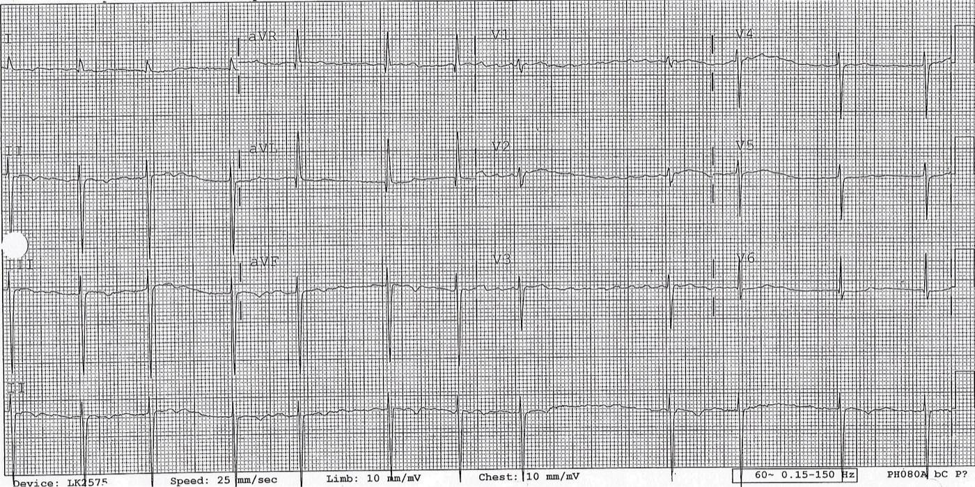

Wide QRS complexes during AF have four explanations: preexisting bundle branch block, rate-related aberrancy, ventricular pacing, and pre-excitation. The Ashman phenomenon is aberrant conduction, usually with right bundle branch morphology, of a beat that ends a short cycle following a long cycle, because the refractory period lengthens after the long cycle. It requires no treatment. Pre-excited AF produces an irregular, very rapid, wide-complex rhythm with beat-to-beat variation in QRS width; a shortest pre-excited R-R interval of 250 ms or less marks a high-risk accessory pathway. AV nodal blocking drugs (adenosine, beta blockers, calcium channel blockers, digoxin, and intravenous amiodarone) are contraindicated in pre-excited AF because they can accelerate conduction over the pathway and precipitate ventricular fibrillation (Class 3: Harm). Hemodynamically unstable patients undergo electrical cardioversion (Class 1), stable patients receive intravenous ibutilide or procainamide (Class 1), and catheter ablation of the pathway is recommended (Class 1).
Atrial flutter is a macroreentrant atrial arrhythmia with regular atrial activation, usually at about 250 to 350 per minute. Typical (cavotricuspid isthmus-dependent) flutter circulates around the tricuspid annulus. In the common counterclockwise form, it produces negative sawtooth flutter waves in II, III, and aVF and positive waves in V1; the clockwise form (reverse typical flutter) produces positive waves in the inferior leads and negative waves in V1. Atypical flutters use circuits around the mitral annulus (perimitral flutter), the left atrial roof, or scar from prior ablation or surgery, and their flutter-wave morphology does not predict the circuit reliably. A regular narrow-complex tachycardia at close to 150 per minute raises the question of flutter with 2:1 conduction, with alternate flutter waves hidden in the QRS complex or T wave. Adenosine or vagal maneuvers transiently increase AV block and expose flutter waves without terminating the arrhythmia.
| Rhythm | Atrial activity | Ventricular rhythm | Distinguishing features |
|---|---|---|---|
| Atrial fibrillation | No organized P waves; f waves of varying morphology | Irregularly irregular | No isoelectric baseline pattern; cycle length varies continuously |
| Typical atrial flutter | Sawtooth F waves, about 250–350/min; negative in II, III, aVF in counterclockwise form | Regular (2:1, 4:1) or regularly irregular with variable block | Fixed F-wave morphology and cycle length |
| Atypical atrial flutter | Organized waves; morphology varies by circuit; may have isoelectric intervals | Regular or variable block | Common after AF ablation, cardiac surgery, or in atrial scar |
| Multifocal atrial tachycardia | Distinct P waves of three or more morphologies; isoelectric baseline | Irregular, rate above 100/min | Associated with lung disease; treated by treating the cause; not anticoagulated as AF |
| Sinus rhythm with frequent premature atrial complexes | Sinus P waves interrupted by premature P waves | Irregular, with a dominant underlying cycle | Premature P waves visible, often in the preceding T wave |
| Pre-excited AF | AF | Irregular, often above 200/min, wide and varying QRS | Delta waves; AV nodal blockers contraindicated |
Differential diagnosis of an irregular or rapid atrial rhythm on the 12-lead electrocardiogram.
Screening, wearable detection, and device-detected atrial fibrillation
Screening identifies more AF but has not yet been shown to reduce stroke in a population. In the LOOP study, implantable loop recorders in older adults with risk factors detected AF about two and a half times as often as usual care (31.8% versus 12.2%) and led to more anticoagulation, without a significant reduction in stroke or systemic embolism. Intermittent handheld electrocardiogram screening of 75- and 76-year-olds in STROKESTOP produced a small benefit on a composite outcome. Consumer devices using photoplethysmography or single-lead electrocardiography identify irregular rhythm with high positive predictive value in older users, and alerts require confirmation on an electrocardiographic tracing. For patients with established AF, a consumer device that provides a high-quality electrocardiographic tracing is reasonable for detecting recurrence (Class 2a). After a stroke or transient ischemic attack of undetermined cause, initial cardiac monitoring and, if needed, an implantable loop recorder are reasonable to detect AF (Class 2a).
Atrial high-rate episodes (AHREs) recorded by pacemakers and defibrillators with an atrial lead, and subclinical AF found by implantable monitors, carry a lower stroke risk than clinical AF, and the risk rises with episode duration and with CHA₂DS₂-VASc score. Stored electrograms are reviewed to confirm AF and exclude far-field oversensing, noise, and other atrial tachycardias before a device counter is treated (Class 1).
| Longest device-detected episode | Stroke risk | 2023 ACC/AHA recommendation | Class |
|---|---|---|---|
| Less than 5 minutes | Any | No oral anticoagulation without another indication | 3: No Benefit |
| 5 minutes to 24 hours | CHA₂DS₂-VASc ≥3 or equivalent | Anticoagulation may be reasonable within shared decision-making | 2b |
| 24 hours or longer | CHA₂DS₂-VASc ≥2 or equivalent | Initiating anticoagulation is reasonable within shared decision-making | 2a |
Recommendations apply to patients without a previous diagnosis of clinical AF. A device-detected episode documented on a surface electrocardiogram is clinical AF and is managed as such.
Two randomized trials published after the guideline define the balance. NOAH-AFNET 6 randomized 2,536 patients (mean age 78 years, median AHRE duration 2.8 hours) to edoxaban or placebo. It was stopped early. The primary composite of cardiovascular death, stroke, or systemic embolism occurred in 3.2% versus 4.0% per patient-year (hazard ratio 0.81, not significant). Stroke occurred at about 1% per year in both groups, and death or major bleeding increased with edoxaban (5.9% versus 4.5% per patient-year). ARTESIA randomized 4,012 patients with subclinical AF lasting 6 minutes to 24 hours (mean CHA₂DS₂-VASc 3.9) to apixaban or aspirin. Stroke or systemic embolism fell from 1.24% to 0.78% per patient-year (hazard ratio 0.63), and major bleeding rose from 0.94% to 1.71% per patient-year (hazard ratio 1.80). A pooled analysis of the two trials found that anticoagulation reduced ischemic stroke by about one-third and increased major bleeding by about 60%. Because absolute stroke rates are low, the net benefit is small and concentrated in patients with higher CHA₂DS₂-VASc scores, longer episodes, or prior stroke. Patients whose episodes progress to 24 hours or longer, or to clinical AF, move into the higher-risk category; in NOAH-AFNET 6, ECG-diagnosed AF developed in 8.7% of patients per year.
Echocardiography and imaging before cardioversion and ablation
Transthoracic echocardiography in new AF establishes left ventricular size and ejection fraction (LVEF), wall thickness, valve disease (above all mitral stenosis and prosthetic valves, which change anticoagulant choice), left atrial size, right ventricular function, and pulmonary pressure. Left atrial volume index is the preferred size measure. A left atrial volume of 73 mL or more, or a diameter of 4.7 cm or more, appears among the guideline's stroke-risk modifiers, and a large, fibrotic atrium predicts recurrence after cardioversion and ablation. Doppler measurements in AF are averaged over several beats.
Transesophageal echocardiography (TEE) is the standard test for left atrial appendage thrombus. The appendage is imaged in multiple planes from about 0 to 135 degrees, because its lobes vary and pectinate muscles can mimic thrombus. Thrombus appears as an echodense mass distinct from the endocardium and pectinate muscles, often with independent motion. Dense spontaneous echo contrast ("smoke") reflects low flow and red-cell aggregation; dense, sludge-like contrast carries a thromboembolic risk close to that of thrombus. Pulsed-wave Doppler at the appendage orifice measures emptying velocity, and velocities below about 20 cm/s are associated with spontaneous echo contrast, thrombus, and embolic events. Ultrasound enhancing agents improve the distinction between thrombus and artifact.
Cardiac CT before ablation defines pulmonary vein number, ostial size, and branching; the relation of the esophagus to the posterior left atrium; and appendage anatomy before occlusion. A filling defect in the appendage on early-phase CT can represent slow flow; delayed imaging, typically at 30 to 60 seconds or later, distinguishes slow flow (which fills in) from thrombus (which persists). Late gadolinium enhancement magnetic resonance imaging quantifies left atrial fibrosis, which was associated with recurrence in observational series, but a randomized trial of fibrosis-guided ablation added to pulmonary vein isolation did not improve outcomes. Intracardiac echocardiography guides transseptal puncture and catheter positioning and is used in some centers to exclude appendage thrombus.
Stroke risk: CHA₂DS₂-VASc and CHA₂DS₂-VA
Thromboembolism in AF arises predominantly in the left atrial appendage, from stasis, endothelial dysfunction, and hypercoagulability. Dose-adjusted warfarin reduces stroke by about two-thirds compared with no treatment, and aspirin offers little protection. The 2023 ACC/AHA guideline recommends evaluating every patient's annual thromboembolic risk with a validated clinical risk score such as CHA₂DS₂-VASc (Class 1). The recommendations are stated in terms of estimated annual risk, not a fixed score, because event rates for the same score differ among cohorts.
| Component | Definition | Points |
|---|---|---|
| C: Congestive heart failure | Clinical heart failure, or objective evidence of moderate to severe left ventricular dysfunction | 1 |
| H: Hypertension | Resting blood pressure above 140/90 mm Hg on two occasions, or current antihypertensive treatment | 1 |
| A₂: Age 75 years or older | 2 | |
| D: Diabetes mellitus | Fasting glucose 126 mg/dL or more, or treatment with a glucose-lowering drug or insulin | 1 |
| S₂: Prior stroke, transient ischemic attack (TIA), or thromboembolism | Ischemic stroke, transient ischemic attack, or systemic embolism | 2 |
| V: Vascular disease | Prior myocardial infarction, coronary revascularization, peripheral artery disease, or complex aortic plaque | 1 |
| A: Age 65 to 74 years | 1 | |
| Sc: Sex category (female) | Omitted in CHA₂DS₂-VA | 1 |
CHA₂DS₂-VASc ranges from 0 to 9 and CHA₂DS₂-VA from 0 to 8. Definitions from the original derivation (Lip et al., 2010) and the 2023 ACC/AHA guideline, Table 10.
Female sex is a risk modifier and not an independent risk factor: it raises risk mainly in women aged 65 years or older with other risk factors, and a woman whose only point is sex is at low risk. The 2023 US guideline therefore uses sex-specific thresholds. The 2024 European Society of Cardiology (ESC) guideline removed sex from the score, renaming it CHA₂DS₂-VA, which applies the same thresholds to men and women and in practice yields the same recommendation as the sex-specific US thresholds.
CHA₂DS₂-VASc and CHA₂DS₂-VA calculator
2023 ACC/AHA and 2024 ESC thresholdsThe scores do not apply to patients with a mechanical heart valve or moderate to severe rheumatic mitral stenosis (warfarin regardless of score), hypertrophic cardiomyopathy (anticoagulation regardless of score), or cardiac amyloidosis (anticoagulation reasonable regardless of score). Moderate or complex congenital heart disease with Fontan circulation or cyanosis may warrant anticoagulation independent of score.
| Estimated annual risk | CHA₂DS₂-VASc equivalent | 2023 ACC/AHA recommendation | Class |
|---|---|---|---|
| 2% or more | Men ≥2; women ≥3 | Oral anticoagulation recommended | 1 |
| 1% to under 2% | Men 1; women 2 | Oral anticoagulation reasonable; risk modifiers inform the decision when the patient is uncertain | 2a |
| Under 1% | Men 0; women 1 | No anticoagulation for stroke prevention; aspirin monotherapy of no benefit | 3: No Benefit (aspirin) |
| Any risk, candidate for anticoagulation | Any | Aspirin alone or aspirin plus clopidogrel as a substitute for anticoagulation is not recommended | 3: Harm |
The need for and choice of stroke-prevention therapy is reassessed at intervals (Class 1), because age and new comorbidities raise the score over time.
At intermediate risk, the guideline lists factors that raise stroke risk beyond the score: higher AF burden or persistent and permanent AF, obesity with BMI of 30 kg/m² or more, poorly controlled hypertension, estimated glomerular filtration rate below 45 mL/min/1.73 m², proteinuria above 150 mg per 24 hours, an enlarged left atrium, and hypertrophic cardiomyopathy. Natriuretic peptides and troponin also predict stroke and are part of biomarker-based scores such as ABC-stroke.
Bleeding risk: HAS-BLED and modifiable bleeding factors
Bleeding scores discriminate poorly between patients who will and will not bleed (c-statistics near 0.6) and contain several factors, such as age, hypertension, kidney disease, and prior stroke, that also raise stroke risk. For this reason the 2023 guideline states that in patients at high stroke risk, bleeding scores are not used in isolation to determine eligibility for anticoagulation (Class 3: No Benefit). Their use is to identify and correct modifiable bleeding risk and to set the frequency of review. The guideline instead emphasizes factors that raise bleeding risk without raising stroke risk: prior bleeding, anemia, thrombocytopenia, and concomitant antiplatelet or nonsteroidal anti-inflammatory drugs (Class 1).
HAS-BLED calculator
Euro Heart Survey derivationOne point per item (maximum 9). In the derivation cohort, a score of 3 or more identified high risk of major bleeding at 1 year. The labile-INR item applies only to patients taking warfarin.
Modifiable bleeding factors are uncontrolled blood pressure, labile INR (corrected by better warfarin management or a switch to a DOAC), concomitant antiplatelet or nonsteroidal anti-inflammatory drugs, alcohol excess, anemia, and inappropriate DOAC dosing. Nonmodifiable factors are age, prior major bleeding, kidney and liver disease, malignancy, and prior stroke. Removing an aspirin that has no current indication is among the most effective bleeding-reduction steps in patients with stable coronary or peripheral artery disease.
Choosing an anticoagulant: DOAC versus warfarin
In patients without moderate to severe rheumatic mitral stenosis or a mechanical heart valve who are candidates for anticoagulation, DOACs are recommended over warfarin to reduce mortality, stroke, systemic embolism, and intracranial hemorrhage (Class 1, Level A). Four phase 3 trials enrolling more than 71,000 patients established the class.
| Trial (drug) | Patients; mean CHADS₂ | Stroke or systemic embolism versus warfarin | Bleeding versus warfarin |
|---|---|---|---|
| RE-LY, 2009 (dabigatran 110 or 150 mg twice daily, open-label warfarin) | 18,113; 2.1 | 150 mg: superior (1.11% versus 1.69% per year); 110 mg: noninferior | Intracranial hemorrhage reduced with both doses; more gastrointestinal bleeding with 150 mg; major bleeding lower with 110 mg |
| ROCKET AF, 2011 (rivaroxaban 20 mg daily, 15 mg if CrCl 30–49 mL/min) | 14,264; 3.5 | Noninferior (per-protocol 1.7% versus 2.2% per year) | Major and clinically relevant bleeding similar; less intracranial and fatal bleeding; more gastrointestinal bleeding |
| ARISTOTLE, 2011 (apixaban 5 mg twice daily; 2.5 mg with 2 of 3 criteria) | 18,201; 2.1 | Superior (1.27% versus 1.60% per year) | Major bleeding lower (2.13% versus 3.09% per year); all-cause mortality lower (3.52% versus 3.94% per year) |
| ENGAGE AF-TIMI 48, 2013 (edoxaban 60 or 30 mg daily, halved for dose-reduction criteria) | 21,105; 2.8 | Both doses noninferior; the 60-mg regimen is the approved US dose | Major bleeding and cardiovascular death lower with both regimens |
In a meta-analysis of the four trials, DOACs reduced stroke or systemic embolism by 19%, intracranial hemorrhage by 52%, and all-cause mortality by 10% compared with warfarin, with a 25% increase in gastrointestinal bleeding. CrCl, creatinine clearance.
Agent choice integrates kidney function, age, body weight, interacting drugs, gastrointestinal bleeding history, dosing frequency, adherence, and cost. Apixaban has the least renal clearance and, in ARISTOTLE, less major bleeding than warfarin. Dabigatran 150 mg twice daily had the strongest ischemic-stroke reduction but depends on renal clearance and causes dyspepsia. Once-daily regimens (rivaroxaban, edoxaban) help some patients adhere. Doses not supported by trial evidence, including empiric dose reduction to lower bleeding, are avoided (Class 3: Harm), because underdosing increases stroke without a consistent fall in bleeding. Warfarin is managed to an INR of 2.0 to 3.0 with routine monitoring and attention to diet and drug interactions (Class 1); time in therapeutic range below 60% to 65% identifies poor control.
Factor XI inhibitors are being tested to separate hemostasis from thrombosis. In OCEANIC-AF, asundexian was inferior to apixaban for stroke prevention and the trial stopped early; other agents in this class remain in phase 3 trials, and none is approved for AF.
DOAC dosing by kidney function, age, weight, and interacting drugs
DOAC doses follow drug-specific labeled criteria and are not adjusted as a class. Creatinine clearance is calculated with the Cockcroft-Gault equation using actual body weight, the method used in the trials and US labeling; estimated glomerular filtration rate from laboratory reports can overstate clearance in older, lighter patients. Kidney function is reassessed at least yearly, and more often when it is reduced or changing.
DOAC dose by creatinine clearance and interacting drugs
US labeling for stroke prevention in AF| Drug | Dose |
|---|---|
| Apixaban | — |
| Rivaroxaban | — |
| Dabigatran | — |
| Edoxaban | — |
Cockcroft-Gault: creatinine clearance (mL/min) = (140 − age) × weight (kg) ÷ (72 × serum creatinine in mg/dL), multiplied by 0.85 in women. The calculator does not address liver disease, pregnancy, mechanical valves, or rheumatic mitral stenosis, where the agent choice changes.
| Drug | CrCl above 95 | 51–95 | 31–50 | 15–30 | Below 15 or dialysis |
|---|---|---|---|---|---|
| Apixaban | 5 mg twice daily; 2.5 mg twice daily with any 2 of age ≥80 years, weight ≤60 kg, serum creatinine ≥1.5 mg/dL (dialysis: 5 mg twice daily, 2.5 mg if age ≥80 years or weight ≤60 kg) | ||||
| Rivaroxaban | 20 mg daily | 20 mg daily | 15 mg daily | 15 mg daily | 15 mg daily (pharmacokinetic data only) |
| Dabigatran | 150 mg twice daily | 150 mg twice daily | 150 mg twice daily | 75 mg twice daily | Not recommended |
| Edoxaban | Not used | 60 mg daily | 30 mg daily | 30 mg daily | Not recommended |
Adapted from the 2023 ACC/AHA guideline, Table 19, and US prescribing information. Rivaroxaban is taken with the evening meal. Edoxaban is not used at CrCl above 95 mL/min because of reduced efficacy relative to warfarin in ENGAGE AF-TIMI 48. ARISTOTLE excluded CrCl below 25 mL/min, RE-LY below 30 mL/min, and ROCKET AF and ENGAGE AF-TIMI 48 below 30 mL/min, so doses in the lowest bands rest on pharmacokinetic data.
Drug interactions act through P-glycoprotein and cytochrome P450 3A4 (CYP3A4). With combined P-glycoprotein and strong CYP3A4 inhibitors (systemic ketoconazole, itraconazole, ritonavir), apixaban 5 mg twice daily is reduced to 2.5 mg twice daily, and apixaban is avoided in patients already taking 2.5 mg; rivaroxaban is avoided. Rivaroxaban is also avoided at CrCl 15 to under 80 mL/min with combined P-glycoprotein and moderate CYP3A4 inhibitors such as erythromycin. Dabigatran is reduced to 75 mg twice daily at CrCl 30 to 50 mL/min with dronedarone or systemic ketoconazole and is avoided with P-glycoprotein inhibitors at CrCl below 30 mL/min. Strong inducers (rifampin, carbamazepine, phenytoin, St. John's wort) lower DOAC levels, and the combination is avoided. Amiodarone, verapamil, diltiazem, and clarithromycin raise DOAC levels modestly without a labeled AF dose change. The 2023 guideline recommends active management of these interactions (Class 1).
In class III obesity (BMI of 40 kg/m² or more), DOACs are reasonable over warfarin (Class 2a); after bariatric surgery, warfarin may be reasonable because of uncertain DOAC absorption (Class 2b). In mild or moderate liver disease (Child-Pugh A or B) without significant coagulopathy, anticoagulation is reasonable (Class 2a). Any DOAC is reasonable in Child-Pugh A. Apixaban, dabigatran, or edoxaban is reasonable in Child-Pugh B (the US edoxaban label does not recommend it in Child-Pugh B), and rivaroxaban is contraindicated in Child-Pugh B (Class 3: Harm). DOACs are avoided in Child-Pugh C. In stage 3 chronic kidney disease, evidence-based DOAC doses are preferred to warfarin (Class 1). In stage 4, warfarin or labeled DOAC doses are reasonable (Class 2a). In kidney failure (CrCl below 15 mL/min) or dialysis, warfarin with an INR of 2.0 to 3.0 or an evidence-based dose of apixaban might be reasonable (Class 2b). Randomized trials in dialysis (RENAL-AF, AXADIA-AFNET 8) were small and underpowered, and observational data show high bleeding rates with any anticoagulant in dialysis.
Indications for warfarin over a DOAC
"Valvular AF" in current usage refers only to AF with a mechanical heart valve or moderate to severe rheumatic mitral stenosis. In these patients warfarin is recommended over DOACs (Class 1). In the INVICTUS trial of rheumatic heart disease, rivaroxaban led to more cardiovascular events and deaths than a vitamin K antagonist, and in RE-ALIGN, dabigatran in patients with mechanical valves produced more thromboembolic and bleeding events than warfarin and the trial stopped early. INR targets with mechanical valves depend on valve position and type.
All other valve disease, including aortic stenosis, mitral regurgitation, prior mitral repair, and bioprosthetic valves, does not exclude DOACs, and the 2023 guideline recommends DOACs over vitamin K antagonists in these patients (Class 1). Transcatheter and surgical bioprostheses in patients with AF are anticoagulated as AF, with timing after surgery set by bleeding risk. Warfarin also remains the choice in antiphospholipid syndrome with arterial events or triple positivity, in which DOACs produced excess arterial thrombosis. It is also used when cost or drug interactions preclude DOACs and in the dialysis and post-bariatric settings described above.
Anticoagulation in older adults, frailty, and falls
Absolute stroke risk, and therefore absolute benefit from anticoagulation, is highest in the oldest patients, and age alone is not a contraindication. In ELDERCARE-AF, Japanese patients aged 80 years or older judged unsuitable for standard-dose anticoagulation received edoxaban 15 mg daily or placebo; stroke or systemic embolism fell from 6.7% to 2.3% per year, and major bleeding was 3.3% versus 1.8% per year, a difference that was not statistically significant, with more gastrointestinal bleeding. The 15-mg dose is not approved in the United States.
Fall risk alone rarely justifies withholding anticoagulation. In a Markov decision analysis of patients aged 65 years or older, a patient taking warfarin would need to fall about 295 times in a year for the risk of subdural hematoma to outweigh the stroke-prevention benefit. The guideline's list of contraindications to long-term anticoagulation includes serious bleeding related to recurrent falls when the cause of the falls cannot be treated. Frailty, cognitive impairment, and polypharmacy are addressed by choosing a DOAC with evidence-based dosing, rationalizing interacting drugs and antiplatelet agents, and arranging support for adherence.
Switching a stable patient from warfarin to a DOAC is not automatically beneficial in frail old age. FRAIL-AF randomized 1,330 frail patients aged 75 years or older (mean age 83) who were stable on INR-guided vitamin K antagonist therapy to continue or switch to a DOAC. The trial stopped for futility: switching increased major or clinically relevant nonmajor bleeding (hazard ratio 1.69) without reducing thromboembolic events. For a frail patient well controlled on warfarin, continuing warfarin is a reasonable choice; for new starts, DOACs remain preferred.
Bleeding on anticoagulation, reversal agents, and resumption
Management of major bleeding combines withholding the anticoagulant, local hemostasis, transfusion, and, for life-threatening or critical-site bleeding, reversal. The DOAC dose and time of the last dose, kidney function, and drug-specific assays determine whether drug effect remains. Idarucizumab, a monoclonal antibody fragment, reverses dabigatran within minutes and is recommended for life-threatening bleeding on dabigatran (Class 1); activated prothrombin complex concentrate is reasonable when idarucizumab is unavailable (Class 2a), and dabigatran is also removed by hemodialysis. For warfarin, 4-factor prothrombin complex concentrate with intravenous vitamin K is recommended over fresh frozen plasma (Class 1, Level A).
For factor Xa inhibitors, the 2023 guideline recommended andexanet alfa or 4-factor prothrombin complex concentrate. In ANNEXA-I, among 530 patients with factor Xa inhibitor-associated intracerebral hemorrhage, andexanet improved hemostatic efficacy compared with usual care (67.0% versus 53.1%), which was mostly prothrombin complex concentrate, but increased thrombotic events (10.3% versus 5.6%), including ischemic stroke (6.5% versus 1.5%), without differences in functional outcome or 30-day death. On the basis of these data and postmarketing reports of thrombotic events, the US Food and Drug Administration concluded that the risks of andexanet alfa outweighed its benefits. The manufacturer withdrew it from the US market. In the United States, 4-factor prothrombin complex concentrate is the reversal agent used for apixaban, rivaroxaban, and edoxaban; andexanet remains available in some other countries.
After major gastrointestinal bleeding, resumption of anticoagulation may be reasonable once reversible causes are corrected and long-term benefit and risk are reassessed by a multidisciplinary team (Class 2b). In a meta-analysis of observational cohorts of patients anticoagulated for AF, venous thromboembolism, or a prosthetic valve, resumption was associated with more recurrent gastrointestinal bleeding (10.1% versus 5.3%) but less thromboembolism and lower mortality. Bleeding from a treatable gastrointestinal, pulmonary, or genitourinary source, from isolated trauma, or from a procedure does not contraindicate long-term anticoagulation.
Anticoagulation after ischemic stroke and intracranial hemorrhage
After an AF-related ischemic stroke, the early recurrence risk competes with the risk of hemorrhagic transformation. The traditional "1-3-6-12 day" rule, which delayed anticoagulation by infarct size, was based on consensus. ELAN randomized 2,013 patients in its primary analysis to early DOAC initiation (within 48 hours for minor or moderate stroke and on day 6 or 7 for major stroke) or later initiation (day 3 or 4, day 6 or 7, and day 12 to 14, respectively). The 30-day composite of recurrent ischemic stroke, systemic embolism, major extracranial bleeding, symptomatic intracranial hemorrhage, or vascular death occurred in 2.9% with early and 4.1% with later treatment, a difference estimated between 2.8 percentage points lower and 0.5 percentage points higher. Symptomatic intracranial hemorrhage occurred in 0.2% in both groups. OPTIMAS and TIMING reported similar findings. Early DOAC initiation after mild to moderate stroke is therefore an accepted strategy, with timing adjusted for infarct size, hemorrhagic transformation, and blood pressure control.
After intracranial hemorrhage, decisions depend on mechanism (traumatic or spontaneous, lobar or deep), the presence of cerebral amyloid angiopathy, and thromboembolic risk. Delayed resumption at 4 to 8 weeks may be considered after careful risk-benefit assessment (Class 2b). In patients with very high thromboembolic risk, above 5% per year, such as those with a mechanical valve or rheumatic heart disease, earlier resumption at 1 to 2 weeks is reasonable (Class 2a). In patients at high risk of recurrent hemorrhage, such as those with cerebral amyloid angiopathy, an anticoagulation-sparing strategy such as left atrial appendage occlusion may be considered (Class 2b). Spontaneous intracranial bleeding from a nonreversible cause is a contraindication to long-term anticoagulation.
Interrupting and restarting anticoagulation for procedures
Most procedures in patients with AF are performed with temporary interruption of the oral anticoagulant and no heparin bridging (Class 1), except in patients with a mechanical valve or recent stroke or transient ischemic attack. In BRIDGE, among 1,884 warfarin-treated patients with AF, no bridging was noninferior to low-molecular-weight heparin bridging for arterial thromboembolism (0.4% versus 0.3%) and caused less major bleeding (1.3% versus 3.2%). Bridging with low-molecular-weight heparin is not administered in these patients (Class 3: Harm). In the PAUSE management study, apixaban, dabigatran, and rivaroxaban were interrupted for 1 day before low-bleeding-risk procedures and 2 days before high-bleeding-risk procedures (longer for dabigatran with reduced kidney function), without bridging. Thirty-day major bleeding was 0.9% to 1.9%, and arterial thromboembolism was 0.6% or less.
| Anticoagulant | Low bleeding risk procedure | High bleeding risk procedure |
|---|---|---|
| Apixaban (CrCl above 25 mL/min) | 1 day | 2 days |
| Rivaroxaban (CrCl above 30 mL/min) | 1 day | 2 days |
| Edoxaban (CrCl above 15 mL/min) | 1 day | 2 days |
| Dabigatran (CrCl above 50 mL/min) | 1 day | 2 days |
| Dabigatran (CrCl 30–50 mL/min) | 2 days | 4 days |
| Warfarin | 5 days for a target INR below 1.5; 2 to 3 days for a target INR below 2 | 5 days |
Full days without any dose before the day of the procedure, with no dose on the procedure day (2023 ACC/AHA guideline, Table 18). For lower clearance than listed, an additional 1 to 3 days is considered for high-bleeding-risk procedures. Neuraxial anesthesia follows the longer intervals in anesthesia society guidance.
After a procedure, a DOAC is generally resumed the day after a low-bleeding-risk procedure and between the evening of the second and the evening of the third day after a high-bleeding-risk procedure, once hemostasis is secure (Class 2a). Warfarin is often restarted the evening of the procedure because its effect takes days to develop. For pacemaker and defibrillator implantation or generator change, continued warfarin is recommended over interruption with heparin bridging in patients with an annual thromboembolic risk of 5% or more (Class 1), because bridging increases pocket hematoma; uninterrupted or interrupted DOAC is reasonable (Class 2a).
Antithrombotic therapy after PCI and in chronic coronary disease
About 5% to 10% of patients undergoing percutaneous coronary intervention (PCI) have AF. Triple therapy with an oral anticoagulant, aspirin, and a P2Y12 inhibitor increases bleeding two- to threefold compared with dual therapy. Four trials (PIONEER AF-PCI, RE-DUAL PCI, AUGUSTUS, and ENTRUST-AF PCI) compared DOAC-based dual therapy with warfarin-based triple therapy. AUGUSTUS was a 2 × 2 factorial trial in 4,614 patients with AF and a recent acute coronary syndrome or PCI on a P2Y12 inhibitor. Apixaban caused less major or clinically relevant nonmajor bleeding than a vitamin K antagonist (10.5% versus 14.7%), and aspirin caused more bleeding than placebo (16.1% versus 9.0%), with similar ischemic events overall. A numerically higher rate of stent thrombosis without aspirin in the first 30 days supports a short aspirin period in patients at high thrombotic risk.
The 2023 AF guideline recommends a DOAC over a vitamin K antagonist in combination with antiplatelet therapy after PCI (Class 1). In most patients, it recommends discontinuing aspirin after 1 to 4 weeks and continuing dual therapy (oral anticoagulant plus a P2Y12 inhibitor) over triple therapy (Class 1). The 2025 ACC/AHA acute coronary syndrome guideline gives the same aspirin window. Clopidogrel is the preferred P2Y12 inhibitor; ticagrelor and prasugrel carry more bleeding in combination with anticoagulation, and prasugrel is not used in triple therapy. The full-intensity stroke-prevention DOAC dose is used, except dabigatran, for which 110 mg twice daily (not approved for AF in the United States) or 150 mg twice daily was tested.
| Phase | Usual regimen | Modifiers |
|---|---|---|
| Periprocedural to 1–4 weeks | DOAC + clopidogrel + aspirin (triple therapy) | Aspirin stopped at discharge in high bleeding risk; up to 30 days in high thrombotic risk (complex PCI, prior stent thrombosis) |
| 1–4 weeks to 6–12 months | DOAC + clopidogrel (dual therapy) | Shorter dual therapy at high bleeding risk; 12 months after acute coronary syndrome |
| Beyond 12 months (chronic coronary disease) | DOAC alone | Antiplatelet therapy added only for a specific indication, such as prior stent thrombosis |
Antithrombotic sequence after PCI in AF (2023 ACC/AHA AF guideline and 2025 ACC/AHA acute coronary syndrome guideline). The DOAC is given at the full stroke-prevention dose appropriate for kidney function.
In chronic coronary disease beyond 1 year after revascularization, or without a revascularization indication, oral anticoagulant monotherapy is recommended over anticoagulation plus a single antiplatelet drug (Class 1). In AFIRE, among 2,236 Japanese patients, rivaroxaban monotherapy was noninferior to rivaroxaban plus an antiplatelet agent for ischemic events and superior for major bleeding, and the trial stopped early because mortality was higher with combination therapy. In EPIC-CAD, among 1,040 patients, edoxaban monotherapy reduced the 12-month net composite of death, myocardial infarction, stroke, systemic embolism, urgent revascularization, and major or clinically relevant nonmajor bleeding (6.8% versus 16.2%), driven by less bleeding. In stable peripheral artery disease, anticoagulant monotherapy is reasonable over dual therapy (Class 2a).
Anticoagulation around cardioversion
Restoring sinus rhythm by any method carries a transient risk of thromboembolism from dislodged thrombus and from atrial stunning, the delayed recovery of atrial mechanical function that peaks in the days after conversion.
| Situation | Recommendation | Class |
|---|---|---|
| AF of 48 hours or longer, or unknown duration, elective cardioversion | 3 weeks of uninterrupted therapeutic anticoagulation, or imaging to exclude intracardiac thrombus, before cardioversion | 1 |
| Any cardioversion | Therapeutic anticoagulation established before cardioversion and continued without interruption for at least 4 weeks afterward; long term by stroke risk | 1 |
| Thrombus on precardioversion imaging | Therapeutic anticoagulation for at least 3 to 6 weeks, then repeat imaging before cardioversion | 1 |
| AF under 48 hours, not anticoagulated, CHA₂DS₂-VASc ≥2 | Precardioversion imaging may be considered | 2b |
| AF under 12 hours, CHA₂DS₂-VASc 0–1 | Benefit of imaging or pericardioversion anticoagulation uncertain because events are rare | 2b |
| Prior left atrial appendage occlusion, off anticoagulation | Imaging to assess the device and exclude device-related thrombus may be reasonable; anticoagulation may be considered if a residual leak is present | 2b |
| Hemodynamic instability | Immediate cardioversion; anticoagulation started as soon as feasible | 1 |
Recommendations for prevention of thromboembolism with cardioversion, 2023 ACC/AHA AF guideline.
The ACUTE trial established the TEE-guided strategy. Among 1,222 patients with AF of more than 2 days, TEE-guided early cardioversion after a brief period of anticoagulation produced embolic event rates similar to those with 3 weeks of conventional anticoagulation (0.8% versus 0.5%), with fewer hemorrhagic events and a shorter time to cardioversion. DOACs were evaluated for cardioversion in X-VeRT, ENSURE-AF, and EMANATE, with low event rates similar to warfarin. Adherence to the DOAC during the preceding 3 weeks is confirmed because, unlike warfarin, there is no INR to document therapeutic anticoagulation.
The 2024 ESC guideline uses a shorter threshold: early cardioversion without 3 weeks of anticoagulation or imaging is not recommended when AF has lasted more than 24 hours.
Rate control: targets and drug choice
Rate control relieves symptoms, prevents tachycardia-mediated cardiomyopathy, and remains part of care whether or not sinus rhythm is pursued. In patients without heart failure, the heart-rate target is guided by symptoms, generally aiming at a resting rate below 100 to 110 per minute (Class 2a). The basis is RACE II, in which 614 patients with permanent AF were assigned to lenient control (resting rate below 110 per minute) or strict control (below 80 per minute at rest and below 110 per minute during moderate exercise). The 3-year composite of cardiovascular death, heart failure hospitalization, stroke, embolism, bleeding, and life-threatening arrhythmia was 12.9% with lenient control and 14.9% with strict control, and lenient control was easier to achieve. Patients with heart failure were few in RACE II. In suspected AF-induced cardiomyopathy or refractory heart failure symptoms, a stricter target (below 80 per minute at rest and below 110 per minute with moderate exercise) may be reasonable (Class 2b). In patients with cardiac resynchronization therapy, rate control aims at near-continuous biventricular pacing.
Beta blockers or nondihydropyridine calcium channel blockers are recommended for long-term rate control, chosen by substrate and comorbidities (Class 1). Diltiazem and verapamil are not given to patients with an LVEF below 40% (Class 3: Harm). Digoxin is reasonable in patients with heart failure symptoms, alone or combined with other agents (Class 2a), and it controls the resting rate better than the exercise rate. In RATE-AF, among older patients with permanent AF and heart failure symptoms, low-dose digoxin and bisoprolol produced similar quality of life at 6 months, with fewer adverse events with digoxin. When serum levels are measured, a digoxin level below 1.2 ng/mL is reasonable (Class 2a), because higher concentrations were associated with increased mortality. Dronedarone is not used for rate control in permanent AF in patients with cardiovascular risk factors (Class 3: Harm); in PALLAS it doubled cardiovascular events.
| Drug | Intravenous dosing | Oral maintenance | Notes |
|---|---|---|---|
| Metoprolol tartrate | 2.5–5 mg over 2 min; up to 3 doses | 25–200 mg twice daily (succinate 50–400 mg daily) | Preferred class in coronary disease and HFrEF |
| Esmolol | 500 µg/kg over 1 min, then 50–300 µg/kg/min | — | Half-life 9 min; useful in unstable or perioperative settings |
| Carvedilol, bisoprolol, atenolol | — | Carvedilol 3.125–25 mg twice daily; bisoprolol 2.5–10 mg daily; atenolol 25–100 mg daily | Atenolol renally eliminated |
| Diltiazem | 0.25 mg/kg (actual body weight) over 2 min; may repeat 0.35 mg/kg; then 5–15 mg/h | 120–360 mg daily (extended release) | Avoid with LVEF below 40%; raises DOAC and digoxin levels |
| Verapamil | 5–10 mg over at least 2 min (may repeat twice); then 5 mg/h (maximum 20 mg/h) | 180–480 mg daily (extended release) | Avoid with LVEF below 40%; raises digoxin and dabigatran levels |
| Digoxin | 0.25–0.5 mg, then 0.25 mg every 6 h (maximum 1.5 mg in 24 h) | 0.0625–0.25 mg daily | Renally eliminated; slow onset; level below 1.2 ng/mL |
| Amiodarone | 150–300 mg over 1 h, then 10–50 mg/h over 24 h | 100–200 mg daily | For critically ill patients or decompensated HF when other agents fail; may cardiovert, so stroke risk is considered |
Rate-control agents in AF, adapted from the 2023 ACC/AHA guideline, Table 21.
Acute management of rapid atrial fibrillation
AF with hemodynamic instability attributable to the arrhythmia (hypotension, ischemia, pulmonary edema, or shock) is treated with immediate synchronized electrical cardioversion (Class 1). Much rapid AF in the emergency department and hospital is driven by another illness, such as sepsis, hypovolemia, bleeding, pain, pulmonary embolism, thyrotoxicosis, or alcohol withdrawal, and the rate falls when the driver is treated.
In hemodynamically stable patients, intravenous beta blockers or nondihydropyridine calcium channel blockers (verapamil or diltiazem, provided the ejection fraction is above 40%) are recommended for acute rate control (Class 1). Intravenous diltiazem achieves faster control than metoprolol in several emergency department studies. Digoxin, alone or combined, can be used when these agents are ineffective or contraindicated (Class 2a). Adding intravenous magnesium to standard rate control is reasonable (Class 2a). In critically ill patients or those with decompensated heart failure, intravenous amiodarone may be considered (Class 2b); because amiodarone can restore sinus rhythm, its use in a patient with AF of 48 hours or longer who is not anticoagulated carries thromboembolic risk.
Atrioventricular node ablation and pacing
AV node (junction) ablation with permanent pacing controls the ventricular rate reliably when medication fails and rhythm control is unsuccessful or not appropriate. It improves symptoms and quality of life (Class 2a), does not stop AF, and leaves the patient pacemaker-dependent, so anticoagulation continues according to stroke risk. The pacemaker is implanted before or on the day of ablation to confirm lead function (Class 1). The lower rate is programmed at 80 to 90 per minute initially to reduce the risk of sudden death from bradycardia-dependent repolarization abnormalities (Class 1), then reduced over subsequent weeks.
The pacing mode matters because long-term right ventricular pacing can cause dyssynchrony and cardiomyopathy. In patients with HFrEF (LVEF below 50%) and refractory rapid ventricular response who are not candidates for rhythm control or in whom it has failed, AV node ablation with biventricular pacing can improve symptoms, quality of life, and ejection fraction (Class 2a). In patients with a normal ejection fraction undergoing AV node ablation, conduction system pacing of the His bundle or left bundle branch area may be reasonable (Class 2b). In patients with a biventricular device whose pacing percentage is inadequate because of AF conduction, AV node ablation improves functional class, reduces defibrillator shocks, and improves survival (Class 2a). The APAF-CRT trial found that AV node ablation with biventricular pacing reduced mortality compared with pharmacologic rate control in older patients with permanent AF and narrow QRS complexes who had been hospitalized for heart failure.
Rate versus rhythm control: trial evidence and decision factors
Early trials found no survival advantage for rhythm control. AFFIRM randomized 4,060 patients aged 65 years or older, or younger with stroke risk factors, to rate control or rhythm control with antiarrhythmic drugs. Five-year mortality was 21.3% with rate control and 23.8% with rhythm control (P=0.08), with more hospitalizations and adverse drug effects in the rhythm-control group. RACE, AF-CHF, and smaller trials agreed. These trials used antiarrhythmic drugs with limited efficacy, often stopped anticoagulation after apparent sinus rhythm, and enrolled patients years into their disease.
EAST-AFNET 4 tested early rhythm control. It randomized 2,789 patients with AF diagnosed within 1 year (median 36 days) and cardiovascular conditions to early rhythm control (antiarrhythmic drugs or ablation) or usual care. After a median of 5.1 years, the composite of cardiovascular death, stroke, or hospitalization for heart failure or acute coronary syndrome occurred at 3.9 versus 5.0 per 100 person-years (hazard ratio 0.79), with no increase in nights in hospital. Serious adverse events related to rhythm-control therapy occurred in 4.9% versus 1.4%. The 2023 guideline therefore states that in patients with AF diagnosed within 1 year, rhythm control can be useful to reduce hospitalizations, stroke, and mortality (Class 2a). Rhythm control is also recommended to evaluate whether persistent or high-burden AF contributes to reduced left ventricular function (Class 1). It is useful to improve symptoms (Class 2a), can reduce progression (Class 2a), and may reduce dementia and structural deterioration (Class 2b). Shared decision-making about the two strategies is recommended (Class 1).
| Clinical feature | Favors rhythm control | Favors rate control |
|---|---|---|
| Symptoms | Symptomatic despite adequate rate (modified EHRA 2b–4); uncertain whether symptoms are from AF (trial of cardioversion) | No or minimal symptoms with controlled rate (EHRA 1–2a) |
| Time since diagnosis | AF diagnosed within 1 year, especially with cardiovascular risk factors | Long-standing persistent AF after failed rhythm-control attempts |
| Left ventricular function | Reduced ejection fraction, suspected arrhythmia-induced cardiomyopathy (Class 1 for a trial of rhythm control); HFrEF on guideline-directed medical therapy, suitable for ablation | Advanced cardiomyopathy unlikely to respond, with rate controlled and symptoms limited |
| Atrial substrate | Small or moderately enlarged atrium; paroxysmal or early persistent AF | Severely enlarged, fibrotic atrium with low chance of maintaining sinus rhythm |
| Age and comorbidity | Younger patients, athletes, adults with congenital heart disease (rhythm control recommended), pulmonary hypertension | Older patients with competing comorbidities who prefer fewer procedures and drugs |
| Rate control achievable | Rate difficult to control, or rate-control drugs not tolerated (bradycardia, hypotension) | Rate easily controlled with well-tolerated drugs |
| Patient preference | Preference for sinus rhythm and acceptance of procedural or drug risk | Preference to avoid antiarrhythmic drugs and procedures |
Decision table for rate versus rhythm control, built from the 2023 ACC/AHA recommendations for rhythm control, rate control, heart failure, and adult congenital heart disease. Stroke prevention is decided separately and is not changed by the strategy chosen.
Electrical and pharmacologic cardioversion
Electrical cardioversion is performed as the initial rhythm-control strategy in stable patients or after unsuccessful pharmacologic conversion (Class 1). Energy delivery is synchronized to the QRS complex to avoid inducing ventricular fibrillation (Class 1). An initial biphasic shock of at least 200 J improves first-shock success (Class 2a). With longer AF duration or a failed first shock, changing the electrode vector (anterior-posterior and anterior-lateral), higher energy, and pretreatment with an antiarrhythmic drug improve success (Class 2a). In patients with obesity, manual pressure on the pads and escalation of energy may help (Class 2b). Sinus node dysfunction can appear after conversion, particularly in older patients, so external pacing capability is maintained.
Pharmacologic cardioversion is reasonable in stable patients or when electrical cardioversion is preferred but cannot be performed (Class 2a). The same anticoagulation rules apply as for electrical cardioversion.
| Drug | Dose | Time to conversion | Principal cautions |
|---|---|---|---|
| Ibutilide (Class 2a) | 1 mg IV over 10 min if 60 kg or more (0.01 mg/kg if under 60 kg); second equal dose after 10 min if needed | 30–90 min | Torsades de pointes in about 2% to 7%, higher with low LVEF; not used with LVEF below 40%; potassium and magnesium corrected; ECG monitoring for at least 4 h |
| Amiodarone (Class 2a) | 5–7 mg/kg or 300 mg IV, then 1,200–3,000 mg by infusion over 24 h | 8–12 h | Hypotension, bradycardia, phlebitis; acceptable in structural heart disease |
| Flecainide, "pill in the pocket" (Class 2a) | 200 mg if under 70 kg, 300 mg if 70 kg or more, single oral dose, with an AV nodal blocker | 3–8 h | Only without structural heart disease or coronary disease; first dose given in a monitored setting |
| Propafenone, "pill in the pocket" (Class 2a) | 450 mg if under 70 kg, 600 mg if 70 kg or more, single oral dose, with an AV nodal blocker | 3–8 h | Same restrictions as flecainide |
| Procainamide (Class 2b) | 1 g IV over 30 min, then 2 mg/min for 1 h | 30–60 min | Hypotension in 5% to 12%; QRS and QT prolongation |
Drugs for pharmacologic cardioversion, adapted from the 2023 ACC/AHA guideline, Table 22. Class IC drugs given without an AV nodal blocker can organize AF into slow atrial flutter conducted 1:1 with a wide QRS complex.
Antiarrhythmic drugs: selection by substrate, dosing, and monitoring
Antiarrhythmic drug choice is determined first by safety: the presence of prior myocardial infarction, scar, significant structural heart disease, or HFrEF; the QT interval; kidney function; and conduction disease. Efficacy is modest, with recurrence common at 1 year, and amiodarone is the most effective and the most toxic.
| Substrate | Reasonable options (2023 ACC/AHA) | Not used |
|---|---|---|
| No prior myocardial infarction, structural heart disease, scar, or fibrosis | Flecainide or propafenone with an AV nodal blocker (2a); dronedarone (2a); dofetilide (2a); sotalol (2b); low-dose amiodarone when others fail (2a) | — |
| Coronary disease, prior myocardial infarction, or ventricular scar with preserved LVEF | Dronedarone (2a); dofetilide (2a); sotalol (2b); amiodarone | Flecainide and propafenone (3: Harm) |
| HFrEF (LVEF 40% or less) | Amiodarone or dofetilide (2a) | Flecainide and propafenone (3: Harm); dronedarone with New York Heart Association (NYHA) class III–IV or recent decompensation (3: Harm); sotalol generally avoided |
| Significant left ventricular hypertrophy | Amiodarone or dronedarone are the usual choices; dofetilide with careful QT monitoring | Class IC drugs; sotalol with marked hypertrophy |
| Permanent AF | Rhythm-control drugs not indicated | Dronedarone (3: Harm) |
Antiarrhythmic drug selection for maintenance of sinus rhythm. The class IC restriction derives from the Cardiac Arrhythmia Suppression Trial, in which flecainide and encainide after myocardial infarction increased arrhythmic and total mortality.
Dosing and initiation. Flecainide is given at 50 to 150 mg twice daily (total 50 to 300 mg/day) and propafenone at 150 to 300 mg every 8 hours or 225 to 425 mg every 12 hours (extended release), both with a beta blocker or calcium channel blocker to prevent 1:1 conduction of slowed flutter. Dronedarone is given at 400 mg twice daily with meals; it raises serum creatinine by inhibiting tubular secretion without reducing glomerular filtration, interacts with digoxin and dabigatran, and requires liver enzyme checks in the first 6 months. Dofetilide is dosed by creatinine clearance (500 µg twice daily above 60 mL/min, 250 µg at 40 to 60 mL/min, 125 µg at 20 to 40 mL/min, contraindicated below 20 mL/min). Initiation, dose increase, or reinitiation requires admission for at least 3 days with continuous electrocardiographic monitoring (Class 1). It is contraindicated with verapamil, hydrochlorothiazide, cimetidine, trimethoprim, ketoconazole, and other drugs that block its renal cation transport. Sotalol is initiated in a monitored facility (Class 2a) with dosing interval by kidney function; an intravenous loading protocol can shorten the hospital stay. Amiodarone is loaded with 6 to 10 g over 1 to 4 weeks and maintained at 100 to 200 mg daily.
Proarrhythmia and QT monitoring. Dofetilide, sotalol, and ibutilide block the rapid delayed rectifier potassium current and cause torsades de pointes, with risk increased by female sex, bradycardia, hypokalemia, hypomagnesemia, kidney dysfunction, and other QT-prolonging drugs. Dofetilide is held or reduced when the QTc rises by more than 15% or exceeds 500 ms (550 ms with ventricular conduction abnormalities) after dosing. Class IC drugs widen the QRS complex, particularly at faster rates, and an exercise test after initiation is often used to detect use-dependent QRS widening. ECG, potassium, magnesium, and creatinine are checked every 3 to 6 months on dofetilide and sotalol.
Amiodarone surveillance. Thyroid function and liver enzymes are measured at baseline, at 3 to 6 months, and every 6 months; an ECG annually; and a chest radiograph at baseline and for unexplained cough or dyspnea. Thyroid dysfunction occurs in 2% to 24% of patients, and pulmonary toxicity in about 1% to 2%, with substantial mortality when it develops. Amiodarone raises warfarin and digoxin levels and interacts with statins metabolized by CYP3A4.
Catheter ablation: indications and outcomes
Catheter ablation is the most effective therapy for maintaining sinus rhythm. Indications in the 2023 guideline:
- Symptomatic AF when antiarrhythmic drugs have been ineffective, contraindicated, not tolerated, or not preferred, and rhythm control is desired: useful to improve symptoms (Class 1, Level A).
- Selected patients with symptomatic paroxysmal AF, generally younger with few comorbidities: useful as first-line therapy to improve symptoms and reduce progression to persistent AF (Class 1, Level A).
- Other patients with symptomatic paroxysmal or persistent AF managed with rhythm control: first-line ablation can be useful (Class 2a).
- AF with HFrEF in appropriate patients on guideline-directed therapy with a reasonable expectation of benefit: ablation is beneficial to improve symptoms, quality of life, ventricular function, and cardiovascular outcomes (Class 1, Level A).
- Symptomatic AF with heart failure with preserved ejection fraction (HFpEF): ablation can be useful to improve symptoms and quality of life (Class 2a).
- Selected asymptomatic or minimally symptomatic younger patients with moderate to high AF burden or persistent AF: ablation may be useful to reduce progression (Class 2b).
- Athletes who develop AF: pulmonary vein isolation is a reasonable rhythm-control strategy (Class 2a).
| Trial | Population and comparison | Principal result |
|---|---|---|
| CABANA, 2019 | 2,204 patients (median age 68) with paroxysmal or persistent AF; ablation versus drug therapy | Primary composite (death, disabling stroke, serious bleeding, cardiac arrest) 8.0% versus 9.2% by intention to treat (hazard ratio 0.86, not significant), with 27.5% crossover to ablation; AF recurrence 49.9% versus 69.5% (hazard ratio 0.52) |
| EARLY-AF, 2021 | 303 patients with untreated symptomatic paroxysmal AF; cryoballoon ablation versus drugs, continuous monitoring | 1-year atrial tachyarrhythmia recurrence 42.9% versus 67.8%; less progression to persistent AF at 3 years |
| STOP AF First, 2021 | 203 patients with untreated paroxysmal AF; cryoballoon versus drugs | 12-month treatment success 74.6% versus 45.0%; 1.9% primary safety events |
| CASTLE-AF, 2018 | 363 patients with AF, LVEF 35% or less, and an implanted defibrillator | Death or heart failure hospitalization 28.5% versus 44.6% (hazard ratio 0.62); all-cause death hazard ratio 0.53 |
| CASTLE-HTx, 2023 | 194 patients with end-stage heart failure referred for transplantation | Death, ventricular assist device implantation, or urgent transplantation 8% versus 30% (hazard ratio 0.24); trial stopped early for efficacy |
| AVANT GUARD, 2026 | 310 patients with untreated persistent AF; pulsed field ablation versus drugs (2:1), implantable monitor | 12-month treatment success 56% versus 30% (hazard ratio for failure 0.46); device- and procedure-related adverse events 5.1% |
Randomized trials of catheter ablation for AF. In CABANA, treatment received and per-protocol analyses favored ablation, and quality of life improved more after ablation.
Persistent AF is harder to treat. Freedom from recurrence after a single procedure is lower than in paroxysmal AF, repeat procedures are more common, and additional lesion sets have not consistently improved outcomes. AVANT GUARD extends first-line ablation evidence to persistent AF with a modern energy source. The trial was paused after 6 neurologic events and resumed after protocol changes that excluded patients with CHA₂DS₂-VASc of 4 or more and added screening for left atrial thrombus. In heart failure, the benefit is greatest in patients with a moderately dilated ventricle, limited ventricular scar, and a recent onset of AF; in patients with suspected arrhythmia-induced cardiomyopathy, restoring sinus rhythm also has diagnostic value. Recurrent symptomatic AF after ablation is treated with repeat ablation or antiarrhythmic drugs (Class 1), and short-term antiarrhythmic therapy after ablation reduces early recurrence and hospitalization (Class 2a).
Ablation technique, energy sources, and complications
Pulmonary vein isolation (PVI) is the primary lesion set for all patients unless a different specific trigger is identified (Class 1). It is performed by transseptal access with radiofrequency energy delivered point by point, guided by electroanatomic mapping and contact force; with a cryoballoon; or with pulsed field ablation. Additional targets beyond PVI, including posterior wall isolation, low-voltage areas, fractionated electrograms, and rotors, are of uncertain value (Class 2b). In FIRE AND ICE, cryoballoon and radiofrequency PVI had similar efficacy and safety in paroxysmal AF.
Pulsed field ablation (PFA) delivers microsecond high-voltage pulses that kill myocytes by irreversible electroporation. Myocardium has a lower threshold for electroporation than esophagus and nerve, which gives PFA its relative tissue selectivity. In ADVENT, among 607 patients with paroxysmal AF, PFA was noninferior to radiofrequency or cryoballoon ablation for 1-year freedom from treatment failure (73.3% versus 71.3%) and for safety (2.1% versus 1.5%). In MANIFEST-17K, a registry of 17,642 patients, major complications occurred in about 1%: tamponade 0.36%, vascular events 0.30%, stroke 0.12%, and death 0.03%. No esophageal fistula, pulmonary vein stenosis, or persistent phrenic palsy was reported. Complications specific to PFA were coronary artery spasm (0.14%) and hemolysis-related acute kidney injury requiring dialysis (0.03%).
| Complication | Frequency | Timing | Presentation and management |
|---|---|---|---|
| Cardiac tamponade | 0.4%–1.5% | During procedure, occasionally delayed | Hypotension; echocardiography; pericardiocentesis, reversal of heparin |
| Stroke or TIA | 0.1%–1.0% | Procedure to 1 week | Neurologic deficit; imaging and stroke-team evaluation |
| Vascular access complications | 1%–7% | Procedure to 1 month | Hematoma, pseudoaneurysm, arteriovenous fistula; ultrasound-guided access reduces risk |
| Atrioesophageal fistula | About 0.2% (thermal energy) | 1 to 4 weeks | Fever, chest pain, odynophagia, neurologic events from air or septic emboli, gastrointestinal bleeding; CT of the chest; endoscopy and TEE are avoided because air insufflation can cause massive air embolism; urgent surgery |
| Pulmonary vein stenosis | 0.1%–0.8% | Months | Dyspnea, cough, hemoptysis, pneumonia-like illness; CT or MRI; angioplasty or stent |
| Phrenic nerve palsy | 0.2%–0.4% | During procedure | Dyspnea, elevated hemidiaphragm; most recover; mainly cryoballoon ablation of right-sided veins |
| Coronary spasm and hemolysis | Uncommon | During procedure | Specific to pulsed field ablation; hydration and limits on application number |
| Death | 0.1%–0.4% | Periprocedural | — |
Complications of catheter ablation for AF, adapted from the 2023 ACC/AHA guideline, Table 26, with PFA-specific events from MANIFEST-17K.
Early recurrence of atrial arrhythmia after ablation is common while lesions mature and inflammation subsides, and it does not by itself predict long-term failure. The 2024 EHRA/HRS/APHRS/LAHRS expert consensus shortened the recommended blanking period, during which recurrences are not counted as failure in clinical trials, from 3 months to 8 weeks. Recurrence after the blanking period is usually caused by pulmonary vein reconnection, and repeat procedures target reconnected veins and non-pulmonary-vein triggers.
Anticoagulation before and after ablation
Ablation is performed on uninterrupted warfarin with an INR of 2.0 to 3.0, or on continuous or minimally interrupted DOAC, with intraprocedural heparin to an activated clotting time typically above 300 seconds (Class 1). Oral anticoagulation continues for at least 3 months after ablation (Class 1), and long-term anticoagulation is determined by the patient's stroke risk (for example, CHA₂DS₂-VASc of 2 or more) and not by apparent procedural success (Class 1), because recurrence is often asymptomatic.
Two randomized trials tested stopping anticoagulation after successful ablation. ALONE-AF randomized 840 Korean patients (mean age 64 years, mean CHA₂DS₂-VASc 2.1) without recurrence for at least 1 year after ablation to discontinue or continue anticoagulation. The 2-year composite of stroke, systemic embolism, and major bleeding occurred in 0.3% versus 2.2%, a difference driven by major bleeding; ischemic stroke occurred in 0.3% versus 0.8%. OCEAN randomized 1,284 patients at least 1 year after successful ablation (mean CHA₂DS₂-VASc 2.2) to rivaroxaban 15 mg daily or aspirin. Stroke, systemic embolism, or new covert embolic infarction occurred in 0.8% versus 1.4% at 3 years (not significant). Fatal or major bleeding occurred in 1.6% versus 0.6% (not significant). Both trials enrolled patients at low to intermediate risk with documented freedom from recurrence. They support individualized discontinuation in such patients and do not apply to patients at high stroke risk, those with prior stroke, or those without systematic rhythm monitoring.
Surgical ablation and left atrial appendage exclusion at cardiac surgery
In patients with AF who undergo cardiac surgery for another indication, concomitant surgical ablation can reduce recurrent AF (Class 2a), and anticoagulation is reasonable for at least 3 months afterward (Class 2a). The Cox-Maze III procedure, a cut-and-sew lesion set, has been replaced largely by the Cox-Maze IV, which uses radiofrequency and cryothermal energy; isolated PVI is used for paroxysmal AF and the biatrial lesion set for persistent AF. For symptomatic persistent AF refractory to drugs, hybrid epicardial and endocardial ablation might be reasonable (Class 2b).
LAAOS III randomized 4,811 patients with AF and a CHA₂DS₂-VASc score of 2 or more undergoing cardiac surgery for another indication to appendage occlusion or no occlusion; most patients continued anticoagulation. Over a mean of 3.8 years, ischemic stroke or systemic embolism occurred in 4.8% versus 7.0% (hazard ratio 0.67), without differences in perioperative bleeding, heart failure, or death. The 2023 guideline recommends surgical exclusion in addition to continued anticoagulation in patients with CHA₂DS₂-VASc of 2 or more or equivalent risk undergoing cardiac surgery (Class 1). The recommended technique leaves no flow across the suture line and a stump shorter than 1 cm on intraoperative TEE (Class 1). Whether surgical exclusion can replace anticoagulation is uncertain (Class 2b). Excision, stapling, and epicardial clip devices achieve complete closure more reliably than suture ligation, which often leaves residual flow.
Percutaneous left atrial appendage occlusion
Percutaneous left atrial appendage occlusion (LAAO) places a self-expanding device in the appendage ostium through transseptal access under TEE or intracardiac echocardiographic guidance. The Watchman FLX and Amulet devices are approved in the United States. After implantation, antithrombotic therapy (a short course of anticoagulation or dual antiplatelet therapy, followed by long-term aspirin) covers the period before endothelialization, and TEE or CT at about 45 days and 1 year assesses device position, peridevice leak, and device-related thrombus.
| Trial | Comparison | Principal result |
|---|---|---|
| PROTECT AF and PREVAIL, 5-year pooled, 2017 | Watchman versus warfarin | Similar composite of stroke, systemic embolism, and cardiovascular death; fewer hemorrhagic and disabling strokes, less nonprocedural bleeding, and lower cardiovascular mortality with the device; more ischemic strokes numerically |
| PRAGUE-17, 2020 | Watchman or Amulet versus DOAC in high-risk patients (402) | Noninferior for a composite of cardiovascular, neurologic, and bleeding events |
| OPTION, 2025 | Watchman FLX versus oral anticoagulation after AF ablation (1,600; mean CHA₂DS₂-VASc 3.5) | Nonprocedural major or clinically relevant nonmajor bleeding 8.5% versus 18.1% at 36 months; death, stroke, or systemic embolism 5.3% versus 5.8% (noninferior) |
| CHAMPION-AF, 2026 | Watchman FLX versus DOAC in patients eligible for anticoagulation (3,000; mean CHA₂DS₂-VASc 3.5) | Cardiovascular death, stroke, or systemic embolism 5.7% versus 4.8% at 3 years (noninferior); nonprocedural bleeding 10.9% versus 19.0% (hazard ratio 0.55); ischemic stroke numerically higher with the device |
Randomized trials of percutaneous left atrial appendage occlusion.
The 2023 guideline rates percutaneous LAAO as reasonable in patients with a CHA₂DS₂-VASc of 2 or more and a contraindication to long-term anticoagulation from a nonreversible cause (Class 2a). It rates LAAO as a possibly reasonable alternative to anticoagulation in patients at high bleeding risk, on the basis of patient preference, with procedural risk considered and the evidence for anticoagulation acknowledged as more extensive (Class 2b). Contraindications to long-term anticoagulation include severe bleeding from a nonreversible gastrointestinal, pulmonary, or genitourinary cause, spontaneous intracranial or intraspinal bleeding from a nonreversible cause, and serious bleeding related to recurrent falls whose cause cannot be treated. OPTION and CHAMPION-AF, published after the guideline, extend the evidence to patients who can take anticoagulants, with less bleeding at the cost of a numerically higher ischemic stroke rate in CHAMPION-AF. Their effect on guideline classes is not yet established. Procedural risks include pericardial effusion, device embolization, and stroke. Device-related thrombus occurs in a few percent of patients and is associated with stroke, and peridevice leaks, including small ones, are associated with thromboembolic events in registry data.
Atrial fibrillation with heart failure
AF and heart failure promote each other. Elevated atrial pressure and stretch, neurohormonal activation, and fibrosis create the atrial substrate, and AF reduces cardiac output through loss of atrial contraction, a rapid ventricular rate, and beat-to-beat irregularity. In a stiff or hypertrophied ventricle, atrial contraction can supply 20% or more of filling, which is why AF often precipitates decompensation in HFpEF and hypertensive heart disease.
Arrhythmia-induced (tachycardia-mediated) cardiomyopathy is left ventricular dysfunction caused by a persistently rapid or irregular rhythm that improves after the arrhythmia is controlled. In a patient with new HFrEF and AF, the guideline recommends suspecting arrhythmia-induced cardiomyopathy and pursuing early and aggressive rhythm control (Class 1). Features that favor it are absence of another cause, a rapid ventricular rate or high AF burden, a mildly dilated ventricle without scar on magnetic resonance imaging, and recovery after rate or rhythm control, usually over weeks to months. After recovery, long-term surveillance is beneficial because recurrence of AF can cause rapid recurrence of dysfunction (Class 2a).
Cardiac amyloidosis frequently presents with AF. Anticoagulation is reasonable regardless of CHA₂DS₂-VASc because atrial infiltration impairs mechanical function and thrombus forms even at low scores.
Atrial fibrillation after cardiac surgery, in acute illness, and with thyroid or lung disease
Postoperative AF occurs in about 20% to 40% of patients after cardiac surgery, peaking on the second to fourth postoperative day, driven by pericardial inflammation, catecholamines, volume shifts, and preexisting atrial disease. In patients at high risk, short-term prophylactic beta blockers or amiodarone are reasonable (Class 2a). Posterior left pericardiotomy, which drains the pericardium into the left pleural space, reduced postoperative AF in a randomized trial and is reasonable during coronary bypass, aortic valve, or ascending aortic surgery (Class 2a). Beta blockers are recommended for rate control (Class 1), with nondihydropyridine calcium channel blockers if beta blockers are ineffective or contraindicated. In stable patients, rate control with a target below 100 per minute, rhythm-control drugs, or both are recommended as initial therapy (Class 1). Poorly tolerated AF is treated with cardioversion and antiarrhythmic drugs, with imaging to exclude appendage thrombus if AF has lasted more than 48 hours without anticoagulation (Class 1).
Anticoagulation for 60 days after surgery is reasonable when surgical bleeding risk allows, with reassessment of the need for long-term anticoagulation at that time (Class 2a). At 30 to 60 days, rhythm is assessed, and persistent AF is cardioverted after adequate anticoagulation (Class 2a). Most postoperative AF resolves, but recurrence and later stroke risk are increased.
AF first detected during acute medical illness or noncardiac surgery (sepsis, pneumonia, pulmonary embolism, thyrotoxicosis, postoperative states) recurs frequently after the illness resolves. Patients are counseled about this risk (Class 1), and outpatient follow-up for stroke-risk assessment, anticoagulation decisions, and rhythm surveillance is beneficial (Class 2a). In critically ill patients with sepsis, the benefit of anticoagulation during the illness is uncertain (Class 2b), because bleeding risk is high and stroke risk during the episode is not well defined. In hyperthyroidism with AF and an elevated stroke-risk score, anticoagulation is recommended until thyroid function has normalized and sinus rhythm can be maintained (Class 1); beta blockers control the rate, and AF often reverts with euthyroidism. In chronic obstructive pulmonary disease, cardioselective beta blockers are reasonable for rate control (Class 2a). In pulmonary hypertension with AF or flutter, rhythm control is reasonable to improve function and possibly survival (Class 2a).
Atrial flutter: circuits, anticoagulation, and ablation
Typical atrial flutter depends on the cavotricuspid isthmus between the inferior vena cava and the tricuspid annulus. Flutter is less responsive to rate-control drugs than AF because the regular atrial input allows the AV node to conduct in fixed ratios, and an abrupt change from 2:1 to 1:1 conduction can occur with exercise or with class IC drugs. Anticoagulation for flutter follows the same risk assessment as for AF (Class 1), including at least 4 weeks after cardioversion or ablation that restores sinus rhythm (Class 1).
Catheter ablation of the cavotricuspid isthmus achieves bidirectional block and cures typical flutter in more than 90% of patients with low complication rates, and ablation of symptomatic or clinically significant flutter is recommended (Class 1). Many patients develop AF after flutter ablation. Patients with previously detected AF continue anticoagulation as for AF after isthmus ablation (Class 1). Patients without known AF who are at high thromboembolic risk receive close follow-up and monitoring for silent AF if anticoagulation is stopped (Class 1), and long-term anticoagulation may be reasonable when the risk of developing AF and stroke is high (Class 2b). Atypical, left atrial flutters after AF ablation or surgery are anticoagulated and managed like AF, and their ablation requires activation and entrainment mapping of the circuit.
Pregnancy, athletes, kidney disease, cancer, hypertrophic cardiomyopathy, and congenital heart disease
Pregnancy. AF in pregnancy is uncommon and usually reflects structural heart disease, thyrotoxicosis, or an accessory pathway. Electrical cardioversion is safe for mother and fetus and is performed as in nonpregnant patients (Class 1). In patients with structurally normal hearts and stable AF, intravenous procainamide may be considered for pharmacologic cardioversion (Class 2b). For maintenance of sinus rhythm, flecainide and sotalol, which have a history of safe use, are reasonable (Class 2a). For rate control, beta blockers such as metoprolol or propranolol, with or without digoxin, are reasonable first-line agents (Class 2a); atenolol is avoided because of fetal growth restriction, and amiodarone is avoided because of fetal thyroid toxicity. Anticoagulation may be considered in pregnant patients at elevated stroke risk after discussion of risks to mother and fetus (Class 2b). DOACs are avoided in pregnancy; low-molecular-weight heparin is the usual agent, with warfarin limited to specific settings such as mechanical valves.
Athletes. Long-term endurance athletes have a higher prevalence of AF than the general population. Beta blockers are often poorly tolerated, class IC drugs carry a risk of 1:1 flutter during exertion, and detraining reduces AF in some athletes. Catheter ablation with PVI is reasonable (Class 2a) because it is effective and does not impair exercise capacity. Anticoagulation follows the usual risk assessment, and contact sports raise bleeding concerns.
Chronic kidney disease and dialysis. Stroke and bleeding rates both rise as kidney function falls. Left atrial appendage occlusion (LAAO) is an option for patients on dialysis with prohibitive bleeding risk, although trial data in dialysis are limited.
Cancer. Cancer and its treatments, including ibrutinib and other Bruton tyrosine kinase inhibitors, anthracyclines, and thoracic surgery, increase AF. Multidisciplinary planning to manage drug interactions, QT prolongation, bleeding, and thromboembolism is recommended (Class 1), and DOACs are reasonable over vitamin K antagonists in most patients (Class 2a). Beta blockers are preferred for rate control with ibrutinib, because amiodarone, dronedarone, verapamil, and diltiazem inhibit CYP3A and raise ibrutinib levels. Thrombocytopenia, intracranial or gastrointestinal malignancy, and severe kidney dysfunction raise bleeding risk.
Hypertrophic cardiomyopathy. AF is common in hypertrophic cardiomyopathy and poorly tolerated because of diastolic dysfunction and outflow obstruction. Anticoagulation is recommended regardless of CHA₂DS₂-VASc score, and DOACs are acceptable. Disopyramide, amiodarone, dofetilide, and sotalol are rhythm-control options, and ablation is less durable than in other patients.
Adult congenital heart disease. AF in adults with congenital heart disease is often poorly tolerated, and an initial rhythm-control strategy is recommended regardless of lesion severity (Class 1). Electrophysiologic procedures in moderate or complex disease are performed by operators with congenital expertise (Class 1). In moderate or severe disease, particularly Fontan circulation, blind-ending chambers, or cyanosis, anticoagulation independent of risk scores may be reasonable (Class 2b). Intra-atrial reentrant tachycardia around atriotomy scars is frequently present alongside AF.
Integrated care frameworks, shared decision-making, and cognitive decline
The 2023 US guideline models AF care as a building. Its foundation is treatment of comorbidities and risk factors in every person, with screening for all risk factors summarized by the mnemonic HEAD 2 TOES. Once AF develops, three care processes are addressed with every patient (SOS: stroke risk assessment and treatment, optimization of all modifiable risk factors, and symptom management by rate and rhythm control), and the overarching principle is access to all aspects of care for all patients (the 4 As). The 2024 ESC guideline uses AF-CARE: comorbidity and risk-factor management, avoidance of stroke and thromboembolism, reduction of symptoms by rate and rhythm control, and evaluation and dynamic reassessment. Its predecessor, the ABC pathway (Avoid stroke, Better symptom control, Cardiovascular and comorbidity optimization), was associated with fewer adverse outcomes in observational analyses and a cluster-randomized trial.
Clinical care pathways such as nurse-led AF clinics are reasonable (Class 2a). Shared decision-making about anticoagulation and about rate versus rhythm control is recommended; evidence-based decision aids might improve decision quality and engagement (Class 2b).
AF is associated with cognitive decline and dementia, independent of clinical stroke, through silent cerebral infarcts, microbleeds, and cerebral hypoperfusion. Anticoagulation is associated with lower dementia incidence in observational data, and rhythm-control strategies may reduce the likelihood of dementia (Class 2b).
Long-term follow-up and monitoring intervals
Follow-up reassesses stroke and bleeding risk, anticoagulant dose and adherence, kidney and liver function, blood count, rate and rhythm control, symptoms, and risk factors. Kidney function is checked at least annually on a DOAC, every 6 months in patients older than 75 years or frail, and more often when creatinine clearance is reduced. The EHRA practical guide sets the interval in months at the creatinine clearance divided by 10 when clearance is below 60 mL/min.
Differences among US, European, and Canadian guidelines
| Topic | 2023 ACC/AHA/ACCP/HRS (US) | 2024 ESC |
|---|---|---|
| Stroke-risk tool | Annual risk by a validated score (CHA₂DS₂-VASc, ATRIA, GARFIELD-AF); anticoagulation recommended at 2% per year or more (men ≥2, women ≥3) and reasonable at 1% to under 2% | CHA₂DS₂-VA (sex removed) or a locally validated tool; anticoagulation recommended at ≥2 and considered at 1 |
| Cardioversion threshold | 48 hours: 3 weeks of anticoagulation or imaging before elective cardioversion | 24 hours: early cardioversion not recommended without 3 weeks of anticoagulation or TEE |
| Care framework | Stages 1–4; risk-factor foundation and SOS processes | AF-CARE |
| Rate target | Resting rate below 100–110/min, guided by symptoms | Lenient control, resting rate below 110/min, as the initial target |
| First-line ablation | Class 1 in selected younger patients with paroxysmal AF; Class 2a in others | Recommended as a first-line option within shared decision-making for paroxysmal AF |
| Device-detected AF | Anticoagulation reasonable for episodes of 24 hours or longer with CHA₂DS₂-VASc ≥2; may be reasonable for 5 minutes to 24 hours with ≥3 | Written after ARTESIA and NOAH-AFNET 6; anticoagulation may be considered in subclinical AF at high stroke risk, excluding patients at high bleeding risk |
Selected differences between the current US and European AF guidelines.
The 2020 Canadian Cardiovascular Society/Canadian Heart Rhythm Society guideline uses the CHADS-65 algorithm: oral anticoagulation for patients aged 65 years or older or with any CHADS₂ risk factor (heart failure, hypertension, diabetes, or prior stroke or TIA); antiplatelet therapy for patients younger than 65 years without those factors who have coronary or arterial vascular disease; and no antithrombotic therapy otherwise. The Canadian approach does not weight female sex.
Self-assessment
A 78-year-old woman with paroxysmal atrial fibrillation, hypertension, and type 2 diabetes weighs 57 kg and has a serum creatinine of 1.4 mg/dL. She takes apixaban 2.5 mg twice daily, prescribed after a nosebleed, and no interacting drugs. What is her CHA₂DS₂-VASc score, and does her apixaban dose meet labeled criteria?
Cardiovascular Medicine Board ReviewReferences
- Joglar JA, Chung MK, Armbruster AL, et al. 2023 ACC/AHA/ACCP/HRS guideline for the diagnosis and management of atrial fibrillation: a report of the American College of Cardiology/American Heart Association Joint Committee on Clinical Practice Guidelines. Circulation. 2024;149:e1–e156. doi:10.1161/CIR.0000000000001193
- Van Gelder IC, Rienstra M, Bunting KV, et al. 2024 ESC guidelines for the management of atrial fibrillation developed in collaboration with the European Association for Cardio-Thoracic Surgery (EACTS). Eur Heart J. 2024;45:3314–3414. doi:10.1093/eurheartj/ehae176
- Andrade JG, Aguilar M, Atzema C, et al. The 2020 Canadian Cardiovascular Society/Canadian Heart Rhythm Society comprehensive guidelines for the management of atrial fibrillation. Can J Cardiol. 2020;36:1847–1948. doi:10.1016/j.cjca.2020.09.001
- Tzeis S, Gerstenfeld EP, Kalman J, et al. 2024 European Heart Rhythm Association/Heart Rhythm Society/Asia Pacific Heart Rhythm Society/Latin American Heart Rhythm Society expert consensus statement on catheter and surgical ablation of atrial fibrillation. Europace. 2024;26:euae043. doi:10.1093/europace/euae043
- Rao SV, O'Donoghue ML, Ruel M, et al. 2025 ACC/AHA/ACEP/NAEMSP/SCAI guideline for the management of patients with acute coronary syndromes. Circulation. 2025;151:e771–e862. doi:10.1161/CIR.0000000000001309
- Haïssaguerre M, Jaïs P, Shah DC, et al. Spontaneous initiation of atrial fibrillation by ectopic beats originating in the pulmonary veins. N Engl J Med. 1998;339:659–666. doi:10.1056/NEJM199809033391003
- Lip GY, Nieuwlaat R, Pisters R, Lane DA, Crijns HJ. Refining clinical risk stratification for predicting stroke and thromboembolism in atrial fibrillation using a novel risk factor-based approach: the Euro Heart Survey on Atrial Fibrillation. Chest. 2010;137:263–272. doi:10.1378/chest.09-1584
- Pisters R, Lane DA, Nieuwlaat R, et al. A novel user-friendly score (HAS-BLED) to assess 1-year risk of major bleeding in patients with atrial fibrillation: the Euro Heart Survey. Chest. 2010;138:1093–1100. doi:10.1378/chest.10-0134
- Connolly SJ, Ezekowitz MD, Yusuf S, et al. Dabigatran versus warfarin in patients with atrial fibrillation (RE-LY). N Engl J Med. 2009;361:1139–1151. doi:10.1056/NEJMoa0905561
- Patel MR, Mahaffey KW, Garg J, et al. Rivaroxaban versus warfarin in nonvalvular atrial fibrillation (ROCKET AF). N Engl J Med. 2011;365:883–891. doi:10.1056/NEJMoa1009638
- Granger CB, Alexander JH, McMurray JJV, et al. Apixaban versus warfarin in patients with atrial fibrillation (ARISTOTLE). N Engl J Med. 2011;365:981–992. doi:10.1056/NEJMoa1107039
- Giugliano RP, Ruff CT, Braunwald E, et al. Edoxaban versus warfarin in patients with atrial fibrillation (ENGAGE AF-TIMI 48). N Engl J Med. 2013;369:2093–2104. doi:10.1056/NEJMoa1310907
- Ruff CT, Giugliano RP, Braunwald E, et al. Comparison of the efficacy and safety of new oral anticoagulants with warfarin in patients with atrial fibrillation: a meta-analysis of randomised trials. Lancet. 2014;383:955–962. doi:10.1016/S0140-6736(13)62343-0
- Okumura K, Akao M, Yoshida T, et al. Low-dose edoxaban in very elderly patients with atrial fibrillation (ELDERCARE-AF). N Engl J Med. 2020;383:1735–1745. doi:10.1056/NEJMoa2012883
- Joosten LPT, van Doorn S, van de Ven PM, et al. Safety of switching from a vitamin K antagonist to a non-vitamin K antagonist oral anticoagulant in frail older patients with atrial fibrillation: results of the FRAIL-AF randomized controlled trial. Circulation. 2024;149:279–289. doi:10.1161/CIRCULATIONAHA.123.066485
- Connolly SJ, Sharma M, Cohen AT, et al. Andexanet for factor Xa inhibitor-associated acute intracerebral hemorrhage (ANNEXA-I). N Engl J Med. 2024;390:1745–1755. doi:10.1056/NEJMoa2313040
- US Food and Drug Administration. Update on the safety of Andexxa by AstraZeneca: FDA safety communication. 2025. fda.gov
- Fischer U, Koga M, Strbian D, et al. Early versus later anticoagulation for stroke with atrial fibrillation (ELAN). N Engl J Med. 2023;388:2411–2421. doi:10.1056/NEJMoa2303048
- Douketis JD, Spyropoulos AC, Kaatz S, et al. Perioperative bridging anticoagulation in patients with atrial fibrillation (BRIDGE). N Engl J Med. 2015;373:823–833. doi:10.1056/NEJMoa1501035
- Douketis JD, Spyropoulos AC, Duncan J, et al. Perioperative management of patients with atrial fibrillation receiving a direct oral anticoagulant (PAUSE). JAMA Intern Med. 2019;179:1469–1478. doi:10.1001/jamainternmed.2019.2431
- Lopes RD, Heizer G, Aronson R, et al. Antithrombotic therapy after acute coronary syndrome or PCI in atrial fibrillation (AUGUSTUS). N Engl J Med. 2019;380:1509–1524. doi:10.1056/NEJMoa1817083
- Yasuda S, Kaikita K, Akao M, et al. Antithrombotic therapy for atrial fibrillation with stable coronary disease (AFIRE). N Engl J Med. 2019;381:1103–1113. doi:10.1056/NEJMoa1904143
- Cho MS, Kang DY, Ahn JM, et al. Edoxaban antithrombotic therapy for atrial fibrillation and stable coronary artery disease (EPIC-CAD). N Engl J Med. 2024;391:2075–2086. doi:10.1056/NEJMoa2407362
- Healey JS, Lopes RD, Granger CB, et al. Apixaban for stroke prevention in subclinical atrial fibrillation (ARTESIA). N Engl J Med. 2024;390:107–117. doi:10.1056/NEJMoa2310234
- Kirchhof P, Toennis T, Goette A, et al. Anticoagulation with edoxaban in patients with atrial high-rate episodes (NOAH-AFNET 6). N Engl J Med. 2023;389:1167–1179. doi:10.1056/NEJMoa2303062
- Klein AL, Grimm RA, Murray RD, et al. Use of transesophageal echocardiography to guide cardioversion in patients with atrial fibrillation (ACUTE). N Engl J Med. 2001;344:1411–1420. doi:10.1056/NEJM200105103441901
- Wyse DG, Waldo AL, DiMarco JP, et al. A comparison of rate control and rhythm control in patients with atrial fibrillation (AFFIRM). N Engl J Med. 2002;347:1825–1833. doi:10.1056/NEJMoa021328
- Van Gelder IC, Groenveld HF, Crijns HJ, et al. Lenient versus strict rate control in patients with atrial fibrillation (RACE II). N Engl J Med. 2010;362:1363–1373. doi:10.1056/NEJMoa1001337
- Kirchhof P, Camm AJ, Goette A, et al. Early rhythm-control therapy in patients with atrial fibrillation (EAST-AFNET 4). N Engl J Med. 2020;383:1305–1316. doi:10.1056/NEJMoa2019422
- Echt DS, Liebson PR, Mitchell LB, et al. Mortality and morbidity in patients receiving encainide, flecainide, or placebo: the Cardiac Arrhythmia Suppression Trial. N Engl J Med. 1991;324:781–788. doi:10.1056/NEJM199103213241201
- Packer DL, Mark DB, Robb RA, et al. Effect of catheter ablation vs antiarrhythmic drug therapy on mortality, stroke, bleeding, and cardiac arrest among patients with atrial fibrillation: the CABANA randomized clinical trial. JAMA. 2019;321:1261–1274. doi:10.1001/jama.2019.0693
- Marrouche NF, Brachmann J, Andresen D, et al. Catheter ablation for atrial fibrillation with heart failure (CASTLE-AF). N Engl J Med. 2018;378:417–427. doi:10.1056/NEJMoa1707855
- Sohns C, Fox H, Marrouche NF, et al. Catheter ablation in end-stage heart failure with atrial fibrillation (CASTLE-HTx). N Engl J Med. 2023;389:1380–1389. doi:10.1056/NEJMoa2306037
- Andrade JG, Wells GA, Deyell MW, et al. Cryoablation or drug therapy for initial treatment of atrial fibrillation (EARLY-AF). N Engl J Med. 2021;384:305–315. doi:10.1056/NEJMoa2029980
- Wazni OM, Dandamudi G, Sood N, et al. Cryoballoon ablation as initial therapy for atrial fibrillation (STOP AF First). N Engl J Med. 2021;384:316–324. doi:10.1056/NEJMoa2029554
- Reddy VY, Gerstenfeld EP, Natale A, et al. Pulsed field or conventional thermal ablation for paroxysmal atrial fibrillation (ADVENT). N Engl J Med. 2023;389:1660–1671. doi:10.1056/NEJMoa2307291
- Wazni OM, Chun KRJ, Nair DG, et al. Pulsed field ablation as initial therapy for persistent atrial fibrillation (AVANT GUARD). N Engl J Med. 2026;394:2407–2418. doi:10.1056/NEJMoa2600929
- Ekanem E, Neuzil P, Reichlin T, et al. Safety of pulsed field ablation in more than 17,000 patients with atrial fibrillation in the MANIFEST-17K study. Nat Med. 2024;30:2020–2029. doi:10.1038/s41591-024-03114-3
- Kim D, Shim J, Choi EK, et al. Long-term anticoagulation discontinuation after catheter ablation for atrial fibrillation: the ALONE-AF randomized clinical trial. JAMA. 2025;334:1246–1254. doi:10.1001/jama.2025.14679
- Verma A, Birnie DH, Jiang C, et al. Antithrombotic therapy after successful catheter ablation for atrial fibrillation (OCEAN). N Engl J Med. 2026;394:323–332. doi:10.1056/NEJMoa2509688
- Whitlock RP, Belley-Cote EP, Paparella D, et al. Left atrial appendage occlusion during cardiac surgery to prevent stroke (LAAOS III). N Engl J Med. 2021;384:2081–2091. doi:10.1056/NEJMoa2101897
- Reddy VY, Doshi SK, Kar S, et al. 5-year outcomes after left atrial appendage closure: from the PREVAIL and PROTECT AF trials. J Am Coll Cardiol. 2017;70:2964–2975. doi:10.1016/j.jacc.2017.10.021
- Wazni OM, Saliba WI, Nair DG, et al. Left atrial appendage closure after ablation for atrial fibrillation (OPTION). N Engl J Med. 2025;392:1277–1287. doi:10.1056/NEJMoa2408308
- Doshi SK, Kar S, Nair DG, et al. Left atrial appendage closure or anticoagulation for atrial fibrillation (CHAMPION-AF). N Engl J Med. 2026;394:2083–2094. doi:10.1056/NEJMoa2517213
- Pathak RK, Middeldorp ME, Meredith M, et al. Long-term effect of goal-directed weight management in an atrial fibrillation cohort: a long-term follow-up study (LEGACY). J Am Coll Cardiol. 2015;65:2159–2169. doi:10.1016/j.jacc.2015.03.002
- Voskoboinik A, Kalman JM, De Silva A, et al. Alcohol abstinence in drinkers with atrial fibrillation. N Engl J Med. 2020;382:20–28. doi:10.1056/NEJMoa1817591
- Man-Son-Hing M, Nichol G, Lau A, Laupacis A. Choosing antithrombotic therapy for elderly patients with atrial fibrillation who are at risk for falls. Arch Intern Med. 1999;159:677–685. doi:10.1001/archinte.159.7.677