Library › Pericardial disease › Constrictive pericarditis
Constrictive pericarditis and its distinction from restrictive cardiomyopathy
Chronic, transient, and effusive-constrictive pericarditis in adults, from physiology and multimodality diagnosis through the comparison with restrictive cardiomyopathy, anti-inflammatory therapy, and pericardiectomy.
Constrictive pericarditis is a physiologic diagnosis: a noncompliant pericardium fixes total cardiac volume, isolates the heart from respiratory changes in intrathoracic pressure, and produces enhanced ventricular interdependence. Respiration-related septal shift with a medial mitral e′ of 9 cm/s or more or a hepatic vein expiratory diastolic reversal ratio of 0.79 or more identifies it on echocardiography. Discordant left and right ventricular systolic pressures confirm it at catheterization. Pericardial thickness is normal in about 18% of surgically proven cases. Inflammatory constriction is treated first with anti-inflammatory therapy; chronic fixed constriction is treated by radical pericardiectomy.
Definition and clinical forms of constrictive pericarditis
Constrictive pericarditis is impaired diastolic filling of the ventricles caused by a thickened, fibrotic, adherent, or calcified pericardium. Systolic function is usually preserved, and the clinical syndrome is predominantly right-sided heart failure with a normal ejection fraction. The diagnosis rests on physiology. An anatomically thick pericardium without constrictive physiology is not constriction, and constrictive physiology can occur with a pericardium of normal thickness.
Several forms are distinguished because their treatment differs.
Chronic (fixed) constriction. Fibrosis or calcification of the pericardium, usually months to years after the injury, with no active inflammation. It does not regress with medical therapy, and pericardiectomy is the only definitive treatment.
Transient (inflammatory) constriction. Constrictive physiology during or soon after acute pericarditis or cardiac surgery, with raised inflammatory markers and pericardial edema or enhancement on cardiac magnetic resonance (CMR). The physiology resolves in a substantial proportion of patients as the inflammation subsides, spontaneously or with anti-inflammatory therapy.
Effusive-constrictive pericarditis. A pericardial effusion under pressure coexists with a constricting visceral pericardium. The patient usually presents with tamponade, and constriction becomes evident when the right atrial pressure remains high after the fluid is drained.
Localized constriction. A band of thickened or calcified pericardium compresses one region, most often the atrioventricular groove or the right ventricular free wall. The hemodynamic findings may be atypical, and CT or CMR defines the extent.
Occult constriction. Constrictive physiology appears only after rapid volume loading, in patients with unexplained fatigue or dyspnea and normal resting filling pressures. It is a rare and debated entity.
Mixed constriction and restriction. Pericardial constraint and myocardial disease coexist, most often after mediastinal radiation and sometimes after cardiac surgery. Pericardiectomy relieves only the pericardial component, and outcomes are worse.
The 2025 American College of Cardiology (ACC) Concise Clinical Guidance on pericarditis classifies constriction by the presence or absence of inflammation, and this distinction directs therapy. Constriction that follows recurrent pericarditis belongs to the continuum of pericardial inflammation described on the acute and recurrent pericarditis page, which also covers tamponade.
Causes and epidemiology of constrictive pericarditis
Any process that inflames the pericardium can cause constriction. The relative frequency of the causes depends on the setting. In the United States and Europe, idiopathic or presumed viral pericarditis, prior cardiac surgery, and mediastinal radiation account for most cases. Where tuberculosis is endemic, tuberculous pericarditis remains the leading cause.
Surgical series document the shift in etiology. In 163 patients undergoing pericardiectomy at a single US center over 24 years, the cause was idiopathic in 46%, prior cardiac surgery in 37%, radiation in 9%, and miscellaneous in 8%. A series of 1,066 patients who had pericardiectomy for constriction between 1936 and 2013 at another US center showed the same change: idiopathic disease accounted for 81% of historical cases, whereas after 1990 nearly half of cases had a nonidiopathic cause, with postoperative constriction in 32% and radiation-associated constriction in 11%. Patients have also become older: in a third US series of 135 patients, the median age was 61 years in the contemporary cohort compared with 45 years in a historical cohort.
| Cause | Features relevant to diagnosis and treatment |
|---|---|
| Idiopathic or presumed viral pericarditis | Most common cause in the United States and Europe; best survival after pericardiectomy |
| Cardiac surgery | Months to years after any operation that opens the pericardium; transient constriction is common in the early postoperative period; dense adhesions and grafts complicate reoperation |
| Mediastinal radiation | Hodgkin lymphoma and breast, lung, and esophageal cancers; latency of years to decades; often combined with myocardial fibrosis, valve disease, coronary disease, and conduction disease; worst prognosis |
| Tuberculosis | Leading cause where tuberculosis is endemic and in people living with human immunodeficiency virus (HIV) infection; frequent calcification and effusive-constrictive phase |
| Purulent bacterial pericarditis | Highest risk of progression; rapid fibrosis after drainage |
| Systemic inflammatory disease | Rheumatoid arthritis, systemic lupus erythematosus, systemic sclerosis, and other connective tissue diseases; IgG4-related disease |
| Post-cardiac injury syndromes | After myocardial infarction, device or ablation procedures, and chest trauma |
| Kidney failure | Uremic and dialysis-associated pericarditis |
| Malignancy | Pericardial metastases (lung, breast, lymphoma, melanoma) and primary pericardial tumors such as mesothelioma; often effusive-constrictive |
| Other | Drugs (methysergide, ergot derivatives), asbestosis, sarcoidosis, fungal and parasitic infection |
Causes of constrictive pericarditis. The proportions in surgical series are given in the text.
The risk of constriction after an episode of acute pericarditis depends on the cause. In a prospective cohort of 500 patients with a first episode of acute pericarditis followed for a median of 72 months, constriction developed in 1.8% overall: 0.48% after idiopathic or viral pericarditis and 8.3% after other causes. Incidence rates per 1,000 person-years were 0.76 for idiopathic or viral pericarditis, 4.40 for connective tissue disease and pericardial injury syndromes, 6.33 for neoplastic pericarditis, 31.65 for tuberculous pericarditis, and 52.74 for purulent pericarditis.
| Cause of acute pericarditis | Constriction per 1,000 person-years | Risk category |
|---|---|---|
| Idiopathic or viral | 0.76 | Low (<1%) |
| Connective tissue disease or pericardial injury syndrome | 4.40 | Intermediate |
| Neoplastic | 6.33 | Intermediate |
| Tuberculous | 31.65 | High |
| Purulent | 52.74 | High |
Risk of constriction after a first episode of acute pericarditis in a prospective cohort of 500 patients (Imazio et al., 2011).
Pathology. The pericardium is fibrotic, with chronic inflammation, fibrin deposition, and, in long-standing disease, dystrophic calcification. The visceral and parietal layers are fused and the pericardial space is obliterated, and the epicardial layer often contributes as much constraint as the parietal layer. Granulomas suggest tuberculosis or fungal infection; malignant cells establish neoplastic disease. In 143 patients with surgically proven constriction, the pericardium was 2 mm or less thick in 26 (18%), yet histology was abnormal in every case, with focal fibrosis, inflammation, calcification, or granulomas. Pericardiectomy was equally effective in patients with and without thickening.
Radiation-associated disease. Mediastinal radiation injures the pericardium, myocardium, valves, coronary arteries, and conduction system. Acute pericarditis can occur during treatment, and chronic constriction or effusive-constrictive disease appears years to decades later. Myocardial fibrosis adds a restrictive component, so patients often have mixed disease, and these features explain the poorer results of surgery in this group.
Tuberculous pericarditis. Constriction can develop during antituberculous treatment, often after an effusive phase. Standard antituberculous therapy reduces progression to constriction. The IMPI trial enrolled 1,400 adults with tuberculous pericarditis, two-thirds of whom had HIV infection. Prednisolone for 6 weeks did not reduce the composite of death, tamponade requiring pericardiocentesis, or constriction (23.8% versus 24.5%), but it reduced constriction alone (4.4% versus 7.8%; hazard ratio 0.56) and hospitalization. Prednisolone was associated with an increase in cancer, mainly HIV-associated cancer (1.8% versus 0.6%). Adjunctive corticosteroids are therefore an option to prevent constriction in tuberculous pericarditis, with caution in people with HIV infection.
Physiology: fixed pericardial volume and ventricular interdependence
The constrictive pericardium sets an upper limit on total cardiac volume. Two consequences follow, and every diagnostic finding is an expression of one or both.
Dissociation of intrathoracic and intracardiac pressures. In a normal heart, the inspiratory fall in intrathoracic pressure is transmitted equally to the pulmonary veins and the left heart chambers. The gradient that fills the left heart therefore changes little. In constriction, the rigid pericardium shields the cardiac chambers from intrathoracic pressure, while the pulmonary veins remain intrathoracic. With inspiration, pulmonary venous pressure falls but left atrial and left ventricular diastolic pressures do not. The pulmonary vein-to-left atrial gradient narrows, mitral inflow decreases, and left ventricular filling and stroke volume fall. Expiration reverses the sequence.
Enhanced ventricular interdependence. Because total volume is fixed, the two ventricles compete for space. When left ventricular filling falls in inspiration, the septum shifts toward the left ventricle and right ventricular filling increases. In expiration, left-sided filling recovers, the septum shifts toward the right ventricle, and right ventricular filling falls. The reciprocal pattern produces respiration-related septal shift on echocardiography, reciprocal mitral and tricuspid inflow variation, expiratory diastolic flow reversal in the hepatic veins, and discordant changes in left and right ventricular systolic pressure at catheterization.
Early rapid filling with an abrupt halt. Myocardial relaxation is normal, and atrial pressure is high, so the ventricles fill rapidly in early diastole until the pericardial limit is reached, when filling stops abruptly. The ventricular pressure trace shows the dip-and-plateau (square root) contour, the atrial and venous trace shows a steep y descent, and the abrupt halt produces the pericardial knock. Most ventricular filling occurs in early diastole, and atrial contraction adds little.
Equalization of diastolic pressures. Because the pericardium constrains all four chambers together, right atrial, right ventricular diastolic, pulmonary artery diastolic, pulmonary capillary wedge, and left ventricular diastolic pressures are elevated and nearly equal, typically within 5 mm Hg.
Preserved longitudinal motion. The pericardium restrains circumferential and lateral expansion. The myocardium is intact, and longitudinal motion of the mitral annulus, particularly at the septal (medial) site, is preserved or exaggerated. The lateral annulus is tethered by the adjacent scarred pericardium. These two observations underlie annulus paradoxus and annulus reversus.
In tamponade, the effusion compresses the heart throughout diastole, so there is no period of unimpeded early filling and the y descent is blunted, whereas pulsus paradoxus is prominent. In constriction, early filling is unimpeded, the y descent is steep, Kussmaul sign is common, and pulsus paradoxus is uncommon unless an effusion coexists.
Symptoms, examination, and laboratory findings
Symptoms develop gradually and are those of systemic venous congestion and a fixed, low cardiac output: edema, abdominal distension from ascites, early satiety, right upper quadrant discomfort, fatigue, exertional dyspnea, and weight loss with muscle wasting in advanced disease. Orthopnea and pulmonary edema are less prominent than the degree of venous congestion suggests. Patients are often first evaluated for liver disease, nephrotic syndrome, or protein-losing enteropathy, and a liver biopsy may show congestive hepatopathy. Atrial fibrillation is common in long-standing disease.
Jugular venous pressure. The jugular venous pressure is markedly elevated, often to the angle of the jaw, and may be missed when the patient is examined supine. The waveform shows a prominent, rapid y descent (Friedreich sign) with a preserved x descent, giving an M or W contour.
Kussmaul sign. The jugular venous pressure fails to fall or rises with inspiration, because the right heart cannot accept the increased venous return. It is common in constriction but is also present in restrictive cardiomyopathy, right ventricular infarction, severe tricuspid regurgitation, and massive pulmonary embolism, so it indicates impaired right heart filling, not its cause.
Pericardial knock. A high-pitched early diastolic sound, heard at the left sternal border or apex, occurs earlier and is higher pitched than a third heart sound. It coincides with the abrupt halt of ventricular filling and the nadir of the y descent. It is present in a minority of patients.
Pulsus paradoxus. An inspiratory fall in systolic pressure of more than 10 mm Hg is uncommon in pure constriction and suggests an effusive-constrictive process or coexisting lung disease.
Abdomen and periphery. Hepatomegaly, sometimes pulsatile, ascites, splenomegaly, and peripheral edema are common. Long-standing congestion causes cardiac cirrhosis, hypoalbuminemia, coagulopathy, and protein-losing enteropathy, and these features increase operative risk.
Laboratory findings. Liver enzymes and bilirubin may be elevated; albumin is often low. Kidney dysfunction and hyponatremia reflect congestion and low output and predict poorer survival after pericardiectomy. C-reactive protein, erythrocyte sedimentation rate, and pericardial inflammation on CMR distinguish inflammatory from fixed constriction. Etiologic testing includes tuberculosis testing, autoimmune serology where clinically suggested, kidney function assessment, and HIV testing.
Electrocardiogram and chest radiograph in constrictive pericarditis
The electrocardiogram is nonspecific. Low QRS voltage, generalized T-wave flattening or inversion, and a left atrial abnormality (a notched or broad P wave) are common. Atrial fibrillation or flutter is frequent in chronic disease. Right axis deviation or a pseudoinfarction pattern can occur when the pericardial process involves the myocardium or a localized band compresses the ventricle. Low voltage does not separate constriction from infiltrative cardiomyopathy, because it also occurs in amyloidosis with thick walls.
The chest radiograph shows a normal or small cardiac silhouette, often with left atrial enlargement, pleural effusions, and a dilated superior vena cava or azygos vein. Pericardial calcification, a curvilinear opacity over the right ventricle and atrioventricular grooves best seen on the lateral view, is highly suggestive of chronic constriction in a patient with venous congestion. It is present in a minority of patients, more often after tuberculous pericarditis, and its absence does not exclude constriction. Calcification without constrictive physiology does not require treatment.
Echocardiographic diagnosis of constrictive pericarditis
Transthoracic echocardiography is the first test for suspected constriction. The 2025 ACC guidance recommends noninvasive hemodynamic assessment by transthoracic echocardiography for the diagnosis of constrictive and effusive-constrictive pericarditis. The study is acquired with a respirometer or nasal respiratory trace and a slow sweep speed, in a spontaneously breathing patient, so each finding can be related to the phase of respiration.
Two-dimensional and M-mode findings. Ventricular size and ejection fraction are usually normal. The atria are mildly to moderately enlarged, less than in restrictive cardiomyopathy. The inferior vena cava and hepatic veins are dilated with little or no inspiratory collapse. The pericardium may appear thickened and bright, but transthoracic measurement of pericardial thickness is unreliable. The septum shows two distinct motions:
- Respiration-related septal shift: the whole septum moves toward the left ventricle with inspiration and back toward the right ventricle with expiration. It is the most sensitive sign and the core of the Mayo Clinic criteria.
- Septal bounce: a brief early diastolic notch, produced by the abrupt change in the transseptal pressure gradient during rapid filling. A bounce also occurs after cardiac surgery, with left bundle branch block and ventricular pacing, and in right ventricular volume or pressure overload, so it supports constriction only together with respiratory shift and Doppler evidence.
Real-time free-breathing imaging, M-mode through the septum, and three-dimensional imaging document the septal motion. Posterior wall flattening in mid and late diastole, the M-mode equivalent of the plateau, is a supportive finding.
Mitral and tricuspid inflow. Mitral inflow typically shows a high E velocity, a short deceleration time, and an E/A ratio above 1.5, a restrictive filling pattern shared with restrictive cardiomyopathy. The discriminating feature is respiratory variation. Mitral E falls with inspiration, and a variation of 25% or more, calculated as (expiratory E − inspiratory E) ÷ expiratory E, supports constriction. Tricuspid E rises reciprocally with inspiration, often by 40% or more. In the first systematic Doppler series, 25 of 28 patients explored surgically had constriction, and Doppler correctly identified it before surgery in 22 (88%).
Respiratory variation in mitral inflow is absent in a substantial minority of patients with constriction. Very high left atrial pressure blunts it, and a repeat recording after preload reduction, with the head elevated, sitting, or after diuresis, can unmask it. Conversely, exaggerated respiratory effort from obstructive lung disease, obesity, or acute asthma produces inflow variation without pericardial disease. Superior vena cava flow distinguishes the two: in lung disease, systolic forward flow in the superior vena cava rises markedly with inspiration, whereas in constriction it changes little.
Hepatic vein flow. Hepatic vein Doppler from the subcostal window shows prominent diastolic forward flow and large diastolic flow reversals after the onset of expiration, when right ventricular filling falls as left ventricular filling recovers. The expiratory diastolic reversal ratio is the reversal velocity divided by the forward diastolic velocity in the same beat. A ratio of 0.79 or more supports constriction. In restrictive cardiomyopathy, diastolic reversals are more prominent in inspiration. Systolic reversal timed with the tricuspid regurgitant jet indicates severe tricuspid regurgitation.
Pulmonary vein flow. Pulmonary vein systolic and diastolic velocities fall with inspiration and rise with expiration in parallel with mitral inflow. Transesophageal recording of pulmonary vein variation is useful when transthoracic mitral inflow is difficult to interpret.
Atrial fibrillation and mechanical ventilation. Atrial fibrillation adds beat-to-beat variation in filling, and mitral inflow variation is then interpretable only from beats of similar cycle length. In the Mayo Clinic cohort, all principal echocardiographic variables except mitral inflow variation remained discriminating in patients with atrial fibrillation or flutter. Positive-pressure ventilation reverses the respiratory pattern, so criteria derived from spontaneous breathing do not apply.
Tissue Doppler and strain: annulus paradoxus and annulus reversus
Tissue Doppler imaging of the mitral annulus is the most useful single discriminator between constriction and myocardial restriction. In constriction, the myocardium relaxes normally and longitudinal motion is preserved, so the early diastolic annular velocity (e′) is normal or increased. In restrictive cardiomyopathy, the myocardium itself is diseased and e′ is reduced.
Annulus paradoxus. In myocardial disease, the ratio of mitral E to e′ rises with filling pressure. In constriction, e′ is preserved or rises with filling pressure, so E/e′ stays low despite a high wedge pressure. In 10 patients with surgically confirmed constriction and a mean wedge pressure of 25 mm Hg, E/e′ was inversely related to wedge pressure and was below 15 in all but one. In a patient with suspected constriction, a normal E/e′ therefore does not indicate normal filling pressure, and the guideline algorithms for estimating left atrial pressure do not apply.
Annulus reversus. In normal hearts, lateral e′ exceeds medial e′. In constriction, the lateral annulus is tethered by the adherent pericardium, and medial e′ equals or exceeds lateral e′. In 99 patients with constriction, medial e′ was equal to or greater than lateral e′ in 74% before pericardiectomy, and the normal lateral-greater-than-medial relationship returned after surgery. All annular velocities were lower in secondary constriction after surgery or radiation than in idiopathic or post-pericarditis constriction, which reflects myocardial involvement. Annulus reversus supports constriction but does not establish it alone, since regional lateral wall disease also lowers lateral e′.
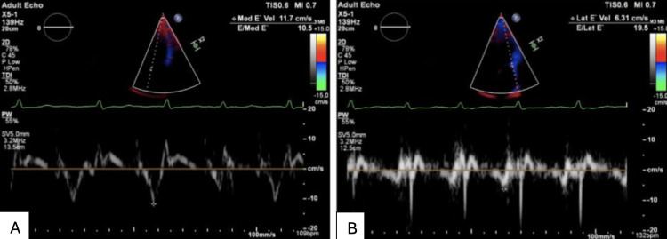
Myocardial deformation. Speckle-tracking strain separates the two conditions by the direction of the deformation abnormality. In 26 patients with constriction and 19 with restrictive cardiomyopathy, constriction reduced circumferential strain, torsion, and early diastolic untwisting while longitudinal strain and basal early diastolic velocities remained normal. Restrictive cardiomyopathy reduced longitudinal displacement and early diastolic velocity while circumferential deformation was preserved. In 52 patients with constriction, longitudinal strain of the left ventricular lateral wall and right ventricular free wall was reduced while septal strain was preserved, giving a lateral-to-septal strain ratio of about 0.8 compared with 1.1 in restrictive cardiomyopathy, and the ratio improved after pericardiectomy. Amyloidosis shows a different regional pattern, with reduced basal longitudinal strain and apical sparing. Strain is supportive and has not replaced tissue Doppler.
Mayo Clinic echocardiographic criteria for constriction
The Mayo Clinic criteria were derived from 130 patients with surgically confirmed constriction and 36 patients in whom constriction was considered and excluded (restrictive myocardial disease or severe tricuspid regurgitation). Five variables were examined, and three were independently associated with constriction: respiration-related ventricular septal shift, medial mitral e′, and the hepatic vein expiratory diastolic reversal ratio.
| Criterion | Threshold | Mechanism | Performance as a single criterion |
|---|---|---|---|
| Respiration-related ventricular septal shift | Present | Enhanced ventricular interdependence | Most sensitive; least specific |
| Medial mitral annular e′ | ≥9 cm/s | Preserved longitudinal myocardial relaxation | Most specific in the validation cohort against cardiac amyloidosis |
| Hepatic vein expiratory diastolic reversal ratio | ≥0.79 | Expiratory fall in right ventricular filling | Independent predictor |
| Combination | Cohort | Sensitivity | Specificity |
|---|---|---|---|
| Septal shift plus either medial e′ ≥9 cm/s or reversal ratio ≥0.79 | Derivation: 130 constriction, 36 restriction or severe tricuspid regurgitation | 87% | 91% |
| All three criteria | Derivation | 64% | 97% |
| Septal shift plus medial e′ ≥9 cm/s | Validation: 107 constriction, 30 cardiac amyloidosis | 80% | 92% |
| All three criteria | Validation | 70% | 97% |
Mayo Clinic echocardiographic criteria for constrictive pericarditis (Welch et al., 2014) and their external validation against cardiac amyloidosis (Qamruddin et al., 2019). The ratio of medial to lateral e′ and the respiratory variation in mitral E velocity differed between groups but were not independent predictors.
The criteria are applied to the comparison they were derived for: constriction against restrictive myocardial disease and severe tricuspid regurgitation in patients with heart failure and preserved ejection fraction. They do not apply during positive-pressure ventilation. A low medial e′ in a patient with radiation or postoperative constriction may reflect coexisting myocardial disease, and these patients often meet the criteria through septal shift and hepatic vein reversal.
Mayo Clinic echocardiographic criteria
Septal shift, medial e′, hepatic vein reversalHepatic vein reversal ratio = expiratory diastolic reversal velocity ÷ forward diastolic velocity of the same beat. Mitral E variation = (expiratory E − inspiratory E) ÷ expiratory E × 100. Thresholds and diagnostic performance from Welch et al. (2014).
CT and cardiac magnetic resonance in constrictive pericarditis
Cross-sectional imaging defines pericardial anatomy, calcification, and inflammation, and real-time CMR adds physiology. The 2025 ACC guidance recommends CMR to identify pericardial inflammation as the cause of constriction and lists CT for pericardial thickness, calcification, and surgical planning. The 2013 American Society of Echocardiography (ASE) multimodality recommendations place CT and CMR as complementary tests when echocardiography is nondiagnostic or windows are poor.
Pericardial thickness. The normal pericardium measures 2 mm or less. A thickness above 4 mm on CT or CMR is the threshold most often used to support constriction in the appropriate clinical setting, and thickening is most marked over the right ventricle and atrioventricular grooves. Thickening without constrictive physiology is not constriction, and normal thickness does not exclude it. A thick pericardium also occurs after uncomplicated cardiac surgery and in acute pericarditis without constriction.
Computed tomography. CT is the most accurate method for detecting and mapping pericardial calcification, which supports chronic disease, suggests prior tuberculous pericarditis, and predicts a more difficult resection. Indirect signs of constriction include a tubular or conical deformity of the ventricles, a straightened or sigmoid septum, atrial enlargement, dilated venae cavae and hepatic veins, reflux of contrast into the hepatic veins, ascites, and pleural effusions. Before pericardiectomy, CT shows the relationship of calcium to the coronary arteries and bypass grafts, the position of the right ventricle and grafts behind the sternum, and coexisting lung, pleural, or mediastinal disease. Retrospectively gated acquisition adds limited functional information.
Cardiac magnetic resonance. CMR measures thickness without the calcium blooming of CT, though calcium itself is poorly seen.
- Inflammation: pericardial edema on T2-weighted short-tau inversion-recovery images and pericardial late gadolinium enhancement identify active inflammation. Enhancement is the strongest imaging predictor that constriction will reverse with anti-inflammatory therapy.
- Ventricular interdependence: real-time free-breathing cine imaging in the short axis shows inspiratory flattening or inversion of the septum toward the left ventricle, the CMR equivalent of respiration-related septal shift. In 42 patients with surgically proven constriction, relative septal excursion had a sensitivity of 93% and a specificity of 95%, and pericardial thickness had a sensitivity of 83% and a specificity of 100%. A model combining thickness and septal excursion had a sensitivity of 100% and a specificity of 90%.
- Pericardial adherence: myocardial tagging shows loss of the normal sliding of the tag lines between pericardium and myocardium in fused, adherent disease.
- Myocardial involvement: native T1, extracellular volume, and myocardial late enhancement detect myocardial fibrosis or infiltration that indicates mixed disease or an alternative diagnosis such as amyloidosis, sarcoidosis, or radiation-induced myocardial fibrosis.
Nuclear imaging. Fluorodeoxyglucose positron emission tomography (FDG PET) can show active pericardial inflammation and is used in selected patients with suspected tuberculous, malignant, or IgG4-related disease. Technetium pyrophosphate or bone-tracer scintigraphy identifies transthyretin cardiac amyloidosis, a common restrictive cause in older adults.
Cardiac catheterization: dip-and-plateau and dynamic discordance
The 2025 ACC guidance recommends invasive catheterization when noninvasive methods are nondiagnostic or equivocal. Simultaneous right and left heart catheterization with high-fidelity or carefully flushed fluid-filled catheters, two transducers zeroed at the same level, and a respiratory trace is required. Measurements are made during quiet spontaneous respiration without breath-holding, and sedation that depresses respiration is minimized. Coronary angiography is performed when surgery is planned or when ischemia could explain the presentation.
Conventional criteria. Elevated and equalized diastolic pressures, a dip-and-plateau contour in both ventricles, and a steep y descent in the right atrium are present in constriction and also in restrictive cardiomyopathy. They establish impaired filling but not its cause. Three conventional criteria were proposed to separate the two conditions.
| Criterion | Favors constriction | Favors restriction | Predictive accuracy |
|---|---|---|---|
| LVEDP − RVEDP | ≤5 mm Hg | >5 mm Hg | 85% |
| RV systolic pressure | <55 mm Hg | ≥55 mm Hg | 70% |
| RVEDP ÷ RV systolic pressure | >1/3 | ≤1/3 | 76% |
| Dip-and-plateau contour | Present in both | Not discriminating | |
| Kussmaul sign (no inspiratory fall in RA pressure) | Present in both | Not discriminating | |
Conventional hemodynamic criteria, with predictive accuracy from a pooled analysis of 82 patients with constriction and 37 with restriction (Vaitkus and Kussmaul, 1991). When all three criteria agreed, more than 90% of patients were correctly classified, but one-quarter could not be classified. In a later series of 100 patients, no conventional criterion reached a predictive accuracy of 75%. LVEDP and RVEDP, left and right ventricular end-diastolic pressure; LV and RV, left and right ventricle; RA, right atrial.
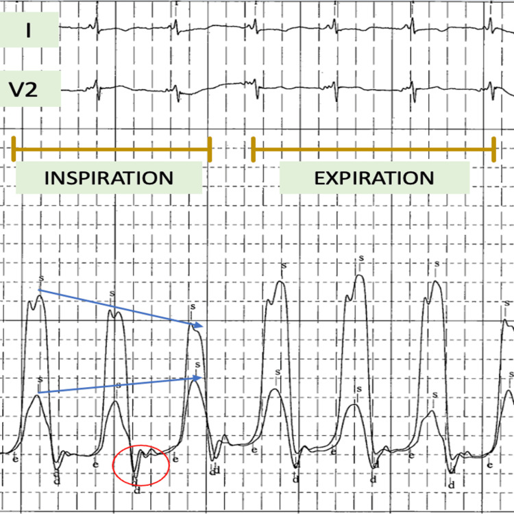
Dynamic respiratory criteria. Enhanced ventricular interdependence is specific for constriction and is the decisive catheterization finding.
- Discordance of ventricular systolic pressures. In constriction, left ventricular systolic pressure falls with inspiration while right ventricular systolic pressure rises, and the pattern reverses in expiration. In restrictive cardiomyopathy and other myocardial disease, both fall together with inspiration (concordance). In 36 patients studied with high-fidelity manometers, discordance accurately separated 15 patients with surgically proven constriction from 21 with other causes of heart failure, whereas conventional criteria did not.
- Systolic area index. The ratio of the right ventricular to the left ventricular systolic pressure-time area in inspiration, divided by the same ratio in expiration, quantifies interdependence and is less affected by heart rate and peak timing than peak pressures. In 100 consecutive patients (59 with surgically documented constriction and 41 with restrictive myocardial disease), a systolic area index above 1.1 had a sensitivity of 97% and a predictive accuracy of 100% for constriction.
- Respiratory change in the wedge-to-left ventricular gradient. The early diastolic gradient between pulmonary capillary wedge and left ventricular pressure falls with inspiration in constriction, the invasive counterpart of mitral inflow variation.
Right atrial to wedge pressure ratio. In pure constriction, right and left filling pressures are nearly equal. When myocardial disease coexists, wedge pressure exceeds right atrial pressure. In 113 surgically confirmed patients, the ratio of right atrial pressure to pulmonary artery wedge pressure correlated directly with pericardial thickness and with medial e′. A higher ratio reflects predominantly pericardial restraint, and a lower ratio a larger myocardial contribution. Patients with a ratio below the median of 0.77 had poorer long-term survival after pericardiectomy, a difference significant in primary but not in mixed constriction.
Provocation. Rapid infusion of 1,000 mL of normal saline over 6 to 8 minutes can unmask constrictive physiology in a patient with normal resting pressures and suspected occult constriction. The response is equalization of diastolic pressures and loss of the inspiratory fall in right atrial pressure. Exercise hemodynamics are used in selected patients with exertional symptoms and equivocal resting studies. Neither provocation is standardized.
Sources of error at catheterization. Hypovolemia from aggressive diuresis can mask equalization and the dip-and-plateau pattern; marked volume overload can blunt respiratory variation. Atrial fibrillation and premature beats confound beat-to-beat comparison, so beats of similar cycle length are compared. Severe tricuspid regurgitation, right ventricular infarction, and severe pulmonary hypertension can produce some constrictive-like features. Endomyocardial biopsy is performed when the hemodynamics suggest restriction and an infiltrative or storage disease that biopsy can identify is suspected.
Constriction versus restrictive cardiomyopathy: side-by-side comparison
Constriction and restrictive cardiomyopathy present with the same syndrome: right-sided congestion, elevated and similar filling pressures, small or normal ventricles, preserved ejection fraction, and a restrictive mitral inflow pattern. The distinction determines treatment. Constriction is often cured by pericardiectomy; restrictive cardiomyopathy is a myocardial disease that surgery cannot help, and a pericardiectomy performed for restriction exposes the patient to operative risk without benefit. The comparison rests on two physiologic questions: whether the ventricles show enhanced interdependence with respiration, and whether myocardial relaxation is intact.
| Feature | Constrictive pericarditis | Restrictive cardiomyopathy |
|---|---|---|
| Site of disease | Pericardium; myocardium normal | Myocardium (infiltration, fibrosis, storage) |
| Common causes | Idiopathic, cardiac surgery, radiation, tuberculosis | Amyloidosis, sarcoidosis, hemochromatosis, Fabry disease, endomyocardial fibrosis, radiation, idiopathic |
| History | Prior pericarditis, cardiac surgery, chest radiation, tuberculosis | Plasma cell dyscrasia, neuropathy, carpal tunnel syndrome, family history, systemic disease |
| Jugular venous pressure | Steep y descent; Kussmaul sign common | Steep y descent; Kussmaul sign may be present |
| Third sound or knock | Pericardial knock | Third heart sound in advanced disease |
| Mitral and tricuspid regurgitation | Usually mild | Often moderate |
| Electrocardiogram | Low voltage, nonspecific T-wave changes, atrial fibrillation | Low voltage with thick walls in amyloidosis, pseudoinfarction, conduction disease, atrial fibrillation |
| Chest radiograph and CT | Pericardial calcification on CT in about half; pericardium often >4 mm | Normal pericardium |
| Wall thickness and atria | Normal walls; mild to moderate atrial enlargement | Often increased wall thickness; marked biatrial enlargement |
| Septal motion | Respiration-related shift and early diastolic bounce | No respiratory shift |
| Mitral E respiratory variation | Often ≥25% | Minimal |
| Medial e′ | Preserved or increased (≥9 cm/s favors constriction) | Reduced |
| Medial versus lateral e′ | Medial ≥ lateral (annulus reversus) | Both reduced; lateral usually ≥ medial |
| E/e′ and filling pressure | Low E/e′ despite high pressure (annulus paradoxus) | High E/e′ tracks high pressure |
| Hepatic vein diastolic reversal | Expiratory (ratio ≥0.79) | Inspiratory |
| Strain | Reduced circumferential strain and twist; longitudinal strain preserved; free wall lower than septum | Reduced longitudinal strain; apical sparing in amyloidosis |
| CMR | Thick pericardium; pericardial enhancement if inflammatory; respiratory septal inversion | Myocardial late enhancement, raised native T1 and extracellular volume, no respiratory septal inversion |
| Natriuretic peptides | Normal or modestly elevated | Markedly elevated |
| LVEDP − RVEDP | ≤5 mm Hg | Often >5 mm Hg |
| RV systolic pressure | Usually <55 mm Hg | Often ≥55 mm Hg |
| RVEDP ÷ RV systolic pressure | >1/3 | ≤1/3 |
| LV and RV systolic pressures with inspiration | Discordant (LV falls, RV rises) | Concordant (both fall) |
| Systolic area index | >1.1 | ≤1.1 |
| Endomyocardial biopsy | Normal or nonspecific | May show amyloid, iron, granulomas, or storage disease |
| Definitive treatment | Anti-inflammatory therapy if inflammatory; pericardiectomy if fixed | Treatment of the underlying myocardial disease; heart failure therapy; transplantation in selected patients |
Constrictive pericarditis and restrictive cardiomyopathy compared. Thresholds from Welch et al. (2014), Vaitkus and Kussmaul (1991), and Talreja et al. (2008). No single feature is decisive; mixed disease after radiation shows features of both.
Natriuretic peptides. Myocardial stretch is limited by the pericardium in constriction, and the myocardium is not the diseased tissue, so B-type natriuretic peptide (BNP) is lower than in restrictive cardiomyopathy at similar filling pressures. In a small series, mean BNP was 128 pg/mL in constriction and 826 pg/mL in restrictive cardiomyopathy despite similar intracardiac pressures. Kidney dysfunction, atrial fibrillation, radiation-associated mixed disease, and older age raise BNP in constriction, so a high value does not exclude it, and the test is supportive only.
Mixed disease. After mediastinal radiation, and less often after cardiac surgery, pericardial constraint and myocardial fibrosis coexist. These patients show respiratory interdependence together with a reduced e′, a wedge pressure above right atrial pressure, and myocardial enhancement on CMR. Pericardiectomy relieves only the pericardial component, and residual restrictive physiology and the poorer survival of radiation-associated disease limit the benefit.
| Condition | Shared features | Distinguishing features |
|---|---|---|
| Cardiac tamponade | Raised, equalized diastolic pressures; ventricular interdependence | Effusion with chamber collapse; blunted y descent; prominent pulsus paradoxus; no dip-and-plateau |
| Severe tricuspid regurgitation | Raised venous pressure, ascites, hepatic vein reversal, septal motion abnormality | Systolic hepatic vein reversal timed with the jet; large right ventricle and right atrium; large v wave |
| Right ventricular infarction | Kussmaul sign, steep y descent, dip-and-plateau in the right ventricle | Acute inferior infarction; right ventricular dilation and hypokinesis; troponin rise |
| Pulmonary hypertension with right ventricular failure | Right-sided congestion, septal flattening | High pulmonary artery pressure and resistance; septal flattening in systole and diastole without respiratory shift |
| Obstructive lung disease | Respiratory variation in inflow | Marked inspiratory increase in superior vena cava systolic flow; preserved inferior vena cava collapse |
| Cirrhosis or nephrotic syndrome | Ascites and edema | Normal or low jugular venous pressure |
Differential diagnosis of constrictive physiology beyond restrictive cardiomyopathy.
The amyloid causes of restriction, their imaging, and treatment are covered on the cardiac amyloidosis page.
Diagnostic sequence for suspected constriction
- Clinical suspicion: unexplained right-sided congestion with a preserved ejection fraction, particularly after pericarditis, cardiac surgery, mediastinal radiation, or tuberculosis, or with recurrent ascites and no primary liver disease.
- Transthoracic echocardiography with a respiratory trace: septal motion, mitral and tricuspid inflow variation, medial and lateral e′, hepatic vein flow, inferior vena cava, and an assessment of myocardial and valvular disease. The Mayo Clinic criteria are applied.
- Natriuretic peptide, C-reactive protein, liver and kidney function, and etiologic testing.
- CMR for pericardial thickness and inflammation, real-time septal motion, and myocardial tissue characterization; CT for calcification and surgical planning.
- Simultaneous right and left heart catheterization with respiratory recording when the noninvasive findings are discordant or equivocal, or when mixed disease is suspected, with coronary angiography before surgery.
- Classification as inflammatory or fixed constriction, and as pure or mixed disease, which determines the choice between anti-inflammatory therapy and pericardiectomy.
Transient inflammatory constriction and anti-inflammatory therapy
Constrictive physiology during active pericardial inflammation can resolve. In 212 patients with echocardiographic constriction, 36 (17%) had resolution without pericardiectomy. The most common cause was prior cardiovascular surgery (25%), and among 22 patients followed with serial studies, constriction resolved an average of 8.3 weeks after diagnosis. Treatment in the transient group included nonsteroidal anti-inflammatory drugs, corticosteroids, and, in some patients, no specific therapy.
Markers of inflammation predict reversibility. In a pilot study of 29 patients with constriction treated with anti-inflammatory therapy, 14 had resolution and 15 had persistent constriction after 13 months. Pericardial late gadolinium enhancement was moderate or severe in 93% of the reversible group and in 33% of the persistent group. An enhanced pericardial thickness of 3 mm or more predicted reversibility with a sensitivity of 86% and a specificity of 80%. Baseline C-reactive protein (mean 59 versus 12 mg/L) and erythrocyte sedimentation rate were higher in the reversible group, and both fell with therapy together with the enhancement.
The 2025 ACC guidance recommends anti-inflammatory therapy as first-line treatment for inflammatory constrictive pericarditis, and radical pericardiectomy for noninflammatory constriction and for inflammatory constriction that fails anti-inflammatory therapy. The 2025 European Society of Cardiology (ESC) guideline similarly recommends anti-inflammatory therapy for newly diagnosed constriction with evidence of active inflammation, to prevent progression and avoid pericardiectomy. The 2025 ESC guideline recommends a trial of anti-inflammatory therapy for 3 to 6 months in newly diagnosed constriction before pericardiectomy is considered, longer than the 2- to 3-month empirical course described in the 2015 ESC guideline.
Regimens follow those for acute and recurrent pericarditis: a nonsteroidal anti-inflammatory drug or aspirin with colchicine, and corticosteroids when these fail, are contraindicated, or inflammation is marked. Response is assessed by symptoms, C-reactive protein, echocardiographic physiology, and repeat CMR. Persistent constrictive physiology after a defined course, or progression with rising venous pressure, hepatic or renal dysfunction, or cachexia, leads to referral for pericardiectomy. Prolonged medical therapy without improvement delays surgery while the risk of operation rises.
Constriction early after cardiac surgery is often transient and inflammatory, and the same approach applies. Pericardiectomy in the early postoperative period is technically difficult and is reserved for persistent or progressive physiology.
Effusive-constrictive pericarditis
Effusive-constrictive pericarditis combines tamponade from a pericardial effusion with constriction by the visceral pericardium. It is defined hemodynamically: after pericardiocentesis lowers intrapericardial pressure to near zero, right atrial pressure fails to fall by 50% or to below 10 mm Hg. The right atrial waveform changes from the blunted y descent of tamponade to the prominent y descent of constriction. The 2025 ACC guidance recommends noninvasive hemodynamic assessment by transthoracic echocardiography for the diagnosis of effusive-constrictive pericarditis.
In a prospective study of 1,184 patients with pericarditis, 218 had tamponade, and 190 of these underwent combined pericardiocentesis and catheterization. Fifteen had effusive-constrictive pericarditis. All presented with clinical tamponade, but concomitant constriction was recognized clinically in only 7. Intrapericardial pressure fell from a median of 12 to −5 mm Hg after drainage, whereas right atrial and ventricular end-diastolic pressures, although slightly reduced, remained elevated with a dip-and-plateau contour. The causes were diverse, including idiopathic, tuberculous, neoplastic, and radiation-associated disease. Pericardiectomy was required in 7 patients, all with visceral pericardial involvement, and 3 idiopathic cases resolved spontaneously.
Echocardiography after drainage shows persistent septal shift, respiratory inflow variation, and expiratory hepatic vein reversal, and these findings support the diagnosis without invasive measurement. CMR shows a thickened, enhancing visceral pericardium. A patient whose dyspnea or venous congestion persists after successful pericardiocentesis is evaluated for effusive-constrictive disease, a residual or loculated effusion, myocardial disease, and pulmonary hypertension.
Management depends on the cause and on inflammation. Inflammatory and idiopathic disease is treated with anti-inflammatory therapy and often resolves. Tuberculous disease is treated with antituberculous therapy. Persistent constriction requires pericardiectomy, and removal of the parietal pericardium alone is insufficient: the visceral layer must be removed (epicardiectomy or decortication), which increases operative difficulty and the risk of bleeding.
Medical therapy for chronic constriction
Medical therapy relieves congestion but does not reverse fixed constriction. Loop diuretics, often with an aldosterone antagonist, and sodium restriction reduce edema and ascites. Overdiuresis lowers the filling pressure on which a constricted heart depends and causes hypotension and kidney dysfunction. Cardiac output depends on heart rate because stroke volume is fixed, so sinus tachycardia is compensatory, and beta-blockers and nondihydropyridine calcium channel blockers that slow the sinus rate are avoided. Atrial fibrillation with a rapid ventricular rate shortens diastolic filling time and is controlled.
Medical therapy alone is appropriate for patients with mild symptoms and stable congestion, and for those in whom surgery carries prohibitive risk (very advanced disease, severe hepatic or kidney failure, cachexia, or extensive radiation-associated myocardial disease). It also serves as a bridge during anti-inflammatory therapy or antituberculous treatment. Paracentesis relieves tense ascites.
Pericardiectomy: selection, risk, and outcomes
Pericardiectomy is the only definitive treatment for chronic fixed constriction. The 2025 ACC guidance recommends radical pericardiectomy for noninflammatory constriction and for inflammatory constriction that fails anti-inflammatory therapy, performed at a high-volume pericardial surgical center. The 2025 ESC guideline also restricts pericardiectomy to expert centers because of its risk.
Selection and timing. Referral is made when symptoms persist despite diuretics or when congestion progresses, and before hepatic dysfunction, cachexia, kidney failure, and New York Heart Association (NYHA) class IV symptoms develop. Operative mortality and late survival are strongly related to functional class at the time of surgery. Patients with predominant myocardial disease, especially after radiation, may gain little.
Technique. Radical (total) pericardiectomy removes the parietal pericardium anteriorly from phrenic nerve to phrenic nerve, the diaphragmatic pericardium, and the pericardium posterior to the left phrenic nerve where accessible, together with the constricting epicardial peel. Median sternotomy gives access to both ventricles and the venae cavae and allows cardiopulmonary bypass when needed; a left anterolateral thoracotomy is an alternative in some centers. Densely calcified plaques adherent to the myocardium or over coronary arteries may be left as islands, with a waffle or grid incision of the residual peel. Cardiopulmonary bypass is used for bleeding, for concomitant procedures, and for posterior access, and its use is associated with higher mortality.
Radical versus partial resection. In 395 patients, most of them young and many with tuberculous disease, operative mortality was 7.6%, and partial pericardiectomy was associated with a 4.5-fold higher risk of death. Long-term actuarial survival was 83.8% after total and 73.9% after partial pericardiectomy, and 96.3% and 79.1% of survivors, respectively, were in NYHA class I or II. Incomplete resection leaves residual or recurrent constriction, which requires a technically harder reoperation.
Concomitant tricuspid surgery. Severe tricuspid regurgitation is common in long-standing constriction and after radiation, and it can worsen after pericardiectomy as the right ventricle dilates. The 2025 ESC guideline recommends tricuspid valve repair at the time of pericardiectomy when tricuspid regurgitation is more than mild and the valve anatomy is suitable, a recommendation based on retrospective surgical series.
| Series | Patients | Early mortality | Long-term outcome | Adverse predictors |
|---|---|---|---|---|
| Ling et al., 1999 (single US center, 1985–1995) | 135 | 6% (historical cohort 14%) | 10-year survival 57%; 83% of late survivors free of symptoms | Older age, NYHA class, radiation (strongest) |
| Bertog et al., 2004 (single US center, 24 years) | 163 | 6% | 7-year survival: idiopathic 88%, postsurgical 66%, postradiation 27% | Radiation, worse kidney function, higher pulmonary artery systolic pressure, LV systolic dysfunction, lower sodium, older age; calcification not predictive |
| Chowdhury et al., 2006 (single center, India) | 395 | 7.6% | Survival 83.8% total versus 73.9% partial pericardiectomy | High right atrial pressure, hyperbilirubinemia, kidney dysfunction, atrial fibrillation, calcification, thoracotomy, partial resection |
| Murashita et al., 2017 (single US center, 1936–2013) | 1,066 | 30-day 5.2% after 1990 (13.5% before) | Overall survival similar between eras after adjustment | NYHA III–IV (HR 2.17), radiation (HR 3.93), prior cardiac surgery (HR 1.47), cardiopulmonary bypass (HR 1.35) |
Outcomes of pericardiectomy for constrictive pericarditis in large surgical series. HR, hazard ratio for overall mortality; LV, left ventricular; NYHA, New York Heart Association.
Low-output syndrome. Low cardiac output after pericardiectomy is common and reflects myocardial atrophy or fibrosis after long-standing constraint, acute ventricular dilation once the restraint is removed, and residual myocardial disease. It occurred in 24% of patients in a series of 127 radical pericardiectomies performed without cardiopulmonary bypass. It is managed with careful volume restriction, inotropic support, and, rarely, mechanical circulatory support. Other complications include bleeding, arrhythmia, phrenic nerve injury, right ventricular or coronary injury, and kidney failure.
Recovery of physiology. Filling pressures and symptoms improve in most survivors, but normalization of Doppler findings may take weeks to months. In the early Doppler series, 14 of 19 patients had normal Doppler findings after pericardiectomy and 12 of these became asymptomatic, whereas all 5 patients with a persistent restrictive pattern remained symptomatic. Persistent restrictive filling after surgery indicates incomplete resection or myocardial disease. After pericardiectomy, all annular velocities fall and the normal relationship of lateral and medial e′ returns.
Radiation-associated constriction. Survival after pericardiectomy for radiation-associated constriction is markedly worse than for idiopathic disease, because of myocardial fibrosis, coronary and valve disease, lung injury, and the underlying cancer. The decision to operate weighs the pericardial contribution to the hemodynamics, measured by the Mayo Clinic criteria, the right atrial to wedge ratio, and CMR, against the extent of myocardial disease.
Prognosis and follow-up after constrictive pericarditis
Untreated chronic constriction progresses, with worsening congestion, hepatic dysfunction, cachexia, and death from heart failure. The prognosis after pericardiectomy depends chiefly on the cause and on the patient's condition at operation. Functional class, kidney function, serum sodium, pulmonary artery systolic pressure, left ventricular systolic function, and age add to risk.
Follow-up after pericardiectomy includes clinical review and echocardiography to document resolution of constrictive physiology, to detect residual or recurrent constriction, and to assess right ventricular function and tricuspid regurgitation. Diuretics are tapered as congestion resolves. Persistent or recurrent symptoms lead to reassessment with echocardiography, CMR or CT, and catheterization for residual constriction, myocardial disease, or tricuspid regurgitation. After radiation, surveillance for coronary, valvular, and conduction disease continues lifelong. In patients managed medically for transient constriction, echocardiography and inflammatory markers are repeated until the physiology has resolved and therapy has been tapered.
Sources of error in diagnosing constrictive pericarditis
- Excluding constriction because the pericardium is not thickened: 18% of surgically proven cases have a pericardium of normal thickness.
- Diagnosing constriction from a thick or calcified pericardium without constrictive physiology: thickening and calcification indicate anatomy, not hemodynamic constraint.
- Diagnosing constriction from a septal bounce alone: a bounce also occurs after cardiac surgery, with left bundle branch block and pacing, and with right ventricular overload. Respiration-related shift is the relevant finding.
- Excluding constriction because mitral inflow variation is absent: very high filling pressure blunts the variation, and preload reduction can unmask it; the Mayo Clinic criteria do not require it.
- Attributing inflow variation to constriction in obstructive lung disease or obesity: superior vena cava flow distinguishes exaggerated respiratory effort from pericardial disease.
- Applying spontaneous-breathing criteria during positive-pressure ventilation, or using inflow variation across beats of different cycle length in atrial fibrillation.
- Using E/e′ to estimate filling pressure in constriction: annulus paradoxus makes the ratio inversely related to wedge pressure.
- Reading hepatic vein systolic reversal from severe tricuspid regurgitation as the expiratory diastolic reversal of constriction: reversal is timed with the electrocardiogram and the regurgitant jet.
- Diagnosing constriction at catheterization from equalized diastolic pressures and a dip-and-plateau contour: both occur in restrictive cardiomyopathy, and respiratory discordance or the systolic area index is required.
- Recording catheterization after aggressive diuresis or with respiratory depression from sedation, which masks constrictive physiology.
- Referring inflammatory constriction for pericardiectomy without a trial of anti-inflammatory therapy, or prolonging medical therapy in fixed constriction until hepatic failure and cachexia raise operative risk.
- Overlooking mixed disease after radiation, in which pericardiectomy relieves only the pericardial component.
Self-assessment
A 58-year-old man treated with mediastinal radiation 20 years ago has ascites and edema. The septum shifts leftward with inspiration, medial e′ is 6 cm/s with lateral e′ 8 cm/s, hepatic vein diastolic reversal is larger in inspiration than in expiration, BNP is 940 pg/mL, and simultaneous ventricular tracings show concordant respiratory changes in systolic pressure with a systolic area index of 0.9. Is pericardiectomy likely to relieve his heart failure?
Cardiovascular Medicine Board ReviewReferences
- Wang TKM, Klein AL, Cremer PC, et al. 2025 concise clinical guidance: an ACC expert consensus statement on the diagnosis and management of pericarditis: a report of the American College of Cardiology Solution Set Oversight Committee. J Am Coll Cardiol. 2025;86:2691–2719. doi:10.1016/j.jacc.2025.05.023
- Schulz-Menger J, Collini V, Gröschel J, et al. 2025 ESC guidelines for the management of myocarditis and pericarditis. Eur Heart J. 2025;46:3952–4041. doi:10.1093/eurheartj/ehaf192
- Adler Y, Charron P, Imazio M, et al. 2015 ESC guidelines for the diagnosis and management of pericardial diseases. Eur Heart J. 2015;36:2921–2964. doi:10.1093/eurheartj/ehv318
- Klein AL, Abbara S, Agler DA, et al. American Society of Echocardiography clinical recommendations for multimodality cardiovascular imaging of patients with pericardial disease: endorsed by the Society for Cardiovascular Magnetic Resonance and Society of Cardiovascular Computed Tomography. J Am Soc Echocardiogr. 2013;26:965–1012.e15. doi:10.1016/j.echo.2013.06.023
- Welch TD, Ling LH, Espinosa RE, et al. Echocardiographic diagnosis of constrictive pericarditis: Mayo Clinic criteria. Circ Cardiovasc Imaging. 2014;7:526–534. doi:10.1161/CIRCIMAGING.113.001613
- Qamruddin S, Alkharabsheh SK, Sato K, et al. Differentiating constriction from restriction (from the Mayo Clinic echocardiographic criteria). Am J Cardiol. 2019;124:932–938. doi:10.1016/j.amjcard.2019.06.002
- Oh JK, Hatle LK, Seward JB, et al. Diagnostic role of Doppler echocardiography in constrictive pericarditis. J Am Coll Cardiol. 1994;23:154–162. doi:10.1016/0735-1097(94)90514-2
- Reuss CS, Wilansky SM, Lester SJ, et al. Using mitral 'annulus reversus' to diagnose constrictive pericarditis. Eur J Echocardiogr. 2009;10:372–375. doi:10.1093/ejechocard/jen258
- Veress G, Ling LH, Kim KH, et al. Mitral and tricuspid annular velocities before and after pericardiectomy in patients with constrictive pericarditis. Circ Cardiovasc Imaging. 2011;4:399–407. doi:10.1161/CIRCIMAGING.110.959619
- Ha JW, Oh JK, Ling LH, et al. Annulus paradoxus: transmitral flow velocity to mitral annular velocity ratio is inversely proportional to pulmonary capillary wedge pressure in patients with constrictive pericarditis. Circulation. 2001;104:976–978. doi:10.1161/hc3401.095705
- Sengupta PP, Krishnamoorthy VK, Abhayaratna WP, et al. Disparate patterns of left ventricular mechanics differentiate constrictive pericarditis from restrictive cardiomyopathy. JACC Cardiovasc Imaging. 2008;1:29–38. doi:10.1016/j.jcmg.2007.10.006
- Talreja DR, Edwards WD, Danielson GK, et al. Constrictive pericarditis in 26 patients with histologically normal pericardial thickness. Circulation. 2003;108:1852–1857. doi:10.1161/01.CIR.0000087606.18453.FD
- Talreja DR, Nishimura RA, Oh JK, Holmes DR. Constrictive pericarditis in the modern era: novel criteria for diagnosis in the cardiac catheterization laboratory. J Am Coll Cardiol. 2008;51:315–319. doi:10.1016/j.jacc.2007.09.039
- Hurrell DG, Nishimura RA, Higano ST, et al. Value of dynamic respiratory changes in left and right ventricular pressures for the diagnosis of constrictive pericarditis. Circulation. 1996;93:2007–2013. doi:10.1161/01.CIR.93.11.2007
- Vaitkus PT, Kussmaul WG. Constrictive pericarditis versus restrictive cardiomyopathy: a reappraisal and update of diagnostic criteria. Am Heart J. 1991;122:1431–1441. doi:10.1016/0002-8703(91)90587-8
- Yang JH, Miranda WR, Borlaug BA, et al. Right atrial/pulmonary arterial wedge pressure ratio in primary and mixed constrictive pericarditis. J Am Coll Cardiol. 2019;73:3312–3321. doi:10.1016/j.jacc.2019.03.522
- Bush CA, Stang JM, Wooley CF, Kilman JW. Occult constrictive pericardial disease: diagnosis by rapid volume expansion and correction by pericardiectomy. Circulation. 1977;56:924–930. doi:10.1161/01.CIR.56.6.924
- Geske JB, Anavekar NS, Nishimura RA, Oh JK, Gersh BJ. Differentiation of constriction and restriction: complex cardiovascular hemodynamics. J Am Coll Cardiol. 2016;68:2329–2347. doi:10.1016/j.jacc.2016.08.050
- Harmon DM, Oh JK. Hemodynamics of pericardial constriction: role of echocardiography and cardiac catheterization. Methodist DeBakey Cardiovasc J. 2026;22(2):74–84. doi:10.14797/mdcvj.1781
- Leya FS, Arab D, Joyal D, et al. The efficacy of brain natriuretic peptide levels in differentiating constrictive pericarditis from restrictive cardiomyopathy. J Am Coll Cardiol. 2005;45:1900–1902. doi:10.1016/j.jacc.2005.03.050
- Bolen MA, Rajiah P, Kusunose K, et al. Cardiac MR imaging in constrictive pericarditis: multiparametric assessment in patients with surgically proven constriction. Int J Cardiovasc Imaging. 2015;31:859–866. doi:10.1007/s10554-015-0616-z
- Bogaert J, Francone M. Cardiovascular magnetic resonance in pericardial diseases. J Cardiovasc Magn Reson. 2009;11:14. doi:10.1186/1532-429X-11-14
- Feng D, Glockner J, Kim K, et al. Cardiac magnetic resonance imaging pericardial late gadolinium enhancement and elevated inflammatory markers can predict the reversibility of constrictive pericarditis after antiinflammatory medical therapy: a pilot study. Circulation. 2011;124:1830–1837. doi:10.1161/CIRCULATIONAHA.111.026070
- Haley JH, Tajik AJ, Danielson GK, et al. Transient constrictive pericarditis: causes and natural history. J Am Coll Cardiol. 2004;43:271–275. doi:10.1016/j.jacc.2003.08.032
- Sagristà-Sauleda J, Angel J, Sánchez A, et al. Effusive-constrictive pericarditis. N Engl J Med. 2004;350:469–475. doi:10.1056/NEJMoa035630
- Imazio M, Brucato A, Maestroni S, et al. Risk of constrictive pericarditis after acute pericarditis. Circulation. 2011;124:1270–1275. doi:10.1161/CIRCULATIONAHA.111.018580
- Mayosi BM, Ntsekhe M, Bosch J, et al. Prednisolone and Mycobacterium indicus pranii in tuberculous pericarditis. N Engl J Med. 2014;371:1121–1130. doi:10.1056/NEJMoa1407380
- Ling LH, Oh JK, Schaff HV, et al. Constrictive pericarditis in the modern era: evolving clinical spectrum and impact on outcome after pericardiectomy. Circulation. 1999;100:1380–1386. doi:10.1161/01.CIR.100.13.1380
- Bertog SC, Thambidorai SK, Parakh K, et al. Constrictive pericarditis: etiology and cause-specific survival after pericardiectomy. J Am Coll Cardiol. 2004;43:1445–1452. doi:10.1016/j.jacc.2003.11.048
- Murashita T, Schaff HV, Daly RC, et al. Experience with pericardiectomy for constrictive pericarditis over eight decades. Ann Thorac Surg. 2017;104:742–750. doi:10.1016/j.athoracsur.2017.05.063
- Chowdhury UK, Subramaniam GK, Kumar AS, et al. Pericardiectomy for constrictive pericarditis: a clinical, echocardiographic, and hemodynamic evaluation of two surgical techniques. Ann Thorac Surg. 2006;81:522–529. doi:10.1016/j.athoracsur.2005.08.009
- Chowdhury UK, George N, Singh S, et al. Total pericardiectomy using a modified left anterolateral thoracotomy without cardiopulmonary bypass. Ann Thorac Surg. 2021;112:1483–1492. doi:10.1016/j.athoracsur.2020.10.045
- Kusunose K, Dahiya A, Popović ZB, et al. Biventricular mechanics in constrictive pericarditis comparison with restrictive cardiomyopathy and impact of pericardiectomy. Circ Cardiovasc Imaging. 2013;6:399–406. doi:10.1161/CIRCIMAGING.112.000078