Library › Cardiomyopathy › Cardiac amyloidosis
Cardiac amyloidosis
Transthyretin and light-chain cardiac amyloidosis in adults, from red flags and multimodality diagnosis through staging, disease-modifying therapy, and management of heart failure, arrhythmia, and conduction disease.
Cardiac amyloidosis is two treatable diseases with a shared phenotype: transthyretin (ATTR) amyloidosis, wild-type or hereditary, and light-chain (AL) amyloidosis from a plasma cell clone. Testing for a monoclonal protein with serum free light chains and serum and urine immunofixation comes first in every patient. When that screen is negative, grade 2 or 3 myocardial uptake on bone scintigraphy, confirmed on single-photon emission computed tomography (SPECT), establishes ATTR cardiomyopathy without biopsy. A monoclonal protein, at any scan grade, leads to tissue diagnosis with amyloid typing and urgent hematology care.
Transthyretin and light-chain amyloidosis: two different diseases
Amyloidosis is the extracellular deposition of misfolded protein as insoluble fibrils with a cross-β-sheet structure that binds Congo red and shows apple-green birefringence under polarized light. More than 30 precursor proteins form amyloid in humans, but two account for nearly all cardiac disease. Light-chain amyloidosis arises from a clonal plasma cell disorder that secretes an unstable immunoglobulin light chain, usually lambda. Transthyretin amyloidosis arises from transthyretin, a transport protein for thyroxine and retinol-binding protein that is synthesized mainly by the liver and circulates as a tetramer.
Transthyretin amyloidosis occurs in two forms. Wild-type disease (ATTRwt), formerly called senile systemic amyloidosis, develops without a pathogenic variant and affects mainly men older than 70 years. Hereditary or variant disease (ATTRv) results from one of more than 130 pathogenic variants in the TTR gene, inherited as an autosomal dominant trait with variable penetrance. Rarer cardiac amyloid types include serum amyloid A (AA) amyloidosis in chronic inflammation, which seldom causes clinically important cardiomyopathy, apolipoprotein A-I and A-IV amyloidosis, and isolated atrial amyloid from atrial natriuretic peptide, which is common in the aging atrium and contributes to atrial myopathy.
The distinction is the first diagnostic decision because treatment, tempo, and prognosis diverge at once. AL amyloidosis with cardiac involvement is a hematologic emergency: untreated advanced disease carries survival measured in months, and treatment is chemotherapy directed at the clone. ATTR cardiomyopathy progresses over years and is treated with drugs that stabilize the transthyretin tetramer or suppress its production. Echocardiography and cardiac magnetic resonance (CMR) cannot reliably separate the two, and bone scintigraphy can be positive in both, so the type is assigned only by the combination of a monoclonal protein screen and scintigraphy or by tissue typing.
| Feature | Wild-type ATTR | Hereditary ATTR | AL amyloidosis |
|---|---|---|---|
| Precursor | Wild-type transthyretin | Variant transthyretin | Monoclonal light chain (lambda more often than kappa) |
| Typical patient | Man older than 70 years | Age and phenotype depend on variant; p.Val142Ile in older Black adults | Adult in the sixth or seventh decade |
| Organs | Heart; tendons and ligaments (carpal tunnel, spinal canal, biceps tendon) | Heart, peripheral and autonomic nerves, eye | Heart, kidney (nephrotic syndrome), liver, nerves, gut, soft tissue |
| Tempo | Slow; years from first symptoms | Variable; neuropathic variants can progress quickly | Rapid; months |
| Bone scintigraphy | Grade 2 or 3 uptake typical | Usually grade 2 or 3; low uptake with some variants | Usually absent or grade 1; grade 2 or 3 occurs |
| Diagnosis | Scintigraphy with negative monoclonal screen, or tissue typing | As for wild-type, plus TTR sequencing | Tissue biopsy with typing by mass spectrometry |
| Disease-specific therapy | Tafamidis, acoramidis, vutrisiran | As for wild-type; silencers also treat polyneuropathy | Daratumumab with bortezomib, cyclophosphamide, and dexamethasone; stem cell transplant in eligible patients |
The main forms of cardiac amyloidosis. Variant names use current protein numbering, which adds the 20-residue signal peptide; p.Val142Ile is the historical Val122Ile.
Epidemiology of wild-type and hereditary transthyretin amyloidosis
Wild-type ATTR is far more common than its clinical diagnosis rate suggests. In a population-based autopsy study of Finns aged 85 years or older, transthyretin amyloid was present in the myocardium of 25%. In a prospective study of patients aged 60 years or older admitted with heart failure with preserved ejection fraction (HFpEF) and a left ventricular wall thickness of 12 mm or more, 13.3% had moderate to severe myocardial uptake on technetium-99m 3,3-diphosphono-1,2-propanodicarboxylic acid (DPD) scintigraphy, and all had wild-type disease. Among older patients with severe aortic stenosis referred for transcatheter aortic valve replacement (TAVR), scintigraphic screening identified ATTR in 16%. In the Mayo Clinic wild-type cohort, median age at diagnosis was 75 years, 91% of patients were men, and median survival from diagnosis without disease-modifying therapy was 3.6 years. Wider use of scintigraphy has moved diagnosis earlier, and the number of patients diagnosed each year has risen steeply since nonbiopsy diagnosis was validated.
Hereditary ATTR has distinct geographic and ancestral patterns. The p.Val142Ile (Val122Ile) variant is carried by about 3% to 4% of Black Americans; in the Atherosclerosis Risk in Communities cohort, 3% of Black participants were carriers, and carriers had a higher risk of incident heart failure (hazard ratio 1.47) than noncarriers. It causes a late-onset cardiomyopathy that resembles wild-type disease and is frequently attributed to hypertensive heart disease. The p.Val50Met (Val30Met) variant, endemic in Portugal, Sweden, Japan, and Brazil, classically causes a length-dependent sensorimotor and autonomic polyneuropathy; early-onset disease is predominantly neuropathic, and late-onset disease more often involves the heart. The p.Thr80Ala (Thr60Ala) variant, common in people of Irish ancestry, produces a mixed cardiac and neurologic phenotype.
AL amyloidosis is rarer, with an incidence of roughly 10 cases per million people per year, but cardiac involvement occurs in most patients and determines survival. It arises from a small clone that often meets criteria only for monoclonal gammopathy, and it can coexist with multiple myeloma. Monoclonal gammopathy of undetermined significance (MGUS) is common in older adults, so a monoclonal protein in an older patient with suspected ATTR does not by itself establish AL disease; it removes the patient from the nonbiopsy pathway.
Pathophysiology: fibril formation and the restrictive myocardium
The transthyretin tetramer dissociates into monomers, which misfold and aggregate into oligomers and fibrils. Dissociation is the rate-limiting step, which is the rationale for stabilizers that bind the thyroxine-binding sites and hold the tetramer together. Pathogenic variants destabilize the tetramer; in wild-type disease, age-related changes in protein homeostasis allow normal transthyretin to aggregate. Because the liver produces nearly all circulating transthyretin, silencing hepatic synthesis lowers the supply of precursor protein; liver transplantation was the first disease-modifying therapy for hereditary disease for the same reason.
Amyloid expands the myocardial interstitium, separates and compresses myocytes, and infiltrates the intramural vessels, the conduction system, the atria, and the valves. The left ventricle thickens without myocyte hypertrophy, its cavity becomes small, and stiffness rises steeply. Longitudinal subendocardial fibers fail first, so mitral annular velocities and longitudinal strain fall while radial thickening and a small cavity keep the ejection fraction normal. Stroke volume is small and relatively fixed, and cardiac output depends on heart rate. Atrial infiltration abolishes atrial contraction, sometimes while the rhythm is still sinus, and contributes to atrial thrombus formation and loss of the atrial contribution to filling.
Light chains add direct toxicity. Circulating amyloidogenic light chains impair myocyte function through oxidative stress and lysosomal dysfunction, independent of fibril mass. AL cardiomyopathy is therefore more severe for a given wall thickness than ATTR cardiomyopathy, and N-terminal pro-B-type natriuretic peptide (NT-proBNP) can fall within weeks of a deep hematologic response before any change in wall thickness.
The vascular and autonomic effects explain intolerance of standard heart failure drugs. Amyloid in small arteries and arterioles causes microvascular dysfunction and angina-like chest pain without epicardial disease. Autonomic neuropathy, prominent in AL and hereditary ATTR, causes orthostatic hypotension. A low, fixed stroke volume and chronotropic dependence make vasodilators and negative chronotropes poorly tolerated.
Clinical presentation and red flags for cardiac amyloidosis
Most patients present with heart failure, usually with a preserved or mildly reduced ejection fraction, or with atrial fibrillation, conduction disease, or an echocardiogram showing unexplained wall thickening. Right-sided congestion with edema, hepatic congestion, and ascites often dominates late disease. Exertional dyspnea and fatigue reflect a fixed stroke volume more than pulmonary congestion. Syncope can be exertional from low output, orthostatic from autonomic failure, or arrhythmic from atrioventricular block or ventricular arrhythmia.
The 2023 American College of Cardiology (ACC) expert consensus decision pathway (ECDP) and the 2025 ACC concise clinical guidance describe a low threshold for testing. Testing is directed at patients with presumed HFpEF, particularly with coexisting aortic stenosis, carpal tunnel syndrome, spinal stenosis, biceps tendon rupture, or neuropathy. Musculoskeletal findings precede cardiac disease by years in ATTR: in a prospective study of men 50 years or older and women 60 years or older undergoing carpal tunnel release, Congo red staining of tenosynovial tissue found amyloid in 10.2%, most often ATTR. Prior hip or knee replacement and shoulder surgery also appear in the histories of patients with wild-type disease.
| Domain | Red flag | Comment |
|---|---|---|
| Heart failure course | HFpEF with wall thickness ≥12 mm; intolerance of beta-blockers or vasodilators; hypotension in a previously hypertensive patient | Fixed stroke volume and autonomic dysfunction |
| Electrocardiogram | Low or normal QRS voltage despite thick walls; pseudoinfarct Q waves; atrioventricular or bundle branch block | Voltage is normal in many ATTR patients |
| Echocardiogram | Thick walls with small cavity; thick valves and interatrial septum; biatrial enlargement; small effusion; relative apical sparing of longitudinal strain | Each finding is nonspecific alone |
| Biomarkers | Persistently elevated troponin; NT-proBNP disproportionate to clinical status | Troponin elevation without coronary cause |
| Valve disease | Low-flow, low-gradient aortic stenosis in an older patient | Coexisting ATTR in about 1 in 6 TAVR candidates in screening studies |
| Musculoskeletal | Bilateral carpal tunnel syndrome; lumbar spinal stenosis; spontaneous distal biceps tendon rupture; trigger finger | Typical of ATTR; often years before heart failure |
| Neurologic | Length-dependent sensorimotor neuropathy; orthostatic hypotension, erectile dysfunction, gastrointestinal dysmotility | Hereditary ATTR and AL |
| Kidney | Proteinuria or nephrotic syndrome | Characteristic of AL |
| Soft tissue and skin | Macroglossia; periorbital purpura; submandibular gland enlargement | Specific for AL |
| Hematologic | Acquired factor X deficiency; monoclonal gammopathy | AL |
| Family history | Heart failure, neuropathy, or pacemaker in relatives; known TTR variant | Hereditary ATTR |
Clinical clues to cardiac amyloidosis, after the 2023 ACC ECDP and the 2021 multimodality imaging consensus. No single feature is diagnostic; the combination raises pretest probability.
Extracardiac involvement. In AL amyloidosis, kidney involvement with albuminuria, often in the nephrotic range, is common; hepatomegaly with a raised alkaline phosphatase, gastrointestinal bleeding, weight loss, and autonomic failure also occur. Macroglossia and periorbital purpura are specific for AL. In hereditary ATTR, neuropathy begins in the feet with small-fiber pain and temperature loss, and autonomic involvement causes orthostatic hypotension, diarrhea alternating with constipation, early satiety, and erectile dysfunction. Vitreous opacities occur with some variants. Wild-type ATTR is largely confined to the heart and musculoskeletal tissue, although mild neuropathy is common in older patients for other reasons.
Natural history. ATTR cardiomyopathy progresses steadily: wall thickness, NT-proBNP, and troponin rise, functional class worsens, and conduction disease and atrial arrhythmia accumulate. Death is usually from progressive heart failure or sudden death. AL cardiomyopathy progresses over months, and early deaths cluster in the first months after diagnosis in patients with advanced cardiac involvement.
Electrocardiogram: low voltage, pseudoinfarction, and the voltage-to-mass mismatch
The electrocardiogram reflects interstitial expansion: myocardial mass rises while the electrically active myocyte mass does not, so QRS voltage is low or normal for the wall thickness on imaging. Low voltage is defined as QRS amplitude of 5 mm or less in all limb leads or 10 mm or less in all precordial leads. It is more frequent in AL than in ATTR. The 2023 ACC ECDP notes that the classic combination of low voltage with echocardiographic hypertrophy is present in only about 30% of patients, so normal voltage does not exclude the disease. Electrocardiographic left ventricular hypertrophy occurs in a minority of patients with ATTR.
The voltage-to-mass relationship is more discriminating than absolute voltage. A normal-voltage tracing in a patient with a 17-mm septum is a mismatch, because hypertensive and sarcomeric hypertrophy of that degree usually produce increased voltage. Indices that divide QRS voltage by wall thickness or left ventricular mass separate amyloidosis from other causes of thickening better than voltage alone.
Pseudoinfarct Q waves or poor R-wave progression in the anterior or inferior leads, without regional wall motion abnormality or coronary disease, are common. Atrial fibrillation and flutter are frequent, especially in ATTR. Conduction disease includes first-degree atrioventricular block, which often reflects infranodal disease, bundle branch block, and progression to high-grade block. A pacemaker for unexplained atrioventricular block in an older patient with a thick ventricle is a common route to diagnosis.
Echocardiography: wall thickness, strain pattern, and diastolic findings
Echocardiography is usually the first test to raise the question. The phenotype combines several findings, and no single finding establishes it. Concentric thickening of the left ventricle with a small or normal cavity is typical, although ATTR often produces asymmetric septal thickening with a sigmoid or reversed septal curvature that can be mistaken for hypertrophic cardiomyopathy. The 2021 multimodality imaging consensus counts three typical echocardiographic features: a left ventricular wall thickness above 12 mm, a relative apical longitudinal strain ratio above 1.0, and grade 2 or worse diastolic dysfunction. Each applies only when hypertension and other causes are reasonably excluded.
Longitudinal function and strain. Global longitudinal strain (GLS) is reduced, often to an absolute value of about 12% or less, while ejection fraction remains normal. The characteristic regional pattern is a base-to-apex gradient with severe impairment of basal and mid segments and relative preservation of the apex, the "cherry on top" bull's-eye map. The relative apical longitudinal strain ratio is average apical strain divided by the sum of average basal and mid strain; in the original study a ratio above 1.0 was 93% sensitive and 82% specific for amyloidosis against hypertrophic cardiomyopathy and aortic stenosis. The ejection fraction-to-GLS ratio captures the same dissociation between preserved ejection and impaired longitudinal shortening. In a head-to-head comparison it was the most accurate echocardiographic discriminator, including in patients with only mild thickening or a normal ejection fraction. Apical sparing is supportive, not specific: it occurs in aortic stenosis, hypertensive heart disease, and hypertrophic cardiomyopathy, and strain values vary by vendor and loading. A low myocardial contraction fraction (stroke volume divided by myocardial volume) quantifies the small output of a heavy ventricle.
Diastolic function and filling pressure. Early disease shows impaired relaxation with reduced septal and lateral e′, often 5 cm/s or less. Advanced disease shows a restrictive mitral inflow with a tall E wave, small A wave, short deceleration time, and an E/A ratio above 2, together with elevated E/e′ and tricuspid regurgitation velocity. A small A wave in sinus rhythm reflects atrial infiltration and mechanical failure, and left atrial reservoir strain is markedly reduced. Restrictive filling identifies elevated pressure but not its cause; the same inflow occurs in constriction, which is separated by preserved or increased medial e′, annulus reversus, respiratory variation, and septal shift (see constrictive pericarditis). The diastolic algorithm and its special-population rules are covered on the HFpEF and diastolic function page.
Valves, atria, right ventricle, and pericardium. Valve leaflets and the interatrial septum thicken from infiltration, and the right ventricular free wall thickens beyond 5 mm. Both atria enlarge. A small circumferential pericardial effusion is common and does not indicate pericarditis. Left atrial appendage emptying velocity falls, and appendage thrombus can form in sinus rhythm. The "granular sparkling" texture of the myocardium carries little weight because harmonic imaging produces a similar appearance in normal and hypertrophied myocardium. Stroke volume index is frequently below 35 mL/m² despite a normal ejection fraction, and any coexisting aortic stenosis gradient then understates severity.
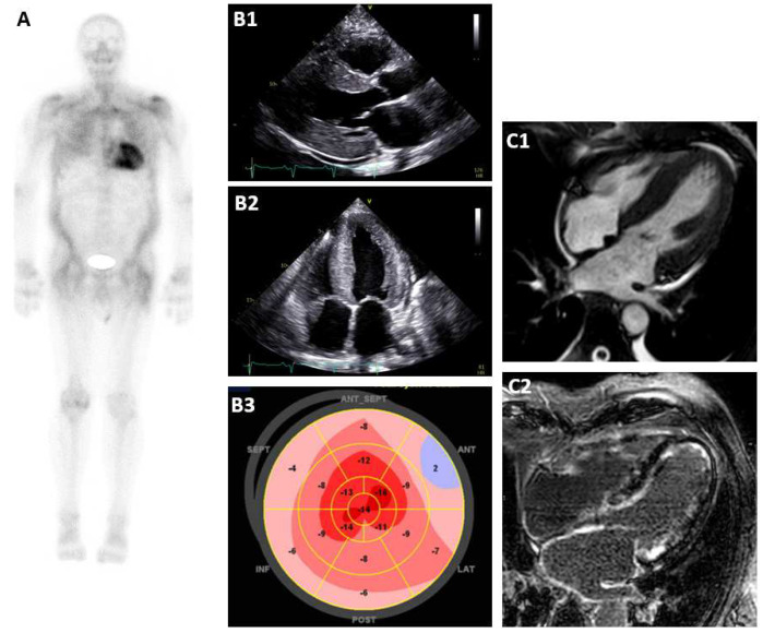
Systolic function. Ejection fraction is preserved until late in most patients, and its fall marks advanced disease. In the Mayo wild-type cohort, lower ejection fraction and pericardial effusion independently predicted mortality. Tissue Doppler s′ velocities are reduced early; an average s′ of about 4 cm/s in patients with coexisting aortic stenosis and ATTR, compared with 6.6 cm/s in patients without amyloid, illustrates the degree of longitudinal impairment.
Cardiac magnetic resonance: late enhancement, myocardial nulling, T1, and extracellular volume
CMR characterizes tissue and is the most informative imaging test when scintigraphy cannot be used, when the monoclonal screen is positive, or when the differential includes hypertrophic cardiomyopathy, Fabry disease, sarcoidosis, or hypertensive heart disease. The 2021 consensus lists four typical CMR features: wall thickness above the sex-specific upper limit, a global extracellular volume fraction (ECV) above 0.40, diffuse late gadolinium enhancement (LGE), and abnormal gadolinium kinetics with myocardial nulling before blood-pool nulling.
Late enhancement and nulling. Gadolinium distributes into the expanded interstitium and clears from the blood pool rapidly because the myocardium acts as a large extracellular reservoir. The blood pool therefore appears dark relative to the myocardium on inversion-recovery images, and on a TI scout (Look-Locker) sequence the myocardium nulls at the same time as or before the blood pool, the reverse of normal. Standard LGE images are difficult to null and can appear diffusely gray; phase-sensitive inversion-recovery reconstruction removes the dependence on choosing the inversion time. The enhancement pattern is global subendocardial or transmural and does not follow a coronary territory. In 263 patients with ATTR, LGE was subendocardial in 29% and transmural in 71%, with right ventricular enhancement in 96%; transmural enhancement and higher ECV were associated with worse survival. Atrial walls also enhance.
T1 mapping and ECV. Native myocardial T1 is markedly elevated in amyloidosis and is measurable without contrast, which matters in patients with an estimated glomerular filtration rate (eGFR) below 30 mL/min/1.73 m², in whom gadolinium is avoided. ECV is calculated from pre- and post-contrast T1 of myocardium and blood corrected for hematocrit: ECV = (1 − hematocrit) × (ΔR1 myocardium ÷ ΔR1 blood), where ΔR1 is the change in 1/T1. Normal ECV is about 25%; values above 40% are typical of amyloid and rarely occur in other cardiomyopathies, and ECV in ATTR independently predicted mortality after adjustment for NT-proBNP, ejection fraction, E/e′, and left ventricular mass. Absolute T1 values depend on scanner field strength and sequence, so each laboratory interprets them against local reference ranges. Native T1 is low in Fabry disease and normal or mildly elevated in hypertrophic cardiomyopathy, a strong pattern clue in unexplained hypertrophy.
Limits of CMR. CMR does not distinguish AL from ATTR. Asymmetric septal hypertrophy is more common in ATTR (79% of patients in the largest ATTR series) than in AL, but no pattern types the amyloid. Atrial fibrillation and inability to breath-hold degrade images, and devices limit access in a population with frequent pacemakers.
Bone scintigraphy: protocol, Perugini grade, and heart-to-contralateral ratio
Tracers and mechanism. Technetium-99m pyrophosphate (PYP), used in the United States, and DPD and hydroxymethylene diphosphonate (HMDP), used in Europe and elsewhere, bind to myocardial transthyretin deposits, probably through microcalcification within the fibrils. Methylene diphosphonate (MDP), the common bone-imaging agent, has low sensitivity for cardiac amyloid and is not used for this purpose. In the original DPD series, all patients with ATTR showed cardiac uptake and none with AL did.
Diagnostic performance. In a multicenter study of 1,217 patients referred for suspected cardiac amyloidosis, any myocardial uptake was more than 99% sensitive and 86% specific for ATTR, and false positives came almost entirely from AL amyloidosis. The combination of grade 2 or 3 uptake with no monoclonal protein in serum or urine had a specificity and positive predictive value of 100% for ATTR cardiomyopathy. This combination is the basis of the nonbiopsy diagnosis endorsed by the 2022 American Heart Association (AHA)/ACC/Heart Failure Society of America (HFSA) heart failure guideline, the 2023 ACC ECDP, and the 2021 multimodality imaging consensus.
Acquisition. The multisociety imaging consensus and its addendum specify 10 to 20 mCi of technetium-99m PYP intravenously, with SPECT of the heart required in every study. Imaging at 2 or 3 hours after injection is recommended for PYP, DPD, and HMDP alike. Imaging at 1 hour is optional for PYP and is followed by 3-hour imaging when blood-pool activity persists, as it does in renal failure. Planar imaging alone at 1 hour is not recommended. SPECT, or SPECT/CT where available, establishes that activity lies in the myocardium and not in the blood pool, overlying ribs, a rib fracture, or the sternum. DPD and HMDP are typically imaged at 2 to 3 hours with whole-body planar images and SPECT.
Visual grade. The Perugini grade compares myocardial activity with rib activity on planar images, interpreted with SPECT. Grade 0 is no myocardial uptake; grade 1 is uptake less than rib; grade 2 is uptake equal to rib; and grade 3 is uptake greater than rib, with attenuated or absent rib uptake. Grades 2 and 3 support ATTR when myocardial localization is confirmed and a monoclonal protein is excluded. Grade 1 is equivocal.
Heart-to-contralateral ratio. For PYP, a circular region of interest over the heart on the anterior planar image is divided by a mirror-image region over the contralateral chest. A ratio of 1.5 or more at 1 hour, or more than 1.3 at 3 hours, identifies ATTR when diffuse myocardial uptake is confirmed on SPECT and AL amyloidosis is excluded. The consensus addendum removed the ratio as a stand-alone criterion for a strongly positive study: visual grading on planar and SPECT images is the primary method, and the ratio mainly helps classify an equivocal grade 1 or grade 2 study. In a validation series, a ratio above 1.5 was 97% sensitive and 100% specific for ATTR against AL, and in a three-center US study PYP imaging was 91% sensitive and 92% specific. The ratio is subject to the same planar overlap as visual grading, depends on rib placement within each region, and its 1.5 threshold was validated with PYP at 1 hour; it does not apply to 3-hour images, where the threshold is 1.3, or to DPD and HMDP. It never converts a SPECT-negative study into a positive one and adds little when visual grade 2 or 3 uptake is unequivocal.
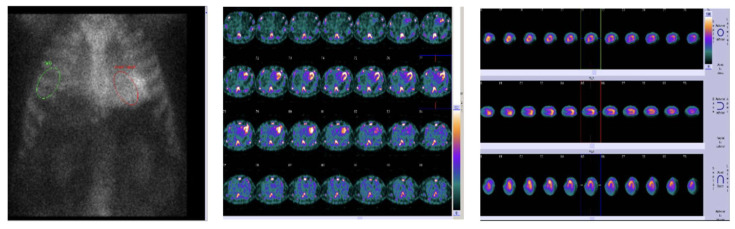
False-positive and false-negative studies. Uptake that is not ATTR occurs with AL amyloidosis and, rarely, apolipoprotein A-I amyloidosis; recent myocardial infarction, which produces focal pyrophosphate uptake; healing rib fractures; persistent blood pool; and hydroxychloroquine cardiotoxicity, reported in biopsy-proven cases with grade 2 or 3 uptake. Uptake is diffuse in ATTR; focal or regional uptake raises an alternative diagnosis. False-negative or low-grade studies occur in early-onset p.Val50Met disease and with the p.Phe84Leu (Phe64Leu) variant, in which only 2 of 19 patients with cardiac involvement had high-grade uptake. Very early ATTR with little myocardial deposit can also be grade 0 or 1. A strong clinical phenotype with a negative or equivocal scan therefore leads to CMR and, when needed, tissue diagnosis.
What scintigraphy does not do. Scintigraphy does not separate wild-type from hereditary ATTR, it does not exclude AL, and change in grade or ratio is not an established measure of treatment response. Higher ratios carry prognostic associations.
Positron emission tomography. Amyloid-binding PET tracers developed for brain imaging (18F-florbetapir, 18F-florbetaben, and 11C-Pittsburgh compound B) bind both AL and ATTR deposits. The 2021 consensus lists a myocardium-to-blood-pool ratio above 1.5 or a retention index above 0.030 min−1 as typical of cardiac amyloidosis, and their use for the heart is off label. Because they image AL, which bone tracers largely miss, they are being developed for early detection and for quantifying whole-body amyloid burden, but they are not part of the standard diagnostic pathway.
Serum free light chains and immunofixation to exclude light-chain amyloidosis
The monoclonal protein screen has three components, all required together: the serum free light chain assay with the kappa-to-lambda ratio, serum immunofixation electrophoresis, and urine immunofixation electrophoresis. Protein electrophoresis of serum or urine without immunofixation is not sensitive enough to exclude a clone. When all three are normal, AL amyloidosis is effectively excluded, and the 2022 AHA/ACC/HFSA guideline recommends bone scintigraphy to confirm ATTR in patients with sufficient clinical suspicion (Class 1). The guideline also gives a Class 1 recommendation for the screen itself whenever cardiac amyloidosis is suspected. The 2025 ACC concise clinical guidance states that monoclonal protein testing is ordered before scintigraphy in every case, because interpreting a scan without it risks labeling AL as ATTR and delaying life-saving therapy.
Interpretation in kidney disease. Chronic kidney disease raises both kappa and lambda free light chain concentrations and widens the ratio, so a modest ratio abnormality with normal immunofixation is common in this population. The ECDP notes that such ratio elevations with normal immunofixation do not by themselves establish a clone. The ratio is read against renal function, and an unequivocal abnormality, or any band on immunofixation, removes the patient from the nonbiopsy pathway.
When the screen is positive. A monoclonal protein in a patient with suspected cardiac amyloidosis has two explanations: AL amyloidosis, or ATTR amyloidosis with an unrelated MGUS. Scintigraphy cannot decide between them, because AL deposits can produce grade 2 or 3 uptake. Tissue is obtained and typed. The urgency is set by the AL possibility: delay in diagnosing cardiac AL carries substantial early mortality, so the evaluation proceeds within days, with hematology involved from the start.
Stepwise diagnostic pathway for suspected cardiac amyloidosis
The pathway below combines the 2023 ACC ECDP, the 2025 ACC concise clinical guidance, the 2022 AHA/ACC/HFSA guideline, and the 2021 multimodality imaging criteria.
| Step | Finding | Interpretation | Next action |
|---|---|---|---|
| 1. Suspicion | Heart failure, wall thickness >12 mm, conduction disease, or atrial fibrillation with one or more red flags | Pretest probability sufficient for testing | Monoclonal protein screen and bone scintigraphy, ordered together; scan read only with screen results |
| 2. Monoclonal screen | Serum free light chains, serum and urine immunofixation all normal | AL effectively excluded | Scintigraphy with SPECT |
| 3. Scintigraphy (screen negative) | Grade 2 or 3 diffuse myocardial uptake confirmed on SPECT | ATTR cardiomyopathy diagnosed without biopsy | TTR sequencing; staging; therapy |
| Grade 1 or equivocal ratio | Nondiagnostic | CMR; endomyocardial biopsy if suspicion persists | |
| Grade 0 | ATTR unlikely; early disease or low-uptake variants not excluded | CMR if phenotype is strong; biopsy if CMR is typical | |
| 4. Screen positive | Abnormal free light chain ratio not explained by kidney disease, or a band on immunofixation, at any scan grade | AL possible; nonbiopsy pathway invalid | Urgent hematology evaluation; tissue biopsy with typing by mass spectrometry |
| 5. Tissue | Fat pad, bone marrow, or other involved organ positive and typed, with typical cardiac imaging | Cardiac involvement by the typed protein | Endomyocardial biopsy when extracardiac tissue is negative or untyped |
| 6. ATTR confirmed | Wild-type or pathogenic variant on TTR sequencing | Subtype assigned | Genetic counseling and cascade testing of relatives when variant present |
Stepwise diagnosis of cardiac amyloidosis. "Typical cardiac imaging" follows the 2021 multimodality consensus: echocardiographic wall thickness >12 mm, relative apical strain ratio >1.0, or grade 2 or worse diastolic dysfunction; or CMR wall thickness above the sex-specific limit, ECV >0.40, diffuse LGE, or abnormal nulling; with hypertension and other causes reasonably excluded.
Endomyocardial and extracardiac biopsy and amyloid typing
Biopsy is required when the monoclonal screen is positive, when scintigraphy is equivocal or negative despite a strong phenotype, when a low-uptake variant is suspected, and when a rare amyloid type is possible. Endomyocardial biopsy is the reference standard. Amyloid deposits throughout the myocardium, so sensitivity of multiple right ventricular septal samples approaches 100%. Complications in experienced centers are uncommon and include perforation with tamponade, tricuspid valve injury, and arrhythmia.
A biopsy of an extracardiac site can establish systemic amyloidosis and its type, and the cardiac diagnosis is then made from typical cardiac imaging or, for AL, abnormal cardiac biomarkers with other causes excluded. Sensitivity depends on type. Fine-needle aspiration of abdominal fat detected amyloid in 84% of patients with cardiac AL, 45% with hereditary ATTR, and only 15% with wild-type ATTR. A negative fat aspirate therefore does not exclude either type, and it is especially weak against wild-type disease. Bone marrow biopsy, performed for the plasma cell evaluation, finds amyloid in a proportion of AL patients and quantifies the clone. Kidney, liver, and nerve biopsies are diagnostic when those organs are involved.
Staining and typing. Congo red staining with apple-green birefringence under polarized light confirms amyloid. Typing identifies the precursor protein. Immunohistochemistry is limited by background staining and by fragments that lose antibody epitopes. Laser microdissection with tandem mass spectrometry identifies the protein in deposits directly; in its development series it typed amyloid with 100% sensitivity and specificity in the training set and 98% in a cardiac validation set. Mass spectrometry is the preferred method, and typing is performed at specialized laboratories.
A biopsy showing ATTR in a patient who also has a monoclonal protein establishes ATTR with coincident MGUS, and the clone is then followed by hematology. A biopsy showing AL directs the patient to plasma cell therapy, whatever the scintigraphy showed.
Genetic testing for hereditary transthyretin amyloidosis
Once ATTR is established, the 2022 AHA/ACC/HFSA guideline recommends TTR gene sequencing to distinguish hereditary from wild-type disease (Class 1). Age, sex, and ancestry do not substitute: hereditary disease occurs in older men, and wild-type disease occurs in women and in people of African ancestry. Scintigraphy, biopsy, and typing all identify transthyretin without revealing whether it is variant or wild-type.
The result matters for three reasons. A pathogenic variant identifies relatives at risk, each with a 50% chance of inheritance. Some variants carry a neurologic phenotype that changes therapy: the 2025 ACC concise clinical guidance prefers a silencer in hereditary ATTR cardiomyopathy with polyneuropathy. Some variants affect test interpretation, such as the low scintigraphic sensitivity of p.Phe84Leu. Testing is performed with genetic counseling, and the counseling covers insurance implications, penetrance, and the variable age of onset.
Cascade testing and carrier surveillance. First-degree relatives of a patient with a pathogenic variant are offered counseling and testing. An international expert consensus recommends that surveillance of identified carriers begin about 10 years before the predicted age of disease onset. The predicted age is based on the age at which the proband and affected relatives developed symptoms, and surveillance includes periodic neurologic and cardiac evaluation. Cardiac surveillance combines electrocardiography, echocardiography with strain, NT-proBNP, and troponin, with scintigraphy or CMR when findings change. In the Transthyretin Amyloidosis Outcomes Survey (THAOS), 36% of 740 asymptomatic carriers, most with p.Val50Met, developed symptomatic disease during a mean follow-up of 6.6 years, a median of 2.2 years after enrollment, which supports structured follow-up. Carriers of p.Val142Ile have a late-onset, largely cardiac phenotype with incomplete penetrance.
Differential diagnosis of increased left ventricular wall thickness
Systemic context separates the causes of a thick ventricle more often than morphology does. Amyloidosis is distinguished from hypertensive and sarcomeric hypertrophy by the mismatch between thick walls and electrical voltage, the strain pattern, the tissue signature on CMR, and the scintigraphy result.
| Condition | Distinguishing features | Confirmatory test |
|---|---|---|
| Cardiac amyloidosis | Low or normal voltage for wall thickness; thick valves and septum; apical sparing; diffuse LGE with abnormal nulling; ECV >0.40; extracardiac red flags | Monoclonal screen and scintigraphy; tissue typing |
| Hypertensive heart disease | Long-standing hypertension; symmetric thickening usually <15 mm; increased voltage; no amyloid red flags; ECV normal or mildly raised | Regression with blood pressure control |
| Hypertrophic cardiomyopathy | Wall ≥15 mm unexplained by loading; asymmetric; systolic anterior motion and outflow obstruction; high voltage; patchy midwall LGE at insertion points; family history | Sarcomere gene testing (see hypertrophic cardiomyopathy) |
| Aortic stenosis | Pressure overload with valve gradient; can coexist with ATTR | Scintigraphy when red flags present |
| Fabry disease | Concentric thickening; low native T1; basal inferolateral LGE; neuropathic pain, angiokeratoma, proteinuria; X-linked | α-Galactosidase A activity (men); GLA sequencing |
| Cardiac sarcoidosis | Basal septal thinning; patchy LGE not following a coronary territory; high-grade block or ventricular tachycardia in a younger adult | Fluorodeoxyglucose (FDG) PET; tissue |
| Glycogen storage (Danon, PRKAG2) | Massive thickening in a young patient; preexcitation; skeletal myopathy | Genetic testing |
| Athlete's heart | Mild symmetric thickening with dilated cavity; normal diastolic function; regression with deconditioning | Detraining; CMR |
| Constrictive pericarditis | Normal wall thickness; preserved medial e′; respiratory interdependence | Simultaneous pressures; CMR (see constrictive pericarditis) |
Differential diagnosis of increased wall thickness or restrictive filling in adults. LGE, late gadolinium enhancement; ECV, extracellular volume fraction.
Staging and prognosis in ATTR and AL cardiac amyloidosis
Staging uses cardiac biomarkers because they integrate myocardial stretch, myocyte injury, and, in AL, the size of the clone. The systems are disease-specific, and an ATTR system is not applied to AL or the reverse.
ATTR staging. The Mayo Clinic system for wild-type ATTR assigns stage I when neither biomarker exceeds its threshold, stage II when one does, and stage III when both do. The UK National Amyloidosis Centre (NAC) system, derived in 869 patients with wild-type or hereditary ATTR and validated in a French cohort, stages by NT-proBNP and eGFR, with stage III requiring both to be abnormal. Age-adjusted hazard ratios were 2.05 for stage II and 3.80 for stage III. The NAC system needs no troponin assay. These survival figures predate disease-modifying therapy and earlier diagnosis, so they overstate risk for patients diagnosed and treated today; they remain useful for comparing patients and for stratifying trials.
AL staging. The Mayo 2004 system stages by NT-proBNP and troponin. In a European series of 346 patients with stage III disease, NT-proBNP above 8,500 ng/L and systolic blood pressure below 100 mm Hg independently identified a subgroup with median survival of only 3 months. Stage III is now commonly divided into IIIa and IIIb by the NT-proBNP threshold. The revised Mayo 2012 system adds the difference between involved and uninvolved free light chains (dFLC) and scores one point for each variable above its threshold, which defines stages I to IV.
| System | Disease | Variables and thresholds | Stages and outcome |
|---|---|---|---|
| Mayo ATTR (2016) | Wild-type ATTR | Troponin T >0.05 ng/mL; NT-proBNP >3,000 pg/mL | I–III; 4-year survival 57%, 42%, 18% |
| NAC (2018) | Wild-type and hereditary ATTR | NT-proBNP >3,000 ng/L; eGFR <45 mL/min/1.73 m² | I–III; median 69.2, 46.7, 24.1 months |
| Mayo 2004 | AL | NT-proBNP ≥332 ng/L; troponin T ≥0.035 µg/L or troponin I ≥0.1 µg/L | I–III; median 26.4, 10.5, 3.5 months |
| Stage III subdivision | AL | NT-proBNP >8,500 ng/L within Mayo 2004 stage III | IIIb; median about 3 months in the subgroup defined by NT-proBNP >8,500 ng/L and systolic pressure <100 mm Hg |
| Mayo 2012 | AL | dFLC ≥18 mg/dL; troponin T ≥0.025 ng/mL; NT-proBNP ≥1,800 pg/mL | I–IV; median 94.1, 40.3, 14, 5.8 months |
Biomarker staging systems in cardiac amyloidosis. NT-proBNP in pg/mL equals ng/L. Troponin thresholds were derived with conventional (not high-sensitivity) troponin T assays; high-sensitivity values reported in ng/L are not interchangeable without assay-specific conversion.
Beyond biomarkers, adverse prognostic features include New York Heart Association (NYHA) class III or IV symptoms, low ejection fraction, low stroke volume index, severely reduced GLS, pericardial effusion, high ECV, a short 6-minute walk distance, and, in AL, systolic hypotension and multiorgan involvement. Early deaths in AL are concentrated among patients with stage IIIb disease, whose treatment is modified to reduce toxicity.
ATTR and AL staging calculator
Biomarker stage in ATTR and AL amyloidosis
NAC, Mayo ATTR, Mayo 2004, Mayo 2012ATTR: NAC stage uses NT-proBNP >3,000 and eGFR <45; Mayo ATTR stage uses NT-proBNP >3,000 and troponin T >0.05. AL: Mayo 2004 stage uses NT-proBNP ≥332 and troponin T ≥0.035, with stage III above an NT-proBNP of 8,500 reported as IIIb; Mayo 2012 stage adds dFLC ≥18 mg/dL with troponin T ≥0.025 and NT-proBNP ≥1,800. Troponin T is the conventional assay in ng/mL. The example (ATTR, NT-proBNP 4,200, eGFR 52, troponin T 0.06) gives NAC stage II and Mayo ATTR stage III.
Transthyretin stabilizers: tafamidis, acoramidis, and diflunisal
Tafamidis. Tafamidis binds the thyroxine-binding sites of transthyretin and slows tetramer dissociation. In the ATTR Transthyretin Amyloidosis Cardiomyopathy Clinical Trial (ATTR-ACT), 441 patients with wild-type or hereditary ATTR cardiomyopathy and NYHA class I to III symptoms were randomized to tafamidis 80 mg, tafamidis 20 mg, or placebo for 30 months. The pooled tafamidis groups had lower all-cause mortality (29.5% vs. 42.9%; hazard ratio 0.70) and fewer cardiovascular hospitalizations (0.48 vs. 0.70 per year; relative risk 0.68), with slower decline in 6-minute walk distance and Kansas City Cardiomyopathy Questionnaire score. The survival curves separated only after about 18 months, consistent with a drug that slows progression without reversing deposits. Adverse events were similar to placebo. Tafamidis is available as tafamidis meglumine 80 mg (four 20-mg capsules) or the bioequivalent tafamidis free acid 61 mg, once daily.
In the NYHA class III subgroup of ATTR-ACT, cardiovascular hospitalizations were more frequent with tafamidis than with placebo, a finding attributed to longer survival of sicker patients. In the long-term extension, continuous tafamidis was associated with lower all-cause mortality than delayed treatment both in patients who had been NYHA class III at baseline (hazard ratio 0.64) and in those who had been class I or II (hazard ratio 0.50). NYHA class IV patients were excluded from the trial.
The 2022 AHA/ACC/HFSA guideline recommends tafamidis for selected patients with wild-type or variant ATTR cardiomyopathy and NYHA class I to III symptoms to reduce cardiovascular morbidity and mortality (Class 1, level of evidence B-R), and it assigned a low economic value at the list price then in effect. The 2021 European Society of Cardiology (ESC) heart failure guideline gives tafamidis a Class I recommendation only for patients in NYHA class I or II.
Acoramidis. Acoramidis is a high-affinity stabilizer designed to mimic a naturally protective transthyretin variant, with more than 90% stabilization across the dosing interval. In ATTRibute-CM, 632 patients were randomized 2:1 to acoramidis or placebo for 30 months, with efficacy analyzed in patients with an eGFR of at least 30 mL/min/1.73 m². The hierarchical primary end point of death, cardiovascular hospitalization, NT-proBNP change, and 6-minute walk distance favored acoramidis (win ratio 1.8). All-cause mortality was 19.3% with acoramidis and 25.7% with placebo, a difference that was not statistically significant in isolation, and cardiovascular hospitalization occurred in 26.7% and 42.6%. It is approved to reduce cardiovascular death and cardiovascular hospitalization in wild-type or variant ATTR cardiomyopathy at a dose of 712 mg (two 356-mg tablets) twice daily. No randomized trial has compared acoramidis with tafamidis.
Diflunisal. The nonsteroidal anti-inflammatory drug diflunisal stabilizes the tetramer at 250 mg twice daily. Its randomized evidence is in hereditary polyneuropathy, where it slowed neurologic progression over 2 years. It has no outcome trial in cardiomyopathy and is used, if at all, where approved agents are unavailable. Renal dysfunction, fluid retention, decompensated heart failure, gastrointestinal bleeding, and thrombocytopenia preclude it, which excludes many patients with ATTR cardiomyopathy. Green tea extract (epigallocatechin-3-gallate) and doxycycline with tauroursodeoxycholic acid lack outcome evidence and are not recommended.
Transthyretin silencers, gene editing, and fibril depletion
Silencers reduce hepatic transthyretin synthesis, lowering circulating levels by about 70% to 90%. Because transthyretin carries retinol-binding protein, serum vitamin A falls, and daily vitamin A supplementation at the recommended dietary allowance accompanies treatment.
Vutrisiran. Vutrisiran is a small interfering RNA conjugated to N-acetylgalactosamine for hepatocyte uptake, given as 25 mg subcutaneously every 3 months. In HELIOS-B, 655 patients with ATTR cardiomyopathy were randomized to vutrisiran or placebo for up to 36 months. Vutrisiran reduced the composite of all-cause death and recurrent cardiovascular events in the overall population (hazard ratio 0.72) and in the monotherapy population not receiving tafamidis at baseline (hazard ratio 0.67), and it reduced all-cause mortality through 42 months (hazard ratio 0.65). Functional capacity and health status declined less than with placebo. In the open-label extension through month 48, patients treated continuously from randomization had a lower risk of death or first cardiovascular event than those who started vutrisiran after placebo (hazard ratio 0.63). Vutrisiran is approved for ATTR cardiomyopathy, wild-type or hereditary, and for hereditary polyneuropathy.
Patisiran. Patisiran, an intravenous small interfering RNA given at 0.3 mg/kg every 3 weeks with premedication for infusion reactions, is approved for hereditary polyneuropathy. In APOLLO-B, 360 patients with ATTR cardiomyopathy had less decline in 6-minute walk distance at 12 months than with placebo (median difference 14.7 m) and a small difference in health status, without a significant effect on the composite of death, cardiovascular events, and walk distance. It is not approved for cardiomyopathy in the United States.
Eplontersen and inotersen. Eplontersen is a ligand-conjugated antisense oligonucleotide given as 45 mg subcutaneously every 4 weeks; it is approved for hereditary polyneuropathy on the basis of NEURO-TTRansform. In CARDIO-TTRansform, 1,432 patients with ATTR cardiomyopathy were randomized to eplontersen or placebo for 140 weeks on top of standard care, and 57% were taking a stabilizer at baseline. The primary composite of cardiovascular death and recurrent cardiovascular events did not differ (rate ratio 0.89; 95% CI 0.73 to 1.09). In a prespecified analysis, the effect differed by background therapy: the rate ratio was 0.71 in patients not taking a stabilizer and 1.14 in those taking one. The subgroup finding is hypothesis-generating, and the trial adds evidence that silencing added to stabilization confers no measurable benefit. Inotersen, an earlier weekly antisense oligonucleotide for hereditary polyneuropathy, requires platelet and renal monitoring because of thrombocytopenia and glomerulonephritis and has no cardiomyopathy indication.
Gene editing. Nexiguran ziclumeran (NTLA-2001) is an in vivo CRISPR-Cas9 therapy delivered in lipid nanoparticles that inactivates the TTR gene in hepatocytes after a single infusion. In a phase 1 study of 36 patients with ATTR cardiomyopathy, serum transthyretin fell by 90% at 12 months and remained low. Phase 3 outcome testing is under way, and severe transaminase elevation has occurred in the program, so liver monitoring is part of its protocols. Editing is permanent, and long-term safety is unknown.
Fibril depletion. Stabilizers and silencers slow new deposition but do not remove existing amyloid. Antibodies directed at misfolded transthyretin are designed to mark deposits for clearance by macrophages. In a phase 1 trial of NI006 (now ALXN2220) in 40 patients, doses of at least 10 mg/kg were associated with reductions in scintigraphic tracer uptake and in ECV, imaging surrogates of cardiac amyloid load; a phase 3 outcome trial is ongoing. None is approved.
| Trial | Agent | Patients | Primary end point | Result |
|---|---|---|---|---|
| ATTR-ACT | Tafamidis 80 or 20 mg daily | 441; NYHA I–III | All-cause death, then cardiovascular hospitalization (hierarchical) | Met; mortality 29.5% vs. 42.9% (HR 0.70) |
| ATTRibute-CM | Acoramidis 712 mg twice daily | 632 | Death, cardiovascular hospitalization, NT-proBNP, 6-minute walk (hierarchical) | Met; win ratio 1.8 |
| HELIOS-B | Vutrisiran 25 mg every 3 months | 655 | All-cause death and recurrent cardiovascular events | Met; HR 0.72 overall, 0.67 monotherapy |
| APOLLO-B | Patisiran 0.3 mg/kg every 3 weeks | 360 | 6-minute walk distance at 12 months | Met (14.7 m); outcome composite not significant |
| CARDIO-TTRansform | Eplontersen 45 mg every 4 weeks | 1,432 | Cardiovascular death and recurrent cardiovascular events | Not met; rate ratio 0.89 |
Phase 3 trials of disease-modifying therapy in ATTR cardiomyopathy. HR, hazard ratio.
Choosing disease-modifying therapy for ATTR cardiomyopathy
The 2025 ACC concise clinical guidance recommends starting an approved stabilizer (tafamidis or acoramidis) or silencer (vutrisiran) without delay once ATTR cardiomyopathy is confirmed in an appropriate patient. Benefit is greatest when treatment begins before advanced functional decline. In hereditary ATTR cardiomyopathy with polyneuropathy, a silencer is preferred because it treats both. Otherwise, the choice among approved agents is guided by access, cost, payer requirements, and patient preference, because no head-to-head trial exists.
Combination therapy. The concise guidance advises against routine combination of a stabilizer with a silencer, because no trial has shown benefit from combination. In HELIOS-B the treatment effect in patients already taking tafamidis was directionally similar to the overall effect, but the trial was not designed to test combination therapy.
Eligibility and functional class. The pivotal trials enrolled patients with NYHA class I to III symptoms and excluded those with very advanced disease, so evidence in NYHA class IV is lacking. Very advanced disease, frailty, severe noncardiac illness, and limited life expectancy weigh against starting therapy whose benefit accrues over 18 months or more. Patients with an eGFR below 30 mL/min/1.73 m² were excluded from the acoramidis efficacy analysis, and severe kidney disease is a limitation for several agents.
Cost, access, and prior authorization. Approved therapies carry high list prices. Access in the United States depends on prior authorization documenting the diagnosis (scintigraphy with a negative monoclonal screen, or tissue typing), the genotype, and functional class, together with Medicare Part D coverage, manufacturer assistance, and foundation support. The ECDP notes that copay assistance is often needed.
Multidisciplinary care. Amyloidosis involves the heart, nerves, kidneys, gut, and musculoskeletal system, and AL involves the bone marrow. The 2023 ECDP organizes care around a multidisciplinary team in an amyloidosis program: cardiology with heart failure and electrophysiology expertise, hematology, neurology, nephrology, gastroenterology, genetics and genetic counseling, nuclear cardiology and imaging, pathology with typing capability, palliative care, and pharmacy support for access. The team coordinates monitoring and decides on devices, valve intervention, and transplantation.
Plasma cell therapy for AL amyloidosis and cardiac monitoring
AL cardiomyopathy is treated by eliminating the clone that produces the toxic light chain. Hematologic response, measured by the fall in involved free light chains, is followed by organ response, and survival tracks the depth of hematologic response. The goal of therapy is at least a very good partial response, and current practice aims for complete response.
First-line therapy. In the ANDROMEDA trial, 388 patients with newly diagnosed AL amyloidosis received bortezomib, cyclophosphamide, and dexamethasone with or without subcutaneous daratumumab, an anti-CD38 antibody. Hematologic complete response was 53.3% with daratumumab and 18.1% without (relative risk 2.9). Cardiac response at 6 months was 41.5% versus 22.2%, and survival free from major organ deterioration or hematologic progression favored daratumumab (hazard ratio 0.58). Most deaths in both groups were from amyloid cardiomyopathy. Daratumumab with bortezomib, cyclophosphamide, and dexamethasone is the standard first-line regimen. The trial excluded patients with NT-proBNP above 8,500 ng/L and NYHA class IIIB or IV disease, so stage IIIb patients are treated with dose-modified regimens and close monitoring.
Autologous stem cell transplant. High-dose melphalan with autologous stem cell transplant produces durable responses in selected patients, either at diagnosis or as consolidation after an incomplete response to induction. Eligibility depends on age, performance status, blood pressure, kidney function, and cardiac biomarkers, and advanced cardiac involvement is the main exclusion because transplant-related mortality rises steeply with it. Pomalidomide, venetoclax for clones with the t(11;14) translocation, and bendamustine are among the options after an inadequate response.
Response criteria. Changes in free light chains and NT-proBNP predict survival as early as 3 months after treatment begins.
| Response | Definition |
|---|---|
| Hematologic complete response | Normal free light chain ratio with negative serum and urine immunofixation |
| Very good partial response | dFLC <40 mg/L |
| Partial response | dFLC decrease >50% |
| Cardiac response | NT-proBNP decrease >30% and >300 ng/L |
| Cardiac progression | NT-proBNP increase >30% and >300 ng/L |
Response criteria in AL amyloidosis, after the validated free light chain and NT-proBNP criteria. dFLC, difference between involved and uninvolved free light chains. Bortezomib, immunomodulatory drugs, and dexamethasone can raise NT-proBNP independent of cardiac status, which complicates interpretation during therapy.
Cardiac care during chemotherapy. Dexamethasone causes fluid retention, bortezomib can worsen heart failure and hypotension, and immunomodulatory drugs raise NT-proBNP. Volume status, blood pressure, rhythm, and conduction are monitored closely in the first cycles, when early deaths cluster. Arrhythmic death and pulseless electrical activity account for many of these early deaths.
Heart failure management in cardiac amyloidosis
Standard neurohormonal therapy for heart failure with reduced ejection fraction is poorly tolerated and of uncertain benefit in amyloidosis, because cardiac output depends on heart rate with a fixed stroke volume. Autonomic neuropathy causes hypotension, and the stiff ventricle cannot augment stroke volume in response to vasodilation. The 2025 ACC concise clinical guidance recommends adjusting heart failure therapy to amyloid physiology.
Diuretics. Loop diuretics are the main symptomatic therapy. The volume window is narrow: a small ventricle with steep pressure-volume relations needs high filling pressure for adequate stroke volume, so overdiuresis causes hypotension, prerenal azotemia, and fatigue, and underdiuresis leaves congestion. Torsemide or bumetanide is often preferred over furosemide because of more predictable absorption in gut wall edema. Mineralocorticoid receptor antagonists help maintain potassium and are associated with better outcomes in observational ATTR cohorts; the concise guidance includes them for relief of congestion.
SGLT2 inhibitors. Patients with ATTR cardiomyopathy were excluded from the major sodium-glucose cotransporter 2 (SGLT2) inhibitor trials. In a propensity-matched multicenter cohort of 220 treated patients and 220 controls, SGLT2 inhibitor therapy was well tolerated (discontinuation 4.5%). Over a median of 28 months it was associated with lower all-cause mortality (hazard ratio 0.57), lower cardiovascular mortality, and fewer heart failure hospitalizations. The concise guidance includes SGLT2 inhibitors for relief of congestion, alongside careful diuretic titration.
Beta-blockers, ACE inhibitors, ARBs, and sacubitril-valsartan. These drugs are used cautiously, if at all. Beta-blockers limit the heart rate on which output depends and often cause fatigue or hypotension; in atrial fibrillation with rapid ventricular rates, a low dose can be tolerated. Angiotensin-converting enzyme inhibitors, angiotensin receptor blockers, and sacubitril-valsartan often cause hypotension, especially with autonomic dysfunction. They are reduced or withdrawn when they cause symptoms, and their withdrawal frequently improves blood pressure and well-being. Hypertension that resolves as amyloid cardiomyopathy progresses is itself a clue.
Agents avoided. Nondihydropyridine calcium channel blockers (verapamil and diltiazem) are avoided because of negative inotropy, hypotension, and high-grade heart block. Digoxin binds amyloid fibrils, and older reports linked it to toxicity; current guidance permits low-dose digoxin with careful monitoring for rate control when other options fail.
Orthostatic hypotension. Midodrine and droxidopa raise standing blood pressure in autonomic failure and can allow continued diuresis. Compression garments, head-of-bed elevation, and slow positional change are adjuncts.
Atrial fibrillation and thromboembolism in cardiac amyloidosis
Atrial fibrillation and flutter are common, particularly in ATTR, and poorly tolerated, because the stiff ventricle depends on atrial contribution and on an adequate rate for output. Atrial infiltration impairs mechanical function and promotes thrombus formation even at low CHA₂DS₂-VASc scores and, in some patients, in sinus rhythm.
Anticoagulation. The 2022 AHA/ACC/HFSA guideline states that anticoagulation is reasonable for patients with cardiac amyloidosis and atrial fibrillation to reduce stroke risk regardless of the CHA₂DS₂-VASc score (Class 2a), and the 2023 ECDP recommends anticoagulation regardless of the score. Direct oral anticoagulants are used unless contraindicated. Anticoagulation in patients with sinus rhythm and severe atrial mechanical failure or appendage sludge is decided individually; there is no randomized evidence.
Cardioversion. Because thrombus is frequent despite therapeutic anticoagulation, the ECDP recommends transesophageal echocardiography before cardioversion in every patient, whatever the duration of anticoagulation. Cardioversion in amyloidosis carries higher rates of procedural failure and of post-shock bradyarrhythmia than in other populations.
Rate and rhythm. Rhythm control is generally favored because rate control with beta-blockers and calcium channel blockers is poorly tolerated. Amiodarone is the most frequently used antiarrhythmic drug. Catheter ablation is feasible, with recurrence rates that are higher in advanced disease and better results earlier in the course. Atrioventricular node ablation with pacing is an option when rate control fails. The broader management of atrial fibrillation is covered on the atrial fibrillation page.
Conduction disease, pacing, and sudden death
Conduction disease is common and progressive. Infiltration of the His-Purkinje system produces infranodal block, so a prolonged PR interval often reflects prolonged HV conduction, and patients with ATTR frequently need pacemakers. High-grade atrioventricular block occurs in both types, and sinus node dysfunction occurs as well. The ECDP emphasizes close rhythm surveillance, including ambulatory monitoring when patients have syncope, presyncope, or worsening conduction intervals.
Pacing. Indications for pacing follow the general bradycardia and conduction guidelines (see bradycardia and atrioventricular block). Because chronic right ventricular pacing can worsen ventricular function, and patients with amyloidosis often need a high pacing burden, conduction system pacing or biventricular pacing is used when a high burden is expected.
Sudden death and defibrillators. Sudden death in amyloidosis, especially AL, is often from electromechanical dissociation or bradyarrhythmia, not ventricular tachyarrhythmia, which limits the benefit of defibrillators. The ECDP notes that defibrillators have not consistently improved survival and that decisions follow standard heart failure and ventricular arrhythmia guideline indications. A defibrillator is appropriate for secondary prevention after sustained ventricular tachycardia or resuscitated ventricular fibrillation in patients with a reasonable expected survival. Primary prevention is individualized, taking into account the type of amyloid, response to therapy, and life expectancy; ejection fraction thresholds derived from other cardiomyopathies do not translate directly.
Aortic stenosis with ATTR amyloidosis
Calcific aortic stenosis and wild-type ATTR share an older population and coexist more often than chance predicts. In a three-center study of 407 TAVR candidates with blinded DPD scintigraphy, uptake was present in 11.8%, with grade 2 or 3 uptake in 7.9%; these patients had worse functional capacity, higher biomarkers, and more biventricular remodeling. A clinical score combining left ventricular remodeling, age, high-sensitivity troponin, systemic involvement, and electrical abnormalities (right bundle branch block or low voltage) predicted dual pathology with an area under the curve of 0.86.
Coexisting amyloid explains many cases of paradoxical low-flow, low-gradient stenosis: the small, infiltrated ventricle ejects a low stroke volume, and the gradient understates valve severity. Patients with dual pathology had a thicker septum (1.3 vs. 1.1 cm), higher left ventricular mass index, lower stroke volume index (30 vs. 36 mL/m²), more restrictive filling, and worse GLS than patients with aortic stenosis alone in the original TAVR screening study.
Valve replacement relieves the obstruction and is not withheld because of amyloid. In that cohort, 1-year mortality was higher with dual pathology than with lone aortic stenosis (24.5% vs. 13.9%), TAVR improved survival compared with medical management, and survival after TAVR did not differ from that in lone aortic stenosis. The ECDP states that aortic valve replacement may improve symptoms and that referral for TAVR is appropriate. Disease-modifying therapy is added for the cardiomyopathy, and the high risk of conduction disease after TAVR in this population is planned for. Grading of stenosis in low-flow states is detailed on the aortic stenosis page.
Heart transplantation and mechanical circulatory support
Heart transplantation is an option for selected patients with advanced disease. In ATTR, candidates are usually younger patients with hereditary disease or fit patients with wild-type disease without major extracardiac involvement; advanced age limits eligibility for many with wild-type disease. Combined heart and liver transplantation was historically used in hereditary ATTR to remove the source of variant protein. Outcomes in carefully selected ATTR patients approach those of other transplant indications.
In AL amyloidosis, transplantation requires control of the clone. The ECDP describes autologous stem cell transplant followed by heart transplant as ideal for AL cardiomyopathy, with heart transplant followed by stem cell transplant as a sequence used in selected patients whose cardiac disease precludes chemotherapy. Recurrence in the graft occurs without hematologic control.
Durable left ventricular assist devices are rarely suitable. The small cavity limits inflow cannula placement and device flow, right ventricular involvement predisposes to right heart failure, and outcomes are worse than in dilated cardiomyopathy. Devices are considered only in carefully selected patients, usually as a bridge to transplant, with a larger ventricle.
Palliative care is part of management from diagnosis in advanced disease. Symptom control in amyloid heart failure relies on diuretic management, attention to orthostatic hypotension and gastrointestinal symptoms, and deactivation planning for defibrillators.
Monitoring disease progression and treatment response
ATTR monitoring. No validated definition of treatment response or failure exists for ATTR cardiomyopathy. The 2025 ACC concise clinical guidance recommends monitoring response clinically, using symptom trajectory, functional status, and clinical judgment, without formal treatment-failure thresholds. An international expert consensus published in 2021 assessed progression across three domains, clinical and functional measures, biomarkers, and imaging and electrocardiographic measures, with at least one marker from each domain as the minimum. Its 2026 update replaced this with six parameters: heart failure hospitalization, outpatient diuretic intensification, NT-proBNP, eGFR, 6-minute walk distance, and quality of life by the Kansas City Cardiomyopathy Questionnaire or NYHA class. Each is assessed every 12 months against the preceding 12 months, and progression is considered when at least two parameters meet the defined thresholds, because a single parameter overestimates progression. Imaging, electrocardiographic measures, and troponin are not among the six parameters. The experts state that it is unknown whether progression should change disease-modifying treatment.
Serial assessment combines NYHA class, diuretic dose, and walk distance with NT-proBNP, troponin, renal function, and electrolytes; an electrocardiogram at each visit for conduction disease; echocardiography with strain at intervals and with clinical change; and ambulatory monitoring for symptoms or new conduction delay. Rising NT-proBNP and troponin, an increasing diuretic requirement, and new conduction disease are the most accessible signals of progression. Repeat scintigraphy is not used to judge response, because uptake changes have not been validated as a response marker.
AL monitoring. Monitoring in AL follows hematologic and organ response criteria: free light chains each treatment cycle and NT-proBNP with troponin at regular intervals, with echocardiography as indicated. Hematologic relapse is detected by a rising dFLC before organ progression. Cardiac response by NT-proBNP lags hematologic response by months, and serial echocardiography shows slow regression of wall thickness in patients with deep, sustained responses.
Sources of error in diagnosing cardiac amyloidosis
- Reading a positive bone scan as ATTR without the monoclonal screen, or with an incomplete screen such as serum protein electrophoresis alone. AL deposits produce grade 2 or 3 uptake, and the error delays chemotherapy.
- Grading planar images without SPECT. Blood pool, ribs, and rib fractures produce apparent cardiac activity on planar images, especially at 1 hour and in renal failure.
- Applying the heart-to-contralateral ratio of 1.5 to 3-hour images, to DPD or HMDP, or to a study with no myocardial uptake on SPECT.
- Excluding amyloidosis because QRS voltage is normal. Low voltage with thick walls is present in only about 30% of patients.
- Excluding ATTR after a negative fat pad aspirate. Fat aspiration detects only 15% of wild-type cardiac ATTR.
- Assigning wild-type disease from age, sex, or ancestry without TTR sequencing, which misses hereditary disease and the need for cascade testing.
- Labeling hypertrophy as hypertensive or as hypertrophic cardiomyopathy when voltage, strain pattern, valve and septal thickening, or extracardiac clues do not fit.
- Treating an unexplained free light chain ratio in chronic kidney disease as proof of a clone, or ignoring a band on immunofixation because the ratio is normal.
- Accepting a negative or grade 1 scan as excluding ATTR in a patient with a strong phenotype or a known low-uptake variant such as p.Phe84Leu.
- Using serial scintigraphy to judge response to therapy.
Self-assessment
A 79-year-old man with heart failure, a septal thickness of 17 mm, and bilateral carpal tunnel release has grade 3 myocardial uptake on technetium-99m pyrophosphate imaging, confirmed on SPECT. Serum free light chains and serum and urine immunofixation are normal. NT-proBNP is 4,200 pg/mL, eGFR 52 mL/min/1.73 m², and troponin T 0.06 ng/mL. What is his National Amyloidosis Centre stage, and which test follows the diagnosis?
Nuclear Cardiology Board ReviewReferences
- Kittleson MM, Ruberg FL, Ambardekar AV, et al. 2023 ACC expert consensus decision pathway on comprehensive multidisciplinary care for the patient with cardiac amyloidosis: a report of the American College of Cardiology Solution Set Oversight Committee. J Am Coll Cardiol. 2023;81:1076–1126. doi:10.1016/j.jacc.2022.11.022
- Kittleson MM, Ambardekar AV, Cheng RK, et al. Transthyretin cardiac amyloidosis evaluation and management: 2025 ACC concise clinical guidance. J Am Coll Cardiol. 2026;87:549–565. doi:10.1016/j.jacc.2025.09.004
- Heidenreich PA, Bozkurt B, Aguilar D, et al. 2022 AHA/ACC/HFSA guideline for the management of heart failure: a report of the American College of Cardiology/American Heart Association Joint Committee on Clinical Practice Guidelines. Circulation. 2022;145:e895–e1032. doi:10.1161/CIR.0000000000001063
- Dorbala S, Ando Y, Bokhari S, et al. ASNC/AHA/ASE/EANM/HFSA/ISA/SCMR/SNMMI expert consensus recommendations for multimodality imaging in cardiac amyloidosis: part 1 of 2—evidence base and standardized methods of imaging. Circ Cardiovasc Imaging. 2021;14:e000029. doi:10.1161/HCI.0000000000000029
- Dorbala S, Ando Y, Bokhari S, et al. ASNC/AHA/ASE/EANM/HFSA/ISA/SCMR/SNMMI expert consensus recommendations for multimodality imaging in cardiac amyloidosis: part 2 of 2—diagnostic criteria and appropriate utilization. Circ Cardiovasc Imaging. 2021;14:e000030. doi:10.1161/HCI.0000000000000030
- American Society of Nuclear Cardiology. 99mTechnetium-pyrophosphate imaging for transthyretin cardiac amyloidosis: practice points, version 2.0. 2019. asnc.org
- McDonagh TA, Metra M, Adamo M, et al. 2021 ESC guidelines for the diagnosis and treatment of acute and chronic heart failure. Eur Heart J. 2021;42:3599–3726. doi:10.1093/eurheartj/ehab368
- Gillmore JD, Maurer MS, Falk RH, et al. Nonbiopsy diagnosis of cardiac transthyretin amyloidosis. Circulation. 2016;133:2404–2412. doi:10.1161/CIRCULATIONAHA.116.021612
- Perugini E, Guidalotti PL, Salvi F, et al. Noninvasive etiologic diagnosis of cardiac amyloidosis using 99mTc-3,3-diphosphono-1,2-propanodicarboxylic acid scintigraphy. J Am Coll Cardiol. 2005;46:1076–1084. doi:10.1016/j.jacc.2005.05.073
- Bokhari S, Castaño A, Pozniakoff T, et al. 99mTc-pyrophosphate scintigraphy for differentiating light-chain cardiac amyloidosis from the transthyretin-related familial and senile cardiac amyloidoses. Circ Cardiovasc Imaging. 2013;6:195–201. doi:10.1161/CIRCIMAGING.112.000132
- Castano A, Haq M, Narotsky DL, et al. Multicenter study of planar technetium 99m pyrophosphate cardiac imaging: predicting survival for patients with ATTR cardiac amyloidosis. JAMA Cardiol. 2016;1:880–889. doi:10.1001/jamacardio.2016.2839
- Musumeci MB, Cappelli F, Russo D, et al. Low sensitivity of bone scintigraphy in detecting Phe64Leu mutation-related transthyretin cardiac amyloidosis. JACC Cardiovasc Imaging. 2020;13:1314–1321. doi:10.1016/j.jcmg.2019.10.015
- Phelan D, Collier P, Thavendiranathan P, et al. Relative apical sparing of longitudinal strain using two-dimensional speckle-tracking echocardiography is both sensitive and specific for the diagnosis of cardiac amyloidosis. Heart. 2012;98:1442–1448. doi:10.1136/heartjnl-2012-302353
- Pagourelias ED, Mirea O, Duchenne J, et al. Echo parameters for differential diagnosis in cardiac amyloidosis: a head-to-head comparison of deformation and nondeformation parameters. Circ Cardiovasc Imaging. 2017;10:e005588. doi:10.1161/CIRCIMAGING.116.005588
- Martinez-Naharro A, Treibel TA, Abdel-Gadir A, et al. Magnetic resonance in transthyretin cardiac amyloidosis. J Am Coll Cardiol. 2017;70:466–477. doi:10.1016/j.jacc.2017.05.053
- Quarta CC, Gonzalez-Lopez E, Gilbertson JA, et al. Diagnostic sensitivity of abdominal fat aspiration in cardiac amyloidosis. Eur Heart J. 2017;38:1905–1908. doi:10.1093/eurheartj/ehx047
- Vrana JA, Gamez JD, Madden BJ, et al. Classification of amyloidosis by laser microdissection and mass spectrometry-based proteomic analysis in clinical biopsy specimens. Blood. 2009;114:4957–4959. doi:10.1182/blood-2009-07-230722
- Grogan M, Scott CG, Kyle RA, et al. Natural history of wild-type transthyretin cardiac amyloidosis and risk stratification using a novel staging system. J Am Coll Cardiol. 2016;68:1014–1020. doi:10.1016/j.jacc.2016.06.033
- Gillmore JD, Damy T, Fontana M, et al. A new staging system for cardiac transthyretin amyloidosis. Eur Heart J. 2018;39:2799–2806. doi:10.1093/eurheartj/ehx589
- Dispenzieri A, Gertz MA, Kyle RA, et al. Serum cardiac troponins and N-terminal pro-brain natriuretic peptide: a staging system for primary systemic amyloidosis. J Clin Oncol. 2004;22:3751–3757. doi:10.1200/JCO.2004.03.029
- Kumar S, Dispenzieri A, Lacy MQ, et al. Revised prognostic staging system for light chain amyloidosis incorporating cardiac biomarkers and serum free light chain measurements. J Clin Oncol. 2012;30:989–995. doi:10.1200/JCO.2011.38.5724
- Wechalekar AD, Schonland SO, Kastritis E, et al. A European collaborative study of treatment outcomes in 346 patients with cardiac stage III AL amyloidosis. Blood. 2013;121:3420–3427. doi:10.1182/blood-2012-12-473066
- Palladini G, Dispenzieri A, Gertz MA, et al. New criteria for response to treatment in immunoglobulin light chain amyloidosis based on free light chain measurement and cardiac biomarkers: impact on survival outcomes. J Clin Oncol. 2012;30:4541–4549. doi:10.1200/JCO.2011.37.7614
- Maurer MS, Schwartz JH, Gundapaneni B, et al. Tafamidis treatment for patients with transthyretin amyloid cardiomyopathy. N Engl J Med. 2018;379:1007–1016. doi:10.1056/NEJMoa1805689
- Elliott P, Gundapaneni B, Sultan MB, et al. Improved long-term survival with tafamidis treatment in patients with transthyretin amyloid cardiomyopathy and severe heart failure symptoms. Eur J Heart Fail. 2023;25:2060–2064. doi:10.1002/ejhf.2974
- Gillmore JD, Judge DP, Cappelli F, et al. Efficacy and safety of acoramidis in transthyretin amyloid cardiomyopathy. N Engl J Med. 2024;390:132–142. doi:10.1056/NEJMoa2305434
- Fontana M, Berk JL, Gillmore JD, et al. Vutrisiran in patients with transthyretin amyloidosis with cardiomyopathy. N Engl J Med. 2025;392:33–44. doi:10.1056/NEJMoa2409134
- Maurer MS, Kale P, Fontana M, et al. Patisiran treatment in patients with transthyretin cardiac amyloidosis. N Engl J Med. 2023;389:1553–1565. doi:10.1056/NEJMoa2300757
- Fontana M, Masri A, Solomon SD, et al. Eplontersen for transthyretin amyloid cardiomyopathy. N Engl J Med. Published online 2026. doi:10.1056/NEJMoa2608510
- García-Pavía P, Cappelli F, Davis MK, et al. Eplontersen with and without background transthyretin stabilizers in transthyretin amyloid cardiomyopathy: secondary analysis of a phase 3, randomized controlled trial. Nat Med. Published online 2026. doi:10.1038/s41591-026-04670-6
- Coelho T, Marques W, Dasgupta NR, et al. Eplontersen for hereditary transthyretin amyloidosis with polyneuropathy. JAMA. 2023;330:1448–1458. doi:10.1001/jama.2023.18688
- Berk JL, Suhr OB, Obici L, et al. Repurposing diflunisal for familial amyloid polyneuropathy: a randomized clinical trial. JAMA. 2013;310:2658–2667. doi:10.1001/jama.2013.283815
- Fontana M, Solomon SD, Kachadourian J, et al. CRISPR-Cas9 gene editing with nexiguran ziclumeran for ATTR cardiomyopathy. N Engl J Med. 2024;391:2231–2241. doi:10.1056/NEJMoa2412309
- Garcia-Pavia P, Aus dem Siepen F, Donal E, et al. Phase 1 trial of antibody NI006 for depletion of cardiac transthyretin amyloid. N Engl J Med. 2023;389:239–250. doi:10.1056/NEJMoa2303765
- Kastritis E, Palladini G, Minnema MC, et al. Daratumumab-based treatment for immunoglobulin light-chain amyloidosis. N Engl J Med. 2021;385:46–58. doi:10.1056/NEJMoa2028631
- Gertz MA. Immunoglobulin light chain amyloidosis: 2024 update on diagnosis, prognosis, and treatment. Am J Hematol. 2024;99:309–324. doi:10.1002/ajh.27177
- Porcari A, Cappelli F, Nitsche C, et al. SGLT2 inhibitor therapy in patients with transthyretin amyloid cardiomyopathy. J Am Coll Cardiol. 2024;83:2411–2422. doi:10.1016/j.jacc.2024.03.429
- García-Pavía P, Witteles RM, Damy T, et al. Monitoring disease progression in patients with transthyretin amyloid cardiomyopathy. JACC Heart Fail. 2026;14:102766. doi:10.1016/j.jchf.2025.102766
- Castaño A, Narotsky DL, Hamid N, et al. Unveiling transthyretin cardiac amyloidosis and its predictors among elderly patients with severe aortic stenosis undergoing transcatheter aortic valve replacement. Eur Heart J. 2017;38:2879–2887. doi:10.1093/eurheartj/ehx350
- Nitsche C, Scully PR, Patel KP, et al. Prevalence and outcomes of concomitant aortic stenosis and cardiac amyloidosis. J Am Coll Cardiol. 2021;77:128–139. doi:10.1016/j.jacc.2020.11.006
- González-López E, Gallego-Delgado M, Guzzo-Merello G, et al. Wild-type transthyretin amyloidosis as a cause of heart failure with preserved ejection fraction. Eur Heart J. 2015;36:2585–2594. doi:10.1093/eurheartj/ehv338
- Tanskanen M, Peuralinna T, Polvikoski T, et al. Senile systemic amyloidosis affects 25% of the very aged and associates with genetic variation in alpha2-macroglobulin and tau: a population-based autopsy study. Ann Med. 2008;40:232–239. doi:10.1080/07853890701842988
- Quarta CC, Buxbaum JN, Shah AM, et al. The amyloidogenic V122I transthyretin variant in elderly black Americans. N Engl J Med. 2015;372:21–29. doi:10.1056/NEJMoa1404852
- Sperry BW, Reyes BA, Ikram A, et al. Tenosynovial and cardiac amyloidosis in patients undergoing carpal tunnel release. J Am Coll Cardiol. 2018;72:2040–2050. doi:10.1016/j.jacc.2018.07.092
- Conceição I, Damy T, Romero M, et al. Early diagnosis of ATTR amyloidosis through targeted follow-up of identified carriers of TTR gene mutations. Amyloid. 2019;26:3–9. doi:10.1080/13506129.2018.1556156
- Garcia-Pavia P, Bengel F, Brito D, et al. Expert consensus on the monitoring of transthyretin amyloid cardiomyopathy. Eur J Heart Fail. 2021;23:895–905. doi:10.1002/ejhf.2198
- Coelho T, Conceição I, Waddington-Cruz M, et al. A natural history analysis of asymptomatic TTR gene carriers as they develop symptomatic transthyretin amyloidosis in the Transthyretin Amyloidosis Outcomes Survey (THAOS). Amyloid. 2022;29:228–236. doi:10.1080/13506129.2022.2070470
- Dorbala S, Ando Y, Bokhari S, et al. Addendum to ASNC/AHA/ASE/EANM/HFSA/ISA/SCMR/SNMMI expert consensus recommendations for multimodality imaging in cardiac amyloidosis: part 1 of 2—evidence base and standardized methods of imaging. J Nucl Cardiol. 2021;28:1769–1774. doi:10.1007/s12350-020-02455-z
- Garcia-Pavia P, Witteles R, Morbach C, et al. Long-term impact of vutrisiran in patients with TTR amyloidosis with cardiomyopathy: 1-year efficacy and safety outcomes from the HELIOS-B open-label extension. Circulation. Published online November 3, 2026. doi:10.1161/CIRCULATIONAHA.126.080735