Library › Heart failure › Cardiogenic shock
Cardiogenic shock
Cardiogenic shock in adults from definition, causes, and SCAI staging through invasive hemodynamics, vasoactive drugs, temporary mechanical circulatory support, weaning, and exit strategies.
Cardiogenic shock is cardiac dysfunction that produces clinical and biochemical evidence of sustained tissue hypoperfusion, with or without hypotension. Severity is graded by the Society for Cardiovascular Angiography and Interventions (SCAI) stages A to E and reassessed after every intervention. Emergency revascularization of the culprit artery is the cornerstone of treatment in infarct-related shock, and norepinephrine is the usual first vasopressor. A microaxial flow pump is Class 2a in selected STEMI-related shock, while routine balloon pump and routine VA-ECMO carry Class 3: No Benefit.
Definition and diagnostic criteria of cardiogenic shock
Cardiogenic shock is a state of inadequate tissue perfusion caused primarily by cardiac dysfunction. The 2025 American College of Cardiology (ACC) concise clinical guidance adopts the Shock Academic Research Consortium (SHARC) definition: a cardiac disorder that produces both clinical and biochemical evidence of sustained tissue hypoperfusion, irrespective of the blood pressure. The definition is broader than the criteria of the landmark trials because hypoperfusion with a preserved systolic pressure (normotensive cardiogenic shock) is common. It carries higher mortality than hypotension without hypoperfusion. Intense vasoconstriction can hold the arterial pressure near normal while cardiac output, urine output, and mentation are already failing.
Trial definitions remain the reference for the evidence. The SHOCK trial required a systolic pressure below 90 mm Hg for at least 30 minutes (or support to maintain 90 mm Hg) and end-organ hypoperfusion. It also required invasive confirmation of a cardiac index of 2.2 L/min/m² or less with a wedge pressure of 15 mm Hg or more. IABP-SHOCK II and CULPRIT-SHOCK dropped the catheter requirement and accepted a lactate above 2.0 mmol/L as one criterion of hypoperfusion; ECLS-SHOCK required a lactate above 3 mmol/L. DanGer Shock required a lactate of 2.5 mmol/L or more and a left ventricular ejection fraction below 45%. The ACC guidance groups the bedside elements in the mnemonic SUSPECT CS: symptoms and signs, urine output, sustained hypotension, perfusion markers, ECG and echocardiogram, congestion, and triage.
| Source | Pressure criterion | Hypoperfusion criterion | Hemodynamic or other requirement |
|---|---|---|---|
| SHOCK trial (1999) | SBP <90 mm Hg for ≥30 min, or support to keep SBP ≥90 mm Hg | Cold extremities or urine output <30 mL/h | Cardiac index ≤2.2 L/min/m² with wedge pressure ≥15 mm Hg |
| IABP-SHOCK II and CULPRIT-SHOCK | SBP <90 mm Hg for ≥30 min, or catecholamines to keep SBP >90 mm Hg | At least one of altered mental status, cold clammy skin, urine output <30 mL/h, lactate >2.0 mmol/L | Pulmonary congestion; infarct-related cause |
| DanGer Shock | SBP <100 mm Hg or vasopressor support | Lactate ≥2.5 mmol/L | STEMI; LVEF <45%; within 24 hours of shock onset; comatose survivors of out-of-hospital arrest excluded |
| ACC 2025 (SUSPECT CS) | SBP <90 mm Hg or MAP <65 mm Hg for >30 min, a >30 mm Hg fall from baseline, or support to keep SBP >90 mm Hg; hypotension not required | Lactate >2 mmol/L, ALT >200 U/L or >3× upper limit, creatinine ≥2× upper limit, pH <7.2, urine output <30 mL/h (<0.5 mL/kg/h) | ECG and echocardiographic evidence of the cardiac cause; congestion profile; triage |
| SHARC (2023) | Not required | Clinical and biochemical evidence of sustained hypoperfusion | Cardiac disorder as the primary cause; classified as acute myocardial infarction (AMI)–related, heart failure–related, postcardiotomy, or secondary |
SBP, systolic blood pressure; MAP, mean arterial pressure; ALT, alanine aminotransferase; LVEF, left ventricular ejection fraction. Trial criteria from the primary publications; ACC criteria from Table 1 of the 2025 concise clinical guidance.
Cardiogenic shock is distinguished from the other shock categories by the combination of low cardiac output, elevated filling pressure, and compensatory vasoconstriction. Hypovolemic shock has low filling pressures. Distributive shock has low systemic vascular resistance with preserved or high output. Obstructive shock from tamponade, massive pulmonary embolism, or tension pneumothorax produces low output from impaired filling or right ventricular afterload and is treated by removing the obstruction. Mixed shock, cardiogenic failure with a vasodilatory component, is common in the cardiac intensive care unit, accounted for one-fifth of shock admissions in a North American registry, and carries the highest organ failure scores.
Epidemiology and outcomes of cardiogenic shock
AMI was historically the dominant cause, and heart failure now accounts for at least as many cases. In the US National Inpatient Sample, cardiogenic shock complicated 7.9% of STEMIs in adults 40 years and older between 2003 and 2010, and the annual rate rose from 6.5% to 10.1% over that period while in-hospital mortality fell from 44.6% to 33.8%. In the Cardiogenic Shock Working Group registry of 3,455 patients from 17 US hospitals, heart failure caused 52% of cases and myocardial infarction 32%, with an overall in-hospital mortality of 35%. In the Critical Care Cardiology Trials Network snapshot of 16 advanced cardiac intensive care units, 22% of admissions met shock criteria; 66% of these were cardiogenic, and only 30% of cardiogenic cases were infarct-related, with ischemic cardiomyopathy without infarction, nonischemic cardiomyopathy, and nonmyocardial cardiac causes making up the rest.
Mortality remains high despite revascularization and increasing device use. Six-year mortality in IABP-SHOCK II was 66.3% with the balloon pump and 67.0% without it, so two-thirds of patients with infarct-related shock died within 6 years. Short-term mortality rises steeply with shock severity, cardiac arrest, age, and lactate. In 10,004 Mayo Clinic cardiac intensive care unit patients classified at admission, unadjusted hospital mortality was 3.0%, 7.1%, 12.4%, 40.4%, and 67.0% in SCAI stages A through E.
Women with infarct-related shock are older, are less often treated with angiography, revascularization, and mechanical support, and have higher adjusted mortality. In a National Inpatient Sample analysis of 134,501 admissions of adults 75 years and older, women received less coronary angiography (49.2% against 55.4%) and mechanical support (27.2% against 34.3%), and female sex independently predicted in-hospital death (adjusted odds ratio 1.05). Women also have a lower cardiac power index than men at presentation, and the SHOCK registry found the difference independent of age.
Causes of cardiogenic shock
The cause determines the treatment that reverses the shock, so the etiologic diagnosis is made in parallel with resuscitation. SHARC classifies cardiogenic shock as AMI-related (with or without ST elevation, including mechanical complications and infarct-related arrhythmias), heart failure–related (de novo or acute-on-chronic), postcardiotomy, and secondary to other cardiac conditions.
| Category | Causes | Features that change treatment |
|---|---|---|
| Acute myocardial infarction | Extensive left ventricular infarction, usually anterior; right ventricular infarction; reinfarction in a previously scarred ventricle | Emergency culprit revascularization; culprit-only percutaneous coronary intervention (PCI); microaxial pump in selected STEMI |
| Mechanical complications of infarction | Ventricular septal rupture, papillary muscle rupture, free wall rupture with tamponade | Echocardiographic diagnosis; temporary support as a bridge to surgical or transcatheter repair |
| Acute-on-chronic heart failure | Ischemic or nonischemic dilated cardiomyopathy with decompensation from infection, arrhythmia, nonadherence, or progression | Often higher filling pressures and lower cardiac power, fewer vasopressors, more inotrope dependence; early advanced heart failure evaluation |
| De novo cardiomyopathy | Fulminant myocarditis (viral, giant cell, eosinophilic, immune checkpoint inhibitor), peripartum cardiomyopathy, takotsubo syndrome, tachycardia-induced cardiomyopathy, cardiotoxic drugs | High potential for recovery; endomyocardial biopsy when histology changes treatment; dynamic outflow obstruction in takotsubo |
| Postcardiotomy | Failure to wean from cardiopulmonary bypass; graft failure; myocardial stunning | Central or peripheral VA-ECMO; early assessment for graft or valve problems |
| Valvular | Critical aortic stenosis, acute aortic regurgitation (endocarditis, dissection), acute mitral regurgitation, prosthetic valve thrombosis or dysfunction | Definitive valve intervention; balloon pump contraindicated with significant aortic regurgitation |
| Arrhythmic | Sustained ventricular tachycardia, rapid atrial fibrillation in a stiff or failing ventricle, complete heart block | Cardioversion, ablation, or pacing; chronotropes in bradycardia |
| Outflow obstruction | Hypertrophic cardiomyopathy, takotsubo with basal hyperkinesis, hypovolemia with a small hyperdynamic ventricle | Inotropes and vasodilators worsen the gradient; volume, beta blockade, and pure vasoconstriction relieve it |
| Obstructive (classified separately) | Massive pulmonary embolism, cardiac tamponade, tension pneumothorax | Reperfusion of the pulmonary artery, pericardial drainage, or decompression |
Classification adapted from the SHARC definitions (2023) and the 2025 ACC concise clinical guidance. High-risk pulmonary embolism is covered on the pulmonary embolism page.
Infarct-related versus heart failure–related shock
AMI-related shock (AMI-CS) and heart failure–related shock (HF-CS) differ in onset, hemodynamics, response to therapy, and evidence base. AMI-CS develops over hours in a ventricle that was functioning normally the day before. The sudden loss of contractile mass produces hypotension with a high vasoconstrictor drive, cardiac arrest is frequent, and the ischemic myocardium may recover after reperfusion. Every randomized trial of revascularization, balloon pump, microaxial pump, and VA-ECMO enrolled infarct-related shock.
HF-CS develops in a ventricle that has remodeled over months or years. Patients tolerate a lower cardiac output and pressure, may appear deceptively well at a given SCAI stage, and present with higher filling pressures, more right ventricular involvement, and severe congestion. In a single-center registry of 520 consecutive patients, those with HF-CS were younger than those with AMI-CS (58.5 against 65.6 years), had fewer cardiac arrests (15.9% against 35.2%), and used fewer vasopressors. They also had a higher wedge pressure (25.4 against 22.2 mm Hg) and a lower cardiac power output (0.64 against 0.77 W). In-hospital mortality was 23.9% in HF-CS and 39.3% in AMI-CS, and 1-year mortality 42.6% against 52.9%. Patients with HF-CS more often need a bridge to durable left ventricular assist device (LVAD) support or heart transplantation, and no randomized trial has tested temporary mechanical support in this population.
The SCAI framework applies to both, but the 2022 update notes that acute-on-chronic patients may show a lower stage than their hemodynamics warrant because of chronic adaptation. The difference narrows at stages C to E, where the clinical pictures converge.
Pathophysiology: the spiral of low output, ischemia, and inflammation
On the pressure-volume loop, loss of contractility flattens the end-systolic pressure-volume relationship. The ventricle ejects less, end-systolic volume rises, and end-diastolic volume and pressure climb onto the steep portion of the diastolic curve. Stroke volume falls and filling pressure rises simultaneously, which is why low output and pulmonary congestion appear together in left ventricular shock.
Left ventricular myocardium is perfused mainly in diastole, driven by the aortic diastolic pressure minus the left ventricular end-diastolic pressure. Shock narrows this gradient from both ends: aortic pressure falls with output, and end-diastolic pressure rises with ventricular dilation. Subendocardial ischemia then reduces contractility further, in a self-reinforcing cycle that can progress over hours. Sympathetic activation and the renin-angiotensin system raise heart rate and systemic vascular resistance, which preserve arterial pressure at the cost of afterload and myocardial oxygen demand.
Prolonged hypoperfusion triggers a systemic inflammatory response, with release of cytokines and inducible nitric oxide synthase activity that dilates the vasculature. The systemic vascular resistance that should be high falls, the circulation becomes warm and vasoplegic, and microcirculatory flow and cellular oxygen use become impaired independent of global cardiac output. Late shock therefore behaves like sepsis, and restoring cardiac output alone may not restore tissue oxygenation. Hypoperfusion of the kidneys, liver, and gut produces acute kidney injury, ischemic hepatitis, and bowel ischemia; lactate accumulates; and acidosis further depresses contractility and vascular responsiveness to catecholamines.
Right ventricular failure follows different physiology. The thin-walled right ventricle tolerates volume and poorly tolerates afterload. When it fails, it dilates, tricuspid regurgitation increases, and the septum shifts toward the left ventricle, which reduces left ventricular filling and output through ventricular interdependence. Right atrial pressure rises disproportionately to wedge pressure, and the pulmonary artery pulse pressure narrows.
Clinical presentation and bedside examination
Presentation combines the cause (chest pain, dyspnea, palpitations, a viral prodrome, recent delivery) with signs of hypoperfusion and congestion. Hypoperfusion produces altered mental status, cool and clammy or mottled extremities, delayed capillary refill (more than 2 seconds), weak pulses, oliguria below 30 mL/h, and nausea, abdominal pain, or early satiety from splanchnic ischemia. Sinus tachycardia above 100 beats/min is the usual compensatory finding and may be absent with beta blockade, conduction disease, or inferior infarction. A pulse pressure below 25% of systolic pressure indicates a small stroke volume.
Congestion produces orthopnea, crackles, an elevated jugular venous pressure, hepatojugular reflux, and edema. Clear lungs with a high jugular venous pressure in a patient with inferior infarction indicate right ventricular infarction. A new holosystolic murmur indicates ventricular septal rupture or acute mitral regurgitation, although the murmur of acute severe mitral regurgitation is often soft or absent because left atrial pressure rises rapidly. Muffled heart sounds, pulsus paradoxus, and distended neck veins indicate tamponade.
The bedside assessment classifies perfusion (warm or cold) and congestion (wet or dry). Most patients with cardiogenic shock are cold and wet. Cold and dry patients include those who were overdiuresed and those with right ventricular infarction whose left side is underfilled. Warm and wet patients with hypotension raise concern for a mixed vasodilatory component, sepsis, or late inflammatory shock.
| Profile | Cardiac index | Wedge pressure | SVR | Typical setting | Initial direction of therapy |
|---|---|---|---|---|---|
| Cold and wet (classic) | Low | High | High | Large anterior infarction; decompensated cardiomyopathy | Vasopressor for perfusion pressure, then inotrope or mechanical support; decongestion |
| Cold and dry | Low | Low or normal | High | Right ventricular infarction; overdiuresis; dynamic outflow obstruction | Preload assessment and cautious volume; treatment of the right ventricle or obstruction |
| Warm and wet (vasodilatory or mixed) | Normal or high | High | Low | Late inflammatory phase; sepsis superimposed on pump failure; postcardiotomy vasoplegia | Vasopressor (norepinephrine, vasopressin); search for infection |
| Euvolemic cold | Low | Normal | High | Chronic heart failure with chronic low output adaptation | Inotrope; assessment for advanced therapies |
Hemodynamic profiles are interpreted together with lactate, urine output, and mentation. SVR, systemic vascular resistance. Profiles change during treatment and are reassessed after each intervention.
Laboratory markers of hypoperfusion and end-organ injury
Lactate is the most accessible marker of global hypoperfusion. A level of 2 mmol/L or more with clinical signs places the patient in SCAI stage C. A rising level above 2 mmol/L despite therapy indicates stage D, and a level of 8 mmol/L or more is a stage E criterion. Serial values carry more information than a single value; in the National Cardiogenic Shock Initiative, lactate and cardiac power output measured 12 to 24 hours after the index procedure predicted mortality. Epinephrine raises lactate through beta-2 metabolic effects, liver failure slows its clearance, and chronic low-output heart failure can coexist with a normal lactate, so lactate is interpreted with the clinical picture.
Organ involvement is quantified by kidney injury (creatinine rise of 0.3 mg/dL or 1.5 times baseline, or a fall of more than 50% in glomerular filtration rate), transaminase elevation from ischemic hepatitis, metabolic acidosis (pH below 7.2 or base deficit above 10 mEq/L in stage E), and a rising natriuretic peptide. Central venous oxygen saturation from a central line and mixed venous saturation from the pulmonary artery reflect the balance of oxygen delivery and consumption. A low value (SCAI lists a pulmonary artery saturation of 65% or more as normal) indicates inadequate delivery. An unexpectedly high value in a patient who appears shocked indicates impaired extraction (sepsis, cytotoxic injury), a left-to-right shunt such as ventricular septal rupture, or a wedged sample. Troponin, a complete blood count, coagulation tests, fibrinogen, and a type and screen are obtained at presentation because many patients will need procedures, anticoagulation, and large-bore access.
Electrocardiogram, echocardiography, and coronary angiography
Every patient with suspected cardiogenic shock has a 12-lead ECG and, where available, transthoracic echocardiography or focused cardiac ultrasound at presentation. ST elevation or an equivalent pattern triggers immediate triage to coronary angiography. Diffuse ST depression with ST elevation in aVR indicates severe multivessel or left main disease. Right-sided leads (ST elevation of 1 mm or more in V4R) identify right ventricular infarction in inferior STEMI, and the finding fades within hours. Ventricular tachycardia, rapid atrial fibrillation, and complete heart block identify arrhythmic causes or contributors.
Echocardiography defines the mechanism and excludes the causes that need a different treatment. The study defines left and right ventricular size and function, regional wall motion, and valve function (acute mitral or aortic regurgitation, critical aortic stenosis, prosthetic dysfunction). It also identifies ventricular septal rupture, pericardial effusion and tamponade, dynamic left ventricular outflow obstruction, and intracardiac thrombus. Doppler measurements include the left ventricular outflow tract velocity-time integral, a surrogate of stroke volume that is followed serially, and estimates of filling pressure. Inferior vena cava size and collapsibility are unreliable during positive-pressure ventilation and with severe tricuspid regurgitation or right ventricular failure. After a device is placed, echocardiography confirms position (microaxial pump inlet within the left ventricular cavity below the aortic valve, clear of the mitral apparatus), aortic valve opening on VA-ECMO, and the effect of support on ventricular size.
Coronary angiography is performed emergently in suspected infarct-related shock, whether or not ST elevation is present. Left heart catheterization measures the left ventricular end-diastolic pressure, which helps select left ventricular support, and right heart catheterization is often performed in the same session. A chest radiograph documents pulmonary edema and the position of the endotracheal tube, pulmonary artery catheter, pacing leads, and support devices.
SCAI SHOCK stages A to E
The SCAI SHOCK classification, published in 2019 and updated in 2022, grades shock severity in five stages. It is endorsed by the ACC, AHA, and other societies, and the 2025 ACC guidance recommends classifying every patient by SCAI stage once the diagnosis is made. The stages are dynamic: patients move up or down with response to therapy, and most change stage within the first 24 hours. Stage D requires time in stage C, because it is defined by failure of an initial intervention, while a catastrophic event can move a patient to stage E from any stage. Patients in stage B are at the highest risk of worsening by 24 hours.
| Stage | Description | Bedside findings | Biochemical markers | Hemodynamics |
|---|---|---|---|---|
| A: At risk | No signs or symptoms of shock; at risk because of large AMI, prior infarction, or acute or acute-on-chronic heart failure | Normal jugular venous pressure; warm, well perfused; strong distal pulses; normal mentation; clear lungs | Normal lactate; normal or baseline renal function | SBP ≥100 mm Hg or baseline; if measured: CI ≥2.5 L/min/m², CVP ≤10 mm Hg, PCWP ≤15 mm Hg, PA saturation ≥65% |
| B: Beginning | Hemodynamic instability (relative hypotension or tachycardia) without hypoperfusion | Elevated jugular venous pressure; warm and well perfused; normal mentation; rales may be present | Normal lactate; minimal acute renal impairment; elevated BNP | SBP <90 mm Hg, MAP <60 mm Hg, or >30 mm Hg fall from baseline; heart rate ≥100 beats/min |
| C: Classic | Hypoperfusion requiring one intervention (pharmacologic or mechanical) beyond volume resuscitation; hypotension usual but not required | Looks unwell; acute alteration in mental status; cold and clammy; ashen, mottled, or dusky extremities; delayed capillary refill; urine output <30 mL/h; extensive rales | Lactate ≥2 mmol/L; creatinine 1.5× baseline (or +0.3 mg/dL) or >50% fall in GFR; elevated liver tests; elevated BNP | If measured (strongly recommended): CI <2.2 L/min/m², PCWP >15 mm Hg |
| D: Deteriorating | Similar to stage C but worsening; failure of the initial support strategy to restore perfusion | Any stage C finding with worsening or nonimproving hypoperfusion despite initial therapy | Any stage C marker with lactate rising and persistently >2 mmol/L; deteriorating renal and liver function; rising BNP | Any stage C finding requiring escalating doses or numbers of vasoactive drugs, or addition of a mechanical support device |
| E: Extremis | Actual or impending circulatory collapse | Typically unconscious; near pulselessness; cardiac collapse; multiple defibrillations | Lactate ≥8 mmol/L; pH <7.2; base deficit >10 mEq/L; CPR | Profound hypotension despite maximal support; bolus doses of vasopressors needed |
Adapted from the SCAI SHOCK stage classification expert consensus update (2022), Table 3, which lists findings that are typically included and those that may be included. Cardiac arrest modifier (A): applied only after arrest with coma (no response to verbal commands or Glasgow Coma Scale <9); brief arrest with full neurologic recovery no longer qualifies. CI, cardiac index; CVP, central venous pressure; PCWP, pulmonary capillary wedge pressure; GFR, glomerular filtration rate; BNP, B-type natriuretic peptide.
The 2022 update clarified three points. First, stage D is generally defined by the need for more than one vasoactive drug or more than one device because of failure of appropriate initial therapy, or by escalating doses or device settings over time; a patient who needs two drugs immediately on presentation can still be in stage C. Second, the cardiac arrest modifier applies only to arrest with coma, because brief arrest with intact neurologic status does not change prognosis. Third, severity is one of three axes of risk. The others are the clinical phenotype (cause, acute or acute-on-chronic presentation, left, right, or biventricular involvement) and risk modifiers (age, frailty, comorbidity, and anoxic brain injury). A young patient with fulminant myocarditis in stage D can have a better prognosis than an older patient with infarct-related shock and coma in stage C.
The Cardiogenic Shock Working Group derived objective stage criteria from 3,455 patients, using systolic pressure, lactate, alanine aminotransferase, pH, and the number of drugs and devices. Both baseline and maximum stage predicted in-hospital mortality, and escalation from a lower baseline stage was common. SCAI stage is a severity grade and not an indication for any particular device.
Hemodynamic phenotypes: left, right, and biventricular shock
The congestion profile defines which ventricle needs support. The 2025 ACC guidance uses three profiles. Left ventricle–dominant shock has a wedge pressure or left ventricular end-diastolic pressure above 15 mm Hg with a right atrial pressure that is not elevated in proportion. Right ventricle–dominant shock has a right atrial or central venous pressure above 15 mm Hg with a relatively normal wedge pressure. Biventricular shock has both pressures elevated. Biventricular and right ventricle–dominant profiles are common in both AMI-CS and HF-CS and are associated with higher mortality and with need for durable LVAD or transplantation.
Right ventricular failure is identified by a right atrial pressure–to–wedge pressure ratio above about 0.6 to 0.8 and a low pulmonary artery pulsatility index (PAPi). In acute inferior infarction, a PAPi of 0.9 or less identified patients who died or needed percutaneous right ventricular support with 100% sensitivity and 98.3% specificity in the derivation study of 20 patients with proximal right coronary occlusion and suspected right ventricular dysfunction, compared with 64 controls. The ACC guidance places the low-PAPi threshold at below 0.9 in AMI-CS and below 1.85 in patients undergoing durable LVAD implantation. It notes that PAPi is most informative when right atrial pressure exceeds 10 mm Hg and pulmonary artery systolic pressure is 50 mm Hg or less. A low PAPi can result from a narrow pulmonary pulse pressure, a very high right atrial pressure, or both, and the components are read separately.
Right ventricular failure is the dominant problem in right ventricular infarction, acute pulmonary embolism, acute decompensation of pulmonary hypertension, right heart failure after LVAD implantation, after heart transplantation, and after cardiotomy. In infarct-related right ventricular shock, the sequence begins with restoration of right coronary flow and maintenance of atrioventricular synchrony (pacing for high-grade block, cardioversion of atrial fibrillation). Volume is given cautiously and only when the right side is underfilled, and nitrates and diuretics are avoided. Norepinephrine maintains right coronary perfusion pressure, and an inotrope treats low output. Pulmonary vascular resistance is reduced by correcting hypoxemia, hypercapnia, and acidosis and by inhaled pulmonary vasodilators. Percutaneous right ventricular support is added when these measures fail. The STEMI page covers the recognition of right ventricular infarction.
Mixed shock combines pump failure with a low systemic vascular resistance (below about 800 dyn·s·cm⁻⁵). It arises from the inflammatory phase of prolonged shock, superimposed infection, postcardiotomy vasoplegia, and drug effects. A warm periphery in a patient with pump failure can be falsely reassuring: the last compensatory mechanism has failed. Treatment combines vasopressors (norepinephrine, with vasopressin as an adjunct) with inotropes or mechanical support for the pump, and an infection search.
| Shock state | RAP | PCWP | Cardiac index | SVR | Mixed venous saturation |
|---|---|---|---|---|---|
| Left ventricle–dominant cardiogenic | Normal or mildly high | High (>15 mm Hg) | Low | High | Low |
| Right ventricle–dominant cardiogenic | High (>15 mm Hg) | Normal or low | Low | High | Low |
| Biventricular cardiogenic | High | High | Low | High | Low |
| Mixed cardiogenic and vasodilatory | Variable | High | Low or normal | Low | Variable |
| Hypovolemic | Low | Low | Low | High | Low |
| Distributive (early sepsis) | Low or normal | Low or normal | Normal or high | Low | Normal or high |
| Tamponade | High | High, equal to RAP | Low | High | Low |
| Massive pulmonary embolism | High | Normal or low | Low | High | Low |
Typical patterns; combinations occur and patterns change with treatment. RAP, right atrial pressure; PCWP, pulmonary capillary wedge pressure; SVR, systemic vascular resistance.
Pulmonary artery catheter in cardiogenic shock
No randomized trial has tested the pulmonary artery catheter in cardiogenic shock. ESCAPE, the trial most often cited against routine catheter use, enrolled patients with severe heart failure who were not in shock, and the ACC guidance states that its results do not apply to the contemporary shock population. Observational data favor invasive hemodynamic assessment. In the Cardiogenic Shock Working Group registry of 1,414 patients, complete hemodynamic profiling before mechanical support was associated with the lowest in-hospital mortality across SCAI stages C, D, and E. Absence of catheter data was associated with higher mortality. In the Critical Care Cardiology Trials Network, centers with shock teams placed more catheters (60% against 49%). The Pulmonary Artery Catheter in Cardiogenic Shock (PACCS) trial is testing early invasive assessment in HF-CS.
The 2022 AHA/ACC/HFSA heart failure guideline states that placement of a pulmonary artery catheter may be considered to define hemodynamic subsets and appropriate management strategies (Class 2b). The 2025 ACC guidance describes invasive hemodynamics as useful to define the ventricular phenotype, grade severity, and guide escalation and weaning of support, and integrates the catheter with echocardiography. The catheter adds most when the phenotype is uncertain, right ventricular involvement would change device choice, mixed shock is suspected, or the response is not as expected. It also adds most while a device is being escalated or weaned. Insertion, waveform interpretation, and the sources of error in thermodilution and Fick output are covered on the right heart catheterization and hemodynamics page.
| Index | Formula | Normal | Value in severe shock | Meaning |
|---|---|---|---|---|
| Cardiac index | CO ÷ BSA | 2.5–4.0 L/min/m² | <2.2 L/min/m² (SCAI stage C) | Forward flow indexed to body size |
| Cardiac power output (CPO) | MAP × CO ÷ 451 | About 1.0 W | <0.6 W | Hydraulic power of the heart; strongest hemodynamic correlate of in-hospital mortality in the SHOCK registry |
| Cardiac power index | CPO ÷ BSA | Not standardized | Lower with older age and in women | CPO indexed to body size |
| Pulmonary artery pulsatility index (PAPi) | (PASP − PADP) ÷ RAP | Above 2 | <0.9 in AMI-CS; <1.85 before durable LVAD | Right ventricular pulsatile output relative to filling pressure |
| RAP-to-PCWP ratio | RAP ÷ PCWP | About 0.5 | >0.6 (higher mortality); about 0.8 or more in right ventricular infarction | Disproportionate right-sided filling |
| Systemic vascular resistance | 80 × (MAP − RAP) ÷ CO | 800–1,200 dyn·s·cm⁻⁵ | High in pure cardiogenic shock; low in mixed shock | Afterload and vasomotor tone |
| Pulmonary vascular resistance | (mPAP − PCWP) ÷ CO | <2 Wood units | High values limit right ventricular recovery and LVAD or transplant candidacy | Right ventricular afterload |
| Mixed venous saturation | Measured in pulmonary artery blood | ≥65% (SCAI stage A) | Low | Balance of oxygen delivery and use |
CO, cardiac output; BSA, body surface area; MAP, mean arterial pressure; PASP and PADP, pulmonary artery systolic and diastolic pressures; RAP, right atrial pressure; PCWP, pulmonary capillary wedge pressure; mPAP, mean pulmonary artery pressure. Thresholds from the SCAI 2022 update, the 2025 ACC guidance, the SHOCK trial registry (cardiac power), and the PAPi derivation study. No single value defines shock or mandates a device.
Cardiac power output and PAPi calculator
The calculator derives mean arterial pressure, cardiac output–based indices, and right ventricular indices from a single set of catheter measurements, and states the congestion profile. In the SHOCK trial registry, cardiac power output was the strongest independent hemodynamic correlate of in-hospital mortality (odds ratio 0.60 per 0.20 W). In a standardized team-based program, a cardiac power output below 0.6 W and a PAPi below 1.0 at 24 hours independently predicted 30-day mortality. In the National Cardiogenic Shock Initiative, a cardiac power output below 0.6 W also predicted death. Values measured on support reflect native plus device output when flow is in series (a microaxial pump), so serial values are compared under the same support conditions.
Mean arterial pressure = diastolic + (systolic − diastolic) ÷ 3, or the monitored mean when available. Thresholds: cardiac power output <0.6 W; cardiac index <2.2 L/min/m²; congestion above 15 mm Hg on either side; RAP-to-PCWP ratio >0.6; PAPi <0.9 in AMI-CS and <1.85 before durable LVAD; SVR <800 dyn·s·cm⁻⁵ suggests a vasodilatory component.
Risk scores and the vasoactive-inotropic score
Risk scores estimate short-term mortality and inform discussions about escalation and goals of care. They do not select a device. The CardShock score was derived from a prospective European cohort of 219 patients with shock of mixed causes (in-hospital mortality 37%). It uses age, confusion at presentation, prior infarction or bypass surgery, acute coronary syndrome as the cause, left ventricular ejection fraction, lactate, and estimated glomerular filtration rate (area under the curve 0.85). The IABP-SHOCK II score applies to infarct-related shock and uses six variables available after PCI.
| IABP-SHOCK II score variable | Points |
|---|---|
| Age >73 years | 1 |
| Prior stroke | 2 |
| Glucose at admission >191 mg/dL (10.6 mmol/L) | 1 |
| Creatinine at admission >1.5 mg/dL (132.6 µmol/L) | 1 |
| Arterial lactate at admission >5 mmol/L | 2 |
| Thrombolysis in Myocardial Infarction (TIMI) flow grade <3 after PCI | 2 |
| Risk category and observed 30-day mortality | Low (0–2): 23.8%; intermediate (3–4): 49.2%; high (5–9): 76.6% |
From the IABP-SHOCK II trial derivation (area under the curve 0.79 in the IABP-SHOCK II registry and 0.73 in the CardShock external validation cohort). The publication assigns 1 or 2 points per variable for a maximum of 9.
The intensity of pharmacologic support is itself prognostic. The vasoactive-inotropic score (VIS) adds the doses of the common agents on a single scale: dopamine + dobutamine + 100 × epinephrine + 10 × milrinone + 100 × norepinephrine (all in mcg/kg/min) + 10,000 × vasopressin (U/kg/min). Derived in infants after cardiac surgery, it was applied in 493 adults with cardiogenic shock, where in-hospital mortality rose from 8.2% with a score of 1 to 10 to 65.7% with a score above 85. A rising score indicates progression to stage D and is one trigger for mechanical support.
Initial management in the first hours
The 2025 ACC guidance provides a 24-hour roadmap (SUSPECT CS) and a phased framework of treatment: recognize and rescue, optimize, stabilize, and de-escalate or exit. In the first hour, recognition, resuscitation, and the search for the cause proceed together. This includes an ECG, echocardiography, an arterial line, laboratory tests including lactate and blood gases, activation of the shock team or contact with a regional shock center, and triage to angiography when infarction is suspected.
Perfusion pressure. A mean arterial pressure above 60 to 65 mm Hg is a reasonable initial target. Higher targets raise afterload and myocardial oxygen demand without proven benefit. Perfusion end points (mentation, urine output, lactate clearance, venous oxygen saturation) take precedence over the pressure alone.
Volume. Routine fluid administration is inappropriate in cardiogenic shock, because most patients are congested. A small fluid challenge is reasonable when the clinical and hemodynamic assessment shows underfilling, as in overdiuresis or right ventricular infarction with low filling pressures, and is repeated only if output improves. In congested patients, intravenous loop diuretics, thiazide augmentation, and ultrafiltration relieve congestion, and lowering the left ventricular end-diastolic pressure also improves the coronary perfusion gradient. Venous congestion itself lowers renal perfusion pressure (mean arterial pressure minus central venous pressure).
Oxygenation and ventilation. Oxygen is titrated to normal saturation. Noninvasive ventilation reduces preload and left ventricular afterload but is used cautiously in patients with altered mentation, vomiting, or right ventricular failure. Intubation is common in infarct-related shock; induction agents and the transition to positive pressure can precipitate collapse in preload-dependent and right ventricle–dominant patients, so vasopressor support is in place before induction. Positive end-expiratory pressure reduces left ventricular afterload and preload and benefits left ventricular failure with or without mitral regurgitation, while it raises pulmonary vascular resistance and can worsen right ventricular failure.
Rhythm. Sustained ventricular arrhythmias and rapid atrial fibrillation are cardioverted. Amiodarone is used for recurrent ventricular arrhythmias; beta blockers and nondihydropyridine calcium channel blockers are avoided in the low-output state. Bradycardia and high-grade block are treated with temporary pacing, and atrioventricular synchrony is preserved in right ventricular infarction.
Drugs to stop. Beta blockers, renin-angiotensin system inhibitors, angiotensin receptor–neprilysin inhibitors, mineralocorticoid antagonists, SGLT2 inhibitors, nitrates, and other vasodilators are held during shock and restarted during recovery. Sedatives and opioids are titrated with attention to their vasodilating effects.
Vasopressors and inotropes: evidence, choice, and doses
Vasoactive drugs restore perfusion pressure and cardiac output, at the cost of myocardial oxygen consumption, arrhythmia, and peripheral and splanchnic ischemia. No vasoactive drug has improved survival in cardiogenic shock. The 2025 ACC guidance recommends the lowest dose that supports adequate perfusion for the shortest possible time. The 2022 heart failure guideline recommends intravenous inotropic support in cardiogenic shock to maintain systemic perfusion and preserve end-organ function (Class 1).
Norepinephrine. Norepinephrine is a reasonable first choice for most hypotensive patients with cardiogenic shock. It raises arterial and coronary perfusion pressure through alpha-1 vasoconstriction, with moderate beta-1 inotropy and less tachycardia than dopamine or epinephrine. The evidence is indirect. SOAP II randomized 1,679 patients with shock of any cause to dopamine or norepinephrine. Overall 28-day mortality did not differ (52.5% against 48.5%), and arrhythmic events were twice as frequent with dopamine (24.1% against 12.4%). In the prespecified subgroup of 280 patients with cardiogenic shock, dopamine was associated with higher 28-day mortality.
Epinephrine. In OptimaCC, 57 patients with infarct-related shock were randomized to epinephrine or norepinephrine. Cardiac index and arterial pressure responses were similar, but refractory shock occurred in 37% of patients given epinephrine and 7% of those given norepinephrine, and the trial was stopped early. Epinephrine raised heart rate, the double product, and lactate. It remains a drug for cardiac arrest and for refractory shock when other agents have failed.
Dobutamine and milrinone. Inotropes are added when output remains inadequate after perfusion pressure is restored, and they lower systemic vascular resistance, so they are usually combined with norepinephrine in hypotensive patients. Dobutamine is a beta-1 and beta-2 agonist with a short half-life. Milrinone, a phosphodiesterase-3 inhibitor, acts downstream of the beta receptor and so remains effective with beta blockade, lowers pulmonary vascular resistance, causes more hypotension, has a longer half-life, and accumulates in renal failure. In DOREMI, 192 patients with SCAI stage B to E shock received milrinone or dobutamine in double-blind fashion. The composite of in-hospital death, resuscitated arrest, transplant or mechanical support, infarction, stroke, or renal replacement occurred in 49% and 54% (relative risk 0.90; P = 0.47), and in-hospital death in 37% and 43%. The choice therefore follows the patient: beta blocker exposure, high pulmonary vascular resistance, and right ventricular failure favor milrinone; hypotension and renal dysfunction favor dobutamine.
Vasopressin. Vasopressin raises vascular tone through V1 receptors without beta-adrenergic stimulation or tachycardia and has little effect on pulmonary vascular resistance. It is a catecholamine-sparing adjunct in mixed shock and is used at a fixed dose. No trial has tested it as a primary agent in cardiogenic shock.
Phenylephrine. A pure alpha-1 agonist raises afterload without inotropy and can cause reflex bradycardia and a fall in cardiac output. The ACC guidance strongly discourages its use as a single first-line infusion. Its specific role is dynamic left ventricular outflow obstruction (hypertrophic cardiomyopathy, takotsubo with basal hyperkinesis), where inotropes and vasodilators are harmful.
Levosimendan. This calcium sensitizer is available in Europe but is not approved by the US Food and Drug Administration, and it has not improved survival in cardiogenic shock.
| Agent | Receptor or mechanism | Usual dose range | Hemodynamic effect | Principal limitations |
|---|---|---|---|---|
| Norepinephrine | Alpha-1 +++, beta-1 ++, beta-2 + | 0.05–1 mcg/kg/min | ↑↑ SVR, ↑↑ BP, ↑ CO, ↑ HR | Raises afterload; peripheral and splanchnic ischemia at high doses |
| Epinephrine | Beta-1 +++, alpha-1 ++, beta-2 ++ | 0.01–0.5 mcg/kg/min | ↑↑ SVR, ↑↑ BP, ↑↑ CO, ↑↑ HR | Tachyarrhythmia, lactic acidosis, refractory shock in OptimaCC |
| Dopamine | D1 +++, beta-1 ++, alpha-1 + | 2–5 (low), 5–10 (intermediate), 10–20 (high) mcg/kg/min | Dose dependent; ↑ BP, ↑ CO, ↑↑ HR | More arrhythmia than norepinephrine; higher mortality in the cardiogenic subgroup of SOAP II |
| Dobutamine | Beta-1 +++, beta-2 ++ | 2–10 mcg/kg/min | ↑↑ CO, ↓ SVR, ↑ HR, BP variable | Hypotension without a vasopressor; tachycardia; ischemia |
| Milrinone | Phosphodiesterase-3 inhibition | 0.125–0.5 mcg/kg/min | ↑↑ CO, ↓↓ SVR, ↓ PVR | Hypotension; atrial and ventricular arrhythmia; renal clearance and long half-life |
| Vasopressin | V1 receptor | 0.01–0.04 U/min | ↑↑ SVR, ↑↑ BP, CO and HR neutral or ↓ | Splanchnic and digital ischemia; no inotropy |
| Phenylephrine | Alpha-1 +++ | 0.1–10 mcg/kg/min | ↑↑ SVR, ↑ BP, ↓ CO, ↓ HR | Reduces output in left ventricular failure; reserved for dynamic outflow obstruction |
| Levosimendan | Troponin C calcium sensitization; potassium channel opening | 0.05–0.2 mcg/kg/min | ↑ CO, ↓↓ SVR | Not approved in the United States; hypotension; long-acting metabolite |
| Nitroprusside | Nitric oxide donor | 0.3–10 mcg/kg/min | ↓ SVR, ↓ preload, ↑ CO | Only with adequate pressure (normotensive shock, acute mitral or aortic regurgitation); cyanide toxicity |
Dose ranges and effects from Table 2 of the 2025 ACC concise clinical guidance. SVR, systemic vascular resistance; BP, blood pressure; CO, cardiac output; HR, heart rate; PVR, pulmonary vascular resistance. Isoproterenol (2–20 mcg/min) is used as a chronotrope in bradycardia-related shock.
Escalating doses of two or more vasoactive drugs define stage D and signal that the strategy is failing. Increasing catecholamine doses raise myocardial oxygen demand and arrhythmia risk without restoring perfusion; the decision point is reassessment of the cause and phenotype and consideration of mechanical support, and transfer to a center that provides it.
Revascularization in infarct-related shock
Emergency revascularization of the culprit artery is the cornerstone of treatment in infarct-related shock, the first intervention shown to improve long-term survival, and it is performed regardless of the time from symptom onset (Class 1 in the 2025 ACC/AHA/ACEP/NAEMSP/SCAI acute coronary syndrome guideline). Reperfusion strategy, time targets, fibrinolysis, and the general conduct of primary PCI are covered on the STEMI page.
The SHOCK trial randomized 302 patients with shock from left ventricular failure complicating infarction, a median of 5.6 hours after infarction, to emergency revascularization by PCI or bypass surgery or to initial medical stabilization; 86% of both groups received a balloon pump. Thirty-day mortality was 46.7% with revascularization and 56.0% with medical stabilization (P = 0.11), and 6-month mortality was 50.3% against 63.1% (P = 0.027). The absolute survival difference of about 13 percentage points persisted at 6 years, when survival was 32.8% against 19.6%. Among hospital survivors, 6-year survival was 62.4% against 44.4%. The benefit emerged after the first month, which is why short-term mortality end points can miss a real effect in shock trials.
Up to 80% of patients with infarct-related shock have multivessel disease. CULPRIT-SHOCK randomized 706 such patients to culprit-lesion-only PCI, with staged revascularization of other lesions as needed, or immediate multivessel PCI. Death or renal replacement therapy at 30 days occurred in 45.9% with culprit-only PCI and 55.4% with multivessel PCI (relative risk 0.83), and death alone was also lower (relative risk 0.84). Routine PCI of nonculprit arteries during the index procedure is therefore not recommended in shock (Class 3: Harm). A nonculprit lesion is treated during the index procedure only when it is itself unstable or the culprit cannot be identified, and nonculprit disease is revascularized after stabilization.
Emergency bypass surgery is used in a small minority of patients with infarct-related shock. Surgery is reserved for anatomy unsuitable for PCI, failed PCI, and mechanical complications that require repair.
Access and procedure. Radial access reduces bleeding in acute coronary syndromes, but shock with vasoconstriction and vasoactive drugs predicts radial failure, and mechanical support needs large-bore femoral or axillary access. Femoral access in shock is obtained with ultrasound and fluoroscopic guidance, and the arteriotomy is checked angiographically before a large-bore sheath is placed. Contrast is limited because acute kidney injury is common.
Antithrombotic therapy. Oral P2Y12 inhibitor absorption is delayed by splanchnic hypoperfusion, opioid-related gastrointestinal dysmotility, hypothermia, and mechanical ventilation, and clopidogrel activation is impaired by hepatic hypoperfusion. Crushed ticagrelor or prasugrel given by gastric tube achieves faster platelet inhibition than whole tablets, and intravenous cangrelor, which reaches steady state within minutes and has a half-life under 6 minutes, bridges the period of unreliable absorption. These choices are extrapolated from stable acute coronary syndromes, because dedicated trials in shock are small.
The 2023 European Society of Cardiology acute coronary syndrome guideline, published before DanGer Shock, rates short-term mechanical support in severe or refractory shock as Class IIb and routine balloon pump use as Class III.
Mechanical complications and valvular causes of shock
Ventricular septal rupture, papillary muscle rupture, and free wall rupture present with abrupt shock, usually days after infarction, and are diagnosed by echocardiography. A new left-to-right shunt raises the pulmonary artery oxygen saturation above the right atrial value (a step-up). Medical therapy aims to reduce afterload and the regurgitant or shunt fraction while preserving perfusion pressure; vasodilators are limited by hypotension. A balloon pump lowers afterload and raises diastolic pressure and is used as a bridge to surgery. VA-ECMO bridges selected patients to repair, with the limitation that it raises left ventricular afterload and can increase the regurgitant or shunt volume; device reviews list ventricular septal rupture as a contraindication to a microaxial pump. Repair (surgical, or transcatheter edge-to-edge repair or septal occlusion in prohibitive surgical risk) is the definitive treatment. The clinical features and timing of each complication are covered on the STEMI page under mechanical complications.
Valve lesions cause shock that devices cannot fix. Acute severe aortic regurgitation from endocarditis or dissection requires emergency surgery; a balloon pump worsens it and is contraindicated, and a microaxial pump is contraindicated with significant aortic regurgitation. Critical aortic stenosis with shock is treated by urgent valve intervention, with balloon aortic valvuloplasty as a bridge in selected patients (see the aortic stenosis page). Acute mitral regurgitation from chordal or papillary muscle rupture responds to afterload reduction and needs surgical or transcatheter repair. Prosthetic valve thrombosis is treated with surgery or thrombolysis.
Myocarditis, takotsubo syndrome, peripartum cardiomyopathy, and postcardiotomy shock
Fulminant myocarditis presents with rapid hemodynamic deterioration, often with ventricular arrhythmias or heart block, and frequently requires temporary mechanical support. Myocardial recovery is common in lymphocytic myocarditis, so aggressive support is justified, and patients are moved early to a center with mechanical support and transplant capability. Endomyocardial biopsy is most valuable in unstable presentations, with malignant arrhythmia or high-grade block, or when giant cell, eosinophilic, or immune checkpoint inhibitor myocarditis would change immunosuppressive treatment. Coronary evaluation excludes infarction when ischemia remains plausible.
Takotsubo syndrome causes shock in a minority of patients, through severe apical dysfunction, right ventricular involvement, or dynamic left ventricular outflow obstruction from basal hyperkinesis with systolic anterior motion of the mitral valve. Echocardiography determines the mechanism before vasoactive drugs are chosen. With outflow obstruction, inotropes, vasodilators, and diuretics increase the gradient; cautious volume when underfilled, a pure vasoconstrictor, and careful beta blockade reduce it, and a balloon pump can worsen it. Without obstruction, severe pump failure is supported with mechanical devices; catecholamine excess is part of the mechanism, so catecholamine inotropes are used sparingly.
Peripartum cardiomyopathy presenting with shock is managed by a team that includes maternal-fetal medicine and advanced heart failure specialists. Delivery is expedited in a hemodynamically unstable pregnant patient. Temporary mechanical support bridges many patients to recovery or to durable therapy.
Postcardiotomy shock, failure to separate from cardiopulmonary bypass or deterioration in the first hours after surgery, is evaluated for graft failure, valve dysfunction, tamponade, and vasoplegia. Central or peripheral VA-ECMO is the usual support when inotropes fail, because it provides biventricular support and oxygenation through cannulas the surgical team can place in the operating room.
Arrhythmic shock from incessant ventricular tachycardia or electrical storm may need mechanical support to permit ablation, and tachycardia-induced cardiomyopathy recovers with rate or rhythm control.
Temporary mechanical circulatory support: goals, timing, and candidacy
Temporary mechanical circulatory support (tMCS) increases systemic flow, unloads the failing ventricle, or both, and allows vasoactive drugs to be reduced. It does not treat the cause. The 2022 heart failure guideline states that temporary support is reasonable when end-organ function cannot be maintained by pharmacologic means (Class 2a). The 2025 acute coronary syndrome guideline recommends a microaxial flow pump in selected patients with STEMI and severe or refractory cardiogenic shock to reduce death (Class 2a). It assigns routine balloon pump and routine VA-ECMO use in infarct-related shock to Class 3: No Benefit. The 2025 ACC guidance strongly discourages routine support in all patients with shock and suggests escalation to a microaxial pump in appropriately selected STEMI-related shock with left ventricle–dominant failure and clinical hypoperfusion or hemodynamic deterioration.
Device selection follows four questions: which ventricle has failed, how much flow is needed, whether gas exchange has failed, and what the exit strategy is. Before placement the team defines the intended outcome: bridge to recovery, to a definitive procedure, to durable LVAD or transplantation, or to a decision when candidacy is unknown. Placement without a plausible exit converts a temporary device into prolonged support with mounting complications. Early palliative care involvement is advised when a patient may not be a candidate for recovery, durable LVAD, or transplantation.
Timing is a recurring question in observational data. In the National Cardiogenic Shock Initiative, a protocol of invasive hemodynamics and rapid microaxial support (74% before PCI, door-to-support time 85 ± 63 minutes) was associated with 72% survival to discharge in 171 patients, without a control group. DanGer Shock required randomization within 24 hours of shock onset. Delay allows hemometabolic derangement and multiorgan failure to become irreversible, while premature use in patients who would have stabilized adds complications without benefit.
| Category | Findings that argue against temporary support |
|---|---|
| Patient and goals | Do-not-resuscitate or comfort-focused goals; terminal illness; no plausible exit to recovery, durable support, or transplantation |
| Neurologic | Prolonged arrest with suspected severe anoxic brain injury; comatose survivors of out-of-hospital arrest were excluded from DanGer Shock |
| Organ failure | Irreversible multiorgan failure; severe established liver failure |
| Bleeding | Active major bleeding or contraindication to anticoagulation; recent intracranial hemorrhage |
| Vascular | Severe peripheral artery disease or small iliofemoral vessels (alternative axillary or central access); aortic dissection |
| Cardiac, device-specific | Balloon pump: significant aortic regurgitation, aortic dissection or aneurysm. Microaxial pump: left ventricular thrombus, mechanical aortic valve, significant aortic regurgitation, ventricular septal rupture, free wall rupture, severe right ventricular failure without right-sided support. VA-ECMO: severe aortic regurgitation, aortic dissection |
Assembled from the 2025 ACC guidance, the 2022 AHA statement on escalation and de-escalation of temporary support, device reviews, and DanGer Shock exclusion criteria. Most contraindications are relative and are weighed against the alternative of death without support.
Intra-aortic balloon pump
The intra-aortic balloon pump (IABP) is a 25- to 50-mL balloon on a catheter placed, usually from the femoral artery, in the descending thoracic aorta with the tip about 2 cm distal to the origin of the left subclavian artery. Helium inflates the balloon at the dicrotic notch, at the onset of diastole, which raises diastolic aortic pressure and coronary perfusion pressure. Deflation just before systole, timed to the R wave or the arterial upstroke, lowers aortic end-diastolic pressure and left ventricular afterload. Cardiac output increases by about 0.5 L/min. The device does not provide flow independent of the heart and depends on some native ejection and a stable rhythm.
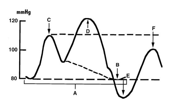

Correct timing is confirmed on the arterial waveform in 1:2 mode. Early inflation, before aortic valve closure, raises afterload and can cause aortic regurgitation. Late inflation reduces diastolic augmentation. Early deflation produces a sharp fall after diastolic augmentation, an assisted end-diastolic pressure equal to or below the unassisted value, suboptimal afterload reduction during systole, and possible retrograde coronary and carotid flow. Late deflation, with the balloon still inflated as the ventricle begins to eject, increases afterload and myocardial oxygen demand, the most harmful timing error. Tachycardia and irregular rhythm reduce the effect.
In IABP-SHOCK II, 600 patients with infarct-related shock and planned early revascularization were randomized to a balloon pump or no balloon pump. Thirty-day mortality was 39.7% and 41.3% (relative risk 0.96; P = 0.69), with no difference in time to hemodynamic stabilization, lactate, catecholamine dose, or renal function. Six-year mortality also did not differ. Major bleeding (3.3% against 4.4%) and peripheral ischemic complications (4.3% against 3.4%) did not differ. Routine balloon pump use in infarct-related shock carries Class 3: No Benefit. The device retains a role as a bridge in mechanical complications and in selected patients with ongoing ischemia awaiting surgery. It also retains a role in heart failure–related shock, where observational data suggest hemodynamic benefit and the device can serve as a bridge to durable LVAD or transplantation. Because it does not require large-bore access, it has fewer vascular complications than the other devices. Complications occur in 6% to 25% of patients and are mainly limb ischemia, bleeding, vascular injury, and thrombocytopenia.
Microaxial flow pumps
A microaxial flow pump (Impella) is a catheter-mounted axial pump that crosses the aortic valve, draws blood from the left ventricle through an inlet in the cavity, and ejects it into the ascending aorta. Flow is continuous and independent of the cardiac cycle. The device directly unloads the left ventricle, lowering end-diastolic pressure, wall stress, and myocardial oxygen demand, while raising mean arterial pressure and cardiac output; on the pressure-volume loop, the loop shifts leftward and becomes triangular as isovolumic phases are lost. The percutaneous Impella CP is placed through a 14 Fr femoral sheath and delivers up to about 4 L/min. The Impella 5.5 is placed by surgical cutdown with a graft to the axillary artery and delivers up to about 6 L/min. It allows the patient to sit and walk and is used for longer support in heart failure–related shock and as a bridge to durable LVAD or transplantation. Adequate right ventricular function and left ventricular filling are required: the pump cannot deliver more than reaches the left ventricle, and suction alarms often signal right ventricular failure or hypovolemia.
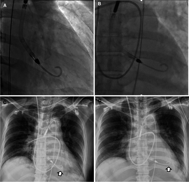
Early randomized trials were small. IMPRESS in Severe Shock randomized 48 mechanically ventilated patients with infarct-related shock to a microaxial pump or balloon pump and found 30-day mortality of 46% and 50%. DanGer Shock randomized 360 patients with STEMI-related shock (lactate of 2.5 mmol/L or more, systolic pressure below 100 mm Hg or vasopressor support, ejection fraction below 45%) within 24 hours of shock onset. Centers in Denmark, Germany, and the United Kingdom participated. Comatose survivors of out-of-hospital arrest, patients with predominant right ventricular failure, and those with mechanical complications were excluded. All-cause death at 180 days occurred in 45.8% with the Impella CP plus standard care and 58.5% with standard care (hazard ratio 0.74; 95% CI 0.55 to 0.99), an absolute reduction of 12.7 percentage points. The composite of severe bleeding, limb ischemia, hemolysis, device failure, or worsening aortic regurgitation occurred in 24.0% and 6.2%, and renal replacement therapy in 41.9% and 26.7%. In long-term follow-up extending to 10 years, the survival difference persisted (hazard ratio 0.70; 95% CI 0.54 to 0.92), with a mean of about 600 additional days alive; causes of death after 6 months were not available.
DanGer Shock applies to the population it enrolled. An individual patient data meta-analysis of nine randomized trials (1,114 patients) found no significant 6-month mortality benefit of early unselected active support overall (hazard ratio 0.87; 95% CI 0.74 to 1.03) or for left ventricular unloading devices as a group, with a signal of benefit confined to STEMI-related shock without risk of hypoxic brain injury. In a Japanese national registry of 3,975 patients with infarct-related shock treated with Impella, only 35.6% met DanGer Shock criteria, and outcomes in the remaining groups varied widely. Large US observational analyses comparing microaxial pumps with balloon pumps found higher bleeding and mortality with the microaxial pump, findings limited by confounding. Use outside the DanGer Shock phenotype, including comatose arrest survivors, right ventricular failure, mechanical complications, and HF-CS, is guided by physiology and expertise and is not supported by randomized evidence.
Management on a microaxial pump includes a heparin-containing purge solution and systemic anticoagulation, with bivalirudin or argatroban in heparin-induced thrombocytopenia; daily echocardiographic position checks; monitoring for hemolysis (plasma free hemoglobin, lactate dehydrogenase, dark urine), which is more frequent with malposition and suction; distal limb perfusion checks; and limb reperfusion strategies when flow to the cannulated leg is threatened.
VA-ECMO and left ventricular unloading
VA-ECMO drains venous blood through a 19 to 25 Fr cannula, usually from the femoral vein into the right atrium, passes it through a centrifugal pump and membrane oxygenator, and returns it through a 15 to 19 Fr arterial cannula, usually into the femoral artery. Flow of 4 to 7 L/min provides full circulatory support and gas exchange independent of both ventricles. It is the device of choice for biventricular failure, combined cardiac and respiratory failure, cardiac arrest refractory to conventional resuscitation, and situations needing rapid bedside support. It does not unload the left ventricle; retrograde aortic flow raises afterload.
The randomized trials of early VA-ECMO were neutral. ECLS-SHOCK randomized 420 patients with infarct-related shock and planned early revascularization; 30-day mortality was 47.8% with VA-ECMO and 49.0% without (relative risk 0.98), moderate or severe bleeding was 23.4% against 9.6%, and peripheral vascular complications needing intervention were 11.0% against 3.8%. ECMO-CS randomized 117 patients with rapidly deteriorating or severe shock of mixed causes to immediate VA-ECMO or an early conservative strategy that allowed downstream ECMO, which 39% of the conservative group received; the composite of death, resuscitated arrest, and another mechanical support device at 30 days occurred in 63.8% and 71.2% (hazard ratio 0.72; P = 0.21). An individual patient data meta-analysis of four trials (567 patients) found no reduction in 30-day mortality (odds ratio 0.93) and more major bleeding (odds ratio 2.44) and peripheral ischemic complications (odds ratio 3.53). Routine VA-ECMO in infarct-related shock is not recommended (Class 3: No Benefit). It remains a rescue therapy for selected refractory shock at experienced centers.
Left ventricular distension. Retrograde flow raises the pressure the left ventricle ejects against. A severely impaired ventricle may stop ejecting, the aortic valve stays closed, and left ventricular and left atrial pressures rise, causing pulmonary edema, myocardial ischemia, and stasis with intracavitary or aortic root thrombus. Loss of arterial pulsatility, a closed aortic valve, a dilating left ventricle with spontaneous contrast, a rising wedge or pulmonary artery diastolic pressure, and worsening pulmonary edema are the signs. The first steps are reduction of ECMO flow to the minimum that maintains perfusion, a lower afterload target, diuresis, and inotropic support to promote ejection. Mechanical unloading uses a microaxial pump (the combination is called ECPELLA or ECMELLA), a balloon pump, atrial septostomy, a transseptal left atrial drainage cannula, or a surgical vent. In a propensity-matched international cohort of 510 patients, unloading with a microaxial pump during VA-ECMO was associated with lower 30-day mortality (hazard ratio 0.79). It was also associated with more severe bleeding (38.4% against 17.9%), access-site ischemia, and renal replacement therapy. No completed randomized trial has tested routine unloading.
Differential hypoxemia. With femoral arterial return, oxygenated ECMO blood flows retrograde up the aorta and meets blood ejected by the recovering left ventricle. If the native lungs are failing, the left ventricle ejects poorly oxygenated blood into the coronary arteries and the vessels of the aortic arch, while the lower body receives well-oxygenated ECMO blood (differential, north-south, or harlequin hypoxemia). Arterial gases and oximetry are monitored from the right radial artery, which reflects the cerebral and coronary supply. Treatment includes optimization of ventilation, an additional return cannula into the internal jugular vein (venoarterial-venous configuration), or central or axillary arterial cannulation.
Extracorporeal cardiopulmonary resuscitation (ECPR). VA-ECMO initiated during refractory arrest restores flow while the cause is treated. In INCEPTION, a pragmatic multicenter trial of 134 patients with refractory out-of-hospital ventricular arrhythmia arrest, survival with favorable neurologic outcome at 30 days was 20% with ECPR and 16% with conventional resuscitation, a nonsignificant difference. ECPR depends on short low-flow times, highly selected patients, and a system that can cannulate quickly.
Right ventricular and biventricular support
Right-sided temporary support moves blood from the venous system to the pulmonary artery, bypassing the failing right ventricle. The Impella RP and RP Flex are microaxial pumps placed from the femoral or internal jugular vein that deliver up to about 4 L/min into the pulmonary artery. The ProtekDuo is a dual-lumen cannula placed through the right internal jugular vein, draining the right atrium and returning blood to the pulmonary artery through an extracorporeal centrifugal pump at 4 to 5 L/min; an oxygenator can be added, and the jugular approach allows mobilization. In RECOVER RIGHT, 30 patients with refractory right ventricular failure after LVAD implantation, cardiotomy, or infarction received the Impella RP; cardiac index rose from 1.8 to 3.3 L/min/m², central venous pressure fell from 19.2 to 12.6 mm Hg, and 30-day survival was 73.3%. No randomized trial has tested right-sided support.
Right ventricular support is considered when right-sided failure persists despite restoration of coronary flow, optimized preload, rhythm, and pulmonary vascular resistance, and inotropes, with a plausible path to recovery or definitive therapy. Contraindications include mechanical tricuspid or pulmonary prostheses and right heart thrombus. A low PAPi supports the decision but does not by itself establish the need. Biventricular failure is supported with VA-ECMO, with left and right microaxial pumps together, or with combinations, chosen by the need for oxygenation, anticipated duration, and access.
Choosing a device: comparison of temporary support
| Feature | IABP | Microaxial pump (Impella CP) | Microaxial pump (Impella 5.5) | TandemHeart | VA-ECMO | Right-sided pump (Impella RP, ProtekDuo) |
|---|---|---|---|---|---|---|
| Mechanism | Diastolic counterpulsation | Left ventricle to aorta, axial | Left ventricle to aorta, axial | Left atrium to femoral artery, centrifugal, transseptal | Right atrium to aorta through an oxygenator, centrifugal | Right atrium or vena cava to pulmonary artery |
| Flow support | About 0.5 L/min | Up to about 4 L/min | Up to about 6 L/min | 3.5–5 L/min | 4–7 L/min | About 4–5 L/min |
| Access | Femoral or axillary artery, small bore | Femoral artery, 14 Fr | Axillary artery surgical graft | Femoral vein with atrial septal puncture plus femoral artery | Femoral vein and artery (19–25 Fr venous, 15–19 Fr arterial), or central | Femoral or internal jugular vein |
| Left ventricular unloading | Modest, indirect | Direct | Direct, greater | Direct (left atrial) | None; raises afterload | Not applicable |
| Oxygenation | No | No | No | Only with added oxygenator | Yes | Only with added oxygenator (ProtekDuo) |
| Requires native rhythm and right ventricle | Yes | Yes (adequate right ventricular output) | Yes | Yes (right ventricle fills left atrium) | No | Left ventricle must handle the added flow |
| Randomized evidence in shock | IABP-SHOCK II: no mortality benefit | DanGer Shock: lower 180-day mortality in selected STEMI shock; IMPRESS: neutral | None | Small trials: hemodynamic improvement, no mortality difference | ECLS-SHOCK, ECMO-CS: no benefit of early routine use | None (RECOVER RIGHT single arm) |
| Guideline position | Routine use in AMI-CS: Class 3: No Benefit | Selected STEMI with severe or refractory shock: Class 2a | Not separately graded | Not separately graded | Routine use in AMI-CS: Class 3: No Benefit | Not separately graded |
| Principal complications | Limb ischemia, bleeding, thrombocytopenia | Bleeding, limb ischemia, hemolysis, kidney injury, aortic valve injury | Bleeding, hemolysis, stroke, arm ischemia | Bleeding, limb ischemia, cannula dislodgement into the right atrium | Bleeding, limb ischemia, stroke, left ventricular distension, differential hypoxemia, infection | Hemolysis, tricuspid or pulmonary valve injury, malposition |
Flows and access from device reviews and the 2025 ACC guidance; guideline classes from the 2025 ACC/AHA/ACEP/NAEMSP/SCAI acute coronary syndrome guideline. Device specifications vary by generation. Combinations (ECPELLA, a left and a right microaxial pump together, VA-ECMO with a balloon pump) are used for biventricular failure and left ventricular distension.
The phenotype and trajectory decide. Left ventricle–dominant infarct-related shock resembling the DanGer Shock population is the setting for a microaxial pump. Left ventricle–dominant HF-CS with a need for longer support or mobilization favors an axillary microaxial pump or a balloon pump as a bridge. Right ventricle–dominant shock needs right-sided support or VA-ECMO. Biventricular failure, refractory hypoxemia, or ongoing arrest needs VA-ECMO, with left ventricular unloading when the ventricle distends. Patients who remain undersupported on one device are escalated by increasing flow, moving to a higher-flow device (for example from Impella CP to 5.5), adding right-sided support, or adding a vent to VA-ECMO.
Complications of temporary mechanical support
Complications are frequent and often decide the outcome. In infarct-related shock, major bleeding occurs in up to 60% of patients receiving temporary support. The risk of limb ischemia is four times higher with support than without it, and limb ischemia is associated with twofold higher in-hospital mortality. Bleeding results from large-bore access, anticoagulation, acquired von Willebrand deficiency and platelet dysfunction from shear stress, thrombocytopenia, and liver dysfunction. Acute kidney injury reflects both shock and device effects, and renal replacement therapy was nearly twice as frequent with the microaxial pump in DanGer Shock.
| Complication | Mechanism | Prevention and management |
|---|---|---|
| Access-site and retroperitoneal bleeding | Large-bore arteriotomy, anticoagulation, coagulopathy | Ultrasound- and fluoroscopy-guided single-wall puncture above the bifurcation; anticoagulation targets kept at the lowest effective level; securement to avoid tenting of the arteriotomy; removal of sheaths ≥12 Fr in the catheterization or operating room |
| Limb ischemia | Arterial occlusion by the cannula or sheath; embolism; dissection | Pulse and Doppler checks every 1–2 hours; distal perfusion cannula for VA-ECMO; external bypass or contralateral perfusion; device removal when ischemia cannot be relieved |
| Hemolysis | Shear stress; pump malposition; suction | Plasma free hemoglobin and lactate dehydrogenase monitoring; echocardiographic repositioning; flow and preload optimization |
| Stroke | Thrombus in the circuit or ventricle; air; hemorrhage on anticoagulation | Anticoagulation monitoring; daily neurologic assessment; imaging for new deficits |
| Left ventricular distension (VA-ECMO) | Retrograde afterload with a nonejecting ventricle | Pulsatility and aortic valve opening monitoring; flow reduction, inotropes, unloading device |
| Differential hypoxemia (femoral VA-ECMO) | Native lung failure with recovering left ventricular ejection | Right radial arterial gas; ventilator optimization; venoarterial-venous configuration |
| Thrombocytopenia and acquired von Willebrand syndrome | Shear, consumption, heparin-induced thrombocytopenia | Platelet monitoring; testing for heparin-induced thrombocytopenia and switch to a direct thrombin inhibitor when present |
| Infection | Prolonged cannulation | Shortest effective duration; access-site care |
Bleeding and limb ischemia data and part of the access measures from the 2025 ACC guidance (Table 4); the remaining measures and complication patterns from the randomized device trials and device reviews.
Shock teams and regional systems of care
A standardized multidisciplinary approach has been associated with better outcomes in observational studies, and the 2022 heart failure guideline states that management by a multidisciplinary team experienced in shock is reasonable (Class 2a). Teams typically include critical care cardiology, advanced heart failure and transplant cardiology, interventional cardiology, and cardiac surgery, with extracorporeal support specialists and palliative care in many centers. They share a single activation call, a coordinating physician, protocols built on SCAI stage and invasive hemodynamics, and daily joint review.
In a single-center program, 30-day survival rose from 47% in 2016 to 57.9% in 2017 and 76.6% in 2018 after a standardized team approach was introduced. In the Critical Care Cardiology Trials Network, the 10 of 24 centers with shock teams obtained more pulmonary artery catheters and used fewer devices overall, with more advanced devices in place of balloon pumps. They had lower risk-adjusted cardiac intensive care unit mortality (23% against 29%; adjusted odds ratio 0.72). These data are observational and cannot separate the team from the institution.
Most US hospitals provide acute cardiac care, and fewer can offer the full range of temporary and durable support. The ACC guidance describes Level 1 centers with 24/7 interventional, surgical, advanced heart failure, and critical care expertise, all temporary devices, and usually durable LVAD and transplantation, and Level 2 and 3 centers with more limited resources. The 2022 heart failure guideline states that triage of patients who do not respond rapidly to initial measures to centers that can provide temporary support may be considered (Class 2b). Patients with infarct-related shock who remain in refractory shock after revascularization at a local hospital are almost always transferred to a Level 1 center. Arrest with uncertain neurologic status, any need for vasoactive drugs, or consideration of temporary support prompts early discussion with the regional center. Observational outcomes of transferred patients are mixed and reflect selection.
Structured reassessment and organ support in the intensive care unit
Cardiogenic shock changes over hours, and the 2025 ACC guidance recommends structured reassessment at defined intervals, particularly in the first 24 hours. Laboratory markers of perfusion are measured as often as every 2 to 4 hours early in care, spacing to every 6 to 8 hours as the patient stabilizes.
| Domain | Measures | Frequency |
|---|---|---|
| Clinical examination | Mental status, extremity perfusion, access sites, distal pulses | Every 6–8 hours; pulses every 1–2 hours with large-bore arterial access |
| Vital signs | Heart rate and rhythm, mean arterial pressure, arterial oxygen saturation | Continuous |
| Physiology | Urine output | Hourly |
| Laboratory | Creatinine, bicarbonate, pH, central or mixed venous saturation, lactate, liver chemistries | Every 2–8 hours depending on phase of care |
| Imaging | Echocardiographic biventricular function, volume status, device position; chest radiograph for device, catheter, pacing lead, and tube position | Echocardiography daily (focused study acceptable); radiograph as needed |
| Invasive hemodynamics | Central venous, pulmonary artery, and wedge pressures; cardiac output | Pressures continuous; output continuous or every 2–8 hours |
| Shock team | Joint review of status, escalation, de-escalation, and goals | Daily, more often with deterioration |
Adapted from Table 3 of the 2025 ACC concise clinical guidance.
Kidney. Acute kidney injury is common and multifactorial (low output, venous congestion, contrast, hemolysis, nephrotoxins). Congestion refractory to diuretics is treated with ultrafiltration or continuous renal replacement therapy. Renal replacement therapy is associated with higher mortality, a marker of severity more than an independent cause.
Ventilation. Lung-protective ventilation is used. Positive pressure helps a failing left ventricle and can harm a failing right ventricle, so pressures are titrated to hemodynamic response.
Other organs. Glucose control, stress ulcer prophylaxis, venous thromboembolism prophylaxis when not anticoagulated, nutrition, and infection surveillance follow general critical care practice. Corticosteroids have no established role in cardiogenic shock outside adrenal insufficiency or coexisting septic shock.
Weaning and escalation of temporary support
Escalation is indicated when perfusion, congestion, or end-organ function fails to improve on the present support. The patient is undersupported (cardiac index or power still low), inadequately unloaded (wedge pressure still high, pulmonary edema), or has a right ventricle that cannot keep up. Failure to improve also prompts a search for an unrecognized cause such as tamponade, a mechanical complication, bleeding, sepsis, or device malposition.
Readiness to wean requires recovery of the cause, minimal or no vasoactive support, euvolemia, and recovering end-organ function (falling lactate, creatinine, and transaminases). Before weaning, an advanced heart failure specialist assesses whether recovery, durable LVAD, or transplantation is the realistic goal, because removing support from a patient who will need durable therapy wastes the window for it. Weaning proceeds stepwise, with assessment of examination, perfusion markers, echocardiography, and hemodynamics at each step.
| Device | Weaning method | Findings supporting removal |
|---|---|---|
| IABP | Reduction of the assist ratio from 1:1 to 1:2 to 1:3, or reduction of augmentation | Stable pressure, output, and lactate at 1:3 |
| Microaxial pump | Reduction of performance level by 1–2 steps every 2–4 hours to the minimum level | Stable mean arterial pressure, cardiac power output, wedge and right atrial pressures, and PAPi at minimal support |
| VA-ECMO | Flow reduction by about 0.5–1 L/min every 2–4 hours to a trial at low flow (below about 1.5–2 L/min) | Pulsatile arterial pressure; aortic velocity-time integral ≥10 cm, LVEF >20–25%, and lateral mitral annular systolic velocity ≥6 cm/s at minimal flow; adequate right ventricular function and oxygenation |
| Right-sided pump or ProtekDuo | Stepwise flow reduction | Stable right atrial pressure, cardiac index, and PAPi at minimal flow |
The 2025 ACC guidance describes weaning by 0.5 to 1 L/min (two Impella performance levels) every 2 to 4 hours. VA-ECMO echocardiographic criteria from a study of 51 patients in which all successfully weaned patients met these thresholds at minimal flow. HF-CS generally needs slower weaning than AMI-CS, and some patients are bridged from devices to inotropes.
Transition to durable therapy, palliative care, and long-term outcomes after shock
Patients who do not recover ventricular function on temporary support are evaluated for durable therapy. Interagency Registry for Mechanically Assisted Circulatory Support (INTERMACS) profile 1 (critical cardiogenic shock) and profile 2 (progressive decline despite inotropes) describe the acuity of patients considered for durable LVAD. Outcomes of durable LVAD implanted in profile 1 are worse than in more stable profiles, so stabilization on temporary support, with recovery of kidney, liver, and neurologic function and assessment of right ventricular function, usually precedes implantation. Heart transplantation candidates supported with temporary devices receive higher allocation priority in the US system, which has increased transplantation directly from temporary support. The evaluation for advanced therapies is covered on the heart failure with reduced ejection fraction page.
Palliative care is part of shock care from the outset, not only after escalation fails. In the 2017 US Nationwide Readmissions Database (133,738 cardiogenic shock admissions), 8.6% of hospital survivors received palliative care services, and their 30-day readmission rate was lower (11.6% against 21.9%). Goals-of-care discussions before device placement define what the patient would accept and what the team will do if no exit exists. When support no longer serves the patient's goals, its withdrawal is planned with the family and the palliative care team.
Long-term outcomes. Survival after discharge from infarct-related shock is better than in-hospital outcomes suggest but remains below that of infarction without shock. In a registry comparing shock types, 30-day readmission after discharge was 19.5% after AMI-CS and 24.5% after HF-CS. Survivors carry the burdens of ischemic or nonischemic cardiomyopathy, kidney injury, deconditioning, cognitive impairment, and post-intensive care syndrome. Discharge care includes guideline-directed therapy for heart failure with reduced ejection fraction once hemodynamics allow, defibrillator decisions after reassessment of ejection fraction, cardiac rehabilitation, and follow-up in a heart failure clinic.
Cardiac arrest, temperature control, and neurologic prognosis
Cardiac arrest complicates a large share of infarct-related shock and was present in about one-third of AMI-CS in a US registry. When it leaves the patient comatose, it changes prognosis more than any hemodynamic variable. The SCAI cardiac arrest modifier applies to arrest with coma. Comatose survivors of out-of-hospital arrest were excluded from DanGer Shock, and the benefit of a microaxial pump does not extend to them. In TTM2, 1,900 comatose adults after out-of-hospital arrest were randomized to targeted hypothermia at 33°C or normothermia with early treatment of fever; 6-month mortality was 50% and 48%, with no difference in functional outcome. Active prevention of fever is the current practice, and hypotension and hypoxemia are avoided because they worsen anoxic injury. Neurologic prognosis is assessed with multimodal testing no earlier than 72 hours after normothermia, and escalation to durable therapies waits for evidence of neurologic recovery. Coronary angiography after arrest without ST elevation is covered on the STEMI page.
Sources of error in managing cardiogenic shock
- Waiting for hypotension: normotensive shock with rising lactate, oliguria, and cool extremities is already stage C.
- Treating a single number: lactate, cardiac power output, and PAPi are interpreted together, serially, and in the clinical context; none mandates a device.
- Applying DanGer Shock outside its population: comatose arrest survivors, right ventricular failure, mechanical complications, and HF-CS were not studied.
- Placing a device without an exit strategy or a goals-of-care discussion.
- Missing right ventricular failure before left-sided support: a microaxial pump cannot deliver flow that the right ventricle does not provide.
- Missing left ventricular distension on VA-ECMO: an adequate mean pressure with a flat arterial waveform indicates a nonejecting ventricle.
- Measuring oxygenation from the wrong arm on femoral VA-ECMO: the right radial artery reflects cerebral and coronary supply.
- Giving inotropes or vasodilators in dynamic outflow obstruction, or fluids to a congested left ventricle.
- Performing multivessel PCI at the index procedure in shock.
- Escalating catecholamines indefinitely: rising doses of two or more vasoactive drugs define stage D and call for reassessment, mechanical support, or transfer.
- Delaying transfer from a hospital without temporary support, durable LVAD, or transplantation.
- Overlooking access-site and limb complications, which double in-hospital mortality when limb ischemia occurs.
Self-assessment
A 62-year-old man has an anterior STEMI treated with primary PCI. Two hours later his blood pressure is 88/66 mm Hg on norepinephrine 0.12 mcg/kg/min, lactate is 4.6 mmol/L, cardiac output is 3.0 L/min, right atrial pressure is 9 mm Hg, wedge pressure is 27 mm Hg, and pulmonary artery pressure is 44/24 mm Hg. He was not resuscitated from cardiac arrest. What are his cardiac power output and PAPi, what is the shock phenotype, and which temporary support device has randomized evidence of a survival benefit in this setting?
Cardiovascular Medicine Board ReviewReferences
- Rao SV, O'Donoghue ML, Ruel M, et al. 2025 ACC/AHA/ACEP/NAEMSP/SCAI guideline for the management of patients with acute coronary syndromes: a report of the American College of Cardiology/American Heart Association Joint Committee on Clinical Practice Guidelines. J Am Coll Cardiol. 2025;85:2135–2237. doi:10.1016/j.jacc.2024.11.009
- Sinha SS, Morrow DA, Kapur NK, Kataria R, Roswell RO. 2025 Concise clinical guidance: an ACC expert consensus statement on the evaluation and management of cardiogenic shock. J Am Coll Cardiol. 2025;85:1618–1641. doi:10.1016/j.jacc.2025.02.018
- Naidu SS, Baran DA, Jentzer JC, et al. SCAI SHOCK stage classification expert consensus update: a review and incorporation of validation studies. J Am Coll Cardiol. 2022;79:933–946. doi:10.1016/j.jacc.2022.01.018
- van Diepen S, Katz JN, Albert NM, et al. Contemporary management of cardiogenic shock: a scientific statement from the American Heart Association. Circulation. 2017;136:e232–e268. doi:10.1161/CIR.0000000000000525
- Heidenreich PA, Bozkurt B, Aguilar D, et al. 2022 AHA/ACC/HFSA guideline for the management of heart failure: a report of the American College of Cardiology/American Heart Association Joint Committee on Clinical Practice Guidelines. Circulation. 2022;145:e895–e1032. doi:10.1161/CIR.0000000000001063
- Geller BJ, Sinha SS, Kapur NK, et al. Escalating and de-escalating temporary mechanical circulatory support in cardiogenic shock: a scientific statement from the American Heart Association. Circulation. 2022;146:e50–e68. doi:10.1161/CIR.0000000000001076
- Waksman R, Pahuja M, van Diepen S, et al. Standardized definitions for cardiogenic shock research and mechanical circulatory support devices: scientific expert panel from the Shock Academic Research Consortium (SHARC). Circulation. 2023;148:1113–1126. doi:10.1161/CIRCULATIONAHA.123.064527
- Byrne RA, Rossello X, Coughlan JJ, et al. 2023 ESC guidelines for the management of acute coronary syndromes. Eur Heart J. 2023;44:3720–3826. doi:10.1093/eurheartj/ehad191
- Hochman JS, Sleeper LA, Webb JG, et al. Early revascularization in acute myocardial infarction complicated by cardiogenic shock (SHOCK). N Engl J Med. 1999;341:625–634. doi:10.1056/NEJM199908263410901
- Hochman JS, Sleeper LA, Webb JG, et al. Early revascularization and long-term survival in cardiogenic shock complicating acute myocardial infarction. JAMA. 2006;295:2511–2515. doi:10.1001/jama.295.21.2511
- Thiele H, Akin I, Sandri M, et al. PCI strategies in patients with acute myocardial infarction and cardiogenic shock (CULPRIT-SHOCK). N Engl J Med. 2017;377:2419–2432. doi:10.1056/NEJMoa1710261
- Thiele H, Zeymer U, Neumann FJ, et al. Intraaortic balloon support for myocardial infarction with cardiogenic shock (IABP-SHOCK II). N Engl J Med. 2012;367:1287–1296. doi:10.1056/NEJMoa1208410
- Thiele H, Zeymer U, Thelemann N, et al. Intraaortic balloon pump in cardiogenic shock complicating acute myocardial infarction: long-term 6-year outcome of the randomized IABP-SHOCK II trial. Circulation. 2019;139:395–403. doi:10.1161/CIRCULATIONAHA.118.038201
- Møller JE, Engstrøm T, Jensen LO, et al. Microaxial flow pump or standard care in infarct-related cardiogenic shock (DanGer Shock). N Engl J Med. 2024;390:1382–1393. doi:10.1056/NEJMoa2312572
- Møller JE, Beske RP, Engstrøm T, et al. Long-term outcomes of the DanGer Shock trial. N Engl J Med. 2025;393:1037–1038. doi:10.1056/NEJMc2508284
- Ouweneel DM, Eriksen E, Sjauw KD, et al. Percutaneous mechanical circulatory support versus intra-aortic balloon pump in cardiogenic shock after acute myocardial infarction (IMPRESS in Severe Shock). J Am Coll Cardiol. 2017;69:278–287. doi:10.1016/j.jacc.2016.10.022
- Thiele H, Zeymer U, Akin I, et al. Extracorporeal life support in infarct-related cardiogenic shock (ECLS-SHOCK). N Engl J Med. 2023;389:1286–1297. doi:10.1056/NEJMoa2307227
- Ostadal P, Rokyta R, Karasek J, et al. Extracorporeal membrane oxygenation in the therapy of cardiogenic shock: results of the ECMO-CS randomized clinical trial. Circulation. 2023;147:454–464. doi:10.1161/CIRCULATIONAHA.122.062949
- Zeymer U, Freund A, Hochadel M, et al. Venoarterial extracorporeal membrane oxygenation in patients with infarct-related cardiogenic shock: an individual patient data meta-analysis of randomised trials. Lancet. 2023;402:1338–1346. doi:10.1016/S0140-6736(23)01607-0
- Thiele H, Møller JE, Henriques JPS, et al. Temporary mechanical circulatory support in infarct-related cardiogenic shock: an individual patient data meta-analysis of randomised trials with 6-month follow-up. Lancet. 2024;404:1019–1028. doi:10.1016/S0140-6736(24)01448-X
- De Backer D, Biston P, Devriendt J, et al. Comparison of dopamine and norepinephrine in the treatment of shock (SOAP II). N Engl J Med. 2010;362:779–789. doi:10.1056/NEJMoa0907118
- Levy B, Clere-Jehl R, Legras A, et al. Epinephrine versus norepinephrine for cardiogenic shock after acute myocardial infarction (OptimaCC). J Am Coll Cardiol. 2018;72:173–182. doi:10.1016/j.jacc.2018.04.051
- Mathew R, Di Santo P, Jung RG, et al. Milrinone as compared with dobutamine in the treatment of cardiogenic shock (DOREMI). N Engl J Med. 2021;385:516–525. doi:10.1056/NEJMoa2026845
- Jentzer JC, van Diepen S, Barsness GW, et al. Cardiogenic shock classification to predict mortality in the cardiac intensive care unit. J Am Coll Cardiol. 2019;74:2117–2128. doi:10.1016/j.jacc.2019.07.077
- Kapur NK, Kanwar M, Sinha SS, et al. Criteria for defining stages of cardiogenic shock severity. J Am Coll Cardiol. 2022;80:185–198. doi:10.1016/j.jacc.2022.04.049
- Kolte D, Khera S, Aronow WS, et al. Trends in incidence, management, and outcomes of cardiogenic shock complicating ST-elevation myocardial infarction in the United States. J Am Heart Assoc. 2014;3:e000590. doi:10.1161/JAHA.113.000590
- Berg DD, Bohula EA, van Diepen S, et al. Epidemiology of shock in contemporary cardiac intensive care units. Circ Cardiovasc Qual Outcomes. 2019;12:e005618. doi:10.1161/CIRCOUTCOMES.119.005618
- Sinha SS, Rosner CM, Tehrani BN, et al. Cardiogenic shock from heart failure versus acute myocardial infarction: clinical characteristics, hospital course, and 1-year outcomes. Circ Heart Fail. 2022;15:e009279. doi:10.1161/CIRCHEARTFAILURE.121.009279
- Fincke R, Hochman JS, Lowe AM, et al. Cardiac power is the strongest hemodynamic correlate of mortality in cardiogenic shock: a report from the SHOCK trial registry. J Am Coll Cardiol. 2004;44:340–348. doi:10.1016/j.jacc.2004.03.060
- Korabathina R, Heffernan KS, Paruchuri V, et al. The pulmonary artery pulsatility index identifies severe right ventricular dysfunction in acute inferior myocardial infarction. Catheter Cardiovasc Interv. 2012;80:593–600. doi:10.1002/ccd.23309
- Garan AR, Kanwar M, Thayer KL, et al. Complete hemodynamic profiling with pulmonary artery catheters in cardiogenic shock is associated with lower in-hospital mortality. JACC Heart Fail. 2020;8:903–913. doi:10.1016/j.jchf.2020.08.012
- Tehrani BN, Truesdell AG, Sherwood MW, et al. Standardized team-based care for cardiogenic shock. J Am Coll Cardiol. 2019;73:1659–1669. doi:10.1016/j.jacc.2018.12.084
- Papolos AI, Kenigsberg BB, Berg DD, et al. Management and outcomes of cardiogenic shock in cardiac ICUs with versus without shock teams. J Am Coll Cardiol. 2021;78:1309–1317. doi:10.1016/j.jacc.2021.07.044
- Basir MB, Kapur NK, Patel K, et al. Improved outcomes associated with the use of shock protocols: updates from the National Cardiogenic Shock Initiative. Catheter Cardiovasc Interv. 2019;93:1173–1183. doi:10.1002/ccd.28307
- Schrage B, Becher PM, Bernhardt A, et al. Left ventricular unloading is associated with lower mortality in patients with cardiogenic shock treated with venoarterial extracorporeal membrane oxygenation: results from an international, multicenter cohort study. Circulation. 2020;142:2095–2106. doi:10.1161/CIRCULATIONAHA.120.048792
- Anderson MB, Goldstein J, Milano C, et al. Benefits of a novel percutaneous ventricular assist device for right heart failure: the prospective RECOVER RIGHT study of the Impella RP device. J Heart Lung Transplant. 2015;34:1549–1560. doi:10.1016/j.healun.2015.08.018
- Aissaoui N, Luyt CE, Leprince P, et al. Predictors of successful extracorporeal membrane oxygenation (ECMO) weaning after assistance for refractory cardiogenic shock. Intensive Care Med. 2011;37:1738–1745. doi:10.1007/s00134-011-2358-2
- Harjola VP, Lassus J, Sionis A, et al. Clinical picture and risk prediction of short-term mortality in cardiogenic shock (CardShock). Eur J Heart Fail. 2015;17:501–509. doi:10.1002/ejhf.260
- Pöss J, Köster J, Fuernau G, et al. Risk stratification for patients in cardiogenic shock after acute myocardial infarction. J Am Coll Cardiol. 2017;69:1913–1920. doi:10.1016/j.jacc.2017.02.027
- Na SJ, Chung CR, Cho YH, et al. Vasoactive inotropic score as a predictor of mortality in adult patients with cardiogenic shock: medical therapy versus ECMO. Rev Esp Cardiol (Engl Ed). 2019;72:40–47. doi:10.1016/j.rec.2018.01.003
- Vallabhajosyula S, Vallabhajosyula S, Dunlay SM, et al. Sex and gender disparities in the management and outcomes of acute myocardial infarction–cardiogenic shock in older adults. Mayo Clin Proc. 2020;95:1916–1927. doi:10.1016/j.mayocp.2020.01.043
- Mehta A, Vavilin I, Nguyen AH, et al. Contemporary approach to cardiogenic shock care: a state-of-the-art review. Front Cardiovasc Med. 2024;11:1354158. doi:10.3389/fcvm.2024.1354158
- Jana S, Wijesinghe M, DiCaro MV, et al. Cardiogenic shock management in the modern era: a narrative review of percutaneous mechanical circulatory support devices. J Cardiovasc Dev Dis. 2025;13:9. doi:10.3390/jcdd13010009
- Dankiewicz J, Cronberg T, Lilja G, et al. Hypothermia versus normothermia after out-of-hospital cardiac arrest (TTM2). N Engl J Med. 2021;384:2283–2294. doi:10.1056/NEJMoa2100591
- Suverein MM, Delnoij TSR, Lorusso R, et al. Early extracorporeal CPR for refractory out-of-hospital cardiac arrest (INCEPTION). N Engl J Med. 2023;388:299–309. doi:10.1056/NEJMoa2204511
- Feng Z, Fonarow GC, Ziaeian B. Palliative care services in patients admitted with cardiogenic shock in the United States: frequency and predictors of 30-day readmission. J Card Fail. 2021;27:560–567. doi:10.1016/j.cardfail.2021.01.020
- Arai R, Kojima K, Fukamachi D, Okumura Y. DanGer shock criteria and outcomes in acute myocardial infarction-related cardiogenic shock treated with Impella: the J-PVAD registry. Eur Heart J. 2026;47:472–486. doi:10.1093/eurheartj/ehaf787
- Dhruva SS, Ross JS, Mortazavi BJ, et al. Association of use of an intravascular microaxial left ventricular assist device vs intra-aortic balloon pump with in-hospital mortality and major bleeding among patients with acute myocardial infarction complicated by cardiogenic shock. JAMA. 2020;323:734–745. doi:10.1001/jama.2020.0254