Library › Pulmonary vascular disease › Pulmonary hypertension
Pulmonary hypertension
Pulmonary hypertension in adults, from hemodynamic definition and clinical classification through diagnosis, risk assessment, treatment of each group, chronic thromboembolic disease, pregnancy, surgery, and acute right ventricular failure.
Pulmonary hypertension is a mean pulmonary artery pressure above 20 mm Hg at rest, measured by right heart catheterization. Wedge pressure and pulmonary vascular resistance then separate pre-capillary disease (wedge 15 mm Hg or less, resistance above 2 Wood units) from post-capillary disease, and the clinical group sets treatment. Pulmonary arterial hypertension is treated with combination therapy aimed at a low-risk profile; chronic thromboembolic disease is screened for with a ventilation-perfusion scan because surgery or angioplasty can relieve it; left heart and lung disease are treated at their source.
Hemodynamic definition of pulmonary hypertension
Pulmonary hypertension is a hemodynamic state, not a single disease. The 7th World Symposium on Pulmonary Hypertension (WSPH, 2024) kept the definition adopted in 2018: a mean pulmonary artery pressure (mPAP) above 20 mm Hg at rest. The 2022 European Society of Cardiology and European Respiratory Society (ESC/ERS) guideline endorsed the same definition. There is no current ACC/AHA pulmonary hypertension guideline, so US practice follows the WSPH documents, the ESC/ERS guideline, and the 2019 CHEST guideline on drug therapy.
The threshold was lowered from 25 mm Hg because normal mPAP is about 14 mm Hg with an upper limit near 20 mm Hg, and because mortality rises in large referral cohorts from values above 20 mm Hg. The upper limit of normal for pulmonary vascular resistance (PVR) was lowered from 3 to 2 Wood units (WU) on the same reasoning. The drugs approved for pulmonary arterial hypertension (PAH) were tested in patients who met the older definition, and no therapy is approved specifically for an mPAP of 21 to 24 mm Hg.
| Hemodynamic type | mPAP | PAWP | PVR | Usual clinical groups |
|---|---|---|---|---|
| Pulmonary hypertension | >20 mm Hg | — | — | All |
| Pre-capillary | >20 mm Hg | ≤15 mm Hg | >2 WU | 1, 3, 4, and 5 |
| Isolated post-capillary (IpcPH) | >20 mm Hg | >15 mm Hg | ≤2 WU | 2 and 5 |
| Combined post- and pre-capillary (CpcPH) | >20 mm Hg | >15 mm Hg | >2 WU | 2 and 5 |
| Unclassified | >20 mm Hg | ≤15 mm Hg | ≤2 WU | High-flow states: congenital shunt, liver disease, hyperthyroidism, anemia |
| Exercise pulmonary hypertension | Normal resting values; mPAP/cardiac output slope >3 mm Hg/L/min between rest and exercise | Early pulmonary vascular or left heart disease | ||
Hemodynamic definitions from the 7th WSPH (2024), unchanged from the 2022 ESC/ERS guideline. PVR = (mPAP − PAWP) ÷ cardiac output. mPAP, mean pulmonary artery pressure; PAWP, pulmonary artery wedge pressure; WU, Wood units.
The three measurements locate the disease. A normal wedge pressure with a high resistance places it in the pulmonary arterioles. A high wedge pressure with a normal resistance indicates passive back-transmission from the left heart. A high wedge pressure with a high resistance indicates left heart disease that has remodeled the pulmonary vessels. The hemodynamic phenotype is not the clinical diagnosis: pre-capillary hemodynamics occur in PAH, in lung disease, and in chronic thromboembolic disease, and the label "pre-capillary pulmonary hypertension" is used until the cause is established.
Wedge pressure at the boundary. The upper limit of normal wedge pressure is 13 mm Hg, but PAH trials enrolled patients with values up to 15 mm Hg and showed benefit in those with 13 to 15 mm Hg. The 7th WSPH therefore kept 15 mm Hg as the cutoff and described 12 to 18 mm Hg as a zone of uncertainty. In that zone the phenotype is assigned from the whole patient: age, obesity, hypertension, diabetes, atrial fibrillation, left atrial size, diastolic indices, and any history of pulmonary edema. Occult left heart disease is unmasked by a wedge pressure above 18 mm Hg after a rapid infusion of 500 mL of saline (or 7 mL/kg over 5 to 10 minutes). A wedge pressure above 25 mm Hg during exercise or a PAWP/cardiac output slope above 2 mm Hg/L/min has the same significance. Catheterization technique, waveform interpretation at end-expiration, and the formulas behind these values are covered on the right heart catheterization and hemodynamics page.
Severity thresholds outside group 1. In left heart disease, a PVR above 5 WU indicates a severe pre-capillary component. In chronic lung disease, the 2022 ESC/ERS guideline defines severe pulmonary hypertension as a PVR above 5 WU, the value with the strongest association with mortality in chronic obstructive pulmonary disease (COPD). In sarcoidosis, an mPAP of 40 mm Hg or more and a PVR of 5 WU or more predict reduced transplant-free survival.
Classifying a hemodynamic study: pre-capillary, post-capillary, and combined
The phenotype follows from three catheter measurements: mean pulmonary artery pressure, wedge pressure, and cardiac output by thermodilution or direct Fick. The diastolic pulmonary gradient is derived when the pulmonary artery diastolic pressure is available.
Hemodynamic phenotype
7th WSPH (2024) definitionsPVR = (mPAP − PAWP) ÷ cardiac output; transpulmonary gradient = mPAP − PAWP; diastolic pulmonary gradient = PA diastolic pressure − PAWP. The example (mPAP 52 mm Hg, PAWP 9 mm Hg, cardiac output 3.9 L/min) gives a PVR of 11.0 WU, pre-capillary pulmonary hypertension with a markedly raised resistance. Values of mPAP 40 mm Hg, PAWP 20 mm Hg, and cardiac output 3.5 L/min give a PVR of 5.7 WU, combined post- and pre-capillary disease with a severe pre-capillary component.
The five clinical groups of pulmonary hypertension
The clinical classification groups conditions that share mechanism, hemodynamics, and treatment. The 7th WSPH kept the five groups and made four changes: long-term responders to calcium channel blockers returned as a subgroup of idiopathic PAH; group 2 was subdivided into heart failure, valvular disease, and other cardiovascular conditions; group 3 was organized by lung disease instead of by functional abnormality; and mitomycin C and carfilzomib joined the drugs with a definite association with PAH.
| Group | Subgroups (7th WSPH, 2024) |
|---|---|
| 1. Pulmonary arterial hypertension | 1.1 Idiopathic (1.1.1 long-term responders to calcium channel blockers); 1.2 heritable; 1.3 associated with drugs and toxins; 1.4 associated with connective tissue disease, human immunodeficiency virus (HIV) infection, portal hypertension, congenital heart disease, or schistosomiasis; 1.5 PAH with features of venous or capillary involvement (pulmonary veno-occlusive disease, pulmonary capillary hemangiomatosis); 1.6 persistent pulmonary hypertension of the newborn |
| 2. Left heart disease | 2.1 Heart failure with preserved, mildly reduced, or reduced ejection fraction, and cardiomyopathies of specific cause (hypertrophic, amyloid, Fabry, Chagas); 2.2 aortic, mitral, or mixed valve disease; 2.3 congenital or acquired cardiovascular conditions leading to post-capillary pulmonary hypertension |
| 3. Lung disease or hypoxia | 3.1 COPD or emphysema; 3.2 interstitial lung disease; 3.3 combined pulmonary fibrosis and emphysema; 3.4 other parenchymal lung disease; 3.5 nonparenchymal restrictive disease (hypoventilation syndromes, pneumonectomy); 3.6 hypoxia without lung disease, such as high altitude; 3.7 developmental lung disease |
| 4. Pulmonary artery obstruction | 4.1 Chronic thromboembolic pulmonary hypertension (CTEPH); 4.2 other obstructions: sarcoma and other tumors, arteritis without connective tissue disease, congenital pulmonary artery stenosis, hydatid disease |
| 5. Unclear or multifactorial mechanisms | 5.1 Hematologic disorders (chronic hemolytic anemia, myeloproliferative neoplasms); 5.2 sarcoidosis, pulmonary Langerhans cell histiocytosis, neurofibromatosis type 1; 5.3 metabolic disorders (glycogen storage disease, Gaucher disease); 5.4 chronic kidney failure with or without dialysis; 5.5 pulmonary tumor thrombotic microangiopathy; 5.6 fibrosing mediastinitis; 5.7 complex congenital heart disease |
Clinical classification of pulmonary hypertension, after the 7th WSPH (Kovacs et al., 2024). Heritable and drug-associated PAH can also include long-term calcium channel blocker responders.
The groups differ in treatment more than in hemodynamics. PAH-targeted drugs are effective in group 1, have defined roles in CTEPH and in pulmonary hypertension with interstitial lung disease, and are not used routinely in groups 2 and 3, where some have caused harm. Patients often have more than one mechanism; the dominant one determines the group, and a group 3 or group 2 label is favored when significant lung or left heart disease is present.
Epidemiology, natural history, and survival
Pulmonary hypertension of any cause affects about 1% of the world's population and up to 10% of people older than 65 years. Groups 2 and 3 account for 90% to 95% of cases, most with mild to moderate disease and limited pulmonary vascular involvement; about 1% to 10% of patients with left heart or lung disease develop severe pulmonary hypertension out of proportion to the underlying condition. In a global systematic review, the prevalence of catheter-confirmed PAH averaged 3.7 cases per 100,000. CTEPH has an estimated incidence of 5 to 6 cases per million per year, rising to about 13 per million where patients are followed systematically after pulmonary embolism.
The demographics of PAH have changed. Early registries described young women with idiopathic disease. Contemporary European registries describe a mean age at diagnosis in the mid-60s, with more men and more cardiopulmonary comorbidity, and a phenotype that overlaps with heart failure with preserved ejection fraction (HFpEF) and mild lung disease. Women still predominate in idiopathic, heritable, and connective tissue disease-associated PAH.
Untreated idiopathic PAH is fatal, and death results from progressive right ventricular failure or sudden death. Modern therapy has lengthened survival, which falls steeply with functional class at enrollment. Survival is shortest in PAH associated with systemic sclerosis and in pulmonary veno-occlusive disease, and longest in long-term calcium channel blocker responders and in repaired congenital shunts.
| Population | Source | Survival |
|---|---|---|
| Idiopathic PAH, untreated | US National Institutes of Health registry, 1980s | Median 2.8 years; 68%, 48%, and 34% at 1, 3, and 5 years |
| PAH, newly diagnosed, modern therapy | US REVEAL registry | 61% at 5 years |
| PAH, previously diagnosed, modern therapy | US REVEAL registry | 65% at 5 years |
| Idiopathic PAH, long-term calcium channel blocker responders | French referral series of 557 patients tested for vasoreactivity | All but 1 alive in functional class I or II after a mean follow-up of 7 years |
| Idiopathic PAH, acute responders in whom calcium channel blockers failed | Same series | 48% at 5 years |
Survival in pulmonary arterial hypertension before and after modern therapy (D'Alonzo et al., 1991; Farber et al., 2015; Sitbon et al., 2005). Survival after pulmonary endarterectomy and angioplasty is given in the section on chronic thromboembolic pulmonary hypertension.
Pulmonary hypertension worsens prognosis in every group. In left heart disease, combined post- and pre-capillary hemodynamics carry higher mortality than isolated post-capillary disease. In COPD and interstitial lung disease, severe pulmonary hypertension predicts death independent of spirometry.
Pathobiology of pulmonary arterial hypertension
PAH is a proliferative vasculopathy of the small muscular pulmonary arteries and arterioles. Medial hypertrophy, intimal proliferation and fibrosis, adventitial thickening, in situ thrombosis, and plexiform lesions (disorganized channels of proliferating endothelial cells at arterial branch points) narrow and obliterate the vessels. Vasoconstriction contributes early and in a minority of patients; in most patients, fixed remodeling dominates, which explains why acute vasodilator responses are uncommon and modest.
Four signaling pathways are deranged, and each is the target of a drug class:
- Endothelin. Endothelin-1 is overexpressed in the lung vasculature and acts on endothelin A and B receptors on smooth muscle cells to cause vasoconstriction and proliferation. Endothelin receptor antagonists block it.
- Nitric oxide. Endothelial nitric oxide synthase activity is reduced. Nitric oxide normally activates soluble guanylate cyclase to generate cyclic guanosine monophosphate (cGMP), which relaxes smooth muscle. Phosphodiesterase type 5 (PDE5) inhibitors prevent cGMP breakdown, and riociguat stimulates soluble guanylate cyclase directly.
- Prostacyclin. Prostacyclin synthase is reduced and thromboxane increased. Prostacyclin analogs and the prostacyclin receptor agonist selexipag raise cyclic adenosine monophosphate, causing vasodilation and inhibiting proliferation and platelet aggregation.
- Bone morphogenetic protein and activin. Loss of signaling through bone morphogenetic protein receptor type 2 (BMPR2) and excess activin and growth differentiation factor signaling through the activin receptor type IIA shift the balance toward proliferation. Sotatercept, which sequesters activins and related growth factors, rebalances this pathway.
Inflammation, altered mitochondrial metabolism with a glycolytic shift, DNA damage, and estrogen signaling contribute, and several are targets of drugs in development.
Genetics. Heterozygous loss-of-function variants in BMPR2 are present in most families with heritable PAH and in about 10% to 20% of patients with apparently idiopathic disease. Penetrance is incomplete and higher in women, and carriers present younger with more severe hemodynamics. An international panel classified 12 genes as having definitive evidence for PAH (BMPR2, ACVRL1, ATP13A3, CAV1, EIF2AK4, ENG, GDF2, KCNK3, KDR, SMAD9, SOX17, and TBX4). Variants in ACVRL1 and ENG link PAH with hereditary hemorrhagic telangiectasia, and biallelic EIF2AK4 variants cause heritable pulmonary veno-occlusive disease. The 2022 ESC/ERS guideline recommends genetic counseling and testing in idiopathic, heritable, and anorexigen-associated PAH and in pulmonary veno-occlusive disease. A pathogenic variant confirms heritable disease, informs relatives, and identifies carriers for annual screening.
Causes and associated forms of pulmonary arterial hypertension
Idiopathic PAH is a diagnosis of exclusion after left heart disease, lung disease, chronic thromboembolism, drugs and toxins, connective tissue disease, HIV, portal hypertension, congenital shunts, and schistosomiasis have been sought.
Drugs and toxins. The anorexigens aminorex, fenfluramine, dexfenfluramine, and benfluramine established the association. Methamphetamine-associated PAH is increasingly recognized in the United States. Dasatinib causes PAH that is often partly reversible after withdrawal, and a switch to another tyrosine kinase inhibitor is usual. Carfilzomib and mitomycin C (which causes a veno-occlusive phenotype) were added as definite associations in 2024.
| Definite association | Possible association |
|---|---|
| Aminorex; benfluorex; carfilzomib; dasatinib; dexfenfluramine; fenfluramine; methamphetamines; mitomycin C*; toxic rapeseed oil | Alkylating agents*; amphetamines; bevacizumab; bortezomib; bosutinib; cocaine; diazoxide; sofosbuvir-based hepatitis C antivirals; indigo naturalis (Qing-Dai); interferon-α and -β; leflunomide; L-tryptophan; phenylpropanolamine; ponatinib; trichloroethylene and other solvents*; St. John's wort |
Drugs and toxins associated with PAH, after the 7th WSPH (2024). *Associated with a veno-occlusive or capillary phenotype.
The other associated forms differ in how they are recognized, which mechanisms coexist, and how standard PAH therapy is modified.
| Associated form | Features and diagnosis | Treatment and course |
|---|---|---|
| Connective tissue disease | Systemic sclerosis is the leading cause; PAH develops in 8% to 12% of patients with it. Systemic lupus erythematosus, mixed connective tissue disease, Sjögren disease, and myositis are less common causes. In systemic sclerosis, interstitial lung disease, left ventricular diastolic dysfunction, and veno-occlusive involvement also cause pulmonary hypertension, and the mechanisms often coexist | Survival is worse than in idiopathic PAH. Immunosuppression is effective in a subset with lupus or mixed connective tissue disease and not in systemic sclerosis |
| Portopulmonary hypertension | Portal hypertension with or without cirrhosis. The hyperdynamic circulation can raise mPAP without raising PVR, so the diagnosis requires a PVR above 2 WU with a normal wedge pressure | Survival tracks the severity of liver disease and is best after liver transplantation. Untreated moderate or severe disease raises transplant risk, and programs require hemodynamic improvement on therapy first. Macitentan lowered PVR by about one-third in the randomized PORTICO trial. Beta-blockers are generally withdrawn because they lower cardiac output, and bosentan is avoided because of hepatotoxicity |
| HIV infection | Declining with effective antiretroviral therapy: in the French registry, from about 6% of new PAH cases in 2007 to less than 2% in 2022. Low CD4 counts and high viral loads predict it | Standard PAH algorithm, with attention to interactions between protease inhibitors or boosters and endothelin receptor antagonists, PDE5 inhibitors, and riociguat |
| Schistosomiasis | Hepatosplenic Schistosoma mansoni infection causes PAH through portal hypertension and pulmonary vascular inflammation | A leading cause of PAH in endemic regions of South America, Africa, and the Middle East |
| Congenital heart disease | Four forms: Eisenmenger syndrome, PAH with persistent systemic-to-pulmonary shunts, PAH with small or coincidental defects, and PAH after defect repair | Patients more than 1 year after repair are treated like those with idiopathic PAH. Eisenmenger syndrome is managed in adult congenital centers: bosentan improved exercise capacity and lowered PVR without worsening oxygen saturation in BREATHE-5, whereas macitentan did not improve exercise capacity in MAESTRO. Phlebotomy is reserved for symptomatic hyperviscosity, and iron deficiency is corrected |
| Pulmonary veno-occlusive disease and capillary hemangiomatosis | Septal veins and capillaries are involved. Diffusing capacity for carbon monoxide is markedly reduced (often below 50% of predicted), hypoxemia is severe, and CT shows septal lines, centrilobular ground-glass opacities, and mediastinal lymphadenopathy. Causes include biallelic EIF2AK4 variants, alkylating chemotherapy, mitomycin C, and organic solvents | Pulmonary vasodilators can precipitate pulmonary edema and are used cautiously in expert centers. Early referral for lung transplantation is the definitive treatment |
Associated forms of PAH (group 1.4 and 1.5), after the 7th WSPH (2024) and the 2022 ESC/ERS guideline. mPAP, mean pulmonary artery pressure; PVR, pulmonary vascular resistance; WU, Wood units.
The resistance thresholds that decide whether a congenital shunt can be closed are covered on the atrial septal defect and patent foramen ovale page.
The right ventricle under pressure load
The right ventricle is a thin-walled chamber built to pump into a low-resistance, high-compliance circuit. It tolerates volume load well and acute pressure load poorly: a previously normal right ventricle cannot generate a mean pressure much above 40 mm Hg, which is why acute pulmonary embolism produces shock at pressures that chronic disease reaches with little symptom.
Chronic pressure load is met first by homeometric adaptation: hypertrophy and increased contractility that preserve coupling between ventricular contractility and arterial load (end-systolic elastance relative to arterial elastance). Stroke volume is maintained at a normal ventricular size. When contractility can no longer match afterload, the ventricle dilates (heterometric adaptation), and coupling is lost. Dilation stretches the tricuspid annulus and adds functional regurgitation, raises wall stress and oxygen demand, and displaces the septum into the left ventricle. Septal shift and pericardial constraint reduce left ventricular filling, so a failing right ventricle lowers left ventricular stroke volume through ventricular interdependence. The septum is flattened in systole by pressure overload and in diastole by volume overload, producing the D-shaped left ventricle in short axis.
Right ventricular coronary flow normally continues through systole. As right ventricular pressure approaches systemic pressure, systolic flow ceases, and systemic hypotension then reduces right ventricular perfusion when wall stress is highest. Ischemia lowers contractility, output falls further, and the spiral of acute right heart failure follows. This mechanism is why systemic pressure is defended in acute decompensation and why every PAH therapy is judged by its effect on the right ventricle. Right atrial pressure, cardiac index, stroke volume index, mixed venous saturation, right atrial area, and pericardial effusion are among the strongest predictors of survival in PAH, and all describe right ventricular adaptation. The pulmonary artery pulsatility index (pulmonary artery pulse pressure divided by right atrial pressure) quantifies how much flow the right ventricle still generates against its load.
Symptoms, examination, ECG, and chest radiograph
Symptoms are nonspecific and appear late. Exertional dyspnea and fatigue come first, from the inability to raise cardiac output with exercise. Exertional chest pain reflects right ventricular ischemia or, less often, compression of the left main coronary artery by a dilated pulmonary artery. Exertional presyncope and syncope indicate a fixed output and a poor prognosis. Bendopnea, abdominal distension, edema, and early satiety reflect right heart failure. Hemoptysis occurs in CTEPH, Eisenmenger syndrome, and veno-occlusive disease, and hoarseness from compression of the left recurrent laryngeal nerve (Ortner syndrome) occurs with a dilated pulmonary artery. Diagnosis is often delayed by years, and asthma, deconditioning, and anxiety are frequent misdiagnoses.
The pulmonary component of the second heart sound is loud and often palpable. Right ventricular hypertrophy produces a left parasternal lift and a right-sided fourth sound. Tricuspid regurgitation produces a holosystolic murmur at the left lower sternal border that increases with inspiration (Carvallo sign), and pulmonary regurgitation produces a high-pitched early diastolic murmur (Graham Steell murmur). The jugular venous pulse shows a prominent a wave from a stiff right ventricle and, with significant tricuspid regurgitation, a large v wave. Hepatomegaly, a pulsatile liver, ascites, and edema follow. Examination also identifies associated conditions: sclerodactyly, telangiectasia, and digital ulcers in systemic sclerosis; spider angiomas and splenomegaly in liver disease; clubbing in congenital heart disease, veno-occlusive disease, and interstitial lung disease; and fine crackles in pulmonary fibrosis. Clubbing is not a feature of idiopathic PAH.
The electrocardiogram shows right axis deviation, right ventricular hypertrophy with repolarization changes in the right precordial leads, right atrial enlargement (P pulmonale), and right bundle branch block. In an adult with dyspnea, right axis deviation has a high positive predictive value for pulmonary hypertension; a normal electrocardiogram with a normal natriuretic peptide and normal gas exchange makes it unlikely. Atrial flutter and atrial fibrillation are poorly tolerated and frequently precipitate decompensation.
The chest radiograph shows enlarged central pulmonary arteries, attenuated peripheral vessels, and filling of the retrosternal space by an enlarged right ventricle. It also identifies emphysema, fibrosis, pulmonary venous congestion, and the septal lines of veno-occlusive disease. A normal radiograph does not exclude pulmonary hypertension.
Natriuretic peptides. B-type natriuretic peptide (BNP) and N-terminal pro-BNP (NT-proBNP) rise with right ventricular wall stress. They are not specific, are lowered by obesity, and are raised by kidney dysfunction and atrial fibrillation, but serial values are central to risk assessment and response to therapy.
Echocardiographic probability of pulmonary hypertension
Transthoracic echocardiography is the first test in suspected pulmonary hypertension. It estimates the probability of pulmonary hypertension, identifies left heart disease, valve disease, and shunts, and measures how the right heart has adapted. It cannot establish the diagnosis or the hemodynamic type, because both pressure and resistance estimates are imprecise in individual patients.
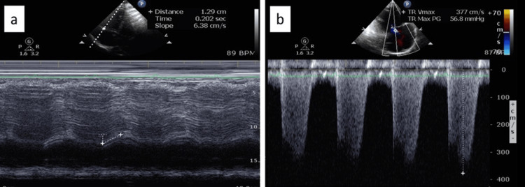
Pulmonary artery systolic pressure is estimated as 4 × (peak tricuspid regurgitant velocity)² plus right atrial pressure in the absence of pulmonic or outflow obstruction. Right atrial pressure is estimated from the inferior vena cava: a diameter of 2.1 cm or less with more than 50% inspiratory collapse indicates about 3 mm Hg, a diameter above 2.1 cm with less than 50% collapse indicates about 15 mm Hg, and other combinations indicate about 8 mm Hg. Because errors in right atrial pressure and in the velocity both propagate, current guidelines grade probability from the velocity itself and from supporting signs, not from the calculated pressure. A velocity error of 10% produces a pressure-gradient error of about 21%.
| Category | Additional echocardiographic signs of pulmonary hypertension |
|---|---|
| A. Ventricles | Right-to-left ventricular basal diameter or area ratio >1.0; septal flattening with left ventricular eccentricity index >1.1 in systole, diastole, or both; TAPSE/PASP ratio <0.55 mm/mm Hg |
| B. Pulmonary artery | Right ventricular outflow tract acceleration time <105 ms, mid-systolic notching, or both; early diastolic pulmonary regurgitant velocity >2.2 m/s; pulmonary artery diameter >25 mm or larger than the aortic root |
| C. Inferior vena cava and right atrium | Inferior vena cava >21 mm with reduced inspiratory collapse; right atrial end-systolic area >18 cm² |
Signs from the 2022 ESC/ERS guideline, reproduced in the 7th WSPH. Signs from at least two categories modify the probability assigned by tricuspid regurgitant velocity. TAPSE, tricuspid annular plane systolic excursion; PASP, pulmonary artery systolic pressure.
The ESC/ERS scheme assigns low probability to a velocity of 2.8 m/s or less (or no measurable jet) without signs from two categories. Intermediate probability is assigned to a velocity of 2.8 m/s or less with signs from two categories, or to 2.9 to 3.4 m/s without them. High probability is assigned to 2.9 to 3.4 m/s with signs from two categories, or to any velocity above 3.4 m/s. The 2025 American Society of Echocardiography (ASE) right heart guideline uses a related rule. A peak velocity of 2.9 m/s or more suggests pulmonary hypertension. A velocity of 2.8 m/s suggests it when at least two supporting signs are present (right ventricular basal diameter greater than left ventricular basal diameter, eccentricity index above 1.1, mid-systolic notching of the outflow tract Doppler signal, end-diastolic pulmonary regurgitant velocity above 2.2 m/s, or an inferior vena cava above 2.1 cm or right atrial area above 18 cm²).
Echocardiographic probability
ESC/ERS 2022 and ASE 2025The pulmonary regurgitant velocity is end-diastolic in the ASE document and early diastolic in the ESC/ERS list, and an enlarged inferior vena cava or right atrium counts once toward the ASE criterion. The example (velocity 3.1 m/s, eccentricity index above 1.1, dilated inferior vena cava with reduced collapse) has signs in two categories and a high ESC/ERS probability.
Right ventricular size and function. A right ventricular basal diameter of 4.1 cm or more is enlarged by ASE 2025 criteria, and right ventricular wall thickness of 0.5 cm or more indicates hypertrophy. Systolic function is reported with at least one quantitative index: TAPSE of 1.7 cm or less, tissue Doppler S′ of 9.5 cm/s or less, fractional area change of 35% or less, three-dimensional ejection fraction of 45% or less, and free-wall longitudinal strain of 20% or less in absolute value are abnormal. TAPSE overestimates function when severe tricuspid regurgitation distorts the annulus. The TAPSE/PASP ratio expresses ventricular-arterial coupling; values below 0.19 mm/mm Hg identify patients with PAH at high risk. A pericardial effusion indicates high right atrial pressure and is an adverse sign.
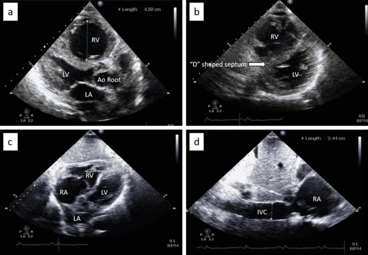
Sources of discordance. A low velocity does not exclude severe disease. In torrential tricuspid regurgitation, right ventricular and right atrial pressures equalize early in systole, and the dense, triangular, early-peaking envelope underestimates the gradient. A failing right ventricle generates less pressure, so a falling estimated pressure with a falling outflow tract velocity-time integral and a lower TAPSE/PASP ratio indicates deterioration, not response. Pulmonic stenosis or outflow obstruction makes the tricuspid gradient report right ventricular pressure, not pulmonary artery pressure. High-flow states raise the gradient without raising resistance; the ratio of tricuspid velocity to outflow tract velocity-time integral helps separate flow from resistance but does not replace catheter PVR.
Pre-capillary or post-capillary. The left heart findings shift the probability toward one phenotype before catheterization. In established pulmonary hypertension, tricuspid velocity reflects pulmonary vascular disease and is not used as a marker of left atrial pressure. Agitated saline contrast identifies intracardiac shunts, and transesophageal echocardiography or cardiac MRI defines sinus venosus defects and anomalous venous return.
| Finding | Favors left heart disease (group 2) | Favors pre-capillary disease |
|---|---|---|
| Left atrium | Enlarged (volume index >34 mL/m²); reduced reservoir strain | Atrial septum bowing toward the left atrium |
| Left ventricle | Hypertrophy or dysfunction | Small and underfilled |
| Mitral inflow and tissue Doppler | High E/A ratio; lateral E/e′ ≥13 | Low E/A ratio |
| Valves | Significant mitral or aortic valve disease | — |
| Rhythm | Atrial fibrillation | — |
| Right ventricle | — | Remodeling out of proportion to the pulmonary pressure |
Echocardiographic findings that favor post-capillary or pre-capillary pulmonary hypertension, after the 2022 ESC/ERS guideline and the 7th WSPH. The findings modify probability; right heart catheterization establishes the phenotype.
Diagnostic pathway and the decision to catheterize
The 7th WSPH proposes a stepwise evaluation whose purpose is to identify patients who need referral to a pulmonary hypertension center and invasive hemodynamics. Patients with a high probability of PAH or CTEPH, especially with signs of right heart failure, rapid progression, syncope, or a low cardiac output, are referred directly at any step.
- Clinical assessment: symptoms, examination, and risk factors (connective tissue disease, portal hypertension, HIV, congenital heart disease, prior venous thromboembolism, left heart disease, lung disease, family history, and drug or stimulant exposure).
- Basic tests: electrocardiogram, chest radiograph, pulse oximetry, blood count, electrolytes, kidney and liver function, and BNP or NT-proBNP.
- Heart and lung assessment: echocardiography, pulmonary function tests with diffusing capacity, and arterial blood gases.
- Further evaluation by suspected cause: ventilation-perfusion scintigraphy, chest CT, cardiac MRI, cardiopulmonary exercise testing, serologies, and abdominal ultrasound.
- Right heart catheterization at a pulmonary hypertension center, with vasoreactivity testing, exercise, or fluid challenge as the phenotype requires.
Ventilation-perfusion scintigraphy. Every patient with newly diagnosed or unexplained pulmonary hypertension is screened for CTEPH, because it is the potentially curable form. A normal perfusion scan essentially excludes CTEPH. In a referral series, planar scintigraphy had a sensitivity above 96% for chronic thromboembolic disease, whereas multidetector CT pulmonary angiography had a sensitivity of 51%. Mismatched segmental defects lead to CT pulmonary angiography and referral to a CTEPH center. Perfusion defects also occur in veno-occlusive disease, pulmonary artery sarcoma, vasculitis, and fibrosing mediastinitis.
Chest CT. High-resolution CT identifies emphysema, interstitial lung disease, and the septal lines, ground-glass nodules, and lymph nodes of veno-occlusive disease; it is recommended particularly in current or former smokers with a diffusing capacity below 45% of predicted. Contrast CT shows an enlarged main pulmonary artery (a ratio of pulmonary artery to ascending aorta diameter above 1 suggests pulmonary hypertension), right ventricular enlargement, mosaic perfusion, bronchial artery collaterals, webs and bands in CTEPH, and anomalous pulmonary venous return. Dual-energy CT perfusion is an alternative to scintigraphy in experienced centers.
Cardiac MRI is the reference standard for right ventricular volumes, mass, and ejection fraction and is used for diagnosis when echocardiography is inconclusive and for risk assessment. MRI also identifies shunts, anomalous veins, and myocardial disease that suggest group 2.
Pulmonary function, gas exchange, and sleep. In PAH, spirometry and lung volumes are normal or mildly reduced, and the diffusing capacity is mildly to moderately reduced. A severely reduced diffusing capacity with near-normal spirometry suggests veno-occlusive disease, systemic sclerosis, or a lung-vascular phenotype associated with smoking. Arterial blood gases show hypocapnia in PAH; hypercapnia suggests hypoventilation. Overnight oximetry or polysomnography is performed when sleep-disordered breathing or hypoventilation is suspected; obstructive sleep apnea alone rarely causes more than mild pulmonary hypertension.
Cardiopulmonary exercise testing. PAH produces a reduced peak oxygen consumption, a high ventilatory equivalent for carbon dioxide, and a low end-tidal carbon dioxide that falls during exercise.
Laboratory tests. Antinuclear antibodies, anticentromere, anti-topoisomerase I (Scl-70), and anti-RNA polymerase III antibodies screen for systemic sclerosis, with other serologies as indicated; HIV and hepatitis serologies, thyroid function, and liver tests are standard. Abdominal ultrasound with Doppler identifies portal hypertension. Testing for antiphospholipid antibodies is part of the CTEPH evaluation.
Who undergoes right heart catheterization. Catheterization is performed when it will change management: when echocardiographic probability is high, or intermediate in a patient with risk factors for PAH or CTEPH, or when symptoms remain unexplained after noninvasive testing. In patients with left heart or lung disease, it is reserved for suspected severe pulmonary hypertension, a suspected second cause, or evaluation for transplantation or valve intervention. Diagnostic catheterization establishes mPAP, wedge pressure, cardiac output, PVR, right atrial pressure, and mixed venous saturation, and includes an oxygen saturation run when a shunt is possible. It is performed at a pulmonary hypertension center because classification depends on precise measurements.
Screening of asymptomatic high-risk groups. Annual screening with echocardiography, electrocardiography, and natriuretic peptide measurement is applied to BMPR2 carriers, first-degree relatives of patients with heritable PAH, patients being evaluated for liver transplantation or a transjugular intrahepatic portosystemic shunt, and patients with systemic sclerosis spectrum disorders. In systemic sclerosis of more than 3 years' duration with a diffusing capacity below 60% of predicted, the two-step DETECT algorithm (serologies, biomarkers, electrocardiogram, and pulmonary function, followed by echocardiography) selects patients for catheterization. Patients with other connective tissue diseases, HIV, or portal hypertension without transplant evaluation undergo testing when symptoms appear.
Acute vasoreactivity testing and calcium channel blockers
Acute vasoreactivity testing identifies the small group of patients whose PAH responds to high-dose calcium channel blockers. It is performed only in the forms in which a positive response predicts long-term benefit. In other forms of PAH and in groups 2 through 5, responses are misleading and calcium channel blockers can cause harm. Empirical calcium channel blockers without a positive test cause hypotension, right ventricular failure, and death.
| Element | Specification |
|---|---|
| Patients tested | Idiopathic, heritable, and drug-associated PAH, at the diagnostic catheterization |
| Agents | Inhaled nitric oxide (20 to 40 ppm), inhaled iloprost, or intravenous epoprostenol |
| Positive acute response | Fall in mPAP of at least 10 mm Hg to an absolute value below 40 mm Hg (7th WSPH; 40 mm Hg or less in the 2022 ESC/ERS guideline), with cardiac output unchanged or increased |
| Frequency of acute response | About 12% in idiopathic or drug-associated PAH; fewer than 5% in heritable PAH |
| Long-term response | Functional class I or II with sustained hemodynamic improvement after at least 1 year on calcium channel blockers alone |
| Reassessment on treatment | At 3 to 4 months: functional class, walk distance, natriuretic peptide, imaging, and repeat catheterization. Goals are a low-risk profile and near-normal hemodynamics; patients who do not reach them start PAH therapy |
Acute vasoreactivity testing and definitions of response, after the 2022 ESC/ERS guideline and the 7th WSPH (2024). mPAP, mean pulmonary artery pressure.
In the largest idiopathic series, 70 of 557 patients (12.6%) were acute responders, and only 38 (6.8%) remained in functional class I or II with sustained hemodynamic improvement after at least 1 year on calcium channel blockers alone. Long-term responders have excellent survival; acute responders who do not meet the long-term criteria are treated with the standard PAH algorithm.
| Drug | Target dose | Preferred setting |
|---|---|---|
| Nifedipine | 20–60 mg two or three times daily | Relative bradycardia |
| Diltiazem | 120–360 mg twice daily | Relative tachycardia |
| Amlodipine | 15–30 mg once daily | Relative bradycardia |
| Felodipine | 15–30 mg once daily | — |
| Verapamil | Not used | Avoided because of its negative inotropic effect |
Calcium channel blockers for vasoreactive PAH, with target doses from the 2022 ESC/ERS guideline and the choice of drug by baseline heart rate from the 2015 ESC/ERS guideline. Doses are started low and titrated to the target.
Risk assessment in pulmonary arterial hypertension
Treatment in PAH is driven by estimated 1-year mortality. At diagnosis, the 2022 ESC/ERS guideline uses a three-strata model that integrates clinical, exercise, biomarker, imaging, and hemodynamic variables. For initial treatment, the 7th WSPH separates patients only into high risk and not high risk.
| Variable | Low risk (<5%) | Intermediate risk (5% to 20%) | High risk (>20%) |
|---|---|---|---|
| Signs of right heart failure | Absent | Absent | Present |
| Symptom progression | None | Slow | Rapid |
| Syncope | None | Occasional | Repeated |
| WHO functional class | I, II | III | IV |
| 6-minute walk distance | >440 m | 165–440 m | <165 m |
| BNP or NT-proBNP | BNP <50 ng/L; NT-proBNP <300 ng/L | BNP 50–800; NT-proBNP 300–1,100 ng/L | BNP >800; NT-proBNP >1,100 ng/L |
| Cardiopulmonary exercise test | Peak VO₂ >15 mL/min/kg; VE/VCO₂ slope <36 | Peak VO₂ 11–15; VE/VCO₂ 36–44 | Peak VO₂ <11; VE/VCO₂ >44 |
| Echocardiography | RA area <18 cm²; TAPSE/PASP >0.32 mm/mm Hg; no effusion | RA area 18–26 cm²; TAPSE/PASP 0.19–0.32; minimal effusion | RA area >26 cm²; TAPSE/PASP <0.19; moderate or large effusion |
| Cardiac MRI | RVEF >54%; RVESVI <42 mL/m² | RVEF 37%–54%; RVESVI 42–54 mL/m² | RVEF <37%; RVESVI >54 mL/m² |
| Hemodynamics | RAP <8 mm Hg; CI ≥2.5 L/min/m²; SVI >38 mL/m²; SvO₂ >65% | RAP 8–14; CI 2.0–2.4; SVI 31–38; SvO₂ 60%–65% | RAP >14; CI <2.0; SVI <31; SvO₂ <60% |
Three-strata risk assessment at diagnosis, after the 2022 ESC/ERS guideline. Percentages are estimated 1-year mortality. At diagnosis, the stratum is assigned from as many variables as possible, with emphasis on disease type, functional class, walk distance, natriuretic peptide, and hemodynamics. RA, right atrium; RAP, right atrial pressure; CI, cardiac index; SVI, stroke volume index; SvO₂, mixed venous oxygen saturation; RVEF, right ventricular ejection fraction; RVESVI, right ventricular end-systolic volume index; WHO, World Health Organization; VO₂, oxygen consumption; VE/VCO₂, ventilatory equivalent for carbon dioxide.
At follow-up, most patients fall in the intermediate stratum of the three-strata model, which therefore separates them poorly. The four-strata model, derived from the COMPERA registry and adopted by the 2022 ESC/ERS guideline and the 7th WSPH, uses functional class, 6-minute walk distance, and BNP or NT-proBNP with refined cutoffs. It splits the intermediate group into intermediate-low and intermediate-high. The US alternative is REVEAL Lite 2, which scores functional class, systolic blood pressure, heart rate, walk distance, natriuretic peptide, and kidney function. A score of 5 or less indicates low risk, and a score of 9 or more defined the high-risk population of the ZENITH trial. Both perform similarly. Neither is validated outside group 1, and in older patients with comorbidities the variables reflect more than PAH.
Four-strata PAH risk at follow-up
ESC/ERS 2022, COMPERA 2.0Grades: functional class I or II, 1; III, 3; IV, 4. Walk distance >440 m, 1; 320–440 m, 2; 165–319 m, 3; <165 m, 4. NT-proBNP <300 ng/L, 1; 300–649, 2; 650–1,100, 3; >1,100, 4 (BNP <50, 50–199, 200–800, >800 ng/L). The mean of available grades is rounded to the nearest integer: 1, low; 2, intermediate-low; 3, intermediate-high; 4, high. The example (class III, 380 m, NT-proBNP 900 ng/L) grades 3, 2, and 3, a mean of 2.67, intermediate-high risk.
Low risk is the treatment goal, yet in a pooled analysis of AMBITION, GRIPHON, and SERAPHIN more than 20% of patients classified as low risk had a clinical worsening event within 3 years. Additional goals proposed by the 7th WSPH include a PVR below 5 WU, a fall in PVR of more than 50%, a normal stroke volume index, and normal or near-normal right ventricular size and function on imaging. Risk is reassessed 3 to 4 months after each change in therapy and every 3 to 6 months in stable patients.
Supportive care in pulmonary arterial hypertension
- Exercise training. Supervised exercise training improves walk distance, peak oxygen consumption, and quality of life in stable patients on medical therapy. Strenuous isometric exertion and exercise to the point of presyncope are avoided.
- Diuretics and fluid balance. Loop diuretics treat congestion, often with a mineralocorticoid receptor antagonist. Sodium and fluid restriction support them. Overdiuresis lowers preload and cardiac output in a preload-dependent right ventricle.
- Oxygen. Long-term oxygen is prescribed when arterial oxygen tension is consistently below 60 mm Hg (8 kPa). In-flight oxygen is advised for patients on oxygen or with a resting arterial oxygen tension below 60 mm Hg at sea level.
- Iron. Iron deficiency is common, impairs exercise capacity, and predicts worse outcome. Iron-deficiency anemia is corrected, preferably with intravenous iron because oral absorption is impaired.
- Vaccination. Immunization against influenza, Streptococcus pneumoniae, and SARS-CoV-2 is recommended, and a single dose of respiratory syncytial virus vaccine is recommended for adults with pulmonary hypertension aged 50 years or older.
- Anticoagulation. Randomized data are lacking, and observational data conflict. Anticoagulation is not generally recommended in idiopathic, heritable, or drug-associated PAH and is not recommended in connective tissue disease-associated PAH, where registry data suggest no benefit and possible harm. It is used when another indication exists, such as atrial fibrillation or a central venous catheter for prostacyclin infusion in selected patients.
- Contraception and pregnancy. Pregnancy is advised against, and clear contraceptive advice is given at diagnosis (see the section on pregnancy).
- Psychosocial support and transplant preparation. Depression and anxiety are common. Weight optimization, tobacco and drug abstinence, and treatment of comorbidities keep transplantation available.
- Arrhythmias. Atrial flutter and fibrillation are treated with early restoration of sinus rhythm; flutter ablation is highly effective. Beta-blockers and nondihydropyridine calcium channel blockers are used cautiously because they lower cardiac output.
PAH drug classes: endothelin, nitric oxide, and prostacyclin pathways
| Class and drug | Adult dosing | Main adverse effects and cautions | Pivotal trial |
|---|---|---|---|
| Endothelin receptor antagonists | All are teratogenic; pregnancy is excluded before treatment and highly reliable contraception is required. Edema, anemia, and nasal congestion are class effects; hemoglobin is monitored. | ||
| Ambrisentan | 5–10 mg once daily | Peripheral edema; contraindicated in idiopathic pulmonary fibrosis | ARIES-1 and -2 |
| Bosentan | 62.5 mg twice daily for 4 weeks, then 125 mg twice daily | Dose-related aminotransferase elevation (monthly liver tests); induces CYP3A4 and CYP2C9, lowering sildenafil and hormonal contraceptive levels | BREATHE-1 |
| Macitentan | 10 mg once daily | Anemia; fixed-dose combination with tadalafil available | SERAPHIN |
| PDE5 inhibitors | Headache, flushing, dyspepsia, epistaxis, rare visual or hearing loss. Contraindicated with nitrates and with riociguat. | ||
| Sildenafil | 20 mg three times daily (approved dose; higher doses are used off label) | Higher doses increase adverse effects | SUPER |
| Tadalafil | 40 mg once daily | Preferred partner for ambrisentan or macitentan | PHIRST |
| Soluble guanylate cyclase stimulator: riociguat | 1 mg three times daily, titrated every 2 weeks to 2.5 mg three times daily | Hypotension, dizziness, dyspepsia; teratogenic; contraindicated with PDE5 inhibitors and nitrates; approved for PAH and inoperable or persistent CTEPH | PATENT-1, CHEST-1 |
| Prostacyclin receptor agonist: selexipag | 200 µg twice daily, titrated weekly to the highest tolerated dose (maximum 1,600 µg twice daily); intravenous form available | Headache, diarrhea, jaw pain, nausea, myalgia, flushing | GRIPHON |
| Oral treprostinil | Titrated three times daily | Prostanoid effects | FREEDOM-EV |
| Inhaled treprostinil or iloprost | Treprostinil 4 times daily; iloprost 6–9 times daily | Cough, throat irritation, prostanoid effects; treprostinil also approved for pulmonary hypertension with interstitial lung disease | TRIUMPH, AIR, INCREASE |
| Subcutaneous or intravenous treprostinil | Continuous infusion, titrated | Infusion-site pain (subcutaneous); line infection and thrombosis (intravenous) | Pivotal subcutaneous trial |
| Intravenous epoprostenol | Continuous infusion via central line, titrated from 1–2 ng/kg/min | Half-life of minutes; interruption causes rebound pulmonary hypertension and can be fatal; catheter sepsis, thrombosis, high-output state with excessive doses | Only PAH drug with a survival benefit in a randomized trial (1996) |
| Activin signaling inhibitor: sotatercept | 0.3 mg/kg subcutaneously, then 0.7 mg/kg every 3 weeks | Raised hemoglobin, thrombocytopenia, epistaxis and other bleeding, telangiectasia, hypertension; hemoglobin and platelets checked before each of the first 5 doses; avoided in pregnancy; possible reduction in fertility | STELLAR, ZENITH, HYPERION |
Drugs approved for PAH in the United States, after the 7th WSPH treatment algorithm (Table 2). Doses are the usual adult regimens from US labeling; individual titration follows tolerance and blood pressure. In April 2025, the US Food and Drug Administration removed the pregnancy-related Risk Evaluation and Mitigation Strategy (REMS) requirements for ambrisentan and macitentan; bosentan remains under a REMS for hepatotoxicity, and all endothelin receptor antagonists keep a boxed warning for embryo-fetal toxicity.
Endothelin receptor antagonists. SERAPHIN was the first event-driven PAH trial. Bosentan was the first oral PAH drug and is now used less because of hepatotoxicity and drug interactions. Ambrisentan is contraindicated in idiopathic pulmonary fibrosis because it increased disease progression in ARTEMIS-IPF.
Nitric oxide pathway. Sildenafil and tadalafil improve exercise capacity and hemodynamics. Riociguat stimulates soluble guanylate cyclase directly and does not require nitric oxide for its effect. Riociguat and PDE5 inhibitors are never combined, because the combination caused hypotension without added benefit. In REPLACE, patients at intermediate risk on a PDE5 inhibitor improved more often after switching to riociguat than after continuing the PDE5 inhibitor.
Prostacyclin pathway. Intravenous epoprostenol improved survival in a 12-week randomized trial in severe idiopathic PAH and remains the most potent therapy. Patients carry a backup pump, and a lost line is an emergency. Subcutaneous treprostinil avoids a central line at the cost of site pain.
| Trial | Treatment | Population | Primary end point and result |
|---|---|---|---|
| ARIES-1 and -2 (2008) | Ambrisentan versus placebo | 202 and 192 patients | 6-minute walk distance at 12 weeks, placebo-corrected: +31 m and +51 m with 5 and 10 mg (ARIES-1); +32 m and +59 m with 2.5 and 5 mg (ARIES-2) |
| PATENT-1 (2013) | Riociguat up to 2.5 mg three times daily versus placebo | 443 patients, treatment-naive or on an endothelin receptor antagonist or a nonintravenous prostanoid | 6-minute walk distance at 12 weeks: +36 m versus placebo. PVR, NT-proBNP, functional class, and time to clinical worsening also improved |
| SERAPHIN (2013) | Macitentan 3 or 10 mg versus placebo | 742 patients; stable oral or inhaled background therapy other than an endothelin receptor antagonist allowed | Death, atrial septostomy, lung transplantation, start of intravenous or subcutaneous prostanoid, or worsening PAH: 31.4% with 10 mg versus 46.4% (hazard ratio 0.55) over a median of 2 years; worsening PAH was the most frequent event |
| AMBITION (2015) | Ambrisentan plus tadalafil versus either drug alone | 500 treatment-naive patients in functional class II or III | Clinical failure: 18% versus 31% with pooled monotherapy (hazard ratio 0.50), driven by fewer hospitalizations |
| GRIPHON (2015) | Selexipag up to 1,600 µg twice daily versus placebo | 1,156 patients, most on an endothelin receptor antagonist, a PDE5 inhibitor, or both | Death or PAH complication: 27.0% versus 41.6% (hazard ratio 0.60), mainly from fewer disease progression and hospitalization events; mortality not different |
| FREEDOM-EV | Oral treprostinil versus placebo | Patients on oral monotherapy | Clinical worsening: hazard ratio 0.74 |
| TRITON (2021) | Initial macitentan, tadalafil, and selexipag versus macitentan, tadalafil, and placebo | 247 newly diagnosed, treatment-naive patients | PVR at 26 weeks: fell 54% versus 52% (ratio of geometric means 0.96); no significant difference |
Pivotal randomized trials of oral PAH therapy. Rates are active treatment versus comparator. PVR, pulmonary vascular resistance; NT-proBNP, N-terminal pro-B-type natriuretic peptide; PDE5, phosphodiesterase type 5.
Sotatercept and the activin pathway
Sotatercept is a fusion protein of the extracellular domain of the activin receptor type IIA and the Fc portion of human immunoglobulin G1. By sequestering activins and growth differentiation factors, it restores the balance between proliferative and antiproliferative signaling that loss of BMPR2 signaling disturbs. Unlike every earlier PAH drug, it has little acute vasodilator effect, and its hemodynamic benefit comes from reversal of remodeling. In the phase 2 PULSAR trial, sotatercept 0.7 mg/kg lowered PVR by about 240 dyn·s·cm⁻⁵ relative to placebo. The US Food and Drug Administration approved it in 2024 for adults with PAH.
| Trial | Population | Primary end point and result |
|---|---|---|
| STELLAR (2023) | 323 adults in functional class II or III on stable background therapy (61% on triple therapy) | Change in 6-minute walk distance at 24 weeks: +40.8 m versus placebo (Hodges-Lehmann estimate). PVR, NT-proBNP, functional class, and time to death or clinical worsening also improved |
| ZENITH (2025) | 172 adults in functional class III or IV at high risk (REVEAL Lite 2 score ≥9) on maximal tolerated therapy | Death, lung transplantation, or PAH hospitalization of at least 24 hours: 17.4% versus 54.7% (hazard ratio 0.24); stopped early for efficacy |
| HYPERION (2025) | 320 adults diagnosed less than 1 year earlier, functional class II or III, intermediate or high risk, on double or triple therapy | Clinical worsening (death, unplanned PAH hospitalization, atrial septostomy, lung transplantation, or worsening exercise performance): 10.6% versus 36.9% (hazard ratio 0.24); driven mainly by fewer declines in exercise performance; all-cause death similar |
Phase 3 trials of add-on sotatercept in PAH. Rates are sotatercept versus placebo.
Hemoglobin and platelet count are measured before each of the first 5 doses and periodically after; a significant rise in hemoglobin or fall in platelets leads to dose delay or reduction, and treatment is not started with a platelet count below 50,000/µL. Telangiectasia developed in about 10% of patients in STELLAR, and epistaxis and gingival bleeding are frequent, so bleeding risk and concurrent anticoagulation or prostacyclin therapy are reviewed before treatment. The drug is self-administered subcutaneously every 3 weeks after training. Its place in therapy is as add-on treatment to established oral or parenteral combination therapy; it has not been tested as initial therapy or in groups 2 through 5.
Initial combination therapy, escalation, and treatment goals
Therapy for confirmed group 1 PAH is directed by risk, delivered in or with a pulmonary hypertension center, and escalated until the goals are met. Vasoreactive patients are treated with calcium channel blockers as above. The 7th WSPH algorithm applies to patients with idiopathic, heritable, drug-associated, and connective tissue disease-associated PAH without major cardiopulmonary comorbidity.
| Setting | Recommended approach (7th WSPH, 2024) |
|---|---|
| At diagnosis, not high risk | Initial dual oral therapy with an endothelin receptor antagonist and a PDE5 inhibitor, preferably macitentan or ambrisentan with tadalafil; same-day start or sequential start over 1 to 2 weeks |
| At diagnosis, high risk | Initial triple therapy including a parenteral prostacyclin (intravenous epoprostenol or subcutaneous or intravenous treprostinil) with an endothelin receptor antagonist and a PDE5 inhibitor; discussion of lung transplant referral |
| Significant cardiopulmonary comorbidity | Initial monotherapy, usually with a PDE5 inhibitor, with individualized escalation |
| Follow-up at 3 to 4 months: low risk | Continue therapy; individualized addition when symptoms, imaging, or hemodynamics remain abnormal |
| Follow-up: intermediate-low risk | Add sotatercept, an oral prostacyclin pathway agent (selexipag or oral treprostinil), or an inhaled prostacyclin, or switch from the PDE5 inhibitor to riociguat |
| Follow-up: intermediate-high risk | Add a parenteral prostacyclin or sotatercept; refer for transplant evaluation |
| Follow-up: high risk | Add a parenteral prostacyclin if not already used, with sotatercept as a fourth drug; refer for transplant evaluation |
Treatment algorithm for PAH, after Chin et al. (7th WSPH). Maximal medical therapy is four drugs acting on four pathways. Clinical deterioration leads to escalation without waiting for the scheduled reassessment.
Evidence for upfront combination. Initial dual oral therapy rests on AMBITION, in which combination therapy halved the risk of clinical failure compared with monotherapy. Initial triple oral therapy with selexipag did not lower PVR further in TRITON and is not recommended. Observational data support initial triple therapy with a parenteral prostacyclin in high-risk patients, with better survival than less intensive regimens.
Patients with comorbidities. Patients with PAH and three or more risk factors for left heart disease (obesity, hypertension, diabetes, coronary disease) or with mild lung disease tolerate combination therapy less well and respond less. The 2022 ESC/ERS guideline recommends initial monotherapy in these patients, with a decision about escalation made individually. In the US, the CHEST guideline's recommendation of initial ambrisentan plus tadalafil for treatment-naive patients in functional class II or III is consistent with the WSPH algorithm.
Mild disease. No therapy is approved for an mPAP of 21 to 24 mm Hg or a PVR of 2 to 3 WU, and these patients are followed closely or enrolled in trials.
Lung transplantation, atrial septostomy, and palliative care
Referral for lung transplant evaluation is made when disease progresses or leads to hospitalization despite therapy, when parenteral prostacyclin is needed, when veno-occlusive disease is known or suspected, or when liver or kidney dysfunction develops from right heart failure. Evaluation is considered at diagnosis in high-risk patients and recommended at follow-up when intermediate-high or high risk persists. Bilateral lung transplantation is the usual operation; the right ventricle recovers after the afterload is removed. Heart-lung transplantation is reserved for complex congenital disease or irreversible left heart disease. Patients with PAH have higher waiting-list mortality than those with parenchymal lung disease, so referral is early. Venoarterial extracorporeal membrane oxygenation, often in an ambulatory configuration, bridges selected patients to transplantation.
Balloon atrial septostomy creates a right-to-left shunt that decompresses the right ventricle and raises left ventricular filling and systemic output at the cost of oxygen saturation. It is used as palliation or as a bridge to transplantation, most often where drug therapy is unavailable, and is avoided when right atrial pressure is very high or baseline saturation is low because of high procedural mortality. Palliative care, with attention to dyspnea, edema, anxiety, and goals of care, is integrated early in advanced disease.
Pulmonary hypertension from left heart disease (group 2)
Left heart disease is the most common cause of pulmonary hypertension. It is common in heart failure with reduced or preserved ejection fraction and in severe mitral disease, and in a community series 53% of patients with aortic stenosis had at least moderately elevated pulmonary pressure. Raised left atrial pressure is transmitted backward (isolated post-capillary disease). With time and in susceptible patients, venous and arterial remodeling add a pre-capillary component (combined disease), which carries a worse prognosis and a higher risk of right ventricular failure. The 7th WSPH proposed a staging of pulmonary hypertension in left heart disease from A (at risk) through D (advanced), parallel to the heart failure stages.
The central diagnostic task is separation from PAH in older patients with cardiometabolic risk factors. A wedge pressure of 12 to 18 mm Hg in a patient with obesity, hypertension, diabetes, atrial fibrillation, left atrial enlargement, or abnormal diastolic indices is resolved with fluid or exercise challenge and the full clinical picture before a group 1 label is assigned; scores such as H2FPEF estimate the probability of HFpEF. The HFpEF and diastolic function page covers this evaluation.
Treatment. The left heart disease is treated: guideline-directed therapy for heart failure with reduced ejection fraction, sodium-glucose cotransporter 2 inhibitors, diuretics, blood pressure and rhythm control, weight loss in obesity, and timely valve intervention. Pulmonary vasodilators are not recommended. In SIOVAC, sildenafil given for persistent pulmonary hypertension after successful valve surgery worsened clinical outcomes. Macitentan in MELODY-1 caused fluid retention without hemodynamic benefit, and trials of endothelin antagonists, prostacyclin, and riociguat in heart failure have been neutral or harmful. Increasing pulmonary flow into a left heart that cannot accept it raises wedge pressure and can cause pulmonary edema. Patients with combined disease and a severe pre-capillary component (PVR above 5 WU) are referred to a pulmonary hypertension center, and trial enrollment is preferred.
Valve disease. In mitral stenosis, pulmonary pressure falls after balloon commissurotomy or surgery, although a severe pre-capillary component may regress only partly. Pulmonary hypertension predicts higher risk after aortic and mitral valve procedures but does not by itself exclude them. In heart transplant candidates, a high PVR that does not fall with vasodilator challenge or mechanical unloading raises the risk of donor right ventricular failure.
Pulmonary hypertension from lung disease and hypoxia (group 3)
Pulmonary hypertension complicates COPD, interstitial lung disease, combined pulmonary fibrosis and emphysema, sarcoidosis, and hypoventilation disorders. Hypoxic vasoconstriction, loss of the capillary bed, inflammation, and vascular remodeling contribute. Most patients have mild or moderate disease with a PVR below 5 WU, in whom pulmonary hypertension marks advanced lung disease; a minority have severe disease (PVR above 5 WU) that limits exercise more than the lung disease explains. A reduced diffusing capacity, desaturation with exercise, and a cardiovascular pattern of exercise limitation (preserved breathing reserve) point to a dominant vascular component. In smokers with a severely reduced diffusing capacity and mild emphysema or fibrosis on CT, the boundary between group 1 and group 3 is uncertain, and these patients are classified at a pulmonary hypertension center.
Treatment. The lung disease is optimized, hypoxemia is corrected with long-term oxygen, sleep-disordered breathing and hypoventilation are treated, pulmonary rehabilitation is offered, and eligible patients are referred for lung transplantation.
- Interstitial lung disease. Inhaled treprostinil is the one drug with randomized benefit (INCREASE) and is approved in the United States for this indication. A PDE5 inhibitor may be considered in severe pulmonary hypertension with interstitial lung disease in an expert center. Ambrisentan and riociguat are contraindicated in fibrotic lung disease.
- COPD. No PAH drug has shown benefit. Pulmonary vasodilators can worsen ventilation-perfusion matching and hypoxemia. Patients with severe pulmonary hypertension and a vascular phenotype are managed individually in expert centers.
| Trial | Population | Treatment | Result |
|---|---|---|---|
| SIOVAC (2018), group 2 | 200 patients with mPAP ≥30 mm Hg at least 1 year after successful valve replacement or repair | Sildenafil 40 mg three times daily versus placebo for 6 months | Harm: fewer patients improved and more worsened on the composite clinical score (odds ratio 0.39); survival free of heart failure admission 76% versus 86% (hazard ratio 2.0) |
| MELODY-1 (2018), group 2 | 63 patients with combined post- and pre-capillary pulmonary hypertension from left heart disease (diastolic pulmonary gradient ≥7 mm Hg, PVR >3 WU) | Macitentan 10 mg versus placebo for 12 weeks | No benefit: fluid retention or worsening functional class in 7 versus 4 patients (not significant), with the excess fluid retention apparent in the first month; no change in PVR or wedge pressure |
| INCREASE (2021), group 3 | 326 patients with interstitial lung disease and catheter-confirmed pulmonary hypertension (mPAP ≥25 mm Hg, PVR >3 WU) | Inhaled treprostinil up to 72 µg four times daily versus placebo for 16 weeks | Benefit: 6-minute walk distance +31 m; NT-proBNP −15% versus +46%; clinical worsening 22.7% versus 33.1% (hazard ratio 0.61) |
| ARTEMIS-IPF (2013), group 3 | 492 patients with idiopathic pulmonary fibrosis, 10% with pulmonary hypertension at baseline | Ambrisentan 10 mg daily versus placebo | Harm; stopped early: disease progression 27.4% versus 17.2% (hazard ratio 1.74); respiratory hospitalization 13.4% versus 5.5% |
| RISE-IIP (2019), group 3 | 147 patients with pulmonary hypertension and idiopathic interstitial pneumonia | Riociguat versus placebo | Harm; stopped early: serious adverse events 37% versus 23%, 8 versus 3 deaths, and no efficacy signal |
| PERFECT (2024), group 3 | 76 patients with COPD, mPAP ≥30 mm Hg, and PVR ≥4 WU | Inhaled treprostinil versus placebo | Harm; stopped early: excess serious adverse events and a signal of increased mortality; walk distance numerically worse |
Randomized trials of pulmonary vasodilators in pulmonary hypertension from left heart disease and lung disease. Rates are active treatment versus placebo. mPAP, mean pulmonary artery pressure; PVR, pulmonary vascular resistance; WU, Wood units; NT-proBNP, N-terminal pro-B-type natriuretic peptide.
High altitude. Chronic hypoxia at high altitude causes pulmonary vasoconstriction and remodeling. Chronic mountain sickness combines excessive erythrocytosis, hypoxemia, and pulmonary hypertension and improves with descent. In patients with established pulmonary hypertension, travel to altitude and air travel without supplemental oxygen can worsen hypoxemia and right ventricular load.
Chronic thromboembolic pulmonary hypertension
CTEPH is the main group 4 condition. It results from failure of thrombus to resolve after pulmonary embolism, with organized fibrotic obstruction of proximal and segmental arteries and a secondary small-vessel arteriopathy in unobstructed territories. Chronic thromboembolic pulmonary disease (CTEPD) is the umbrella term for symptomatic patients with persistent mismatched perfusion defects and chronic organized obstruction after at least 3 months of anticoagulation, with or without pulmonary hypertension at rest. CTEPH is CTEPD with pulmonary hypertension by the current hemodynamic definition. A quarter or more of patients have no recognized prior embolism. Risk factors include unprovoked or recurrent embolism, large perfusion defects, antiphospholipid syndrome (about 10% of patients), splenectomy, ventriculoatrial shunts, infected intravenous lines, inflammatory bowel disease, and cancer. Persistent dyspnea or exercise limitation 3 to 6 months after acute embolism leads to echocardiography and, when the probability of pulmonary hypertension is intermediate or high, ventilation-perfusion scintigraphy. Management of acute embolism is covered on the pulmonary embolism page.
Diagnosis requires mismatched perfusion defects on scintigraphy or CT perfusion, characteristic findings on CT pulmonary angiography or invasive pulmonary angiography (webs, bands, pouch defects, abrupt tapering, and chronic total occlusions), and catheter-confirmed pulmonary hypertension after at least 3 months of therapeutic anticoagulation. Every patient is reviewed by a multidisciplinary CTEPH team with a pulmonary endarterectomy surgeon, a balloon angioplasty interventionalist, a pulmonary hypertension specialist, and a radiologist.
Anticoagulation. Lifelong therapeutic anticoagulation is recommended. Vitamin K antagonists are the established standard and are required in antiphospholipid syndrome; direct oral anticoagulants are widely used in patients without it, although observational data suggest a higher recurrence rate in some series.
Pulmonary endarterectomy. Surgery is the treatment of choice for operable disease. The operation removes a cast of the organized material to the subsegmental level. Operability depends on lesion accessibility, the match between the obstruction and the PVR, comorbidity, and above all surgical experience; disease judged inoperable at one center can be operable at another.
Balloon pulmonary angioplasty and medical therapy. Inoperable disease and persistent or recurrent pulmonary hypertension after endarterectomy are treated with staged angioplasty, drugs, or both. Riociguat is the only drug approved for CTEPH in the United States. Macitentan lowered PVR in MERIT-1, and subcutaneous treprostinil improved walk distance in CTREPH. Many centers give medical therapy before angioplasty, and many patients receive surgery, angioplasty, and drugs in sequence. Five-year survival in inoperable patients treated with angioplasty and medical therapy now approaches 90%.
| Feature | Pulmonary endarterectomy | Balloon pulmonary angioplasty | Riociguat |
|---|---|---|---|
| Indication | Operable disease; treatment of choice | Inoperable disease; persistent or recurrent pulmonary hypertension after endarterectomy | Inoperable disease; persistent or recurrent pulmonary hypertension after endarterectomy; pretreatment before angioplasty |
| Method | Cardiopulmonary bypass with periods of deep hypothermic circulatory arrest; dissection in a plane within the media | Staged dilation of distal webs and stenoses over several sessions | 1 mg three times daily, titrated to 2.5 mg three times daily |
| Results | Operative mortality about 2% at expert centers; pulmonary pressure normalizes in about 70% to 75%; survival >90% at 3 years and >70% at 10 years | Lower mPAP and PVR and better functional class at experienced centers. In RACE, PVR at 26 weeks fell to 40% of baseline versus 67% with riociguat (ratio of geometric means 0.60) | In CHEST-1 (261 patients), 6-minute walk distance +46 m and PVR −246 dyn·s·cm⁻⁵ versus placebo at 16 weeks |
| Main complications | Reperfusion pulmonary edema; residual pulmonary hypertension (mPAP ≥38 mm Hg with PVR ≥5 WU at 3 to 6 months predicts worse survival) | Lung injury from wire perforation or reperfusion in about 10% of sessions in earlier series and 7.7% in recent ones, more often when mPAP is ≥40 mm Hg; fewer complications when riociguat precedes angioplasty | Hypotension, dizziness, dyspepsia; teratogenic |
Treatment options in CTEPH, after the 7th WSPH (Kim et al., 2024), CHEST-1 (2013), and RACE (2022). mPAP, mean pulmonary artery pressure; PVR, pulmonary vascular resistance; WU, Wood units.
Pulmonary hypertension with unclear or multifactorial mechanisms (group 5)
Group 5 gathers conditions in which several mechanisms combine. In sarcoidosis, pulmonary hypertension arises from fibrosis, granulomatous vasculitis, extrinsic compression of pulmonary arteries or veins by nodes, and left ventricular involvement; it carries a poor prognosis, and lung transplantation outcomes are similar to those for other indications. In chronic hemolytic anemias, including sickle cell disease, pulmonary hypertension can be pre-capillary or post-capillary and is associated with high mortality; PDE5 inhibitors increased painful crises in sickle cell disease. Myeloproliferative neoplasms cause pulmonary hypertension through thrombosis, extramedullary hematopoiesis, and drug effects. In chronic kidney failure, arteriovenous fistula flow, volume overload, and left heart disease contribute. Fibrosing mediastinitis, usually from histoplasmosis, compresses central pulmonary arteries and veins and is treated with stenting in selected patients. Pulmonary tumor thrombotic microangiopathy, most often from gastric or breast adenocarcinoma, causes rapidly progressive pulmonary hypertension and hypoxemia. Treatment in every group 5 condition is directed at the underlying disorder; PAH drugs are used only in selected patients at expert centers.
Pregnancy and contraception in pulmonary hypertension
Pregnancy imposes a 30% to 50% rise in cardiac output and plasma volume, a fall in systemic vascular resistance, a hypercoagulable state, and abrupt shifts in preload at delivery. A right ventricle facing a fixed high resistance cannot accommodate these changes. In a systematic review of cohorts from 2008 to 2018, maternal mortality in PAH was 12% overall and 20% in idiopathic PAH, down from 30% to 56% in older series but still prohibitive. Most deaths occur late in pregnancy, at delivery, or in the first weeks after delivery, from right ventricular failure, pulmonary hypertensive crisis, arrhythmia, or thromboembolism. Pulmonary hypertension of any cause with significant elevation of PVR carries a similar risk, and pulmonary arterial hypertension is in the highest category (modified World Health Organization class IV) of maternal cardiovascular risk.
Contraception. Pregnancy is advised against at diagnosis, and contraception is discussed with every woman of childbearing potential. Endothelin receptor antagonists and riociguat are teratogenic, and sotatercept is avoided in pregnancy; pregnancy is excluded before treatment, highly reliable contraception is used throughout, and pregnancy-testing requirements follow each drug's current labeling. Women considering pregnancy receive genetic counseling when heritable disease is present.
| Method | Use in pulmonary hypertension |
|---|---|
| Intrauterine device (levonorgestrel or copper) | Among the most effective options. Insertion can provoke a vasovagal reaction, which is poorly tolerated, and is performed with monitoring in severe disease |
| Progestin implant | Among the most effective options |
| Surgical sterilization | Among the most effective options |
| Estrogen-containing contraceptives | Raise thrombotic risk |
| Any hormonal method with bosentan | Efficacy reduced by bosentan, so a second method is used |
Contraception in women with pulmonary hypertension, after the 2022 ESC/ERS guideline.
Pregnancy in a woman with PAH. A woman who becomes pregnant is informed of the risk and offered termination, which is safest early. A woman who continues pregnancy is managed in a center with pulmonary hypertension, obstetric, anesthetic, and intensive care expertise. Teratogenic drugs are stopped, and PDE5 inhibitors and inhaled or parenteral prostacyclins are continued or started; women who are untreated often begin parenteral prostacyclin. Delivery is planned, usually by cesarean section with carefully titrated neuraxial anesthesia, and general anesthesia is avoided when possible. Monitoring continues for several weeks after delivery, the period of highest risk. Prophylactic anticoagulation is often used in the peripartum period.
Noncardiac surgery and anesthesia in pulmonary hypertension
Pulmonary hypertension increases perioperative mortality, heart failure, arrhythmia, and respiratory failure after noncardiac surgery. Risk rises with severity of pulmonary hypertension, right ventricular dysfunction, right atrial pressure, low walk distance, functional class III or IV, emergency surgery, and high-risk procedures. The 2023 AHA scientific statement on pulmonary hypertension in noncardiac surgery recommends preoperative assessment of the pulmonary hypertension group, severity, and right ventricular function; optimization of therapy before elective surgery; and surgery at a center with pulmonary hypertension expertise for patients with PAH or CTEPH.
PAH therapy is continued without interruption; parenteral prostacyclin is never stopped, and oral PDE5 inhibitors can be replaced by intravenous sildenafil when patients cannot take oral drugs. Anesthetic management maintains systemic vascular resistance and right ventricular perfusion pressure, avoids hypoxemia, hypercapnia, acidosis, hypothermia, and high airway pressures, all of which raise PVR, and avoids abrupt falls in preload. Induction of general anesthesia and the start of positive-pressure ventilation are the moments of highest risk, and vasopressors and inhaled pulmonary vasodilators are prepared before induction. Laparoscopic insufflation raises PVR through hypercapnia and impaired venous return. Monitoring continues after surgery, when fluid shifts and pain raise the risk of decompensation.
Acute right ventricular failure in pulmonary hypertension
Acute decompensated right heart failure in a patient with pulmonary hypertension carries high in-hospital mortality and is managed in an intensive care unit with pulmonary hypertension expertise. The usual triggers are infection, arrhythmia (especially atrial flutter or fibrillation), interruption of PAH therapy, pulmonary embolism, anemia, pregnancy, surgery, and nonadherence to diuretics or sodium restriction.
Management addresses preload, systemic pressure, contractility, and right ventricular afterload together.
| Target | Management | Cautions |
|---|---|---|
| Preload | Diuresis is the usual first step, with ultrafiltration when diuretics fail | Most patients are congested, and an overdistended right ventricle worsens tricuspid regurgitation, septal shift, and left ventricular filling. Hypotension alone is not an indication for fluid; small boluses are given only when underfilling is shown |
| Systemic pressure | Norepinephrine restores mean arterial pressure and right ventricular coronary perfusion. Vasopressin, which raises systemic more than pulmonary resistance, is an alternative or addition | — |
| Contractility | Dobutamine at low to moderate doses improves right ventricular contractility and output | Milrinone lowers PVR but causes systemic hypotension and usually requires a vasopressor |
| Afterload | Hypoxemia, hypercapnia, and acidosis are corrected. Inhaled nitric oxide or inhaled epoprostenol lowers PVR without systemic hypotension. In PAH, intravenous prostacyclin is started or increased | Intravenous prostacyclin is given with care in hypotensive patients |
| Rhythm | Atrial arrhythmias are converted promptly to sinus rhythm | The failing right ventricle depends on both atrial contraction and rate control |
| Airway | High-flow nasal oxygen or noninvasive ventilation is preferred | Intubation and positive-pressure ventilation reduce venous return, raise PVR, and can precipitate cardiac arrest; when intubation is unavoidable, vasopressor support and inhaled vasodilators are in place first |
| Mechanical support | Venoarterial extracorporeal membrane oxygenation as a bridge to recovery when a reversible trigger exists, or as a bridge to transplantation in listed or listable patients | Not a destination therapy |
Management of acute right ventricular failure in pulmonary hypertension, after the 2022 ESC/ERS guideline and the 2018 AHA scientific statement on right-sided heart failure. PVR, pulmonary vascular resistance.
Monitoring includes lactate, urine output, central venous saturation, and, in selected patients, a pulmonary artery catheter. Rising right atrial pressure, falling cardiac index, worsening kidney or liver function, and refractory hypotension identify patients who need escalation, transplant evaluation, or a transition to palliative care. Shock from other causes is covered on the cardiogenic shock page.
Self-assessment
A 66-year-old woman with obesity, hypertension, and paroxysmal atrial fibrillation has exertional dyspnea. Right heart catheterization shows mean pulmonary artery pressure 34 mm Hg, pulmonary artery wedge pressure 14 mm Hg, and cardiac output 5.0 L/min, and echocardiography shows a left atrial volume index of 42 mL/m². How are these hemodynamics classified, and what test is appropriate before pulmonary arterial hypertension therapy is started?
Cardiovascular Medicine Board ReviewReferences
- Kovacs G, Bartolome S, Denton CP, et al. Definition, classification and diagnosis of pulmonary hypertension. Eur Respir J. 2024;64:2401324. doi:10.1183/13993003.01324-2024
- Chin KM, Gaine SP, Gerges C, et al. Treatment algorithm for pulmonary arterial hypertension. Eur Respir J. 2024;64:2401325. doi:10.1183/13993003.01325-2024
- Dardi F, Boucly A, Benza R, et al. Risk stratification and treatment goals in pulmonary arterial hypertension. Eur Respir J. 2024;64:2401323. doi:10.1183/13993003.01323-2024
- Humbert M, Kovacs G, Hoeper MM, et al. 2022 ESC/ERS guidelines for the diagnosis and treatment of pulmonary hypertension. Eur Heart J. 2022;43:3618–3731. doi:10.1093/eurheartj/ehac237
- Maron BA, Bortman G, De Marco T, et al. Pulmonary hypertension associated with left heart disease. Eur Respir J. 2024;64:2401344. doi:10.1183/13993003.01344-2024
- Shlobin OA, Adir Y, Barbera JA, et al. Pulmonary hypertension associated with lung diseases. Eur Respir J. 2024;64:2401200. doi:10.1183/13993003.01200-2024
- Kim NH, D'Armini AM, Delcroix M, et al. Chronic thromboembolic pulmonary disease. Eur Respir J. 2024;64:2401294. doi:10.1183/13993003.01294-2024
- Mukherjee M, Rudski LG, Addetia K, et al. Guidelines for the echocardiographic assessment of the right heart in adults and special considerations in pulmonary hypertension: recommendations from the American Society of Echocardiography. J Am Soc Echocardiogr. 2025;38:141–186. doi:10.1016/j.echo.2025.01.006
- Klinger JR, Elliott CG, Levine DJ, et al. Therapy for pulmonary arterial hypertension in adults: update of the CHEST guideline and expert panel report. Chest. 2019;155:565–586. doi:10.1016/j.chest.2018.11.030
- Ruopp NF, Cockrill BA. Diagnosis and treatment of pulmonary arterial hypertension: a review. JAMA. 2022;327:1379–1391. doi:10.1001/jama.2022.4402
- Simonneau G, Montani D, Celermajer DS, et al. Haemodynamic definitions and updated clinical classification of pulmonary hypertension. Eur Respir J. 2019;53:1801913. doi:10.1183/13993003.01913-2018
- Hoeper MM, Humbert M, Souza R, et al. A global view of pulmonary hypertension. Lancet Respir Med. 2016;4:306–322. doi:10.1016/S2213-2600(15)00543-3
- D'Alonzo GE, Barst RJ, Ayres SM, et al. Survival in patients with primary pulmonary hypertension: results from a national prospective registry. Ann Intern Med. 1991;115:343–349. doi:10.7326/0003-4819-115-5-343
- Farber HW, Miller DP, Poms AD, et al. Five-year outcomes of patients enrolled in the REVEAL Registry. Chest. 2015;148:1043–1054. doi:10.1378/chest.15-0300
- Hoeper MM, Pausch C, Olsson KM, et al. COMPERA 2.0: a refined four-stratum risk assessment model for pulmonary arterial hypertension. Eur Respir J. 2022;60:2102311. doi:10.1183/13993003.02311-2021
- Benza RL, Kanwar MK, Raina A, et al. Development and validation of an abridged version of the REVEAL 2.0 risk score calculator, REVEAL Lite 2, for use in patients with pulmonary arterial hypertension. Chest. 2021;159:337–346. doi:10.1016/j.chest.2020.08.2069
- Sitbon O, Humbert M, Jaïs X, et al. Long-term response to calcium channel blockers in idiopathic pulmonary arterial hypertension. Circulation. 2005;111:3105–3111. doi:10.1161/CIRCULATIONAHA.104.488486
- Tunariu N, Gibbs SJ, Win Z, et al. Ventilation-perfusion scintigraphy is more sensitive than multidetector CTPA in detecting chronic thromboembolic pulmonary disease as a treatable cause of pulmonary hypertension. J Nucl Med. 2007;48:680–684. doi:10.2967/jnumed.106.039438
- Coghlan JG, Denton CP, Grünig E, et al. Evidence-based detection of pulmonary arterial hypertension in systemic sclerosis: the DETECT study. Ann Rheum Dis. 2014;73:1340–1349. doi:10.1136/annrheumdis-2013-203301
- Galiè N, Barberà JA, Frost AE, et al. Initial use of ambrisentan plus tadalafil in pulmonary arterial hypertension (AMBITION). N Engl J Med. 2015;373:834–844. doi:10.1056/NEJMoa1413687
- Chin KM, Sitbon O, Doelberg M, et al. Three- versus two-drug therapy for patients with newly diagnosed pulmonary arterial hypertension (TRITON). J Am Coll Cardiol. 2021;78:1393–1403. doi:10.1016/j.jacc.2021.07.057
- Pulido T, Adzerikho I, Channick RN, et al. Macitentan and morbidity and mortality in pulmonary arterial hypertension (SERAPHIN). N Engl J Med. 2013;369:809–818. doi:10.1056/NEJMoa1213917
- Galiè N, Olschewski H, Oudiz RJ, et al. Ambrisentan for the treatment of pulmonary arterial hypertension: results of the ambrisentan in pulmonary arterial hypertension, randomized, double-blind, placebo-controlled, multicenter, efficacy (ARIES) study 1 and 2. Circulation. 2008;117:3010–3019. doi:10.1161/CIRCULATIONAHA.107.742510
- Ghofrani HA, Galiè N, Grimminger F, et al. Riociguat for the treatment of pulmonary arterial hypertension (PATENT-1). N Engl J Med. 2013;369:330–340. doi:10.1056/NEJMoa1209655
- Sitbon O, Channick R, Chin KM, et al. Selexipag for the treatment of pulmonary arterial hypertension (GRIPHON). N Engl J Med. 2015;373:2522–2533. doi:10.1056/NEJMoa1503184
- Hoeper MM, Badesch DB, Ghofrani HA, et al. Phase 3 trial of sotatercept for treatment of pulmonary arterial hypertension (STELLAR). N Engl J Med. 2023;388:1478–1490. doi:10.1056/NEJMoa2213558
- Humbert M, McLaughlin VV, Badesch DB, et al. Sotatercept in patients with pulmonary arterial hypertension at high risk for death (ZENITH). N Engl J Med. 2025;392:1987–2000. doi:10.1056/NEJMoa2415160
- McLaughlin VV, Hoeper MM, Badesch DB, et al. Sotatercept for pulmonary arterial hypertension within the first year after diagnosis (HYPERION). N Engl J Med. 2025;393:1599–1611. doi:10.1056/NEJMoa2508170
- Galiè N, Beghetti M, Gatzoulis MA, et al. Bosentan therapy in patients with Eisenmenger syndrome: a multicenter, double-blind, randomized, placebo-controlled study (BREATHE-5). Circulation. 2006;114:48–54. doi:10.1161/CIRCULATIONAHA.106.630715
- Bermejo J, Yotti R, García-Orta R, et al. Sildenafil for improving outcomes in patients with corrected valvular heart disease and persistent pulmonary hypertension: a multicenter, double-blind, randomized clinical trial (SIOVAC). Eur Heart J. 2018;39:1255–1264. doi:10.1093/eurheartj/ehx700
- Vachiéry JL, Delcroix M, Al-Hiti H, et al. Macitentan in pulmonary hypertension due to left ventricular dysfunction (MELODY-1). Eur Respir J. 2018;51:1701886. doi:10.1183/13993003.01886-2017
- Waxman A, Restrepo-Jaramillo R, Thenappan T, et al. Inhaled treprostinil in pulmonary hypertension due to interstitial lung disease (INCREASE). N Engl J Med. 2021;384:325–334. doi:10.1056/NEJMoa2008470
- Nathan SD, Argula R, Trivieri MG, et al. Inhaled treprostinil in pulmonary hypertension associated with COPD: PERFECT study results. Eur Respir J. 2024;63:2400172. doi:10.1183/13993003.00172-2024
- Raghu G, Behr J, Brown KK, et al. Treatment of idiopathic pulmonary fibrosis with ambrisentan: a parallel, randomized trial (ARTEMIS-IPF). Ann Intern Med. 2013;158:641–649. doi:10.7326/0003-4819-158-9-201305070-00003
- Nathan SD, Behr J, Collard HR, et al. Riociguat for idiopathic interstitial pneumonia-associated pulmonary hypertension (RISE-IIP): a randomised, placebo-controlled phase 2b study. Lancet Respir Med. 2019;7:780–790. doi:10.1016/S2213-2600(19)30250-4
- Ghofrani HA, D'Armini AM, Grimminger F, et al. Riociguat for the treatment of chronic thromboembolic pulmonary hypertension (CHEST-1). N Engl J Med. 2013;369:319–329. doi:10.1056/NEJMoa1209657
- Ghofrani HA, Simonneau G, D'Armini AM, et al. Macitentan for the treatment of inoperable chronic thromboembolic pulmonary hypertension (MERIT-1): results from the multicentre, phase 2, randomised, double-blind, placebo-controlled study. Lancet Respir Med. 2024;12:e21–e30 (retracted and republished; original 2017;5:785–794). doi:10.1016/S2213-2600(24)00027-4
- Jaïs X, Brenot P, Bouvaist H, et al. Balloon pulmonary angioplasty versus riociguat for the treatment of inoperable chronic thromboembolic pulmonary hypertension (RACE): a multicentre, phase 3, open-label, randomised controlled trial and ancillary follow-up study. Lancet Respir Med. 2022;10:961–971. doi:10.1016/S2213-2600(22)00214-4
- Low TT, Guron N, Ducas R, et al. Pulmonary arterial hypertension in pregnancy: a systematic review of outcomes in the modern era. Pulm Circ. 2021;11:20458940211013671. doi:10.1177/20458940211013671
- Rajagopal S, Ruetzler K, Ghadimi K, et al. Evaluation and management of pulmonary hypertension in noncardiac surgery: a scientific statement from the American Heart Association. Circulation. 2023;147:1317–1343. doi:10.1161/CIR.0000000000001136
- Konstam MA, Kiernan MS, Bernstein D, et al. Evaluation and management of right-sided heart failure: a scientific statement from the American Heart Association. Circulation. 2018;137:e578–e622. doi:10.1161/CIR.0000000000000560