Library › Valvular heart disease › Mitral regurgitation
Mitral regurgitation
Primary, ventricular secondary, and atrial mitral regurgitation in adults, from the mitral apparatus and its failure modes through multimodality grading, timing and choice of surgery or transcatheter repair, special populations, and surveillance.
Severe mitral regurgitation is defined by an effective regurgitant orifice area of 0.40 cm² or more, a regurgitant volume of 60 mL or more, a regurgitant fraction of 50% or more, or a vena contracta of 0.7 cm or more. These values are integrated with valve anatomy and chamber size. In severe primary regurgitation, symptoms, an ejection fraction of 60% or less, or an end-systolic diameter of 40 mm or more are Class 1 indications for mitral surgery, and repair is preferred to replacement when it is durable. In secondary regurgitation, the ventricle is treated first, and edge-to-edge repair is added for selected patients whose severe regurgitation and symptoms persist on optimal therapy.
The mitral apparatus and its leaflet segments
Mitral regurgitation (MR) is systolic flow from the left ventricle into the left atrium through an incompetent mitral valve. Competent closure depends on six structures acting together: a saddle-shaped annulus, two leaflets with enough surface to overlap, the primary and secondary chordae tendineae, two papillary muscles, and the left ventricular wall that anchors them. The left atrium, whose wall is continuous with the posterior annulus, is the seventh component that matters clinically, because atrial enlargement stretches the annulus. Regurgitation can begin in any of these parts, and a structurally normal leaflet leaks when the chamber around it remodels.
The annulus is a nonplanar saddle. Its high points lie anteriorly, at the fibrous aorto-mitral continuity anchored by the right and left fibrous trigones, and posteriorly; its low points lie at the commissures. The anterior annulus is fibrous and rarely dilates. The posterior annulus is thinner and partly muscular, and it is the segment that dilates in atrial and ventricular remodeling and the usual site of annular calcification.
The anterior leaflet attaches to about one-third of the annular circumference but is the taller leaflet. The posterior leaflet attaches to the remaining two-thirds and is divided by two indentations into three scallops: P1 (anterolateral, next to the left atrial appendage), P2 (middle), and P3 (posteromedial, next to the septum and inferior wall). The facing anterior segments are labeled A1, A2, and A3, and the two commissures are anterolateral and posteromedial. The leaflets meet over a coaptation zone several millimeters long. This overlap is the reserve that allows the annulus to dilate or the leaflets to be tethered before a gap opens.
Secondary (strut) chordae bend the anterior leaflet into a "seagull" shape in ventricular remodeling. Each papillary muscle supplies chordae to the corresponding half of both leaflets: the anterolateral muscle to A1, P1, and the lateral halves of A2 and P2, and the posteromedial muscle to A3, P3, and the medial halves. The anterolateral muscle usually has a dual blood supply from the left anterior descending and circumflex arteries. The posteromedial muscle usually depends on a single artery, most often the posterior descending artery, and is the muscle that ruptures after inferior infarction.
| Component | Normal anatomy | Failure and effect |
|---|---|---|
| Annulus | Saddle shape; fibrous anteriorly, partly muscular posteriorly; contracts in systole | Dilation and flattening reduce coaptation (atrial and ventricular secondary MR); posterior calcification restricts the leaflet base |
| Leaflets | Tall anterior leaflet; posterior leaflet in three scallops; two commissures | Myxomatous excess, fibroelastic deficiency, rheumatic thickening, perforation, cleft, radiation or drug-induced fibrosis |
| Primary chordae | Insert on the free edge | Elongation produces prolapse; rupture produces a flail segment |
| Secondary chordae | Insert on the ventricular surface of the leaflet body | Tether the anterior leaflet body in ventricular remodeling |
| Papillary muscles | Anterolateral (usually dual supply) and posteromedial (usually single supply); each supplies both leaflets | Apical and lateral displacement tethers leaflets; rupture produces an acute flail |
| Left ventricle | Anchors the papillary muscles and generates closing force | Dilation, infarction, or dyssynchrony displaces the papillary muscles and lowers closing force |
| Left atrium | Continuous with the posterior annulus | Enlargement stretches the annulus and can displace the posterior leaflet hinge |
Failure modes of the mitral apparatus.
Carpentier functional classification of mitral regurgitation
The Carpentier classification groups regurgitation by the motion of the leaflet margins in relation to the annular plane. It describes mechanism and repair strategy and says nothing about severity. The type is assigned from leaflet motion, with jet direction as supporting evidence: a perforation with an eccentric jet remains type I, because the leaflets move normally. Types coexist. Ventricular secondary MR typically combines type IIIb tethering with type I annular dilation, and a Barlow valve can combine type II prolapse with type I annular dilation.
| Type | Leaflet motion | Common lesions | Echocardiographic findings and usual jet | Usual surgical approach |
|---|---|---|---|---|
| I | Normal | Annular dilation (atrial or ventricular remodeling), perforation, cleft | Normal excursion; central gap from dilation, or a jet through the leaflet body or slit | Annuloplasty; patch closure of a perforation; suture closure of a cleft |
| II | Excessive | Prolapse from chordal elongation; flail from chordal or papillary rupture | Free edge above the annular plane; jet directed away from the diseased leaflet | Resection or neochordae with annuloplasty |
| IIIa | Restricted in systole and diastole | Rheumatic disease; radiation, drug-induced, or inflammatory valvulitis | Thickened leaflets and chordae, commissural fusion, diastolic doming | Repair in selected rheumatic valves at experienced centers; replacement more often |
| IIIb | Restricted in systole | Ischemic or nonischemic ventricular remodeling | Thin leaflets tethered toward the apex; central jet with symmetric tethering, posterior jet with posterior tethering | Chordal-sparing replacement or annuloplasty with subvalvular procedures; TEER |
Carpentier classification. TEER, transcatheter edge-to-edge repair. A posterior leaflet flail directs the jet anteriorly along the interatrial septum and aortic root; anterior leaflet prolapse and posterior leaflet restriction both direct it posteriorly, so direction alone does not identify the lesion.
Primary, ventricular secondary, and atrial secondary mitral regurgitation
The 2020 ACC/AHA valve guideline separates chronic primary from chronic secondary MR because the two diseases differ in mechanism, natural history, grading, and treatment. Primary MR is a disease of the valve: one or more components of the apparatus (leaflets, chordae, papillary muscles, or annulus) are structurally abnormal, and correcting the valve treats the disease. Secondary MR is a disease of the ventricle or atrium: the leaflets are normal or nearly so, and the regurgitation is a consequence of chamber remodeling. Restoring valve competence in secondary MR is not curative, because the myocardial or atrial disease persists.
Primary and secondary mechanisms can coexist, for example degenerative prolapse in a patient with an ischemic cardiomyopathy.
| Feature | Primary | Ventricular secondary | Atrial secondary |
|---|---|---|---|
| Leaflets | Abnormal: prolapse, flail, thickening, perforation, cleft | Normal; tethered toward the apex | Normal; may be relatively short for the dilated annulus |
| Carpentier type | II most often; I or IIIa | IIIb with type I annular dilation | I (occasionally posterior leaflet restriction from hinge displacement) |
| Typical jet | Eccentric, away from the affected leaflet; central in rheumatic disease | Central with symmetric tethering; posterior with inferior or inferolateral infarction | Central |
| Left ventricle | Dilates as a consequence of chronic severe MR; ejection fraction normal or high until late | Dilated and dysfunctional first | Normal size and ejection fraction |
| Left atrium | Enlarges with chronic severe MR | Enlarged | Markedly enlarged; tricuspid regurgitation often coexists |
| Regurgitant orifice | Round or irregular | Crescentic along the coaptation line; dynamic through systole | Crescentic or central |
| First treatment | Valve repair or replacement when indicated | Guideline-directed medical therapy, revascularization, and resynchronization | Rhythm and volume control |
| Valve intervention | Surgery (repair preferred); TEER at high or prohibitive surgical risk | TEER in selected patients; surgery with CABG or when anatomy suits it | Surgery with annuloplasty and ablation in selected patients; TEER in selected patients |
Mechanistic phenotypes of mitral regurgitation. CABG, coronary artery bypass grafting; TEER, transcatheter edge-to-edge repair.
Prevalence and mortality of mitral regurgitation
Mitral regurgitation is the most common valve lesion in population-based echocardiographic studies of adults in the United States. In pooled population cohorts of nearly 12,000 adults, moderate or severe valve disease of any type increased from 0.7% at ages 18 to 44 years to 13.3% at 75 years and older, and mitral regurgitation was the most frequent lesion. The age- and sex-adjusted national prevalence of all moderate or severe valve disease was 2.5%. Valve disease was associated with an adjusted mortality risk ratio of 1.36 in the population sample.
Mitral valve prolapse affected 2.4% of the Framingham offspring cohort when current two-dimensional criteria were applied. Most participants with prolapse had trace or mild regurgitation, and their rates of heart failure, atrial fibrillation, stroke, and syncope did not differ from those of participants without prolapse. Prolapse is nevertheless the leading cause of primary MR referred for surgery in high-income countries, because a minority progress to chordal rupture and severe regurgitation, typically in the sixth and seventh decades.
Secondary MR is common in heart failure with reduced ejection fraction and after myocardial infarction. In patients studied more than 16 days after Q-wave infarction, 5-year total mortality was 62% with ischemic MR and 39% in matched patients without MR. The risk rose with an effective regurgitant orifice area (EROA) of 20 mm² or more and a regurgitant volume of 30 mL or more, independent of ejection fraction. Rheumatic MR remains the predominant form in young adults in low- and middle-income regions and in immigrant populations in the United States.
Causes of primary mitral regurgitation
Degenerative (myxomatous) disease. Mitral valve prolapse is the most common cause of chronic primary MR in high-income countries. The 2020 ACC/AHA guideline describes two presentations at the ends of a spectrum: Barlow disease in younger patients and fibroelastic deficiency in older patients, with intermediate forms between them.
| Feature | Fibroelastic deficiency | Barlow disease |
|---|---|---|
| Typical patient | Older adult (often 60 years or older), short history of a murmur | Younger adult, murmur or click known for decades |
| Leaflet tissue | Thin, translucent, deficient; thickening only in the prolapsing segment | Thick, redundant, billowing excess tissue |
| Segments involved | Usually one, most often P2 | Several, often both leaflets |
| Chordae | Thin, elongated, or ruptured | Thick and elongated; rupture less frequent |
| Annulus | Normal or mildly enlarged | Large; mitral annular disjunction common; annular calcification possible |
| Jets | Usually a single eccentric jet | Often several jets, or a central jet from bileaflet prolapse; frequently late systolic |
| Repair | Usually straightforward: limited resection or neochordae with annuloplasty | Complex; experienced reference surgeon; higher risk of systolic anterior motion after repair |
Degenerative phenotypes of mitral valve prolapse.
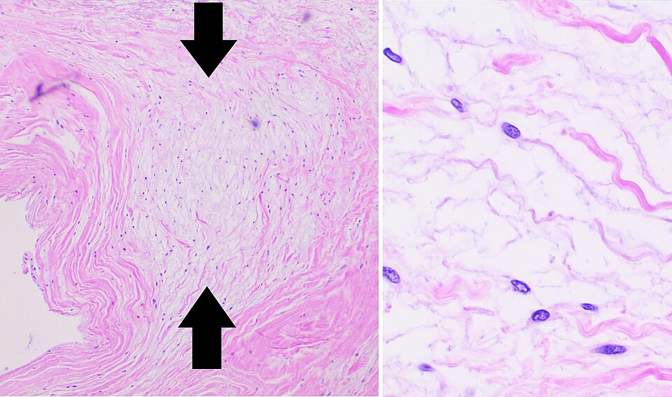

Flail leaflet and chordal rupture. Rupture of one or more primary chordae allows the free edge of the corresponding segment to evert into the left atrium. The flail segment loses coaptation entirely and almost always produces severe, eccentric regurgitation. Spontaneous rupture usually complicates myxomatous disease; other causes are endocarditis, trauma, and ischemic papillary muscle rupture. Rupture can convert chronic moderate MR into acute severe MR in hours. A flail leaflet is listed among the specific findings for severe MR in the 2017 American Society of Echocardiography (ASE) recommendations.
Rheumatic heart disease. Rheumatic carditis thickens the leaflet edges, fuses the commissures, and shortens and fuses the chordae. Restriction in systole and diastole (Carpentier IIIa) produces mixed stenosis and regurgitation. Isolated rheumatic MR without stenosis is characteristic of younger patients and of the posterior leaflet, which retracts and becomes nearly immobile. Repair is less durable than in degenerative disease: the 2020 guideline cites freedom from reoperation of 50% to 60% at 20 years even in experienced hands.
Infective endocarditis. Endocarditis destroys tissue by leaflet perforation, chordal rupture, or abscess of the annulus, and a vegetation can prevent coaptation. Regurgitation is often acute, and a perforation produces a type I jet through the leaflet body. Surgical timing for endocarditic MR is detailed under infective endocarditis.
Mitral annular calcification. Calcification of the fibrous annulus is common in older adults and is associated with chronic kidney disease, hypertension, diabetes, and atherosclerosis. It begins in the posterior annulus and extends into the leaflet bases, restricting leaflet motion and reducing annular contraction. It causes regurgitation, stenosis, or both, with open commissures and mobile leaflet tips, and its acoustic shadow hides jets from apical windows. Extensive calcification complicates surgical repair, raises the risk of atrioventricular groove injury during replacement, and is the main indication for transcatheter valve-in-annular-calcification procedures. Caseous calcification, a smooth posterior mass with an echolucent center, can mimic a tumor or abscess.
Other primary causes. Radiation-associated valve disease produces leaflet and annular fibrosis and calcification 10 to 20 years after mediastinal radiation, with frequent aorto-mitral curtain calcification and coexisting coronary, conduction, and pericardial disease. Drugs with serotonin 5-HT2B receptor agonist activity (the ergot-derived dopamine agonists pergolide and cabergoline at high doses, and the anorectic agents fenfluramine and benfluorex) cause a fibrotic valvulopathy that resembles rheumatic restriction without commissural fusion. Systemic lupus erythematosus and the antiphospholipid syndrome produce Libman-Sacks lesions and leaflet thickening. An isolated cleft of the anterior leaflet occurs with normal atrioventricular connections, distinct from the cleft of an atrioventricular septal defect. Heritable connective tissue disorders (Marfan syndrome, Loeys-Dietz syndrome, Ehlers-Danlos syndrome) cause myxomatous prolapse, and an aortic root evaluation accompanies the mitral assessment in these patients.
Hypertrophic cardiomyopathy. Systolic anterior motion of the anterior leaflet or chordae in obstructive hypertrophic cardiomyopathy produces a posteriorly directed, mid-to-late systolic jet that varies with the outflow gradient. Intrinsic leaflet elongation and anomalous papillary muscle insertion contribute. This MR usually resolves when outflow obstruction is relieved by myectomy, alcohol septal ablation, or myosin inhibition, and an anteriorly or centrally directed jet in a patient with hypertrophic cardiomyopathy indicates an additional primary leaflet lesion. Obstruction is detailed under hypertrophic cardiomyopathy.
Mitral valve prolapse: definition, billowing, and genetics
Mitral valve prolapse is systolic displacement of one or both leaflets at least 2 mm beyond the mitral annular plane into the left atrium. The 2017 ASE recommendations specify that the diagnosis is made in the parasternal long-axis view, with the apical long-axis (three-chamber) view as an alternative when parasternal windows are poor. Both views cut the annulus through its high points. The apical four-chamber and two-chamber views cross the low points of the saddle, where normal leaflets can appear to rise above a line joining the hinge points. These views localize segments but are not used to diagnose prolapse. Apical four-chamber criteria accounted for most of the overdiagnosis in older literature.
Framingham investigators separated classic prolapse (displacement of more than 2 mm with maximal diastolic leaflet thickness of at least 5 mm) from nonclassic prolapse (displacement of more than 2 mm with thinner leaflets). Surgical and echocardiographic terminology then distinguishes three positions of the leaflet. In billowing, the leaflet body bulges above the annular plane while the free edge and coaptation point stay on the ventricular side, and the valve is often competent. In prolapse, the free edge itself overrides the annular plane, although the tip still points toward the ventricle. In a flail leaflet, the tip everts and points toward the atrial roof, coaptation is lost, and a ruptured chord is often visible as a thin mobile echo on the free edge.
Mitral valve prolapse is heritable, most often with autosomal dominant inheritance, incomplete penetrance, and variable expression. Familial X-linked myxomatous valve dystrophy is caused by mutations in FLNA, the gene encoding filamin A, which also affects the aortic and tricuspid valves in some families. Mutations in DCHS1 segregate with nonsyndromic prolapse in multigenerational families. Genetic testing has no routine role in isolated prolapse; syndromic features (aortic root dilation, skeletal findings, lens dislocation, skin laxity) lead to evaluation for heritable connective tissue disease.
Adverse outcomes in prolapse concentrate in patients with moderate or severe regurgitation, a flail leaflet, a reduced ejection fraction, left atrial enlargement, and atrial fibrillation. The minority with arrhythmic features form a separate risk group.
Arrhythmic mitral valve prolapse and mitral annular disjunction
Arrhythmic mitral valve prolapse is prolapse accompanied by frequent or complex ventricular arrhythmia, with or without significant regurgitation, and it accounts for a small but real burden of sudden cardiac death in young adults. In a pathology registry of 650 adults aged 40 years or younger with sudden cardiac death, prolapse was the only structural finding in 43 (7% of all cases and 13% of women). Bileaflet involvement was present in 70%, and fibrosis of the papillary muscles in every case and of the inferobasal wall in 88%. Among the 12 with an available ECG, 10 had inverted T waves in the inferior leads. In living patients with prolapse and complex ventricular arrhythmias, late gadolinium enhancement on cardiac magnetic resonance (CMR) was present in 93%, compared with 14% of controls without arrhythmias.
Mitral annular disjunction is a systolic separation between the atrial wall–mitral valve junction and the top of the left ventricular myocardium, so the posterior leaflet hinge point sits on the atrial wall. It is measured in the parasternal long-axis view, or circumferentially by CMR, as the distance from the hinge point to the ventricular myocardium at end systole. Disjunction is common in Barlow disease and produces paradoxical systolic expansion of the annulus and a "curling" motion of the posterior basal wall. In a cohort of 116 patients with disjunction, 12% had aborted cardiac arrest or sustained ventricular tachycardia, and these patients were younger, had lower ejection fractions, and more often had papillary muscle fibrosis. Disjunction was arrhythmogenic even in the 22% of patients without prolapse.
The 2022 European Heart Rhythm Association (EHRA) expert consensus statement, endorsed by the Heart Rhythm Society, defines the arrhythmic mitral valve prolapse–disjunction complex and lists risk markers. Primary prevention implantable cardioverter-defibrillator decisions rest on the whole phenotype and specialist judgment, and secondary prevention indications are the standard ones for survivors of cardiac arrest or sustained ventricular tachycardia. No single feature has a validated threshold for implanting a defibrillator.
| Domain | Findings associated with higher arrhythmic risk |
|---|---|
| Clinical | Unexplained syncope; young adult, female sex in pathology series; family history of sudden death |
| Electrocardiogram | T-wave inversion or ST depression in inferior and inferolateral leads |
| Ambulatory monitoring | Frequent ventricular ectopy, polymorphic or right bundle branch block morphology ectopy, nonsustained ventricular tachycardia, especially rapid or repetitive |
| Echocardiography | Bileaflet myxomatous prolapse, mitral annular disjunction, Pickelhaube sign (high-velocity lateral annular tissue Doppler systolic spike), reduced ejection fraction |
| CMR | Late gadolinium enhancement of the papillary muscles or inferobasal wall; increased native T1 or extracellular volume |
Features of the arrhythmic mitral valve prolapse–mitral annular disjunction phenotype, summarized from the 2022 EHRA consensus statement and its source studies. The Pickelhaube sign is a marker of abnormal annular mechanics and is not a stand-alone risk threshold.
Mitral surgery in arrhythmic prolapse corrects the regurgitation and the mechanical stretch on the papillary muscles. Reductions in ventricular ectopy after repair have been reported, but surgery is not an established treatment for ventricular arrhythmia in a patient without an indication for valve intervention. Management of documented ventricular tachycardia follows the ventricular tachycardia and sudden cardiac death framework, with catheter ablation of papillary muscle foci in selected patients.
Ventricular secondary mitral regurgitation: ischemic and nonischemic
In ventricular secondary MR, a dilated or regionally scarred left ventricle displaces one or both papillary muscles apically, posteriorly, and laterally. The chordae, which do not lengthen, hold the leaflets toward the apex in systole. The coaptation point moves into the ventricle, the leaflets form a tent below the annular plane, and the coaptation length shortens until a gap opens. Annular dilation and flattening add a type I component, and reduced contractility, dyssynchrony, and left bundle branch block lower the force that closes the leaflets. Tenting height (coaptation depth) and tenting area measured in mid-systole quantify the tethering. A large tenting area, a coaptation depth of about 1 cm or more, and a markedly dilated ventricle predict recurrence after annuloplasty alone.
Global dilation in a nonischemic cardiomyopathy displaces both papillary muscles and tethers both leaflets symmetrically, producing a central jet. An inferior or inferolateral infarction displaces the posteromedial papillary muscle and restricts the posterior leaflet asymmetrically. The anterior leaflet tip then overrides the restricted posterior leaflet, the jet is directed posteriorly, and the appearance can be mistaken for anterior leaflet prolapse unless leaflet motion is examined in two planes.
Secondary MR is dynamic. Its orifice widens with higher afterload, larger ventricular volumes, ischemia, and dyssynchrony, and it narrows with diuresis, vasodilation, beta-blockade, and resynchronization. The 2017 ASE recommendations show a typical biphasic pattern, with the orifice largest in early and late systole and smallest in mid-systole. Severity measured during decompensation therefore overstates the chronic lesion, and the 2020 ACC/AHA guideline reassesses severity after medical therapy has been optimized.
Acute ischemic MR follows papillary muscle rupture, a mechanical complication of infarction discussed with acute MR below (ST-elevation myocardial infarction). Transient ischemic MR from reversible papillary muscle and wall dysfunction during ischemia can produce episodic pulmonary edema with a normal resting echocardiogram, and stress echocardiography reproduces it.
Atrial functional mitral regurgitation
Atrial functional (atrial secondary) MR occurs when the left atrium and mitral annulus enlarge while the left ventricle stays normal in size and function. Long-standing persistent atrial fibrillation and heart failure with preserved ejection fraction are the usual substrates. The annulus dilates mainly in the anteroposterior direction, flattens, and loses its systolic contraction. The leaflets, although normal, do not enlarge enough to cover the larger orifice, and coaptation is lost at the annular level without apical displacement of the coaptation point. In some patients the enlarged atrium pulls the posterior annulus outward over the ventricular crest, so the posterior leaflet hinges against the atrial wall ("atriogenic tethering") and the jet becomes posteriorly directed.
A 2022 expert panel convened by JACC: Cardiovascular Imaging proposed defining atrial functional MR by a normal or near-normal left ventricular ejection fraction, normal or mildly increased left ventricular volumes, left atrial enlargement, no significant leaflet pathology or regional wall motion abnormality, a central jet, and a dilated, flattened annulus. Atrial functional MR is graded with the same parameters as other MR, but low forward flow, atrial fibrillation, and a very compliant atrium blunt the supportive signs, so quantitative and volumetric data carry more weight.
Treatment begins with the atrium. Restoration and maintenance of sinus rhythm by antiarrhythmic therapy or catheter ablation, diuresis, and blood pressure control can reduce left atrial and annular size and the severity of regurgitation. The 2020 ACC/AHA guideline gives a Class 2b recommendation for mitral surgery in chronic severe atrial secondary MR. The criteria are a preserved ejection fraction (50% or more) and severe persistent New York Heart Association (NYHA) class III or IV symptoms despite therapy for heart failure and atrial fibrillation. A ring annuloplasty combined with surgical ablation and left atrial appendage management is the usual operation. Edge-to-edge repair is used in selected patients at high surgical risk, but the anatomy of a large annulus with relatively short leaflets differs from that of ventricular secondary MR, and the COAPT selection criteria do not transfer directly. The 2025 ESC/EACTS guideline gives surgery for symptomatic severe atrial secondary MR a Class IIa recommendation and considers edge-to-edge repair in patients not eligible for surgery.
Acute severe mitral regurgitation
Acute severe MR is a mechanical emergency. The causes are spontaneous chordal rupture in a myxomatous valve, papillary muscle rupture after infarction, leaflet perforation or chordal destruction from endocarditis, blunt chest trauma, and acute dysfunction or dehiscence of a prosthesis or repair. Takotsubo syndrome and acute ischemia can produce acute secondary MR, sometimes with dynamic outflow obstruction and systolic anterior motion.
A sudden regurgitant volume enters a left atrium of normal size and low compliance, so left atrial and pulmonary venous pressures rise abruptly, with a large v wave, and pulmonary edema follows. Total stroke volume and ejection fraction are high, but forward stroke volume falls because much of each beat enters the low-impedance atrium. The ventricle is normal in size and hyperdynamic. Left atrial pressure approaches ventricular pressure in late systole, so the driving gradient collapses, the murmur is short, soft, or inaudible, and the color jet on transthoracic imaging can look brief and unimpressive. A quiet precordium in a patient with flash pulmonary edema or shock and a hyperdynamic ventricle points to acute MR.
Papillary muscle rupture usually follows an inferior or inferoposterior infarction, often a small one, within the first week. Complete rupture of the trunk is usually rapidly fatal; rupture of one head produces severe regurgitation with a flail segment and a mobile mass on the chordal tips. Because the posteromedial muscle supplies both leaflets, the flail usually involves medial segments of both. The 2020 ACC/AHA guideline notes that transesophageal echocardiography (TEE) is especially helpful when a hemodynamically unstable patient after infarction has a hyperdynamic ventricle on transthoracic imaging and no other explanation, because it detects papillary muscle or chordal rupture, vegetations, and annular abscess.
Vasodilators such as sodium nitroprusside or nicardipine lower aortic impedance, which reduces regurgitant volume and increases forward output, but hypotension limits their use. Intra-aortic balloon counterpulsation lowers systolic afterload while raising diastolic and mean aortic pressure, and percutaneous circulatory support devices stabilize patients in shock (cardiogenic shock). Prompt mitral surgery, with repair when possible, is lifesaving in symptomatic acute severe primary MR. Transcatheter edge-to-edge repair has been used as a bridge or alternative in patients with papillary muscle rupture or acute ischemic MR who are prohibitive surgical candidates, but the evidence consists of registries and case series.
Pathophysiology of chronic mitral regurgitation: left ventricular and atrial volume overload
Chronic MR imposes a pure volume load. The ventricle ejects its total stroke volume into two outlets, the high-impedance aorta and the low-impedance left atrium, and regurgitation begins in isovolumic contraction, before the aortic valve opens. To maintain forward output, the ventricle enlarges by eccentric hypertrophy, adding sarcomeres in series. The larger end-diastolic volume, through the Frank-Starling mechanism, increases total stroke volume, and the forward stroke volume is preserved. Wall thickness rises roughly in proportion to cavity radius, so systolic wall stress stays normal during compensation.
Ejection into the atrium lowers the effective afterload. Ejection fraction is therefore higher in compensated MR than in a normal heart, and the 2020 ACC/AHA guideline notes that a "normal" ejection fraction in MR is about 70%. As myocardial contractility declines, the ejection fraction falls toward the normal range while it still looks reassuring, and the end-systolic dimension, which is less dependent on preload, enlarges. This is why the guideline sets intervention thresholds at an ejection fraction of 60% or less and an end-systolic diameter of 40 mm or more, values that would be normal in other conditions. By the time ejection fraction falls below 50%, contractile dysfunction is advanced and often only partly reversible after surgery. Afterload rises abruptly after the valve is corrected, because the low-impedance outlet is closed, and the ejection fraction commonly falls after surgery.
The left atrium dilates to accommodate the regurgitant volume, which keeps atrial pressure near normal in compensated disease. Chronic stretch produces atrial fibrosis and atrial fibrillation, and atrial fibrillation in turn worsens regurgitation by enlarging the annulus and removing atrial systole. Pulmonary venous pressure rises with decompensation, during exercise, or with atrial stiffening, and secondary pulmonary hypertension, tricuspid annular dilation, and right ventricular dysfunction follow. Atrial fibrillation, pulmonary hypertension, and secondary tricuspid regurgitation mark advanced disease and bear on the timing of surgery.
Natural history and prognosis of mitral regurgitation
The prognosis of chronic primary MR is determined by lesion severity, symptoms, ventricular function, and whether the valve can be repaired. In a prospective study of 456 patients with asymptomatic organic MR quantified by Doppler, the estimated 5-year rates of death from any cause, cardiac death, and cardiac events with medical management were 22%, 14%, and 33%. Patients with an EROA of 40 mm² or more had a 5-year survival of 58%, lower than the 78% expected from census data, and an adjusted risk of death 2.9 times that of patients with an EROA below 20 mm².
The flail leaflet is the clearest natural-history model. In 229 patients with isolated MR from a flail leaflet, medically treated patients died at 6.3% per year, more than expected: 34% per year in those ever in NYHA class III or IV and 4.1% per year in those in class I or II. At 10 years, the rates of heart failure, atrial fibrillation, and death or surgery were 63%, 30%, and 90%, and surgery was associated with a lower mortality (hazard ratio 0.29). In the multicenter Mitral Regurgitation International Database registry of patients with flail leaflets and no Class 1 trigger, early surgery within 3 months was associated with 10-year survival of 86%, compared with 69% with initial medical management. The 2020 guideline notes that chronic severe primary MR reaches a trigger for surgery at an average rate of about 8% per year.
Secondary MR carries the prognosis of the underlying cardiomyopathy, worsened by the regurgitation.
Symptoms and physical examination in mitral regurgitation
Chronic MR is tolerated for years without symptoms. When symptoms appear, they are exertional dyspnea, fatigue and reduced exercise capacity from limited forward output, orthopnea, and palpitations from atrial fibrillation. Late disease brings right-sided heart failure with edema, ascites, and hepatic congestion. Hemoptysis and systemic embolism are uncommon and suggest atrial fibrillation or coexisting stenosis. Acute severe MR presents with abrupt dyspnea, pulmonary edema, or cardiogenic shock.
The murmur of chronic significant MR is holosystolic, blowing, high-pitched, loudest at the apex, and radiates to the axilla and left infrascapular area. It begins with the first heart sound and extends to or through the aortic component of the second sound, and its intensity correlates only loosely with severity. Jet direction changes radiation. A posterior leaflet flail directs the jet anteriorly against the atrial septum near the aortic root, so the murmur radiates to the base and neck and can mimic aortic stenosis. Anterior leaflet disease directs the jet posteriorly, and the murmur radiates to the back and occasionally the spine. A third heart sound reflects rapid early diastolic filling with a large volume and does not by itself indicate ventricular failure in MR. A short diastolic flow rumble without an opening snap indicates large transmitral diastolic flow. The apical impulse is displaced laterally and is hyperdynamic. The first heart sound is often soft, and wide splitting of the second sound can occur because the left ventricle empties early.
Mitral valve prolapse produces a mid-systolic click as the redundant leaflet and chordae become taut, followed by a late systolic murmur. Maneuvers that reduce left ventricular volume move the click earlier and lengthen the murmur; maneuvers that increase volume or afterload delay the click and shorten the murmur.
| Maneuver | Main hemodynamic effect | Holosystolic MR murmur | Mitral valve prolapse click and murmur | Obstructive HCM murmur |
|---|---|---|---|---|
| Isometric handgrip | Increased afterload | Louder | Click later; murmur shorter | Softer |
| Squatting from standing | Increased venous return and afterload | Louder or unchanged | Click later; murmur shorter | Softer |
| Standing from squatting | Reduced venous return | Softer or unchanged | Click earlier; murmur longer | Louder |
| Valsalva strain | Reduced venous return and ventricular volume | Softer | Click earlier; murmur longer | Louder |
| Amyl nitrite inhalation | Reduced afterload | Softer | Click earlier; murmur longer | Louder |
Dynamic auscultation of systolic murmurs. HCM, hypertrophic cardiomyopathy. Changes in volume and afterload act in opposite directions on prolapse, so the net effect of squatting on intensity is variable while the click timing is consistent.
| Condition | Murmur | Distinguishing findings |
|---|---|---|
| Chronic primary MR | Apical holosystolic, radiating to the axilla | Displaced hyperdynamic apex; S3; louder with handgrip |
| Mitral valve prolapse | Mid-systolic click and late systolic murmur | Click moves with maneuvers that change ventricular volume |
| Acute severe MR | Short, early systolic, often soft or absent | Pulmonary edema or shock; normal-sized hyperdynamic ventricle |
| Tricuspid regurgitation | Holosystolic at the left lower sternal border | Louder with inspiration (Carvallo sign); large jugular v waves; pulsatile liver |
| Ventricular septal defect or post-infarction septal rupture | Harsh holosystolic at the left sternal border | Thrill common; oxygen step-up at the right ventricle |
| Aortic stenosis | Crescendo-decrescendo at the right upper sternal border, radiating to the carotids | Delayed, diminished carotid upstroke; soft A2; Gallavardin radiation to the apex mimics MR |
| Obstructive hypertrophic cardiomyopathy | Harsh mid-systolic at the left sternal border | Louder with Valsalva and standing; brisk bifid carotid pulse; may coexist with posteriorly directed MR |
Differential diagnosis of an apical or left-sided systolic murmur.
Electrocardiogram, chest radiograph, natriuretic peptides, and strain in mitral regurgitation
The electrocardiogram in chronic MR shows left atrial abnormality (a broad, notched P wave in lead II and a deep terminal negative P wave in V1) while sinus rhythm persists, and atrial fibrillation is common in advanced disease. Voltage criteria for left ventricular hypertrophy can be met in chronic volume overload. Right ventricular hypertrophy suggests pulmonary hypertension or coexisting mitral stenosis. In ventricular secondary MR, Q waves localize a prior infarction, and left bundle branch block identifies dyssynchrony that resynchronization may correct. In arrhythmic prolapse, inferior and inferolateral T-wave inversion and ventricular ectopy of papillary muscle or inferobasal origin are characteristic.
The chest radiograph in chronic severe MR shows cardiomegaly from left atrial and ventricular enlargement. Findings include a straightened left heart border from the atrial appendage, a double density at the right heart border, splaying of the carina, and elevation of the left main bronchus. Annular calcification appears as a C- or J-shaped density. In acute severe MR, the cardiac silhouette is normal in size and the lungs show pulmonary edema. A posteriorly directed jet aimed into the right upper pulmonary vein can produce edema predominantly in the right upper lobe, a pattern that is easily mistaken for pneumonia.
Natriuretic peptides rise in chronic severe MR as forward output becomes more dependent on preload. In asymptomatic patients with severe primary MR, elevated B-type natriuretic peptide is associated with a higher risk of symptoms, heart failure, and death, independent of conventional markers. The 2020 ACC/AHA guideline gives a Class 2b recommendation for serum biomarkers and novel measures of left ventricular function, such as global longitudinal strain, as adjuncts for timing intervention in asymptomatic severe primary MR (stages B and C1). They support a decision to operate when imaging is borderline and do not replace the standard triggers.
Global longitudinal strain measures longitudinal fiber shortening, which declines before ejection fraction in chronic volume overload. Although also load dependent, it is less inflated by the low-impedance outlet than ejection fraction, and in observational surgical cohorts impaired preoperative strain identified patients whose ejection fraction fell after repair despite a normal preoperative value. Vendor differences in strain values mean that serial studies use the same platform.
Echocardiographic grading of mitral regurgitation
Transthoracic echocardiography is the Class 1 initial test in known or suspected primary MR. It establishes the mechanism, the severity, left ventricular size and function, left atrial size, right ventricular function, and pulmonary artery pressure, and it is repeated when symptoms appear or change. In secondary MR, it also establishes the cause (regional scar or global cardiomyopathy) and the extent of ventricular remodeling. Mechanism is defined before severity, because it predicts the jet direction, the shape of the regurgitant orifice, the duration of regurgitation, and therefore which quantitative methods will be valid.
The 2017 ASE recommendations, developed with the Society for Cardiovascular Magnetic Resonance, grade MR by integrating structural, qualitative, semiquantitative, and quantitative parameters. Certain findings are designated specific for their grade. If MR is clearly mild by specific signs, quantitation is not required. If the signs suggest more than mild MR, quantitation is performed whenever data quality allows, and it divides moderate regurgitation into mild-to-moderate (grade II) and moderate-to-severe (grade III). Concordant findings establish the grade with confidence. Discordant findings prompt a search for technical or physiologic explanations, and the most reliable data for the specific valve and jet are weighted. Values from different methods are never averaged.
| Parameter | Mild | Moderate | Severe |
|---|---|---|---|
| Mitral valve morphology | None or mild leaflet abnormality (mild thickening, calcification, prolapse, or tenting) | Moderate leaflet abnormality or moderate tenting | Primary: flail leaflet, ruptured papillary muscle, severe retraction, large perforation. Secondary: severe tenting, poor coaptation |
| LV and LA size (chronic primary MR) | Usually normal | Normal or mildly dilated | Dilated |
| Color jet area (Nyquist 50–70 cm/s) | Small, central, narrow, often brief | Variable | Large central jet (>50% of LA) or eccentric wall-impinging jet of variable size |
| Flow convergence (Nyquist 30–40 cm/s) | Not visible, transient, or small (radius usually <0.3 cm) | Intermediate in size and duration | Large throughout systole (radius usually ≥1.0 cm) |
| CW Doppler jet | Faint, partial, or parabolic | Dense but partial or parabolic | Holosystolic, dense, triangular |
| Vena contracta width | <0.3 cm | Intermediate | ≥0.7 cm (>0.8 cm biplane average) |
| Pulmonary vein flow | Systolic dominance (may be blunted in LV dysfunction or AF) | Normal or systolic blunting | Minimal to no systolic flow or systolic flow reversal |
| Mitral inflow | A-wave dominant | Variable | E-wave dominant (E >1.2 m/s) |
| EROA by 2D PISA | <0.20 cm² | 0.20–0.29 cm² (II); 0.30–0.39 cm² (III) | ≥0.40 cm² |
| Regurgitant volume | <30 mL | 30–44 mL (II); 45–59 mL (III) | ≥60 mL |
| Regurgitant fraction | <30% | 30–39% (II); 40–49% (III) | ≥50% |
Grading of chronic MR by echocardiography, from the 2017 ASE recommendations (Table 8). Shaded entries are the signs the document designates as specific for severe MR; four or more of them (flail leaflet or ruptured papillary muscle, vena contracta ≥0.7 cm, flow convergence radius ≥1.0 cm at a Nyquist limit of 30–40 cm/s, central jet >50% of the LA, systolic pulmonary vein flow reversal, and an enlarged LV with normal function) establish severe MR. The specific signs of mild MR are a small, narrow central jet, a vena contracta of 0.3 cm or less, flow convergence absent or with a radius of 0.3 cm or less, A-wave dominant inflow, a soft or incomplete continuous-wave signal, and normal LV and LA size; four or more establish mild MR without quantitation, and two or three specific signs of either kind lead to quantitation. An EROA in the severe range may be lower in secondary MR with an elliptical orifice, and a severe regurgitant volume may be lower in low-flow states. The mitral inflow criterion is most valid in patients older than 50 years. AF, atrial fibrillation; CW, continuous-wave; EROA, effective regurgitant orifice area; LA, left atrium; LV, left ventricle; PISA, proximal isovelocity surface area.
The ASE algorithm counts specific criteria. Severe MR is certain when four or more specific criteria for severe MR are present: a flail leaflet, a vena contracta of 0.7 cm or more, a PISA radius of 1.0 cm or more at a Nyquist limit of 30 to 40 cm/s, a large central jet occupying more than 50% of the left atrium, systolic flow reversal in the pulmonary veins, and an enlarged ventricle with normal function. Mild MR is certain when four or more specific criteria for mild MR are present. Intermediate patterns are quantified, and when quantitation is not possible or remains discordant with the clinical picture, TEE or CMR follows.
Color Doppler, vena contracta, and supportive Doppler signs
Color jet area. Jet area depends on regurgitant volume and also on the Nyquist limit, color gain, driving pressure, atrial size, and impingement on the atrial wall. An eccentric jet that adheres to the atrial wall (the Coanda effect) occupies a fraction of the area of a free central jet of the same volume. Jet area is therefore a screening tool: a small central jet with no visible convergence zone is usually mild, and any eccentric jet, or any large jet, is quantified.
Vena contracta. The vena contracta is the narrowest portion of the jet, at or just downstream of the orifice, measured in a zoomed parasternal long-axis view perpendicular to the jet. A width below 0.3 cm indicates mild MR and a width of 0.7 cm or more indicates severe MR. The width is relatively independent of driving pressure and flow rate and applies to eccentric jets, but small measurement errors are large relative to the range, and widths of separate jets cannot be added. Averaging the width in the apical four-chamber and two-chamber views reduces the error from a crescentic orifice, and an average above 0.8 cm is consistent with severe MR. Three-dimensional color Doppler planimetry of the vena contracta area measures the orifice directly, without assumptions about its shape, and is particularly useful in secondary MR and multiple jets.
Continuous-wave Doppler. Peak MR velocity is usually 4 to 6 m/s and reflects the systolic pressure difference between the left ventricle and left atrium (4V²), not the regurgitant volume. A low peak velocity, for example 4 m/s, indicates low systolic blood pressure or a high left atrial pressure. A dense signal approaching the density of antegrade flow indicates a large regurgitant volume, and a faint or incomplete signal indicates a small one. A truncated, triangular contour with early peaking reflects a large left atrial v wave that narrows the pressure difference in late systole and is characteristic of severe or acute MR. The tricuspid regurgitant velocity provides the pulmonary artery systolic pressure, an indirect marker of the hemodynamic burden.
Pulmonary vein flow. Systolic flow reversal in more than one pulmonary vein is specific for severe MR. An eccentric jet aimed into one vein can reverse flow in that vein alone, so the right and left upper veins are sampled when possible, most reliably by TEE. Systolic blunting is not specific, because atrial fibrillation, elevated left atrial pressure, and a stiff atrium blunt the systolic wave without severe MR. A normal, systolic-dominant pattern argues against severe MR.
Mitral inflow. Severe MR increases diastolic flow across the valve, so the early (E) velocity is usually 1.2 m/s or higher and inflow is E-dominant. An A-dominant pattern with a low E velocity virtually excludes severe MR in patients older than 50 years. The sign is less useful in secondary MR, in which elevated filling pressure alone produces E dominance, and it is confounded by mitral stenosis, annular calcification, and an annuloplasty ring. In significant MR, the ratio of E to annular e′ velocity does not estimate filling pressure reliably.
PISA, EROA, and regurgitant volume calculator
The proximal isovelocity surface area (PISA) or flow convergence method rests on conservation of mass. Blood accelerating toward a small orifice forms concentric, roughly hemispheric shells of increasing velocity. On color Doppler, the boundary at which flow first aliases marks one shell whose velocity is the aliasing velocity (Va). Flow through that shell equals flow through the orifice. For a hemispheric shell of radius r, regurgitant flow rate = 2πr² × Va. Dividing by the simultaneous peak regurgitant velocity gives the effective regurgitant orifice area (EROA), and multiplying the EROA by the velocity-time integral (VTI) of the regurgitant jet gives the regurgitant volume.
The method is performed in the apical four-chamber view, zoomed on the convergence zone. The color baseline is shifted toward the direction of the jet (downward on transthoracic imaging, upward on TEE) to an aliasing velocity of about 30 to 40 cm/s, which enlarges and rounds the hemisphere. The radius is measured from the aliasing boundary to the orifice in mid-systole, at the time of peak regurgitant velocity, and the peak velocity and VTI come from a continuous-wave recording of the same jet. When the leaflets form a funnel and the convergence surface is constrained by the leaflets or the wall, the angle (α) between the leaflets on the ventricular side is measured and the hemisphere is corrected by α/180.
EROA (cm²) = 2π × r² × Va × (α ÷ 180) ÷ peak MR velocity, with velocities in cm/s. Regurgitant volume (mL) = EROA × MR VTI. Regurgitant fraction = regurgitant volume ÷ (regurgitant volume + LVOT stroke volume), where LVOT stroke volume = π × (LVOT diameter ÷ 2)² × LVOT VTI; the fraction is valid only without significant aortic regurgitation. Grades follow the 2017 ASE thresholds. With the default values (radius 1.0 cm, aliasing velocity 40 cm/s, peak velocity 5.0 m/s, VTI 150 cm), EROA is 0.50 cm² and regurgitant volume 75 mL. LVOT, left ventricular outflow tract.
Because the radius is squared, a 1-mm error in a 1.0-cm radius changes the EROA by about 20%. With an aliasing velocity of about 40 cm/s and a regurgitant velocity of about 5 m/s, which corresponds to a systolic gradient of 100 mm Hg, the 2017 ASE document describes a validated simplification: EROA ≈ r²/2. A radius of 1.0 cm then corresponds to an EROA of about 0.5 cm², and a radius of 0.3 cm to about 0.05 cm². The approximation holds for most patients with jet velocities between 4 and 6 m/s and fails at extremes of blood pressure.
PISA is most accurate for a single, holosystolic jet through a round orifice, the typical lesion of a flail or prolapsing segment. The EROA it reports is instantaneous, and the orifice varies through systole in secondary MR and in prolapse with late systolic regurgitation. For non-holosystolic jets, the 2017 ASE recommendations prefer the regurgitant volume, calculated as the maximal EROA multiplied by the VTI of the densest part of the continuous-wave signal, as the descriptor of severity.
Volumetric quantification by Doppler and ventricular volumes
The volumetric method is independent of jet geometry and suits eccentric, multiple, and crescentic jets. Regurgitant volume is the difference between total left ventricular stroke volume and forward stroke volume through the outflow tract. Total stroke volume is measured from biplane or three-dimensional left ventricular volumes, or from pulsed-wave Doppler at the mitral annulus (annular area × annular VTI). Forward stroke volume is the outflow tract area multiplied by the outflow tract VTI. Regurgitant fraction is the regurgitant volume divided by total stroke volume.
Total stroke volume = end-diastolic volume − end-systolic volume; forward stroke volume = π × (LVOT diameter ÷ 2)² × LVOT VTI; regurgitant volume = total − forward stroke volume; regurgitant fraction = regurgitant volume ÷ total stroke volume. With the default values, total stroke volume is 120 mL, forward stroke volume 57 mL, regurgitant volume 63 mL, and regurgitant fraction 53%.
Each input carries error. Two-dimensional echocardiography underestimates ventricular volumes through foreshortening and endocardial dropout, which lowers the total stroke volume and the calculated regurgitant volume; contrast and three-dimensional volumes reduce this bias. The outflow tract diameter is squared, so a 1-mm error in a 2.0-cm diameter changes forward stroke volume by about 10%. With coexisting aortic regurgitation, total stroke volume from ventricular volumes minus outflow tract stroke volume remains valid, because both terms include the aortic regurgitant volume. Mitral annular stroke volume minus outflow tract stroke volume underestimates MR, because only the outflow term includes it.
Sources of error in quantifying mitral regurgitation
- Late systolic regurgitation in prolapse: single-frame color, vena contracta, and peak EROA measured in late systole overstate severity. The duration of regurgitation is reported, and regurgitant volume describes the lesion.
- A crescentic orifice in secondary MR: the vena contracta in one plane and two-dimensional PISA underestimate the orifice. Biplane vena contracta, three-dimensional vena contracta area, volumetric methods, and CMR are used.
- Multiple jets: single-jet methods undercount the total regurgitant volume, and vena contracta widths cannot be added. Volumetric methods, three-dimensional imaging, and CMR quantify the total.
- Misplaced PISA radius: an error in locating the orifice or the aliasing boundary is squared in the EROA. The hemisphere is enlarged by baseline shifting, and the orifice is identified on zoomed imaging.
- Mismatched timing: combining a late-systolic convergence radius with a mid-systolic peak velocity distorts the EROA. Radius and velocity are measured at the same moment.
- Constrained convergence: wall or leaflet constraint flattens the hemisphere and overestimates flow if uncorrected. The aliasing velocity is raised to shrink the hemisphere, an angle correction is applied, or the method is not reported.
- Hypertension at the time of the study: higher driving pressure enlarges the color jet and can enlarge a functional orifice. Studies are compared at similar blood pressure.
- Atrial fibrillation: beat-to-beat variation in stroke volume and orifice size requires averaging of several beats, and pulmonary vein blunting loses its meaning. Specific signs and averaged quantitation carry the grade.
- General anesthesia during intraoperative TEE: lower preload and afterload reduce secondary and some primary MR. Severity is judged under the loading conditions of the awake state, with pressor support when necessary.
Grading secondary mitral regurgitation and the proportionality concept
The 2020 ACC/AHA guideline defines severe secondary MR with the same thresholds as severe primary MR: an EROA of 0.40 cm² or more and a regurgitant volume of 60 mL or more, with a regurgitant fraction of 50% or more. This reversed the 2014 guideline, which had used an EROA of 0.20 cm² and a regurgitant volume of 30 mL on the basis of prognosis. The 2020 guideline adds two footnotes: two-dimensional PISA underestimates the true EROA in secondary MR because of the crescentic convergence, and the regurgitant volume for severe MR may be lower in low-flow states. The 2017 ASE recommendations took the same position and add that an EROA of 0.30 cm² or more by two-dimensional PISA may still represent severe MR because of these limitations. The 2025 ESC/EACTS guideline defines severe secondary MR at an EROA of 0.30 cm² or more and a regurgitant volume of 45 mL or more.
In secondary MR the forward stroke volume is low, so regurgitant volume is low even when a large fraction of each beat regurgitates. Regurgitant fraction accounts for low flow but carries larger relative errors because the numbers are small. The cardiomyopathy degrades the supportive signs. Systolic blunting of pulmonary vein flow and E-dominant inflow are common without severe MR, and systolic reversal is uncommon even with severe MR because the atrium is large and compliant, although it remains specific when present. Volume status, blood pressure, and medical therapy change severity from one study to the next.
The proportionality concept relates the regurgitation to the size of the ventricle that produces it. In a ventricle with an ejection fraction of 30%, an end-diastolic volume of 220 to 250 mL, and a regurgitant fraction of 50%, hydraulic principles predict an EROA of about 0.3 cm² independent of specific leaflet tethering. Such MR is proportionate to ventricular dilation and is expected to respond to therapies that shrink the ventricle. An EROA of 0.3 to 0.4 cm² in a ventricle of only 160 to 200 mL is disproportionate and is the lesion most likely to benefit from treatment of the valve. The framework was proposed to explain why COAPT, with a larger mean EROA and smaller ventricles, was positive, whereas MITRA-FR, with a smaller EROA and larger ventricles, was neutral. Secondary analyses of COAPT did not confirm that the ratio predicted benefit within the trial, and no single ratio defines eligibility for intervention.
Transesophageal and three-dimensional echocardiography before repair
TEE is indicated (Class 1) in primary MR when transthoracic imaging provides insufficient or discordant information about severity, mechanism, or ventricular function, and intraoperative TEE is indicated (Class 1) in severe primary MR undergoing intervention to establish the anatomic basis of the lesion and to guide repair. In secondary MR with severe symptoms unresponsive to medical therapy, TEE is indicated (Class 1) to determine suitability for transcatheter intervention, and intraprocedural TEE guidance is recommended (Class 1).
Segments are localized in two planes. The mid-esophageal long-axis view at 120° to 140° shows A2 and P2, and the commissural view at 50° to 70° shows P3, A2, and P1 from left to right. The transgastric basal short-axis view shows all six segments en face. A segment is assigned only when two imaging planes agree.
Three-dimensional TEE displays the valve en face from the left atrium in the "surgeon's view," with the aortic valve at the 12 o'clock position and the left atrial appendage at 9 o'clock. It localizes prolapsing and flail segments, commissural lesions, clefts, and multisegment Barlow disease more accurately than two-dimensional imaging, and it measures annular dimensions, leaflet areas, and tenting volume. Thin tissue can drop out on three-dimensional rendering and mimic a cleft or perforation, so a true defect is confirmed in orthogonal source planes at adequate gain and by a color jet through it.
The preoperative and intraoperative TEE report states the Carpentier type, the diseased segments, leaflet thickness and mobility, chordal rupture, annular size and calcification, the number and direction of jets, and the predictors of systolic anterior motion after repair. Repair is most predictable with degenerative disease, thin pliable leaflets, a single posterior segment, intact commissures, and little calcium. Anterior or bileaflet involvement, commissural disease, Barlow complexity, extensive annular calcification, rheumatic thickening, severe tethering, and tissue destroyed by endocarditis make repair less predictable and favor a reference center.
After repair, intraoperative TEE judges the result once physiologic filling and blood pressure are restored. More than trivial residual MR usually leads to revision, because even mild residual regurgitation increases the likelihood of later repair failure. Systolic anterior motion with outflow obstruction after repair is recognized by a posteriorly directed jet, a late-peaking outflow gradient, and the anterior leaflet or chordae moving into the outflow tract. Risk factors are a long posterior leaflet (with a short anterior-to-posterior leaflet length ratio), a small coaptation-to-septum distance, a small hyperdynamic ventricle, a basal septal bulge, and an undersized ring. Most cases resolve with volume loading, discontinuation of inotropes, beta-blockade, and phenylephrine; persistent obstruction requires surgical revision. A mean transmitral gradient is also measured to exclude stenosis from an undersized ring.
Cardiac magnetic resonance quantification of mitral regurgitation
CMR quantifies MR without geometric assumptions about the jet. The standard method subtracts aortic forward flow, measured by phase-contrast velocity mapping in the proximal ascending aorta, from total left ventricular stroke volume, measured by planimetry of a short-axis cine stack: mitral regurgitant volume = left ventricular stroke volume − aortic forward flow. Regurgitant fraction is the regurgitant volume divided by left ventricular stroke volume. Right ventricular stroke volume or pulmonary artery forward flow provides an internal control when no other regurgitant lesion or shunt is present. Phase-contrast acquisition requires a velocity encoding just above the peak velocity, an imaging plane orthogonal to flow, and background phase-offset correction. Arrhythmia, especially atrial fibrillation, degrades segmented cine and phase-contrast data.
The 2020 ACC/AHA guideline gives CMR a Class 1 recommendation in primary MR to assess left and right ventricular volumes and function and to help assess MR severity when clinical assessment and echocardiography disagree. CMR measures ventricular volumes with high interstudy reproducibility, which makes it well suited to serial assessment. In a prospective multicenter study of 103 patients, agreement between CMR and echocardiographic grading was modest. In the 38 who underwent isolated mitral surgery, the reduction in left ventricular end-diastolic volume after surgery correlated strongly with CMR-derived regurgitant volume and fraction (r = 0.85) and not significantly with echocardiographic severity (r = 0.32). CMR is less useful than echocardiography for defining leaflet anatomy.
No outcome-based CMR thresholds are universally accepted. The 2017 ASE recommendations note an early proposal of a regurgitant fraction of 48% for severe regurgitation, an outcome study in which a regurgitant fraction above 40% predicted progression to symptoms or surgery, and adopt the general echocardiographic thresholds (regurgitant volume of 60 mL or more and fraction of 50% or more) in the absence of better data. Late gadolinium enhancement identifies replacement fibrosis in the papillary muscles and inferobasal wall in arrhythmic prolapse and myocardial scar and viability in ischemic secondary MR, and native T1 mapping and extracellular volume quantify diffuse fibrosis in chronic volume overload.
Cardiac CT for transcatheter planning and annular calcification
Cardiac CT has a limited role in grading MR and a central role in planning transcatheter mitral replacement and in defining annular calcification. ECG-gated, contrast-enhanced CT across the cardiac cycle measures the mitral annulus (saddle-shaped or D-shaped contour, intercommissural and septal-lateral diameters, area, and perimeter) by the method specified for the intended device. It maps the circumferential extent, thickness, and distribution of annular calcification, the relationship of the circumflex artery and coronary sinus to the annulus, the transseptal trajectory, and the left ventricular cavity and septum.
Left ventricular outflow tract obstruction is the defining complication of transcatheter mitral replacement. The valve displaces the anterior mitral leaflet toward the septum and creates a "neo-LVOT" between the leaflet or valve frame and the septum. Virtual implantation of the planned device on CT predicts the smallest systolic neo-LVOT area. In a multicenter registry of 194 patients undergoing valve-in-valve, valve-in-ring, and valve-in-annular-calcification procedures, outflow obstruction (an increase in mean outflow gradient of 10 mm Hg or more) occurred in 13.4% overall and in 54.1% of valve-in-calcification cases. Procedural mortality was 34.6% with obstruction compared with 2.4% without it. A predicted neo-LVOT area of 1.7 cm² or less identified obstruction with a sensitivity of 96.2% and specificity of 92.3%. The threshold depends on the device, its implantation depth, and the cardiac phase analyzed. Risk factors for a small neo-LVOT include a long anterior leaflet, an acute aorto-mitral angle, basal septal hypertrophy, and a small ventricular cavity. Preemptive laceration of the anterior leaflet (the LAMPOON procedure) and septal reduction are used when the predicted neo-LVOT is too small.
CT coronary angiography can exclude coronary disease before mitral surgery in patients at low pretest probability, and the 2020 guideline lists it among the tests that establish an ischemic cause in secondary MR (Class 1).
Invasive hemodynamics and left ventriculography
Noninvasive imaging is adequate in most patients. Left ventriculography and hemodynamic measurement are useful when clinical assessment and noninvasive tests are inconclusive or discordant regarding the severity of MR, ventricular function, or the need for surgery. A normal hemodynamic study in a symptomatic patient with apparently less than severe MR suggests a noncardiac cause. Exercise or leg raising during catheterization exposes dynamic regurgitation and filling pressures when resting data are equivocal.
The pulmonary artery wedge pressure in severe MR often shows a large v wave, but the v wave is neither sensitive nor specific. Its height depends on left atrial compliance and volume as well as regurgitant volume. A large, compliant atrium in chronic severe MR can have a small v wave. Large v waves occur without MR in a stiff atrium, in mitral stenosis, after surgical ablation, in ventricular septal rupture with increased pulmonary venous return, and in volume overload. In acute MR, a large v wave with a hyperdynamic ventricle and pulmonary edema supports the diagnosis.
Contrast left ventriculography grades MR semiquantitatively by the density and clearing of contrast in the left atrium. Grade 1+ is contrast that clears with each beat and never opacifies the whole atrium. Grade 2+ is faint opacification of the whole atrium that does not clear with each beat. Grade 3+ is atrial opacification equal to that of the ventricle. Grade 4+ is dense atrial opacification within one beat, greater than that of the ventricle, with reflux into the pulmonary veins. The 2020 guideline stages equate angiographic grades 1+ to 2+ with progressive MR and 3+ to 4+ with severe MR. The method depends on catheter position, contrast volume, injection rate, ventricular ectopy, and atrial size, and premature beats induced by the catheter create spurious regurgitation. Combined with thermodilution or Fick output, the angiographic total stroke volume minus forward stroke volume yields a regurgitant volume and fraction.
Exercise testing and stress echocardiography in mitral regurgitation
Symptoms define stage D, but many patients with chronic MR limit their activity without recognizing it. In primary MR (stages B and C) with symptoms that might be attributable to MR, hemodynamic exercise testing by Doppler echocardiography or catheterization, or cardiopulmonary exercise testing, is reasonable (Class 2a). A treadmill test with measured exercise capacity establishes objective functional status and a baseline for later comparison, and peak oxygen consumption separates cardiac from noncardiac limitation.
Exercise echocardiography adds hemodynamic information. Regurgitation that increases during exercise, a rise in pulmonary artery systolic pressure, failure of the ventricle to augment its ejection fraction (limited contractile reserve), and a fall in right ventricular longitudinal function identify physiology that resting data miss. In 78 patients with asymptomatic, at least moderate degenerative MR, exercise pulmonary hypertension (systolic pulmonary pressure above 60 mm Hg) occurred in 46% and independently predicted the onset of symptoms (hazard ratio 3.4). Exercise pulmonary hypertension above 60 mm Hg was an indication for surgery in the 2006 ACC/AHA guideline; the 2014 guideline retained only resting pulmonary hypertension, and the 2020 recommendations treat exercise pulmonary hypertension as supporting information.
In secondary MR, exercise echocardiography exposes dynamic regurgitation that increases markedly with exercise and explains exertional dyspnea or pulmonary edema out of proportion to resting findings. In 98 patients with ischemic left ventricular dysfunction, an exercise-induced increase in EROA of 0.13 cm² or more independently predicted cardiac death, and half of the patients with such an increase who died had only moderate MR at rest. Measurements are made during exercise or immediately at peak, because dynamic MR regresses within minutes of recovery. Dobutamine stress also assesses viability and contractile reserve in ischemic cardiomyopathy.
ACC/AHA stages of primary and secondary mitral regurgitation
The 2020 ACC/AHA guideline classifies chronic MR in four stages that combine valve anatomy, valve hemodynamics, the hemodynamic consequences, and symptoms. Asymptomatic severe primary MR is divided into stage C1 (normal ventricular function) and stage C2 (left ventricular systolic dysfunction), the distinction that determines the indication for surgery.
| Stage | Definition | Valve anatomy | Valve hemodynamics | Consequences and symptoms |
|---|---|---|---|---|
| A | At risk of MR | Mild prolapse with normal coaptation; mild leaflet thickening and restriction | No jet or small central jet <20% of LA area; vena contracta <0.3 cm | None |
| B | Progressive MR | Moderate to severe prolapse with normal coaptation; rheumatic restriction with loss of central coaptation; prior endocarditis | Central jet 20–40% of LA or late systolic eccentric jet; vena contracta <0.7 cm; RVol <60 mL; RF <50%; EROA <0.40 cm²; angiographic grade 1+ to 2+ | Mild LA enlargement; no LV enlargement; normal pulmonary pressure; no symptoms |
| C1 | Asymptomatic severe MR with normal LV function | Severe prolapse with loss of coaptation or flail leaflet; rheumatic restriction; prior endocarditis; radiation thickening | Central jet >40% of LA or holosystolic eccentric jet; vena contracta ≥0.7 cm; RVol ≥60 mL; RF ≥50%; EROA ≥0.40 cm²; angiographic grade 3+ to 4+ | Moderate or severe LA enlargement; LV enlargement; pulmonary hypertension possible; LVEF >60% and LVESD <40 mm; no symptoms |
| C2 | Asymptomatic severe MR with LV dysfunction | LVEF ≤60% and/or LVESD ≥40 mm; no symptoms | ||
| D | Symptomatic severe MR | As for stage C | As for stage C | Moderate or severe LA enlargement; LV enlargement; pulmonary hypertension; decreased exercise tolerance; exertional dyspnea |
Stages of chronic primary MR, adapted from the 2020 ACC/AHA guideline (Table 17 in the JACC version). EROA, effective regurgitant orifice area; LA, left atrium; LV, left ventricle; LVEF, left ventricular ejection fraction; LVESD, left ventricular end-systolic diameter; RF, regurgitant fraction; RVol, regurgitant volume.
| Stage | Definition | Valve anatomy | Valve hemodynamics | Associated findings and symptoms |
|---|---|---|---|---|
| A | At risk of MR | Normal leaflets, chords, and annulus in a patient with coronary disease or cardiomyopathy | No jet or small central jet <20% of LA area; vena contracta <0.30 cm | Normal or mildly dilated LV with fixed or inducible regional wall motion abnormality, or primary myocardial disease; ischemic or heart failure symptoms that respond to revascularization and medical therapy |
| B | Progressive MR | Regional wall motion abnormality with mild tethering; annular dilation with mild loss of central coaptation | EROA <0.40 cm²; RVol <60 mL; RF <50% | Regional wall motion abnormality with reduced LV function, or LV dilation and dysfunction from primary myocardial disease |
| C | Asymptomatic severe MR | Regional wall motion abnormality or LV dilation with severe tethering; annular dilation with severe loss of central coaptation | EROA ≥0.40 cm²; RVol ≥60 mL (may be lower in low-flow states); RF ≥50% | As for stage B; symptoms, when present, respond to revascularization and medical therapy |
| D | Symptomatic severe MR | As for stage C | As for stage C | Heart failure symptoms attributable to MR persist after revascularization and optimization of medical therapy; decreased exercise tolerance; exertional dyspnea |
Stages of chronic secondary MR, adapted from the 2020 ACC/AHA guideline (Table 18 in the JACC version). Two-dimensional PISA underestimates the true EROA in secondary MR because of the crescentic convergence zone.
Medical therapy, anticoagulation, and rhythm management in mitral regurgitation
Primary MR. No drug has been shown to delay the need for surgery in primary MR. In asymptomatic patients with normal ventricular function (stages B and C1) who are normotensive, vasodilator therapy is not indicated (Class 3: No Benefit). Vasodilators reduce ventricular size and closing force and can increase prolapse, so they may worsen regurgitation. Hypertension is treated, because systolic pressure drives the regurgitant flow and worsens MR. In patients with severe primary MR and ventricular systolic dysfunction (stages C2 and D) in whom surgery is not possible or must be delayed, guideline-directed medical therapy for systolic dysfunction is reasonable (Class 2a), with beta-blockers having the strongest supporting data. Diuretics relieve congestion but do not defer an intervention indicated by symptoms.
Secondary MR. Patients with chronic severe secondary MR and heart failure with reduced ejection fraction receive standard guideline-directed medical therapy, including angiotensin receptor–neprilysin inhibition or an angiotensin-converting enzyme inhibitor or receptor blocker, an evidence-based beta-blocker, a mineralocorticoid receptor antagonist, and biventricular pacing when indicated (Class 1, Level A). The 2022 AHA/ACC/HFSA heart failure guideline adds a sodium-glucose cotransporter 2 inhibitor to this foundation. A cardiologist expert in heart failure leads the optimization of therapy (Class 1). Reverse remodeling with these therapies reduces tethering and often reduces MR, and cardiac resynchronization in patients with left bundle branch block and a wide QRS restores coordinated papillary muscle contraction and closing force. Revascularization is performed when indicated by coronary anatomy, symptoms, and viability. MR severity is reassessed after therapy has been optimized, and only regurgitation that persists, with persistent symptoms, is considered for intervention (heart failure with reduced ejection fraction).
Atrial fibrillation and anticoagulation. Atrial fibrillation is common in MR and is both a consequence of atrial remodeling and a driver of annular dilation. A non-vitamin K oral anticoagulant is an effective alternative to a vitamin K antagonist in patients with atrial fibrillation and native valve disease other than rheumatic mitral stenosis, or with a bioprosthetic valve implanted more than 3 months earlier (Class 1). The indication is based on the CHA2DS2-VASc score. MR itself is not an indication for anticoagulation, and native MR, including rheumatic MR without stenosis, does not require a vitamin K antagonist.
Indications and timing of intervention in primary mitral regurgitation
The aim in chronic severe primary MR is to correct the valve before irreversible ventricular dysfunction, atrial fibrillation, pulmonary hypertension, or right ventricular damage develops. The 2020 ACC/AHA guideline sets the indications in the order of the stages.
| Clinical situation | Recommendation | Class |
|---|---|---|
| Symptomatic severe primary MR (stage D) | Mitral valve intervention, irrespective of LV systolic function | 1 |
| Asymptomatic severe primary MR with LV dysfunction: LVEF ≤60% or LVESD ≥40 mm (stage C2) | Mitral valve surgery | 1 |
| Severe primary MR from degenerative disease with an indication for surgery | Repair in preference to replacement if a successful and durable repair is possible | 1 |
| Asymptomatic severe primary MR with normal LV function (stage C1) | Repair is reasonable when the likelihood of a successful and durable repair without residual MR is >95% with an expected mortality <1%, at a Primary or Comprehensive Valve Center | 2a |
| Stage C1 with a progressive increase in LV size or decrease in LVEF on 3 or more serial imaging studies | Mitral valve surgery may be considered irrespective of the probability of a durable repair | 2b |
| Severely symptomatic (NYHA III or IV) severe primary MR at high or prohibitive surgical risk | TEER is reasonable if anatomy is favorable and life expectancy is at least 1 year | 2a |
| Symptomatic severe rheumatic MR with an indication for surgery | Repair may be considered at a Comprehensive Valve Center by an experienced team if a durable repair is likely | 2b |
| Leaflet pathology limited to less than one-half of the posterior leaflet | Replacement should not be performed unless repair has been attempted at a Primary or Comprehensive Valve Center and was unsuccessful | 3: Harm |
Indications for intervention in chronic primary MR, from the 2020 ACC/AHA guideline. LVEF, left ventricular ejection fraction; LVESD, left ventricular end-systolic diameter; TEER, transcatheter edge-to-edge repair.
Symptoms. The onset of symptoms caused by severe MR worsens prognosis even when the ventricle appears normal, and the adverse effect extends to mild symptoms. Mitral surgery improves the natural history after symptoms appear even if medication has relieved them, so symptom relief with diuretics does not defer surgery. The 2020 guideline recommends intervention irrespective of ventricular function. In patients with severely reduced function, the benefit of surgery is weighed against operative risk and the probability of ventricular recovery, with edge-to-edge repair as an alternative when surgical risk is prohibitive.
Ventricular dysfunction in asymptomatic patients. Because ejection into the left atrium lowers afterload, the guideline treats an ejection fraction of 60% or less or an end-systolic diameter of 40 mm or more as systolic dysfunction (stage C2). One study cited by the guideline found that ventricular size and function returned to normal after repair only when the preoperative ejection fraction exceeded 64% and the end-systolic diameter was below 37 mm. When serial studies show the ejection fraction falling toward 60% or the end-systolic diameter rising toward 40 mm, intervention is reasonable, and transthoracic studies are repeated every 3 to 6 months as the ventricle enlarges.
Early repair in asymptomatic patients with normal ventricular function. Early repair avoids intensive surveillance, the risk that patients are lost to follow-up, and the accumulation of atrial and ventricular damage. Its value depends entirely on the certainty of a durable repair at low risk, which comes from the documented results of the surgical team for the specific lesion. In patients with flail leaflets treated at experienced centers, early operation has been associated with lower rates of heart failure and death than watchful waiting. Additional findings that support earlier surgery in an apparently asymptomatic patient are new atrial fibrillation, resting pulmonary artery systolic pressure above 50 mm Hg, marked left atrial enlargement, at least moderate secondary tricuspid regurgitation, elevated natriuretic peptides, and impaired global longitudinal strain.
The 2025 ESC/EACTS guideline differs in three ways. It adds an indexed end-systolic diameter of 20 mm/m² or more as a Class I trigger in asymptomatic patients. It supports early repair in low-risk asymptomatic patients with a high likelihood of durable repair at a Heart Valve Center, particularly with atrial fibrillation, resting systolic pulmonary pressure above 50 mm Hg, or significant left atrial dilation. It gives edge-to-edge repair a Class IIa recommendation in symptomatic patients who are not candidates for surgery.
Mitral valve repair versus replacement
Why repair is preferred. Repair preserves the native apparatus and ventricular geometry, avoids lifelong anticoagulation with a mechanical valve and degeneration of a bioprosthesis, and carries lower operative mortality than replacement. When only annuloplasty and posterior leaflet repair are needed, the 2020 guideline expects an operative mortality below 1%, long-term survival equivalent to that of the age-matched population, about 95% freedom from reoperation, and more than 80% freedom from recurrent moderate or severe MR at 15 to 20 years. For isolated posterior leaflet disease, repair is the standard of care, and the guideline sets a success rate of 95% or more as the expectation of every surgeon who performs mitral procedures. Anterior or bileaflet disease requires a more complex repair: freedom from reoperation is about 80%, and freedom from recurrent moderate or severe MR about 60% at 15 to 20 years, results that remain superior to replacement at high-volume centers. Replacement is preferable to a poor repair.
Techniques. A complete or partial annuloplasty ring or band restores the size and shape of the annulus and increases coaptation, and nearly every degenerative repair includes one. Posterior leaflet prolapse is corrected by triangular or quadrangular resection, with sliding plasty when the leaflet is tall, or by implantation of expanded polytetrafluoroethylene neochordae, which preserves leaflet tissue. Anterior leaflet prolapse is corrected with neochordae or chordal transfer. Commissural prolapse is closed with a commissural suture. The edge-to-edge (Alfieri) stitch joins the free edges of the middle segments to create a double orifice and is the surgical counterpart of transcatheter edge-to-edge repair. Clefts and perforations are closed directly or with a patch, often of autologous pericardium.
Chordal preservation in replacement. When replacement is necessary, preservation of the posterior chordae, and of the anterior chordae when they do not obstruct the outflow tract, maintains the connection between the annulus and papillary muscles and preserves ventricular geometry and function better than excision of the apparatus.
Choice of prosthesis. Prosthesis choice is a shared decision that accounts for the patient's values, the risks of anticoagulation, and the potential need for reintervention (Class 1). For patients younger than 65 years who need mitral replacement, cannot undergo repair, and have no contraindication to anticoagulation, a mechanical prosthesis is reasonable (Class 2a). For patients 65 years or older who cannot undergo repair, a bioprosthesis is reasonable (Class 2a). For women who are considering pregnancy, the type of prosthesis is chosen through shared decision-making that weighs the maternal and fetal risks of anticoagulation with a mechanical valve against earlier structural deterioration and reoperation with a bioprosthesis. Mechanical mitral prostheses require a vitamin K antagonist with a target international normalized ratio (INR) of 3.0 (Class 1). Management of prosthetic valves follows the prosthetic heart valves guidance.
Concomitant procedures. Tricuspid valve surgery is recommended at the time of mitral surgery in patients with severe tricuspid regurgitation (Class 1). It can be beneficial with progressive (stage B) tricuspid regurgitation when the tricuspid annulus measures more than 4.0 cm in diastole or right-sided heart failure has occurred (Class 2a), because secondary tricuspid regurgitation left untreated often progresses and reoperation for it carries high risk. In symptomatic paroxysmal or persistent atrial fibrillation, surgical pulmonary vein isolation or a maze procedure at the time of valve surgery can reduce symptoms and recurrence (Class 2a). In patients with atrial fibrillation and a CHA2DS2-VASc score of 2 or more, surgical exclusion of the left atrial appendage in addition to continued anticoagulation is indicated to reduce stroke and systemic embolism (Class 1, 2023 ACC/AHA/ACCP/HRS atrial fibrillation guideline). Coronary bypass grafting is performed for significant coronary disease.
Heart valve team and valve centers. Probability of repair and overall outcome correlate with surgeon-specific mitral volume. Hospital mortality is, on average, 50% lower in the highest-volume hospitals that perform about 50 repairs per year, but some low-volume hospitals outperform the median high-volume hospital, so surgeon-specific repair rates and outcomes are better guides than volume alone. Complex repairs and early repair in asymptomatic patients are assigned to experienced mitral surgeons at a Primary (Level II) or Comprehensive (Level I) Valve Center. A multidisciplinary heart valve team of cardiologists with valve, imaging, and heart failure expertise, cardiac surgeons, interventionalists, and anesthesiologists evaluates patients being considered for intervention, and treatment decisions are reached by shared decision-making.
Transcatheter edge-to-edge repair for primary mitral regurgitation
Transcatheter edge-to-edge repair (TEER) approximates the free edges of the anterior and posterior leaflets with one or more devices delivered from the femoral vein across the interatrial septum, creating a double-orifice valve analogous to the surgical Alfieri stitch. The MitraClip system (Abbott) and the PASCAL system (Edwards Lifesciences) are approved in the United States. The procedure is performed under general anesthesia with continuous TEE and fluoroscopic guidance.
EVEREST II randomized 279 patients with grade 3+ or 4+ MR, about three-quarters of whom had degenerative disease, in a 2:1 ratio to percutaneous repair or conventional surgery. At 12 months, the primary efficacy end point (freedom from death, surgery for mitral valve dysfunction, and grade 3+ or 4+ MR) was met by 55% of the percutaneous group and 73% of the surgical group, and major adverse events at 30 days occurred in 15% compared with 48%, a difference driven largely by blood transfusion. At 5 years, freedom from death, surgery, or 3+ or 4+ MR was 44.2% compared with 64.3%, and mortality was 20.8% compared with 26.8% (not significantly different). Beyond 6 months, rates of surgery and moderate-to-severe MR were similar.
The trial established that TEER is safer than surgery and less effective at eliminating MR. In a patient at low or intermediate surgical risk with a repairable degenerative valve, expert surgical repair remains the treatment of choice. TEER is reasonable (Class 2a) in severely symptomatic (NYHA class III or IV) patients with severe primary MR at high or prohibitive surgical risk, favorable anatomy, and a life expectancy of at least 1 year. In the CLASP IID trial of patients with 3+ or 4+ degenerative MR at prohibitive surgical risk, the PASCAL system was noninferior to the MitraClip system at a prespecified interim analysis of 180 patients. Thirty-day major adverse event rates were 3.4% and 4.8%, and MR of 2+ or less at 6 months was present in 96.5% and 96.8%.
Transcatheter edge-to-edge repair for secondary mitral regurgitation
Two randomized trials of the same device in apparently the same disease, published in 2018, reached opposite conclusions, and their reconciliation shaped current selection criteria.
COAPT enrolled 614 patients at 78 sites in the United States and Canada with heart failure and moderate-to-severe or severe secondary MR (3+ or 4+ by core laboratory) who remained symptomatic despite maximally tolerated guideline-directed medical therapy confirmed by a central eligibility committee. Key criteria were an ejection fraction of 20% to 50% and an end-systolic diameter of 70 mm or less. Over 24 months, the annualized rate of heart failure hospitalization was 35.8% with TEER plus medical therapy and 67.9% with medical therapy alone (hazard ratio 0.53), and all-cause mortality was 29.1% compared with 46.1% (hazard ratio 0.62). Freedom from device-related complications at 12 months was 96.6%. At 5 years, all-cause mortality was 57.3% compared with 67.2% (hazard ratio 0.72), and the annualized heart failure hospitalization rate was 33.1% compared with 57.2%.
MITRA-FR enrolled 304 patients in France with severe secondary MR, defined by an EROA greater than 0.20 cm² or a regurgitant volume greater than 30 mL, an ejection fraction of 15% to 40%, and symptomatic heart failure. At 12 months, death or unplanned heart failure hospitalization occurred in 54.6% with TEER and 51.3% with medical therapy alone (odds ratio 1.16), with no difference in death (24.3% compared with 22.4%) or hospitalization.
RESHAPE-HF2 randomized 505 patients with heart failure, an ejection fraction of 20% to 50%, and moderate-to-severe secondary MR, a population with less severe regurgitation than COAPT. Over 24 months, first or recurrent heart failure hospitalization or cardiovascular death occurred at 37.0 compared with 58.9 events per 100 patient-years (rate ratio 0.64), and heart failure hospitalization at 26.9 compared with 46.6 per 100 patient-years (rate ratio 0.59). The Kansas City Cardiomyopathy Questionnaire overall summary score improved by 10.9 points more with TEER at 12 months. Device-specific safety events occurred in 1.6%. The benefit was driven by fewer hospitalizations, without a demonstrated reduction in cardiovascular mortality.
MATTERHORN, conducted in Germany, compared TEER with surgical repair or replacement in 210 patients with heart failure and secondary MR who remained symptomatic despite medical therapy (mean age 70.5 years, mean ejection fraction 43%). The 1-year composite of death, heart failure hospitalization, mitral reintervention, assist device implantation, or stroke occurred in 16.7% after TEER and 22.5% after surgery, meeting the noninferiority criterion, and major adverse events at 30 days occurred in 14.9% compared with 54.8%.
| Trial | Population | Comparison | Primary result |
|---|---|---|---|
| EVEREST II (2011) | 279 patients, grade 3+ or 4+ MR, mostly degenerative, surgical candidates | TEER vs surgery (2:1) | Freedom from death, surgery, or 3+/4+ MR at 12 months 55% vs 73%; 30-day major adverse events 15% vs 48% |
| CLASP IID (2022) | Degenerative 3+ or 4+ MR at prohibitive surgical risk; interim analysis of 180 | PASCAL vs MitraClip (2:1) | Noninferior: major adverse events 3.4% vs 4.8%; MR ≤2+ at 6 months 96.5% vs 96.8% |
| COAPT (2018) | 614 patients, secondary 3+ or 4+ MR, LVEF 20–50%, LVESD ≤70 mm, symptomatic on maximal medical therapy | TEER + medical therapy vs medical therapy | Heart failure hospitalization 35.8% vs 67.9% per patient-year; death 29.1% vs 46.1% at 24 months |
| MITRA-FR (2018) | 304 patients, secondary MR with EROA >0.20 cm² or RVol >30 mL, LVEF 15–40% | TEER + medical therapy vs medical therapy | Death or heart failure hospitalization at 12 months 54.6% vs 51.3% (no difference) |
| RESHAPE-HF2 (2024) | 505 patients, moderate-to-severe secondary MR, LVEF 20–50% | TEER + medical therapy vs medical therapy | Heart failure hospitalization or cardiovascular death 37.0 vs 58.9 per 100 patient-years at 24 months |
| MATTERHORN (2024) | 210 patients, secondary MR, symptomatic on medical therapy, surgical candidates | TEER vs mitral surgery | 1-year composite 16.7% vs 22.5% (noninferior); 30-day major adverse events 14.9% vs 54.8% |
Randomized trials of transcatheter edge-to-edge repair. EROA, effective regurgitant orifice area; LVEF, left ventricular ejection fraction; LVESD, left ventricular end-systolic diameter; RVol, regurgitant volume.
Why COAPT and MITRA-FR differed. The 2020 ACC/AHA guideline attributes the difference to patient selection. MITRA-FR enrolled patients with greater ventricular enlargement (mean left ventricular end-diastolic volume index 135 mL/m², compared with 101 mL/m² in COAPT) and less severe MR (mean EROA 0.31 cm², compared with 0.41 cm² in COAPT). MITRA-FR set no upper limit on ventricular size, whereas COAPT excluded an end-systolic diameter above 70 mm. COAPT also required maximally tolerated medical therapy, adjudicated centrally, before enrollment and allowed little titration afterward, whereas medical therapy in MITRA-FR was adjusted during follow-up. Procedural results differed as well, with more residual MR in MITRA-FR.
Recommendation. In chronic severe secondary MR related to left ventricular systolic dysfunction (ejection fraction below 50%) with persistent NYHA class II, III, or IV symptoms on optimal guideline-directed medical therapy (stage D), TEER is reasonable (Class 2a) when the following criteria are met. These are appropriate anatomy defined by TEE, an ejection fraction of 20% to 50%, an end-systolic diameter of 70 mm or less, and a pulmonary artery systolic pressure of 70 mm Hg or less. The guideline describes the COAPT enrollment criteria as the current standard selection criteria. Patients with advanced heart failure (inotrope dependence, severe right ventricular dysfunction, very large ventricles) are evaluated for ventricular assist devices or transplantation, in which the mitral valve is not the target. The 2025 ESC/EACTS guideline gives TEER a Class I recommendation in hemodynamically stable symptomatic patients with severe ventricular secondary MR despite medical therapy and resynchronization when indicated who meet criteria suggesting a high chance of benefit.
Anatomic selection and procedural imaging for edge-to-edge repair
TEE determines anatomic suitability. The device must grasp enough leaflet tissue to close the gap without creating stenosis, tearing a leaflet, or entangling the chordae. Clinical trials excluded valves with a coaptation length below 2 mm in tethered leaflets, calcification in the grasping zone of A2 or P2, a significant cleft in A2 or P2, and lack of both primary and secondary chordal support. Newer devices and experienced operators have extended treatment to anatomy once considered unsuitable, including commissural lesions, wide flail segments, and short posterior leaflets.
| Feature | Favorable | Challenging | Usually unsuitable |
|---|---|---|---|
| Location of the jet | Central (A2–P2) | Commissural or multiple jets | Extensive multisegment disease without a graspable target |
| Mitral valve area | >4.0 cm² | 3.0–4.0 cm² | <3.0 cm² |
| Baseline mean gradient | <4 mm Hg | 4–5 mm Hg | Higher gradients predict iatrogenic stenosis |
| Posterior leaflet length | >10 mm | 7–10 mm | <7 mm |
| Flail gap (degenerative MR) | <10 mm | ≥10 mm at experienced centers | — |
| Flail width (degenerative MR) | <15 mm | ≥15 mm (multiple devices) | — |
| Coaptation depth (secondary MR) | <10 mm | >10 mm | — |
| Coaptation length (secondary MR) | ≥2 mm | — | <2 mm |
| Calcification | None in the grasping zone | Annular calcification | Severe calcification in the grasping zone |
| Cleft or perforation | None | Small cleft with adequate leaflet length | Cleft in the grasping zone with a short leaflet; perforation |
| Leaflet tissue | Thin, pliable | Mild thickening | Rheumatic or severely thickened restricted leaflets |
Anatomic criteria for mitral edge-to-edge repair, summarized from the 2020 ACC/AHA guideline and a 2025 review of screening and procedural guidance. Thresholds vary by device generation and operator experience.
During the procedure, TEE guides the transseptal puncture to a posterior and superior site with adequate height above the mitral annulus (about 4 cm for degenerative MR, less for secondary MR). It steers the device coaxial to the line of coaptation, confirms leaflet insertion in the grasping arms, and assesses the result before release. Success is a reduction of MR to mild or less with a mean transmitral gradient that does not exceed about 5 mm Hg at a normal heart rate. Grading residual MR after TEER is limited by multiple jets, a double orifice, and device shadowing, which restrict vena contracta and PISA measurements. The pulmonary vein flow pattern, three-dimensional vena contracta area, and volumetric methods carry more weight. Single leaflet device attachment, leaflet tear, and an iatrogenic atrial septal defect are recognized complications; most septal defects are small and close spontaneously, and closure is reserved for large defects with right-to-left shunting or right ventricular volume overload.
Surgery for secondary mitral regurgitation
The 2020 ACC/AHA guideline gives surgery a limited role in ventricular secondary MR. Mitral surgery is reasonable (Class 2a) in severe secondary MR (stages C and D) when coronary bypass grafting is undertaken to treat ischemia. Isolated mitral surgery may be considered (Class 2b) for chronic severe secondary MR with an ejection fraction below 50% and persistent NYHA class III or IV symptoms on optimal medical therapy. In patients with coronary disease and chronic severe secondary MR undergoing mitral surgery for such symptoms, chordal-sparing replacement may be reasonable to choose over downsized annuloplasty repair (Class 2b).
The Cardiothoracic Surgical Trials Network studies define the evidence. In severe ischemic MR, 251 patients were randomized to restrictive annuloplasty repair or chordal-sparing replacement. There was no significant difference in left ventricular end-systolic volume index at 1 or 2 years, and 2-year mortality was 19.0% after repair and 23.2% after replacement, but moderate or severe MR recurred over 2 years in 58.8% after repair compared with 3.8% after replacement, with more heart failure events and cardiovascular readmissions in the repair group. In moderate ischemic MR, 301 patients were randomized to coronary bypass grafting alone or with mitral repair. At 2 years, ventricular end-systolic volume index and mortality (10.6% compared with 10.0%) did not differ, moderate or severe residual MR was less common with repair (11.2% compared with 32.3%), and neurologic events and supraventricular arrhythmias were more frequent with the combined procedure. Repair of moderate ischemic MR at the time of bypass grafting is therefore not routine.
Subvalvular procedures (papillary muscle approximation or relocation, and division of secondary chordae) and ventricular reshaping have been added to annuloplasty to address tethering, with promising observational results. In surgical candidates with secondary MR, noninferior 1-year outcomes with far fewer early adverse events in MATTERHORN favor TEER when anatomy is suitable and no other cardiac surgery is needed.
Transcatheter mitral valve replacement and other devices
Transcatheter mitral valve replacement addresses patients whose anatomy is unsuitable for edge-to-edge repair, including those with extensive annular calcification, short or calcified leaflets, a small valve area, or complex multisegment disease, and those with failed surgical prostheses or rings. Replacement is technically harder than at the aortic valve. The annulus is large, saddle shaped, and noncircular; most patients have no calcified ring to anchor a valve; the device must resist high systolic pressure; and the displaced anterior leaflet can obstruct the outflow tract.
In the United States, the SAPIEN M3 system (Edwards Lifesciences), a balloon-expandable valve deployed inside a nitinol dock delivered from the femoral vein across the septum, received premarket approval on December 22, 2025, for symptomatic moderate-to-severe or severe MR in patients judged by a heart team to be unsuitable for surgery or TEER. The Tendyne system (Abbott), a self-expanding valve delivered through the left ventricular apex and anchored by an apical tether, was approved in 2025 for patients with MR or stenosis associated with annular calcification who are unsuitable for surgery or TEER. Valve-in-valve implantation of a balloon-expandable transcatheter valve within a failed surgical mitral bioprosthesis, usually by a transseptal approach, is an established alternative to reoperation, and valve-in-ring and valve-in-annular-calcification procedures carry higher risks of residual regurgitation, embolization, and outflow obstruction.
Mitral regurgitation in pregnancy
Most women with chronic MR, including asymptomatic severe MR with normal ventricular function, tolerate pregnancy. The fall in systemic vascular resistance offsets the rise in blood volume and cardiac output, and the 2020 ACC/AHA guideline notes no evidence that pregnancy accelerates ventricular dysfunction in MR. High-risk features for heart failure during pregnancy are reduced left ventricular systolic function and pulmonary hypertension, defined in the guideline as a pulmonary artery systolic pressure above 50 mm Hg. Atrial fibrillation, prior heart failure, and symptoms before conception add risk.
Pre-pregnancy counseling by a cardiologist with expertise in valve disease and pregnancy includes a baseline echocardiogram and a review of medications. In symptomatic women with severe valve disease who are considering pregnancy, intervention before pregnancy is recommended on standard indications (Class 1). The threshold for operating on asymptomatic severe MR is higher in a woman who may become pregnant, because a failed repair would require a prosthesis and a mechanical valve carries high maternal and fetal risk. For asymptomatic high-risk women with severe MR, referral to a Comprehensive Valve Center allows the likelihood of a durable repair and the surgical risk to inform the decision. When valve replacement is needed before pregnancy, the type of prosthesis is chosen through shared decision-making with the patient.
During pregnancy, symptoms are treated with diuretics and, when needed, beta-blockers. Angiotensin-converting enzyme inhibitors, angiotensin receptor blockers, angiotensin receptor–neprilysin inhibitors, and mineralocorticoid receptor antagonists are avoided because of fetal toxicity. Valve surgery during pregnancy is reasonable (Class 2a) only for severe regurgitation with NYHA class IV symptoms refractory to medical therapy, because cardiopulmonary bypass carries substantial fetal risk. Surgery in the absence of severe refractory symptoms is harmful (Class 3: Harm). Vaginal delivery with regional anesthesia and hemodynamic monitoring tailored to risk is appropriate for most women.
Athletes and exercise in mitral regurgitation
The 2025 AHA/ACC scientific statement on competitive sports participation shifted eligibility decisions from fixed disqualification criteria to individualized shared decision-making. Athletes with mild MR participate without restriction. For moderate or severe MR, the evaluation includes left ventricular size, ejection fraction and end-systolic dimension, left atrial size, pulmonary artery pressure at rest and during exercise, rhythm, and exercise capacity on symptom-limited testing. Athletic training enlarges the left ventricle, so a ventricular size that would indicate decompensation in a sedentary person can be physiologic in a trained athlete, and serial measurements and exercise testing help separate the two. Severe MR with ventricular dysfunction or pulmonary hypertension carries risk during intense competition, and intervention follows standard indications. Athletes with arrhythmic prolapse features (unexplained syncope, complex ventricular ectopy, inferolateral T-wave inversion, papillary muscle fibrosis on CMR) undergo ambulatory monitoring, exercise testing, and CMR before decisions about participation.
Endocarditis prophylaxis and antithrombotic therapy after intervention
Native MR, including mitral valve prolapse, does not require antibiotic prophylaxis before dental procedures. After intervention, prophylaxis is reasonable (Class 2a) before dental procedures that manipulate gingival tissue or the periapical region of teeth, or perforate the oral mucosa. It applies to patients with prosthetic valves, including transcatheter prostheses, and to patients with prosthetic material used for valve repair, such as annuloplasty rings, chords, or clips. Prophylaxis is also reasonable after previous endocarditis. It is not recommended for nondental procedures such as TEE, endoscopy, or cystoscopy in the absence of active infection (Class 3: No Benefit).
After surgical bioprosthetic mitral replacement, aspirin 75 to 100 mg daily is reasonable in the absence of another indication for anticoagulation (Class 2a). A vitamin K antagonist with a target INR of 2.5 is reasonable for at least 3 and as long as 6 months in patients at low bleeding risk (Class 2a). After mechanical mitral replacement, a vitamin K antagonist with a target INR of 3.0 is indicated (Class 1); direct oral anticoagulants are not recommended with mechanical valves (Class 3: Harm), and dabigatran is contraindicated. New atrial fibrillation within 3 months of bioprosthetic replacement is reasonably treated with a vitamin K antagonist (Class 2a).
Echocardiographic surveillance intervals in mitral regurgitation
Periodic echocardiography is the basis of management in asymptomatic MR, because symptoms develop insidiously and ventricular decline can precede them. The 2020 ACC/AHA guideline sets intervals for asymptomatic patients with normal ventricular function, and repeat imaging is performed whenever symptoms, examination findings, or rhythm change.
| Status | Echocardiographic interval |
|---|---|
| Mild MR (stage B) | Every 3 to 5 years |
| Moderate MR (stage B) | Every 1 to 2 years |
| Asymptomatic severe MR with normal LV function (stage C1) | Every 6 to 12 months; more often when the left ventricle is dilating |
| Severe MR with LVEF approaching 60% or LVESD approaching 40 mm | Every 3 to 6 months, or more frequently as the ventricle enlarges |
| New or changing symptoms, new atrial fibrillation, or a change in the murmur | Prompt repeat transthoracic echocardiography |
| After mitral repair or replacement | Baseline study after recovery; then according to residual MR, ventricular function, and prosthesis type, with prompt imaging for symptoms or a new murmur |
| After TEER | Baseline study before discharge and at about 30 days, then at 1 year and annually |
Surveillance intervals from the 2020 ACC/AHA guideline (Table 5 and supportive text). Mixed valve disease and secondary MR with changing ventricular function warrant shorter intervals. The post-TEER schedule reflects clinical trial and registry practice.
A change in end-systolic diameter of a few millimeters is within measurement variability, so serial comparison requires the same windows, the same method of measuring end-systolic diameter and volumes, and similar blood pressure, and a trigger reached on a single study is confirmed before surgery is scheduled unless the trend is unequivocal.
Residual, recurrent, and prosthetic mitral regurgitation
Recurrent MR after surgical repair results from progression of degenerative disease in segments that were not repaired, suture or ring dehiscence, failure of neochordae, endocarditis, or, in secondary MR, continued ventricular remodeling and tethering. Re-repair is often possible, particularly when the mechanism is focal, and transcatheter options include edge-to-edge repair across a ring and valve-in-ring replacement.
Mitral prosthetic regurgitation is central (within the sewing ring) or paravalvular (outside it). Small central jets are normal in mechanical valves ("washing jets"), with a characteristic pattern for each design. Paravalvular regurgitation is common after replacement in a calcified annulus and after endocarditis. Its consequences include heart failure and hemolysis, and hemolytic anemia with a small paravalvular jet is a recognized presentation. Acoustic shadowing from the prosthesis hides mitral jets from transthoracic apical windows, so TEE, often with three-dimensional imaging, is required to localize and grade the defect. Indirect transthoracic signs of significant prosthetic MR include a high early mitral velocity, a high ratio of the mitral to the outflow tract velocity-time integral, a dense continuous-wave MR signal, an increased tricuspid regurgitant velocity, and flow convergence on the ventricular side of the prosthesis. Transcatheter paravalvular leak closure with plug devices is the usual treatment for symptomatic leaks in patients at high surgical risk. Prosthetic valve assessment is detailed under prosthetic heart valves.
Self-assessment
A 58-year-old man without symptoms has a flail P2 segment with an eccentric anterior jet. Vena contracta is 0.8 cm, PISA radius 1.1 cm at an aliasing velocity of 38 cm/s, peak MR velocity 5.2 m/s, and MR VTI 160 cm. LVEF is 62%, LV end-systolic diameter 38 mm, and he is in sinus rhythm with a pulmonary artery systolic pressure of 34 mm Hg. What are the EROA and regurgitant volume, what ACC/AHA stage applies, and what does the 2020 guideline recommend?
Echocardiography Board ReviewReferences
- Otto CM, Nishimura RA, Bonow RO, et al. 2020 ACC/AHA guideline for the management of patients with valvular heart disease: a report of the American College of Cardiology/American Heart Association Joint Committee on Clinical Practice Guidelines. Circulation. 2021;143:e72–e227. doi:10.1161/CIR.0000000000000923
- Praz F, Borger MA, Lanz J, et al. 2025 ESC/EACTS guidelines for the management of valvular heart disease. Eur Heart J. 2025;46:4635–4736. doi:10.1093/eurheartj/ehaf194
- Zoghbi WA, Adams D, Bonow RO, et al. Recommendations for noninvasive evaluation of native valvular regurgitation: a report from the American Society of Echocardiography developed in collaboration with the Society for Cardiovascular Magnetic Resonance. J Am Soc Echocardiogr. 2017;30:303–371. doi:10.1016/j.echo.2017.01.007
- Bonow RO, O'Gara PT, Adams DH, et al. 2020 focused update of the 2017 ACC expert consensus decision pathway on the management of mitral regurgitation: a report of the American College of Cardiology Solution Set Oversight Committee. J Am Coll Cardiol. 2020;75:2236–2270. doi:10.1016/j.jacc.2020.02.005
- Heidenreich PA, Bozkurt B, Aguilar D, et al. 2022 AHA/ACC/HFSA guideline for the management of heart failure: a report of the American College of Cardiology/American Heart Association Joint Committee on Clinical Practice Guidelines. Circulation. 2022;145:e895–e1032. doi:10.1161/CIR.0000000000001063
- Nkomo VT, Gardin JM, Skelton TN, et al. Burden of valvular heart diseases: a population-based study. Lancet. 2006;368:1005–1011. doi:10.1016/S0140-6736(06)69208-8
- Freed LA, Levy D, Levine RA, et al. Prevalence and clinical outcome of mitral-valve prolapse. N Engl J Med. 1999;341:1–7. doi:10.1056/NEJM199907013410101
- Enriquez-Sarano M, Avierinos JF, Messika-Zeitoun D, et al. Quantitative determinants of the outcome of asymptomatic mitral regurgitation. N Engl J Med. 2005;352:875–883. doi:10.1056/NEJMoa041451
- Ling LH, Enriquez-Sarano M, Seward JB, et al. Clinical outcome of mitral regurgitation due to flail leaflet. N Engl J Med. 1996;335:1417–1423. doi:10.1056/NEJM199611073351902
- Suri RM, Vanoverschelde JL, Grigioni F, et al. Association between early surgical intervention vs watchful waiting and outcomes for mitral regurgitation due to flail mitral valve leaflets. JAMA. 2013;310:609–616. doi:10.1001/jama.2013.8643
- Grigioni F, Enriquez-Sarano M, Zehr KJ, et al. Ischemic mitral regurgitation: long-term outcome and prognostic implications with quantitative Doppler assessment. Circulation. 2001;103:1759–1764. doi:10.1161/01.cir.103.13.1759
- Basso C, Perazzolo Marra M, Rizzo S, et al. Arrhythmic mitral valve prolapse and sudden cardiac death. Circulation. 2015;132:556–566. doi:10.1161/CIRCULATIONAHA.115.016291
- Dejgaard LA, Skjølsvik ET, Lie ØH, et al. The mitral annulus disjunction arrhythmic syndrome. J Am Coll Cardiol. 2018;72:1600–1609. doi:10.1016/j.jacc.2018.07.070
- Sabbag A, Essayagh B, Barrera JDR, et al. EHRA expert consensus statement on arrhythmic mitral valve prolapse and mitral annular disjunction complex in collaboration with the ESC Council on valvular heart disease and the European Association of Cardiovascular Imaging, endorsed by the Heart Rhythm Society, by the Asia Pacific Heart Rhythm Society, and by the Latin American Heart Rhythm Society. Europace. 2022;24:1981–2003. doi:10.1093/europace/euac125
- Durst R, Sauls K, Peal DS, et al. Mutations in DCHS1 cause mitral valve prolapse. Nature. 2015;525:109–113. doi:10.1038/nature14670
- Kyndt F, Gueffet JP, Probst V, et al. Mutations in the gene encoding filamin A as a cause for familial cardiac valvular dystrophy. Circulation. 2007;115:40–49. doi:10.1161/CIRCULATIONAHA.106.622621
- Zoghbi WA, Levine RA, Flachskampf F, et al. Atrial functional mitral regurgitation: a JACC: Cardiovascular Imaging expert panel viewpoint. JACC Cardiovasc Imaging. 2022;15:1870–1882. doi:10.1016/j.jcmg.2022.08.016
- Deferm S, Bertrand PB, Verbrugge FH, et al. Atrial functional mitral regurgitation: JACC review topic of the week. J Am Coll Cardiol. 2019;73:2465–2476. doi:10.1016/j.jacc.2019.02.061
- Uretsky S, Gillam L, Lang R, et al. Discordance between echocardiography and MRI in the assessment of mitral regurgitation severity: a prospective multicenter trial. J Am Coll Cardiol. 2015;65:1078–1088. doi:10.1016/j.jacc.2014.12.047
- Magne J, Lancellotti P, Piérard LA. Exercise pulmonary hypertension in asymptomatic degenerative mitral regurgitation. Circulation. 2010;122:33–41. doi:10.1161/CIRCULATIONAHA.110.938241
- Lancellotti P, Troisfontaines P, Toussaint AC, Piérard LA. Prognostic importance of exercise-induced changes in mitral regurgitation in patients with chronic ischemic left ventricular dysfunction. Circulation. 2003;108:1713–1717. doi:10.1161/01.CIR.0000087599.49332.05
- Grayburn PA, Sannino A, Packer M. Proportionate and disproportionate functional mitral regurgitation: a new conceptual framework that reconciles the results of the MITRA-FR and COAPT trials. JACC Cardiovasc Imaging. 2019;12:353–362. doi:10.1016/j.jcmg.2018.11.006
- Feldman T, Foster E, Glower DD, et al. Percutaneous repair or surgery for mitral regurgitation. N Engl J Med. 2011;364:1395–1406. doi:10.1056/NEJMoa1009355
- Feldman T, Kar S, Elmariah S, et al. Randomized comparison of percutaneous repair and surgery for mitral regurgitation: 5-year results of EVEREST II. J Am Coll Cardiol. 2015;66:2844–2854. doi:10.1016/j.jacc.2015.10.018
- Lim DS, Smith RL, Gillam LD, et al. Randomized comparison of transcatheter edge-to-edge repair for degenerative mitral regurgitation in prohibitive surgical risk patients. JACC Cardiovasc Interv. 2022;15:2523–2536. doi:10.1016/j.jcin.2022.09.005
- Stone GW, Lindenfeld J, Abraham WT, et al. Transcatheter mitral-valve repair in patients with heart failure. N Engl J Med. 2018;379:2307–2318. doi:10.1056/NEJMoa1806640
- Obadia JF, Messika-Zeitoun D, Leurent G, et al. Percutaneous repair or medical treatment for secondary mitral regurgitation. N Engl J Med. 2018;379:2297–2306. doi:10.1056/NEJMoa1805374
- Stone GW, Abraham WT, Lindenfeld J, et al. Five-year follow-up after transcatheter repair of secondary mitral regurgitation. N Engl J Med. 2023;388:2037–2048. doi:10.1056/NEJMoa2300213
- Anker SD, Friede T, von Bardeleben RS, et al. Transcatheter valve repair in heart failure with moderate to severe mitral regurgitation. N Engl J Med. 2024;391:1799–1809. doi:10.1056/NEJMoa2314328
- Baldus S, Doenst T, Pfister R, et al. Transcatheter repair versus mitral-valve surgery for secondary mitral regurgitation. N Engl J Med. 2024;391:1787–1798. doi:10.1056/NEJMoa2408739
- Acker MA, Parides MK, Perrault LP, et al. Mitral-valve repair versus replacement for severe ischemic mitral regurgitation. N Engl J Med. 2014;370:23–32. doi:10.1056/NEJMoa1312808
- Goldstein D, Moskowitz AJ, Gelijns AC, et al. Two-year outcomes of surgical treatment of severe ischemic mitral regurgitation. N Engl J Med. 2016;374:344–353. doi:10.1056/NEJMoa1512913
- Kron IL, Hung J, Overbey JR, et al. Predicting recurrent mitral regurgitation after mitral valve repair for severe ischemic mitral regurgitation. J Thorac Cardiovasc Surg. 2015;149:752–761.e1. doi:10.1016/j.jtcvs.2014.10.120
- Michler RE, Smith PK, Parides MK, et al. Two-year outcomes of surgical treatment of moderate ischemic mitral regurgitation. N Engl J Med. 2016;374:1932–1941. doi:10.1056/NEJMoa1602003
- Yoon SH, Bleiziffer S, Latib A, et al. Predictors of left ventricular outflow tract obstruction after transcatheter mitral valve replacement. JACC Cardiovasc Interv. 2019;12:182–193. doi:10.1016/j.jcin.2018.12.001
- Zygouri A, Rasmeehirun P, L'Official G, et al. Screening and procedural guidance for mitral transcatheter edge-to-edge repair (M-TEER). J Clin Med. 2025;14:4902. doi:10.3390/jcm14144902
- Kim JH, Baggish AL, Levine BD, et al. Clinical considerations for competitive sports participation for athletes with cardiovascular abnormalities: a scientific statement from the American Heart Association and American College of Cardiology. J Am Coll Cardiol. 2025;85:1059–1108. doi:10.1016/j.jacc.2024.12.025
- US Food and Drug Administration. Premarket approval P250019: SAPIEN M3 transcatheter mitral valve replacement system. Decision date December 22, 2025. accessdata.fda.gov
- Joglar JA, Chung MK, Armbruster AL, et al. 2023 ACC/AHA/ACCP/HRS guideline for the diagnosis and management of atrial fibrillation: a report of the American College of Cardiology/American Heart Association Joint Committee on Clinical Practice Guidelines. Circulation. 2024;149:e1–e156. doi:10.1161/CIR.0000000000001193