Library › Coronary artery disease › STEMI
ST-elevation myocardial infarction
ST-elevation myocardial infarction from coronary occlusion and ECG recognition through reperfusion, antithrombotic therapy, complications, special populations, and discharge care.
ST-elevation myocardial infarction (STEMI) is acute myocardial infarction with new ST elevation at the J point in two contiguous leads, at least 1 mm in most leads and 1.5 to 2.5 mm in V2 and V3 by sex and age, or an equivalent ECG pattern of acute coronary occlusion. Reperfusion is not delayed for troponin. Primary percutaneous coronary intervention (PCI) is the Class 1 strategy when first-medical-contact-to-device time is 90 minutes or less at a PCI-capable hospital or 120 minutes or less after transfer; when primary PCI cannot be performed within 120 minutes of first medical contact and onset was within 12 hours, fibrinolysis is given within 30 minutes of arrival and followed by transfer for angiography.
Definition of STEMI and the occlusion myocardial infarction concept
The Fourth Universal Definition of Myocardial Infarction (2018) defines myocardial infarction as acute myocardial injury with clinical evidence of acute ischemia. Injury is a rise or fall of cardiac troponin with at least one value above the 99th-percentile upper reference limit. Evidence of ischemia is any of the following: ischemic symptoms, new ischemic ECG changes, new pathologic Q waves, imaging evidence of new loss of viable myocardium or new regional wall-motion abnormality in an ischemic pattern, or coronary thrombus at angiography or autopsy. STEMI is the subset of acute myocardial infarction in which the presenting ECG shows persistent ST elevation meeting the threshold criteria, or an accepted equivalent, in a patient with a clinical syndrome of ischemia. The ECG diagnosis is the trigger for emergency reperfusion; troponin confirms the infarction afterward.
The universal definition classifies infarction by mechanism. Type 1 infarction results from atherothrombosis: plaque rupture or erosion with intraluminal thrombus. Type 2 infarction results from an imbalance between oxygen supply and demand without acute atherothrombosis, as in sustained tachyarrhythmia, severe anemia, hypoxemia, hypotension, or coronary spasm. Type 3 is cardiac death with ischemic symptoms and presumed new ischemic ECG changes before biomarkers are obtained. Types 4 and 5 are infarctions related to PCI (4a periprocedural, 4b stent thrombosis, 4c restenosis) and to coronary artery bypass surgery. Nearly every STEMI is a type 1 event; stent thrombosis (type 4b) presents identically and is managed identically.
| Type | Mechanism | Relevance to STEMI |
|---|---|---|
| 1 | Atherothrombosis: plaque rupture or erosion with thrombus | The usual substrate of STEMI; occlusive thrombus in an epicardial artery |
| 2 | Supply-demand imbalance without acute atherothrombosis | Rarely produces ST elevation; spasm, embolism, and dissection can, and are classified by mechanism |
| 3 | Sudden cardiac death with presumed new ischemia before biomarkers | Includes fatal STEMI presenting as arrest |
| 4a | Periprocedural infarction with PCI | Procedural; not the presenting event |
| 4b | Stent or scaffold thrombosis | Presents and is treated as STEMI |
| 4c | Restenosis | Usually non-ST-elevation presentation |
| 5 | Infarction related to coronary artery bypass surgery | Graft occlusion after surgery |
Classification of myocardial infarction, after the Fourth Universal Definition of Myocardial Infarction (2018).
The occlusion myocardial infarction concept. ST elevation is a surrogate for acute coronary occlusion with ongoing transmural ischemia, and an imperfect one. A meaningful fraction of patients with an acutely occluded culprit artery do not meet millimeter criteria on the presenting tracing, most often with circumflex occlusion, posterior or high lateral infarction, and small or distal territories. Conversely, ST elevation that meets criteria can be caused by conditions other than occlusion. The occlusion myocardial infarction (OMI) framework, developed largely in emergency medicine, classifies infarction by the presence or absence of acute occlusion and recognizes occlusion from the full ECG (T-wave morphology, proportionality, reciprocity, and specific patterns) as well as from ST amplitude. Current American College of Cardiology/American Heart Association (ACC/AHA) and European Society of Cardiology (ESC) guidelines retain the STEMI and non-ST-elevation acute coronary syndrome (NSTE-ACS) classification. Guideline-based care incorporates the principle in two ways. Equivalent patterns of occlusion, such as isolated posterior infarction and left bundle branch block or ventricular pacing meeting Sgarbossa criteria, are managed as STEMI. Patients with NSTE-ACS who have refractory ischemia, hemodynamic instability, or electrical instability undergo immediate angiography regardless of the ECG.
Epidemiology and mortality trends in STEMI
STEMI incidence has fallen in the United States since the late 1990s while non-ST-elevation myocardial infarction (NSTEMI) has become a larger share of all infarctions. In a large integrated health system in Northern California, the age- and sex-adjusted incidence of STEMI fell from 133 to 50 cases per 100,000 person-years between 1999 and 2008, and 30-day mortality after myocardial infarction fell over the same period. Reasons include reduced smoking, wider use of statins and antihypertensive therapy, and more sensitive troponin assays that reclassify smaller infarctions as NSTEMI. STEMI patients are on average younger than NSTEMI patients and more often male and current smokers.
Short-term mortality depends on age, infarct territory and size, Killip class, time to reperfusion, cardiac arrest at presentation, and the presence of cardiogenic shock. In-hospital mortality in contemporary US registries is in the mid-single digits overall and exceeds one-third in patients with cardiogenic shock. Cardiogenic shock and mechanical complications account for a disproportionate share of deaths. After discharge, the risk of recurrent ischemic events is highest in the first year, and survivors with a reduced ejection fraction carry the long-term risks of heart failure and ventricular arrhythmia.
Plaque rupture, erosion, and other causes of acute coronary occlusion
Plaque rupture. Most STEMIs result from disruption of the fibrous cap of a lipid-rich atheroma, a thin-cap fibroatheroma with a large necrotic core and macrophage infiltration. Rupture exposes tissue factor and collagen to flowing blood, platelets adhere and aggregate, and the coagulation cascade generates a fibrin-rich red thrombus on a platelet-rich white core. Many ruptured plaques were not flow-limiting before the event, which explains why infarction often occurs without prior angina and why stress testing does not predict the culprit site. Inflammation, shear stress at the shoulder of the plaque, and sympathetic surges (morning hours, emotional stress, heavy exertion) contribute to rupture.
Plaque erosion. In erosion, the endothelium is lost over an intact, often proteoglycan-rich and lipid-poor plaque, and thrombus forms on the denuded surface. Erosion is more common in younger patients, in women, and in smokers, and it causes a larger share of NSTE-ACS than of STEMI. Optical coherence tomography distinguishes erosion (thrombus over an intact fibrous cap) from rupture (a cavity in a disrupted cap). The distinction has research and possibly therapeutic implications; current guidelines treat both with standard reperfusion and antithrombotic therapy.
Calcified nodule. Eruptive calcified nodules that protrude through the cap cause a small share of acute coronary syndromes, mainly in older patients and those with chronic kidney disease, and are associated with tortuous, heavily calcified arteries.
| Cause | Typical patient | Angiographic and clinical features | Management principle |
|---|---|---|---|
| Spontaneous coronary artery dissection | Women under 60 years, peripartum period, fibromuscular dysplasia | Long smooth narrowing or intramural hematoma, mid-to-distal vessels; no atherosclerosis elsewhere | Conservative care when flow is preserved and the patient is stable; PCI for ongoing ischemia or left main involvement |
| Coronary embolism | Atrial fibrillation, prosthetic valve, endocarditis, left ventricular thrombus, paradoxical embolism | Abrupt distal cutoff in an otherwise normal artery; multiple territories | Aspiration of embolus when appropriate; anticoagulation and treatment of the source |
| Coronary vasospasm | Smokers; East Asian ancestry; cocaine or amphetamine use | Transient ST elevation resolving with nitrates; normal or minimally diseased arteries | Nitrates and calcium channel blockers; removal of triggers |
| Cocaine and sympathomimetic drugs | Younger adults | Spasm, thrombosis, and accelerated atherosclerosis | Benzodiazepines, nitrates, and calcium channel blockers; standard reperfusion for persistent ST elevation |
| Aortic dissection involving a coronary ostium | Hypertension, connective tissue disease | Most often right coronary involvement with inferior ST elevation | Emergency surgery; fibrinolysis and anticoagulation are harmful |
| Iatrogenic and traumatic occlusion | After catheterization, valve procedures, or blunt chest trauma | Dissection or compression of a coronary artery | Revascularization of the affected artery |
Nonatherosclerotic causes of acute coronary occlusion that present with ST elevation.
Pathophysiology: the wavefront of necrosis and reperfusion injury
After abrupt occlusion, myocardial contraction in the affected territory ceases within seconds, and anaerobic metabolism, potassium efflux, and loss of membrane integrity produce the current of injury recorded as ST elevation. Irreversible injury begins in the subendocardium after about 20 minutes of severe ischemia and advances toward the epicardium over the next several hours as a wavefront of necrosis. The rate of progression depends on the size of the area at risk, collateral flow, residual antegrade flow, myocardial oxygen demand, and ischemic preconditioning from prior brief episodes. The salvageable myocardium shrinks with every minute of occlusion; the greatest salvage occurs when flow is restored in the first 2 to 3 hours, and salvage beyond 12 hours is limited except in patients with collateral flow or stuttering occlusion.
Reperfusion stops the wavefront but introduces injury of its own. Restored flow brings calcium overload, reactive oxygen species, and neutrophil infiltration, and some myocytes viable at the moment of reperfusion die afterward. Microvascular obstruction (no-reflow) is the failure of myocardial perfusion despite an open epicardial artery, caused by distal embolization of thrombus and plaque debris, capillary compression by edema, and endothelial injury. It is recognized angiographically by reduced Thrombolysis in Myocardial Infarction (TIMI) flow or myocardial blush, electrocardiographically by incomplete ST-segment resolution, and on cardiac magnetic resonance as a dark core within enhanced infarct. Intramyocardial hemorrhage is the most severe form. Microvascular obstruction and hemorrhage predict adverse remodeling, heart failure, and death independently of infarct size.
Infarct expansion, the thinning and dilation of the necrotic segment in the first days, precedes ventricular remodeling. Large anterior infarcts dilate the left ventricle, raise wall stress, and activate the renin-angiotensin-aldosterone and sympathetic systems; neurohormonal blockade after infarction is directed at this process. Stunned myocardium, viable tissue with prolonged contractile dysfunction after reperfusion, recovers over days to weeks, which is why left ventricular ejection fraction (LVEF) is reassessed before defibrillator implantation is considered.
Coronary anatomy and the infarct-related artery
Infarct location is determined by the distribution of the occluded artery, which varies with coronary dominance and individual anatomy. The left anterior descending artery supplies the anterior wall, the anterior two-thirds of the septum, and usually the apex; septal perforators supply the bundle branches, and diagonal branches supply the anterolateral wall. A wrap-around left anterior descending artery continues around the apex to supply the inferior apex, so its occlusion produces anterior and inferior ST elevation together. The right coronary artery is dominant in about 80% to 85% of people; it gives off the posterior descending artery and the atrioventricular nodal artery and supplies the inferior wall, the inferior septum, and, through right ventricular marginal branches, the right ventricle. A proximal right coronary occlusion before the right ventricular branches infarcts the right ventricle. The left circumflex artery supplies the lateral and posterolateral walls and, when dominant, the inferior wall and posterior descending artery. The sinoatrial node artery arises from the right coronary artery in most people.
The posteromedial papillary muscle usually has a single blood supply from the posterior descending artery, whereas the anterolateral papillary muscle has a dual supply from the left anterior descending and circumflex arteries. This anatomy explains why papillary muscle rupture complicates inferior infarction far more often than anterior infarction. Collateral circulation from chronic stenosis can limit infarct size, preserve viability beyond 12 hours, and blunt ST elevation, which is one reason occlusion can be present without millimeter criteria.
Symptoms, examination, and Killip class
The typical symptom is retrosternal pressure, tightness, or heaviness that lasts more than 20 minutes, radiates to the arm, jaw, neck, or back, and is accompanied by dyspnea, diaphoresis, nausea, or lightheadedness. Pain is not relieved by rest and responds incompletely to nitroglycerin. Many patients describe discomfort without pain. Women, older adults, and patients with diabetes or chronic kidney disease more often present with dyspnea, fatigue, epigastric discomfort, nausea, or syncope, and with longer delays from symptom onset to first medical contact. Inferior infarction commonly produces nausea, vomiting, bradycardia, and hypotension through vagal activation (the Bezold-Jarisch reflex). Cardiac arrest is the first manifestation in a substantial minority.
Examination establishes hemodynamic status and identifies complications. Findings include an S4, a transient apical systolic murmur of papillary muscle dysfunction, a pericardial friction rub in large transmural infarcts after the first day, and pulmonary crackles with heart failure. Hypotension with elevated jugular venous pressure and clear lungs in inferior infarction indicates right ventricular involvement. A new harsh holosystolic murmur suggests ventricular septal rupture or papillary muscle rupture. Pulse and blood pressure differentials, a diastolic murmur of aortic regurgitation, or neurologic deficit point to aortic dissection, which must be excluded before fibrinolysis or anticoagulation.
The Killip classification, derived in a coronary care unit cohort before the reperfusion era, grades heart failure at presentation and remains a strong independent predictor of mortality in contemporary registries and in the Global Registry of Acute Coronary Events (GRACE) risk score.
| Killip class | Clinical findings | Implication |
|---|---|---|
| I | No clinical signs of heart failure | Lowest risk |
| II | Crackles in the lower half of the lung fields, S3, or elevated jugular venous pressure | Mild to moderate heart failure; higher mortality |
| III | Frank pulmonary edema | High risk; echocardiography for mechanical complications and ventricular function |
| IV | Cardiogenic shock: hypotension with signs of hypoperfusion | Highest risk; emergency revascularization irrespective of time from onset |
Killip classification. Mortality rises steeply from class I to class IV in the original cohort and in reperfusion-era registries.
Differential diagnosis of chest pain with ST elevation. Acute aortic dissection, acute pulmonary embolism, acute pericarditis and myopericarditis, stress (takotsubo) cardiomyopathy, and esophageal rupture are the life-threatening alternatives. Dissection is the alternative in which reperfusion therapy causes catastrophic harm: tearing pain maximal at onset and radiating to the back, pulse deficits, aortic regurgitation, neurologic signs, and a widened mediastinum are indications for immediate aortic imaging.
ECG criteria for STEMI
A 12-lead ECG is acquired and interpreted within 10 minutes of first medical contact in every patient with suspected acute coronary syndrome, and serial tracings are recorded when the first is nondiagnostic and symptoms persist. ST deviation is measured at the J point, the junction of the end of the QRS complex and the beginning of the ST segment, relative to the PR segment (or the TP segment in the absence of PR-segment depression). Contiguous leads are anatomically adjacent leads: the precordial leads V1 to V6 in sequence, the inferior leads II, III, and aVF, and the lateral leads I, aVL, V5, and V6. Lead aVR, which faces the cavity of the left ventricle, is interpreted separately.
| Leads | Population | New J-point ST elevation in two contiguous leads |
|---|---|---|
| V2–V3 | Men 40 years and older | ≥2 mm (0.2 mV) |
| V2–V3 | Men younger than 40 years | ≥2.5 mm (0.25 mV) |
| V2–V3 | Women of any age | ≥1.5 mm (0.15 mV) |
| All other leads | All adults | ≥1 mm (0.1 mV) |
| V7–V9 (posterior) | All adults; men younger than 40 years | ≥0.5 mm; ≥1 mm in men younger than 40 years |
| V3R–V4R (right ventricular) | All adults; men younger than 30 years | ≥0.5 mm; ≥1 mm in men younger than 30 years |
ECG criteria for acute ST elevation in the absence of left ventricular hypertrophy and left bundle branch block, after the Fourth Universal Definition of Myocardial Infarction and the 2009 American Heart Association, American College of Cardiology Foundation, and Heart Rhythm Society ECG standardization recommendations. Thresholds in V2 and V3 are higher because J-point elevation is normal in these leads, especially in young men.
Evolution of the infarction ECG. The earliest change after occlusion is the hyperacute T wave: broad-based, symmetric, and tall relative to the preceding QRS complex. ST elevation follows within minutes, often with a straight or convex upward ST segment that merges with the T wave. As necrosis progresses, R-wave amplitude falls and Q waves develop, usually within hours. After reperfusion, or as the infarct completes, the ST segment returns toward baseline and the T wave inverts, often deeply. Persistent ST elevation beyond several weeks, with Q waves, indicates a left ventricular aneurysm or a large akinetic scar. Rapid ST-segment resolution with terminal T-wave inversion during or after treatment is a marker of reperfusion.
Reciprocal ST depression. ST depression in leads facing away from the injured wall is recorded in most inferior infarctions (in I and aVL) and in many anterior infarctions (in the inferior leads). Reciprocal change increases the specificity of ST elevation for acute infarction and is absent in pericarditis and early repolarization, with the exception of aVR and V1 in pericarditis. ST depression in aVL can precede diagnostic ST elevation in the inferior leads.
Old infarction and new ischemia. Q waves and persistent ST elevation from a prior infarction or aneurysm complicate interpretation. T waves that are large relative to the QRS complex in the leads with ST elevation favor acute infarction over aneurysm, and comparison with a previous tracing is the most reliable discriminator. New left bundle branch block alone is no longer regarded as a STEMI equivalent; patients with left bundle branch block are classified by the Sgarbossa criteria, hemodynamics, and ongoing ischemia.
ECG localization of the infarct territory and the culprit artery
The leads with ST elevation identify the infarct territory, and the pattern of elevation and reciprocal depression often predicts the culprit artery and the level of occlusion. Localization guides recognition of right ventricular involvement, conduction risk, and the likelihood of mechanical complications.
| Territory | ST elevation | Reciprocal ST depression | Usual culprit and level |
|---|---|---|---|
| Septal | V1–V2 | — | Left anterior descending, septal perforator region |
| Anterior | V2–V4 | Inferior leads with proximal occlusion | Left anterior descending |
| Anteroseptal, proximal | V1–V4, often aVR; new right bundle branch block | II, III, aVF | Proximal left anterior descending, before the first septal perforator |
| Extensive anterior (anterolateral) | V1–V6, I, aVL | II, III, aVF | Proximal left anterior descending, before the first diagonal |
| High lateral | I, aVL; sometimes V2 alone | III, aVF | First diagonal or obtuse marginal branch |
| Lateral | I, aVL, V5–V6 | Inferior leads | Left circumflex or diagonal |
| Inferior, right coronary | II, III, aVF with ST elevation in III greater than in II | I and aVL | Right coronary artery |
| Inferior, circumflex | II, III, aVF with ST elevation in II equal to or greater than in III; often V5–V6 | Minimal or absent in I; V1–V3 depression | Left circumflex (dominant) |
| Inferior with right ventricular involvement | II, III, aVF; V1 and V4R | I and aVL | Proximal right coronary artery, before the right ventricular branches |
| Posterior (inferobasal) | V7–V9 | V1–V3 (horizontal, maximal in V2–V3) with tall R and upright T waves | Left circumflex or right coronary posterolateral branch |
| Inferior and anterior together | II, III, aVF and V2–V5 | Often none | Wrap-around left anterior descending, or right coronary supplying a large territory |
ECG localization of ST-elevation infarction. Individual coronary anatomy, dominance, collaterals, and prior infarction modify these patterns.
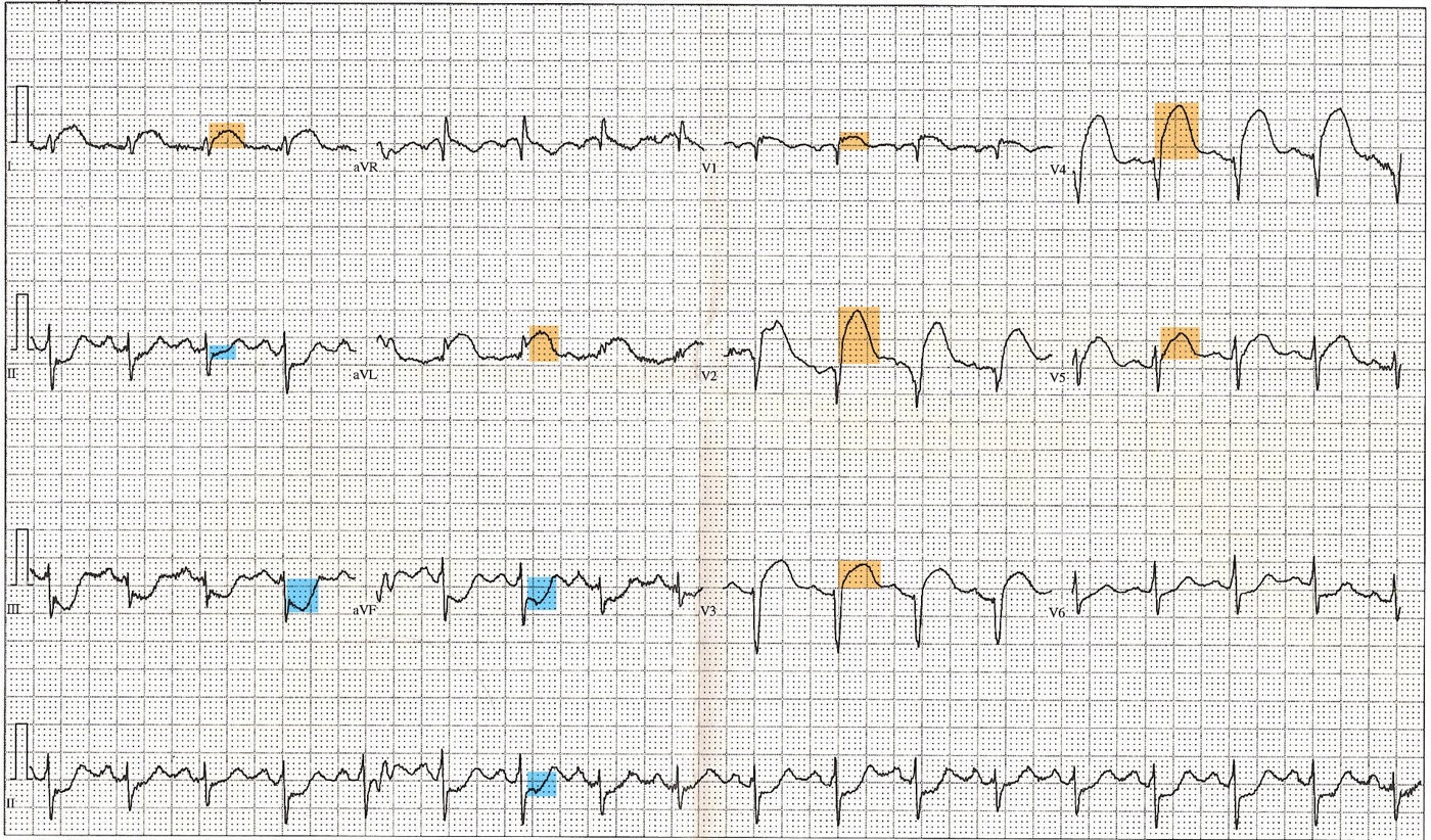
In inferior infarction, ST elevation greater in lead III than in lead II with ST depression in lead I favors the right coronary artery, because the injury vector points rightward toward lead III. ST elevation in lead II equal to or greater than in lead III, ST elevation in lateral leads, and an isoelectric or elevated ST segment in lead I favor a dominant circumflex artery. ST depression in V1 to V3 accompanying inferior elevation indicates posterior extension, which is common with circumflex occlusion.

Posterior and right-sided ECG leads
Posterior leads. The standard 12-lead ECG has no lead facing the inferobasal (formerly posterior) wall, which is supplied by the circumflex artery or the posterolateral branches of a dominant right coronary artery. Occlusion of these vessels produces ST depression in V1 to V3, maximal in V2 or V3, with upright T waves and, as the infarct evolves, tall broad R waves (the mirror image of ST elevation and Q waves). Leads V7, V8, and V9 are placed in the fifth intercostal space at the posterior axillary line, the tip of the left scapula, and the left paraspinal border. ST elevation of 0.5 mm or more in these leads (1 mm or more in men younger than 40 years) confirms posterior infarction. Posterior leads are recorded in every patient with ST depression maximal in V1 to V3, because isolated posterior infarction otherwise passes as NSTEMI and is denied emergency reperfusion.
Right-sided leads. Right ventricular infarction complicates up to half of inferior infarctions from proximal right coronary occlusion. Lead V4R, placed in the fifth intercostal space at the right midclavicular line, is the most sensitive lead; ST elevation of 0.5 mm or more (1 mm or more in men younger than 30 years) indicates right ventricular involvement. ST elevation in V1 in an inferior infarction, or ST elevation in V1 greater than in V2, also suggests it. Right-sided ST elevation is transient and can resolve within hours, so V4R is recorded at presentation in every inferior STEMI.
STEMI equivalents: posterior infarction, de Winter T waves, and hyperacute T waves
A STEMI equivalent is an ECG pattern that indicates acute coronary occlusion with a high probability despite the absence of ST elevation meeting threshold criteria. Patients with an equivalent pattern and an ischemic syndrome are managed with emergency reperfusion.
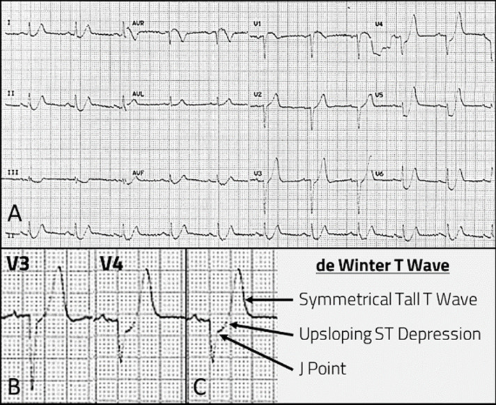
de Winter T waves. The de Winter pattern consists of 1 to 3 mm of upsloping ST depression at the J point in the precordial leads, continuing into tall, positive, symmetric T waves, often with 1 to 2 mm of ST elevation in aVR and loss of precordial R-wave progression. It was described in about 2% of patients with acute anterior infarction in a primary PCI database, and angiography showed proximal left anterior descending occlusion. The pattern is static, does not evolve into classic ST elevation in many patients, and can persist until the artery is opened. It is easily missed because the tracing appears benign, and it is managed as STEMI.
Hyperacute T waves. In the first minutes of occlusion, T waves in the territory become broad at the base, symmetric, and tall relative to the QRS complex, sometimes with a straightened ST segment, before ST elevation reaches threshold. Hyperacute T waves are relative, so they are identified by proportion to the QRS complex, by regional distribution, by reciprocal change, and by evolution on serial tracings. Hyperkalemia, early repolarization, and left ventricular hypertrophy produce tall T waves that are narrow-based and peaked or diffuse.
| Pattern | ECG features | Usual lesion | Management |
|---|---|---|---|
| Posterior STEMI | ST depression maximal in V1–V3, upright T waves; ST elevation ≥0.5 mm in V7–V9 | Circumflex or posterolateral branch occlusion | Emergency reperfusion |
| de Winter T waves | Upsloping J-point depression into tall symmetric precordial T waves; often ST elevation in aVR | Proximal left anterior descending occlusion | Emergency reperfusion |
| Hyperacute T waves | Broad, symmetric T waves disproportionate to the QRS in a territory, with reciprocal change | Early acute occlusion | Serial ECGs every 15 to 30 minutes; emergency angiography when occlusion is evident |
| Left bundle branch block or paced rhythm meeting Sgarbossa or Smith-modified Sgarbossa criteria | Concordant ST elevation or depression, or excessive discordant ST elevation | Acute occlusion | Emergency reperfusion |
| Wellens pattern | Biphasic or deeply inverted T waves in V2–V3 after pain resolves; preserved R waves | Critical proximal left anterior descending stenosis, reperfused | Early angiography; stress testing contraindicated |
| ST elevation in aVR with diffuse ST depression | ST depression ≥1 mm in six or more leads, maximal in V4–V6, with ST elevation in aVR | Left main or three-vessel disease, or global subendocardial ischemia from another cause | Urgent angiography when ischemic and refractory; not a STEMI equivalent for fibrinolysis |
ECG patterns of acute coronary occlusion and high-risk ischemia that do not meet standard ST-elevation criteria.
Wellens pattern and ST elevation in aVR with diffuse ST depression
Wellens pattern. The Wellens pattern, recorded in a pain-free interval after an episode of angina, consists of biphasic T waves (type A, an initial positive and terminal negative deflection) or deep symmetric T-wave inversion (type B, the more common form) in V2 and V3, sometimes extending to V1 through V6, with little or no ST elevation, preserved precordial R waves, and no Q waves. It reflects reperfusion of the territory of a critical proximal left anterior descending stenosis after transient occlusion. In the original series, 26 of 145 patients admitted with unstable angina (18%) had the pattern. Of the 16 treated without surgery, 12 (75%) developed an anterior infarction, usually extensive, within weeks despite nitrates and beta-blockade. The pattern is a reperfusion finding: if the artery reoccludes, the T waves pseudonormalize and ST elevation returns. Patients with the Wellens pattern undergo early angiography, and stress testing is contraindicated because it can precipitate occlusion. Persistent juvenile T-wave inversion, right ventricular strain, cerebral T waves, and post-pacing memory T waves produce similar morphology in the absence of an ischemic history.
ST elevation in aVR with diffuse ST depression. ST depression of 1 mm or more in six or more leads, maximal in V4 to V6, with ST elevation in aVR (and often V1) indicates diffuse subendocardial ischemia. In a patient with ongoing ischemic symptoms it is associated with left main or three-vessel disease, and it also occurs with severe aortic stenosis, hypotension, profound anemia, tachyarrhythmia, and after resuscitation. It does not, by itself, indicate acute left main occlusion: an acutely occluded left main artery more often produces shock, arrest, or extensive anterolateral ST elevation. The pattern is classified with NSTE-ACS; patients with refractory ischemia or hemodynamic instability undergo immediate angiography, and the probability of needing surgical revascularization informs the timing of P2Y12 inhibitor loading.
Infarction in left bundle branch block and ventricular paced rhythm
Left bundle branch block and right ventricular pacing alter ventricular depolarization and produce secondary repolarization changes: ST segments and T waves are normally discordant, directed opposite to the main QRS deflection. ST elevation of several millimeters in V1 to V3 is therefore expected and does not meet standard criteria. In patients with ischemic symptoms, the 2013 American College of Cardiology Foundation/American Heart Association (ACCF/AHA) STEMI guideline withdrew the earlier practice of treating presumed new left bundle branch block as a STEMI equivalent, because most such patients do not have an acute occlusion. Infarction in these rhythms is recognized by repolarization that departs from the expected discordance.
Original Sgarbossa criteria. Derived from patients with left bundle branch block in the GUSTO-1 trial, the rule assigns 5 points for concordant ST elevation of 1 mm or more in any lead, 3 points for concordant ST depression of 1 mm or more in V1, V2, or V3, and 2 points for discordant ST elevation of 5 mm or more. A score of 3 or more is highly specific for acute infarction. A meta-analysis of 10 studies with 1,614 patients found that a score of 3 or more had a pooled sensitivity of 20% and specificity of 98%. A score of 0 does not exclude infarction, and the 5-mm discordance criterion alone (a score of 2) has poor positive predictive value.
Smith-modified Sgarbossa criteria. The modification replaces the absolute 5-mm discordance criterion with a proportional one, recognizing that large QRS complexes carry large discordant ST segments. The rule is positive with any one of three findings: concordant ST elevation of 1 mm or more in any lead; concordant ST depression of 1 mm or more in V1 to V3; or discordant ST elevation of 1 mm or more with an ST/S ratio of −0.25 or less in any lead (ST elevation at the J point at least one-quarter of the depth of the preceding S wave). In the derivation study, sensitivity for angiographic occlusion was 91% with specificity of 90%, against 52% and 98% for the weighted original rule. In a retrospective validation of 45 occlusion cases and 249 controls, sensitivity was 80% against 49% for the weighted rule, with specificity of 99% and 100%.
Ventricular paced rhythm. In a multicenter study of patients with ventricular paced rhythm, the modified criteria had a sensitivity of 81% for occlusion myocardial infarction against 56% for the original criteria, with specificity of 96% and 97% in emergency department controls without occlusion. Adding concordant ST depression in V4 to V6 raised sensitivity to 86%.
| Rule | Positive findings | Reported sensitivity | Reported specificity |
|---|---|---|---|
| Original Sgarbossa, weighted (score ≥3) | Concordant ST elevation ≥1 mm (5 points); concordant ST depression ≥1 mm in V1–V3 (3 points); discordant ST elevation ≥5 mm (2 points) | 20% (meta-analysis); 49%–52% (occlusion studies) | 98%–100% |
| Smith-modified Sgarbossa (any one criterion) | Concordant ST elevation ≥1 mm; concordant ST depression ≥1 mm in V1–V3; discordant ST elevation ≥1 mm with ST/S ratio ≤ −0.25 | 80%–91% | 90%–99% |
| Modified Sgarbossa in ventricular paced rhythm | As above | 81% | 96% |
| Barcelona algorithm | Concordant ST deviation ≥1 mm in any lead; discordant ST deviation ≥1 mm in leads with maximal QRS amplitude ≤6 mm | 93%–95% | 89%–94% |
ECG rules for acute coronary occlusion in left bundle branch block and ventricular paced rhythm, from the derivation and validation studies cited. Performance varies with the reference standard (angiographic occlusion or any infarction) and the control population.
Sgarbossa and Smith-modified Sgarbossa calculator
The calculator applies both rules to measurements from a tracing with left bundle branch block or ventricular pacing. ST deviation is measured at the J point relative to the PR segment, to the nearest 0.5 mm. For the proportional criterion, the lead with the greatest ratio of discordant ST elevation to the depth of the preceding S wave is entered; in V1 to V3 this is usually the lead with the largest ST elevation relative to its S wave, not necessarily the lead with the largest absolute ST elevation.
Original weighted rule: 5 points for concordant ST elevation ≥1 mm in any lead, 3 points for concordant ST depression ≥1 mm in V1–V3, and 2 points for discordant ST elevation ≥5 mm; positive at ≥3. Smith-modified rule: positive with concordant ST elevation ≥1 mm, concordant ST depression ≥1 mm in V1–V3, or discordant ST elevation ≥1 mm with ST/S ratio ≤ −0.25. The ratio is reported as negative by convention because the ST segment and S wave are of opposite polarity.
Conditions that mimic STEMI on the ECG
ST elevation without acute coronary occlusion is common in emergency department patients with chest pain. The distinction relies on morphology, distribution, reciprocal change, evolution on serial tracings, comparison with prior ECGs, and bedside echocardiography. When doubt persists in a patient with ongoing ischemic symptoms, emergency angiography resolves it; fibrinolysis is not given for an uncertain diagnosis.
| Condition | Distinguishing ECG features | Other findings |
|---|---|---|
| Acute pericarditis | Diffuse concave ST elevation across coronary territories; PR depression (PR elevation in aVR); no reciprocal depression except in aVR and V1; no Q waves; evolution through T-wave inversion only after ST segments normalize | Pleuritic, positional pain; friction rub; effusion without regional wall-motion abnormality |
| Benign early repolarization | Concave ST elevation with J-point notching or slurring, maximal in V2–V4; tall T waves proportionate to QRS; stable over time | Young adults, athletes; unchanged from prior tracings |
| Left ventricular hypertrophy | Discordant ST elevation in V1–V3 proportional to deep S waves; lateral strain pattern | Hypertension, aortic stenosis; prior tracings identical |
| Left ventricular aneurysm | Persistent ST elevation with deep Q waves in an old infarct territory; small T waves relative to QRS | History of infarction; dyskinetic segment on echocardiography |
| Brugada pattern (type 1) | Coved ST elevation ≥2 mm in V1–V2 with descending ST into negative T wave; pseudo-right bundle branch block | Fever or sodium-channel blocking drugs unmask it; syncope or family history of sudden death |
| Takotsubo syndrome | ST elevation, usually precordial, with little reciprocal change; later deep diffuse T-wave inversion with QT prolongation | Postmenopausal women; emotional or physical trigger; apical ballooning beyond one coronary territory; diagnosis requires angiography |
| Hyperkalemia | Peaked narrow-based T waves; QRS widening; pseudo-ST elevation in V1–V2 | Kidney disease; corrected with potassium lowering |
| Pulmonary embolism | ST elevation in V1–V3 or aVR; right ventricular strain with precordial T-wave inversion; sinus tachycardia | Hypoxemia; right ventricular dilation |
| Myocarditis | Regional or diffuse ST elevation that can mimic infarction | Viral prodrome; subepicardial or midwall enhancement on cardiac magnetic resonance |
| Aortic dissection | Inferior ST elevation when the dissection involves the right coronary ostium | Pulse deficit, aortic regurgitation, mediastinal widening; reperfusion therapy harmful |
| Intracranial hemorrhage and other neurologic catastrophes | Deep T-wave inversion, QT prolongation, occasional ST elevation | Neurologic findings; fibrinolysis contraindicated |
Differential diagnosis of ST elevation.
Troponin, echocardiography, and imaging in suspected STEMI
Troponin. High-sensitivity cardiac troponin is drawn at presentation, but reperfusion decisions in STEMI are made from the ECG and the clinical syndrome without waiting for the result. Troponin can be normal in the first hour after occlusion. The troponin peak correlates roughly with infarct size, and serial values confirm the diagnosis, identify type 2 infarction or nonischemic injury when the angiogram shows no culprit, and establish a baseline for recognizing reinfarction. Reinfarction after STEMI is diagnosed by a rise of more than 20% from a stable or falling value with new ischemic features; when the baseline value is still rising, the diagnosis rests on recurrent symptoms and ECG changes.
Echocardiography. Bedside transthoracic echocardiography is not required before primary PCI but is valuable when the ECG is nondiagnostic (a regional wall-motion abnormality supports occlusion, and normal regional function in a patient with ongoing pain makes a large acute infarction unlikely), when the patient is hypotensive, and when a mechanical complication or alternative diagnosis is suspected. Echocardiography during the hospitalization assesses left ventricular ejection fraction, regional wall motion and its correspondence to the culprit territory, right ventricular function, mitral regurgitation and its mechanism, pericardial effusion, and left ventricular thrombus. A wall-motion score index derived from 16 or 17 segments quantifies the extent of dysfunction. Ultrasound-enhancing agents improve endocardial definition and the detection of apical thrombus.
Angiographic grading of flow. Antegrade flow in the infarct-related artery is graded by the TIMI scale. TIMI 3 flow after reperfusion is associated with lower mortality than TIMI 2 flow.
| TIMI flow grade | Definition |
|---|---|
| 0 | No antegrade flow beyond the occlusion |
| 1 | Contrast penetrates the occlusion but does not opacify the distal bed |
| 2 | Contrast opacifies the entire distal vessel, but filling or clearance is slower than in a normal artery |
| 3 | Normal antegrade filling and clearance |
TIMI flow grades.
Cardiac magnetic resonance. Late gadolinium enhancement with a subendocardial-to-transmural distribution in a coronary territory confirms ischemic infarction and quantifies infarct size and transmurality; segments with more than 50% transmural enhancement are unlikely to recover function. T2-weighted imaging or T1 and T2 mapping delineate edema, and the difference between the edematous area at risk and the infarct defines myocardial salvage. Microvascular obstruction appears as a hypoenhanced core within enhanced infarct, and intramyocardial hemorrhage as a hypointense core on T2-star imaging. Cardiac magnetic resonance also detects small left ventricular thrombi missed by echocardiography and distinguishes infarction from myocarditis and takotsubo syndrome when the angiogram shows no culprit.
Coronary CT angiography has no role in the evaluation of suspected STEMI, which proceeds directly to invasive angiography. CT angiography of the aorta is obtained when aortic dissection is suspected.
Prehospital ECG and regional STEMI systems of care
The total ischemic time, from symptom onset to restoration of flow, determines the extent of necrosis, and most of it elapses before the patient reaches a hospital. Delay is reduced at three points: by the patient (recognition of symptoms and a call to emergency medical services instead of self-transport), by the system (prehospital ECG, field activation, and direct transport), and by the hospital (door-to-device and door-in–door-out processes).
The 2025 ACC/AHA guideline recommends that patients with suspected STEMI be transported by emergency medical services directly to a PCI-capable hospital, with a first-medical-contact-to-device goal of 90 minutes or less (Class 1). A prehospital 12-lead ECG, transmitted or interpreted in the field, allows activation of the interventional team before arrival and bypass of the emergency department, which shortens door-to-device times. Regional systems link emergency medical services, non-PCI-capable hospitals, and PCI centers through prespecified transfer protocols, single-call activation, and shared quality data. Hospitals without PCI capability operate a protocol that specifies the reperfusion strategy by the anticipated transfer time, and the decision is made at first medical contact.
Reperfusion time targets
| Interval | Target | Setting |
|---|---|---|
| First medical contact to ECG | ≤10 min | All settings |
| First medical contact to device | ≤90 min | Direct presentation or transport to a PCI-capable hospital (Class 1) |
| First medical contact to device | ≤120 min | Transfer from a non-PCI-capable hospital (Class 1) |
| Door-in–door-out at the referring hospital | ≤30 min | Interhospital transfer for primary PCI |
| Door to needle | ≤30 min | Fibrinolysis when primary PCI cannot be achieved within 120 min |
| Symptom onset to fibrinolysis | ≤12 h | Fibrinolytic window; benefit greatest in the first 2 to 3 hours |
| Fibrinolysis to assessment of reperfusion | 60–90 min | ST-segment resolution ≥50% (anterior) or ≥70% (inferior) with symptom relief; otherwise rescue PCI |
| Fibrinolysis to routine angiography | 2–24 h | After successful fibrinolysis (Class 1) |
| Primary PCI 12 to 24 hours after onset | Reasonable | Class 2a, especially with ongoing ischemia |
| Stable, occluded artery more than 24 hours after onset | No routine PCI | Class 3: No Benefit |
Reperfusion time targets in STEMI, after the 2025 ACC/AHA/ACEP/NAEMSP/SCAI acute coronary syndrome guideline and the 2013 ACCF/AHA STEMI guideline (door-in–door-out and door-to-needle). The 2023 ESC guideline measures delay from the time of STEMI diagnosis: primary PCI is preferred when the anticipated time from diagnosis to wire crossing is 120 minutes or less, and fibrinolysis is otherwise given within 10 minutes of diagnosis.
The 90- and 120-minute targets are system goals. Mortality rises with door-to-device time in registries, and the advantage of primary PCI over fibrinolysis narrows as the PCI-related delay lengthens, particularly in young patients with large anterior infarctions presenting within 2 hours. Patients with cardiogenic shock or acute severe heart failure undergo emergency angiography regardless of time from onset or of prior fibrinolysis, and patients with a contraindication to fibrinolysis are transferred for primary PCI regardless of the anticipated delay.
Choosing primary PCI or fibrinolysis
The choice of reperfusion strategy depends on the time from symptom onset, the anticipated first-medical-contact-to-device time, contraindications to fibrinolysis, and the presence of shock or ongoing ischemia.
Strategy by the 2025 ACC/AHA/ACEP/NAEMSP/SCAI acute coronary syndrome guideline. Primary PCI within 12 hours: Class 1 when first-medical-contact-to-device time is ≤90 minutes at a PCI-capable hospital or ≤120 minutes with transfer. Fibrinolysis within 12 hours when the anticipated delay exceeds 120 minutes and no contraindication exists: Class 1. Primary PCI 12 to 24 hours after onset: Class 2a. PCI of a totally occluded artery beyond 24 hours in a stable patient without ongoing ischemia: Class 3: No Benefit. Emergency revascularization in cardiogenic shock regardless of time from onset: Class 1.
Landmark trials of reperfusion in STEMI
| Trial | Comparison | Principal result |
|---|---|---|
| ISIS-2 (1988) | Streptokinase 1.5 million units, aspirin 160 mg daily, both, or neither in 17,187 patients within 24 hours of onset | 5-week vascular mortality 13.2% with neither and 8.0% with both; each agent reduced mortality independently |
| GUSTO-I (1993) | Accelerated alteplase with intravenous heparin against streptokinase regimens | 30-day mortality 6.3% with accelerated alteplase against 7.2% and 7.4% with streptokinase; hemorrhagic stroke 0.72% against 0.49% and 0.54% |
| Meta-analysis of 23 trials (2003) | Primary angioplasty against fibrinolysis | Short-term death 7% against 9%, nonfatal reinfarction 3% against 7%, and stroke 1% against 2% |
| DANAMI-2 (2003) | Transfer for primary angioplasty against on-site fibrinolysis | Death, reinfarction, or disabling stroke at 30 days 8.5% against 14.2% in patients randomized at referral hospitals; 96% transferred within 2 hours; benefit driven by reinfarction |
| TRANSFER-AMI (2009) | Routine transfer for PCI within 6 hours of fibrinolysis against standard care in high-risk patients | 30-day composite 11.0% against 17.2%; no difference in major bleeding |
| STREAM (2013) | Prehospital tenecteplase with angiography 6 to 24 hours later against primary PCI within 3 hours of onset when PCI could not be performed within 1 hour | 30-day composite 12.4% against 14.3%; intracranial hemorrhage 1.0% against 0.2%, falling to 0.5% against 0.3% after the dose was halved in patients 75 years and older |
Landmark trials of reperfusion in STEMI.
Primary PCI: access, stents, thrombus aspiration, and intravascular imaging
Primary PCI restores TIMI 3 flow in most patients, treats the culprit lesion definitively, defines the coronary anatomy and left ventricular filling pressure, and carries a lower risk of intracranial hemorrhage than fibrinolysis. Its benefit over fibrinolysis depends on prompt delivery by experienced operators in high-volume systems.
Radial versus femoral access. Radial access is recommended over femoral access in patients with acute coronary syndrome undergoing PCI to reduce bleeding, vascular complications, and death (Class 1). In MATRIX, which enrolled 8,404 patients with acute coronary syndrome, radial access reduced net adverse clinical events (9.8% against 11.7%), driven by less major bleeding (1.6% against 2.3%) and lower all-cause mortality (1.6% against 2.2%). Femoral access, ideally guided by ultrasound, remains appropriate when large-bore mechanical circulatory support is anticipated or radial access fails.
Drug-eluting versus bare-metal stents. Drug-eluting stents are standard for primary PCI and are superior to bare-metal stents in reducing target-vessel revascularization without an increase in stent thrombosis. Balloon angioplasty alone is reserved for vessels too small to stent or for deferred stenting in exceptional circumstances.
Thrombus aspiration. Routine manual aspiration thrombectomy before primary PCI is not recommended (Class 3: No Benefit). In TOTAL, which randomized 10,732 patients, cardiovascular death, recurrent infarction, cardiogenic shock, or class IV heart failure at 180 days occurred in 6.9% with thrombectomy and 7.0% with PCI alone, and stroke within 30 days was more frequent with thrombectomy (0.7% against 0.3%). A large registry-based trial found no mortality benefit. Selective or bailout aspiration remains available for a large residual thrombus after initial wire passage or balloon dilation.
Intravascular imaging. Intravascular ultrasound or optical coherence tomography is recommended to guide stent implantation in the left main artery or in complex lesions in patients with acute coronary syndrome (Class 1). Imaging defines vessel size, landing zones, calcium, and the mechanism of stent failure, and it confirms stent expansion and apposition.
Pharmacologic and procedural adjuncts. Adjunctive glycoprotein IIb/IIIa inhibitors are reasonable for large thrombus burden, slow flow, or no-reflow (Class 2a), and routine use is harmful because of excess bleeding without ischemic benefit (Class 3: Harm). No-reflow is treated with intracoronary vasodilators (adenosine, nitroprusside, or verapamil) delivered distally. Deferred stenting, routine direct stenting, and routine postconditioning have not improved clinical outcomes in randomized trials.
Culprit-only versus complete revascularization
About half of STEMI patients have significant stenoses in arteries other than the culprit. In hemodynamically stable patients with STEMI and multivessel disease, PCI of significantly stenosed nonculprit arteries after successful PCI of the infarct-related artery is recommended to reduce death or myocardial infarction and to improve angina-related quality of life (Class 1). Significant stenosis is defined as a visually estimated diameter stenosis of 70% or more (50% or more in the left main). In patients with low-complexity multivessel disease not intended for surgery, multivessel PCI at the time of primary PCI may be preferred over a staged approach (Class 2b). In COMPLETE, which enrolled 4,041 patients, cardiovascular death or myocardial infarction at a median of 3 years occurred in 7.8% with complete revascularization and 10.5% with culprit-only PCI (hazard ratio 0.74). The benefit was consistent whether nonculprit PCI was performed during the index admission or within 45 days. In FIRE, which enrolled 1,445 patients 75 years and older with myocardial infarction (35% with STEMI), physiology-guided complete revascularization reduced death, myocardial infarction, stroke, or ischemia-driven revascularization at 1 year (15.7% against 21.0%).
Cardiogenic shock is the exception. In patients with STEMI complicated by cardiogenic shock, routine PCI of nonculprit arteries at the time of primary PCI should not be performed because of a higher risk of death or kidney failure (Class 3: Harm). In CULPRIT-SHOCK, death or renal-replacement therapy at 30 days occurred in 45.9% with culprit-only PCI and 55.4% with immediate multivessel PCI. Nonculprit lesions in shock survivors are revascularized later, when the patient is stable.
Coronary artery bypass surgery is the primary reperfusion strategy only in rare circumstances: anatomy unsuitable for PCI with a large territory at risk and ongoing ischemia, failed PCI, or a mechanical complication that requires surgical repair, at which time bypass grafting accompanies the repair. Surgery for multivessel disease after STEMI is usually performed after several days, when the infarct has stabilized and the effect of the P2Y12 inhibitor has waned.
Fibrinolytic agents and dosing
In the absence of contraindications, fibrinolytic therapy is recommended for STEMI within 12 hours of symptom onset when primary PCI cannot be performed within 120 minutes of first medical contact (Class 1). Fibrinolysis is most effective early, when thrombus is fresh and fibrin-rich, and its benefit declines with time; beyond 12 hours, its benefit is not established, and it is considered only when PCI is unavailable and a large area of myocardium is at risk or the patient is hemodynamically unstable. A fibrin-specific agent (tenecteplase, reteplase, or alteplase) is preferred. Fibrinolysis is not given for ST depression alone, except when true posterior STEMI is suspected (Class 3: Harm), and it is not used in NSTE-ACS.
| Agent | Dose | Features |
|---|---|---|
| Tenecteplase | Single intravenous bolus by weight: <60 kg 30 mg; 60–69 kg 35 mg; 70–79 kg 40 mg; 80–89 kg 45 mg; ≥90 kg 50 mg | Fibrin-specific; single bolus suited to prehospital use; half dose used in patients 75 years and older in STREAM |
| Reteplase | 10 units intravenous bolus, repeated after 30 minutes | Fibrin-specific; double bolus |
| Alteplase (accelerated) | 15 mg bolus, then 0.75 mg/kg over 30 minutes (maximum 50 mg), then 0.5 mg/kg over 60 minutes (maximum 35 mg); total no more than 100 mg | Fibrin-specific; 90-minute infusion |
| Streptokinase | 1.5 million units over 30 to 60 minutes | Not fibrin-specific; antigenic; hypotension; not available in the United States |
Fibrinolytic agents for STEMI, after the 2013 ACCF/AHA STEMI guideline. All are given with aspirin, clopidogrel, and a parenteral anticoagulant.
Adjunctive antithrombotic therapy with fibrinolysis. Aspirin (162 to 325 mg loading dose) and clopidogrel are given with fibrinolysis. Clopidogrel is the only P2Y12 inhibitor with a Class 1 recommendation in this setting. The loading dose is 300 mg in patients younger than 75 years; patients 75 years and older receive 75 mg without a loading dose. In CLARITY-TIMI 28, clopidogrel added to aspirin and fibrinolysis in patients up to 75 years old reduced the composite of an occluded infarct artery, death, or reinfarction from 21.7% to 15.0% without more major bleeding. Ticagrelor and prasugrel have not been established as adjuncts to fibrinolysis. A parenteral anticoagulant is given for the hospitalization (up to 8 days) or until revascularization (Class 1). Enoxaparin is preferred over unfractionated heparin when no invasive strategy is planned.
| Anticoagulant with fibrinolysis | Dose |
|---|---|
| Enoxaparin, age <75 years | 30 mg intravenous bolus, then 1 mg/kg subcutaneously every 12 hours 15 minutes later (first two doses no more than 100 mg) |
| Enoxaparin, age ≥75 years | No bolus; 0.75 mg/kg subcutaneously every 12 hours (first two doses no more than 75 mg) |
| Enoxaparin, creatinine clearance <30 mL/min | 1 mg/kg subcutaneously every 24 hours |
| Unfractionated heparin | 60 U/kg bolus (maximum 4,000 U), then 12 U/kg/h (maximum 1,000 U/h), adjusted to an activated partial thromboplastin time (aPTT) of 1.5 to 2.0 times control, for 48 hours or until revascularization |
| Fondaparinux | 2.5 mg intravenously, then 2.5 mg subcutaneously daily, when creatinine clearance is above 30 mL/min; not used as the sole anticoagulant during PCI |
Parenteral anticoagulation with fibrinolytic therapy, after the 2013 ACCF/AHA STEMI guideline and the 2025 ACC/AHA acute coronary syndrome guideline.
Contraindications to fibrinolysis
| Absolute contraindications | Relative contraindications |
|---|---|
| Any prior intracranial hemorrhage | History of chronic, severe, poorly controlled hypertension |
| Known structural cerebral vascular lesion, such as an arteriovenous malformation | Significant hypertension at presentation (systolic >180 mm Hg or diastolic >110 mm Hg) |
| Known malignant intracranial neoplasm, primary or metastatic | Ischemic stroke more than 3 months earlier, dementia, or known intracranial pathology not listed as absolute |
| Ischemic stroke within 3 months, except acute ischemic stroke within 4.5 hours | Traumatic or prolonged (more than 10 minutes) cardiopulmonary resuscitation |
| Suspected aortic dissection | Major surgery within 3 weeks |
| Active bleeding or bleeding diathesis, excluding menses | Recent internal bleeding, within 2 to 4 weeks |
| Significant closed-head or facial trauma within 3 months | Noncompressible vascular punctures |
| Intracranial or intraspinal surgery within 2 months | Pregnancy |
| Severe uncontrolled hypertension unresponsive to emergency therapy | Active peptic ulcer |
| For streptokinase, prior treatment within the previous 6 months | Oral anticoagulant therapy |
Contraindications to fibrinolytic therapy in STEMI, after the 2013 ACCF/AHA STEMI guideline (Table 6). A relative contraindication is weighed against the expected benefit, which is largest with a large anterior infarction presenting early.
Intracranial hemorrhage occurs in about 0.5% to 1% of patients treated with fibrinolysis. Its risk rises with older age, female sex, low body weight, prior cerebrovascular disease, hypertension at presentation, and excessive anticoagulation. A sudden change in neurologic status within 24 hours of fibrinolysis is managed as intracranial hemorrhage until excluded by emergency imaging, with discontinuation of all antithrombotic therapy.
Pharmacoinvasive strategy and rescue PCI
After fibrinolysis, every patient is transferred to a PCI-capable hospital (Class 1). Reperfusion is assessed 60 to 90 minutes after the fibrinolytic is given. Successful reperfusion is indicated by relief of pain, resolution of ST elevation by 50% or more in anterior infarction or 70% or more in inferior infarction, measured in the lead with the greatest initial elevation or as the sum of ST deviation, and often an accelerated idioventricular rhythm or abrupt early troponin peak. Persistent pain, ST resolution below these thresholds, hemodynamic or electrical instability, or recurrent ischemia indicates failed reperfusion, and immediate angiography with rescue PCI is recommended (Class 1). Repeat fibrinolysis is not used. In patients with successful fibrinolysis, routine angiography with the intent to perform PCI between 2 and 24 hours afterward is recommended (Class 1). The 2-hour lower limit avoids the period of greatest procedural thrombotic and bleeding risk, and the 24-hour upper limit prevents reocclusion of a residual stenosis.
In STREAM, patients presenting within 3 hours of onset who could not undergo primary PCI within 1 hour of first medical contact had similar 30-day outcomes with prehospital tenecteplase followed by angiography as with primary PCI; emergency angiography was required in about one-third of the fibrinolysis group.
When PCI follows fibrinolysis, the P2Y12 inhibitor is continued or changed according to timing: clopidogrel or ticagrelor (in patients younger than 75 years) can be used within 24 hours of the fibrinolytic, and prasugrel is an option after 24 hours. Patients who received enoxaparin need no additional anticoagulant if the last subcutaneous dose was within 8 hours; an additional 0.3 mg/kg intravenous dose is given if it was 8 to 12 hours earlier.
Antiplatelet and anticoagulant therapy in primary PCI
All patients receive aspirin at presentation and a P2Y12 inhibitor before or at the time of primary PCI. In patients with STEMI managed with primary PCI, prasugrel or ticagrelor is recommended to reduce major adverse cardiovascular events and stent thrombosis (Class 1). The 2023 ESC guideline gives prasugrel preference over ticagrelor (Class 2a) in patients proceeding to PCI, on the basis of ISAR-REACT 5; the 2025 ACC/AHA guideline does not rank the two. Dual antiplatelet therapy with aspirin and an oral P2Y12 inhibitor continues for at least 12 months in patients who are not at high bleeding risk (Class 1).
| Agent | Loading dose | Maintenance | Comments |
|---|---|---|---|
| Aspirin | 162–325 mg chewed (non-enteric-coated) | 75–100 mg daily indefinitely | Given at first medical contact unless allergic |
| Ticagrelor | 180 mg | 90 mg twice daily; 60 mg twice daily beyond 12 months when extended | Reversible; dyspnea and ventricular pauses; aspirin dose above 100 mg reduces its efficacy |
| Prasugrel | 60 mg | 10 mg daily; 5 mg if weight <60 kg | Contraindicated with prior stroke or transient ischemic attack; generally avoided at 75 years or older |
| Clopidogrel | 600 mg with primary PCI | 75 mg daily | When ticagrelor and prasugrel are unavailable or contraindicated, or with oral anticoagulation |
| Cangrelor | 30 µg/kg intravenous bolus | 4 µg/kg/min for at least 2 hours or the duration of PCI | Intravenous, rapid onset and offset; P2Y12-naive patients (Class 2b); transition to an oral agent |
| Unfractionated heparin | 70–100 U/kg bolus; 50–70 U/kg with a glycoprotein IIb/IIIa inhibitor | Additional boluses to the target activated clotting time | Standard procedural anticoagulant |
| Bivalirudin | 0.75 mg/kg bolus | 1.75 mg/kg/h during PCI, continued at full dose for 2–4 hours afterward | Alternative to heparin with less bleeding; infusion reduced to 1 mg/kg/h with creatinine clearance <30 mL/min |
Antiplatelet and anticoagulant dosing in primary PCI for STEMI, after the 2013 ACCF/AHA STEMI guideline and the 2025 ACC/AHA acute coronary syndrome guideline.
Unfractionated heparin is the standard anticoagulant for primary PCI. Bivalirudin is an alternative. In BRIGHT-4, which enrolled 6,016 patients undergoing primary PCI, 93% by radial access, bivalirudin with a high-dose infusion for 2 to 4 hours after PCI reduced the 30-day composite of death or major bleeding (3.06% against 4.39%), all-cause mortality (2.96% against 3.92%), and major bleeding (0.17% against 0.80%) compared with heparin alone. The post-procedural infusion addresses the excess of acute stent thrombosis observed in earlier bivalirudin trials. Fondaparinux is not used as the sole anticoagulant for primary PCI because of catheter thrombosis. Parenteral anticoagulation is generally discontinued after uncomplicated primary PCI.
Oxygen, nitrates, analgesia, beta-blockers, and other early therapy
Oxygen. Supplemental oxygen is recommended when the oxygen saturation is below 90%, to a target of 90% or more (Class 1), and routine oxygen is not recommended when saturation is 90% or more (Class 3: No Benefit). In DETO2X-AMI, which enrolled 6,629 patients with suspected infarction and normal saturation, 1-year mortality was 5.0% with oxygen and 5.1% with ambient air, and troponin peaks were similar.
Nitrates. Sublingual nitroglycerin relieves ischemic pain, and intravenous nitroglycerin is used for persistent pain, hypertension, or pulmonary congestion. Nitrates do not reduce mortality. They are withheld with systolic pressure below 90 mm Hg or 30 mm Hg or more below baseline, marked bradycardia or tachycardia, suspected right ventricular infarction, and use of a phosphodiesterase-5 inhibitor within 24 hours (48 hours for tadalafil). Nitrates do not substitute for reperfusion: ST elevation that resolves with nitroglycerin still warrants urgent angiography, because it indicates a dynamic occlusion.
Analgesia. Intravenous opioids relieve pain and anxiety but delay gastrointestinal absorption of oral P2Y12 inhibitors and reduce their early antiplatelet effect, and they can mask ongoing ischemia. Morphine is used at the lowest effective dose for pain refractory to nitrates. Nonsteroidal anti-inflammatory drugs other than aspirin are not given during hospitalization for STEMI because they increase the risk of reinfarction, rupture, heart failure, and death.
Beta-blockers. Early oral beta-blocker therapy within 24 hours is recommended in patients with acute coronary syndrome without contraindications (Class 1). Intravenous beta-blockers are avoided in patients with signs of heart failure, a low-output state, or risk factors for cardiogenic shock (age over 70 years, systolic pressure below 120 mm Hg, sinus tachycardia above 110 beats/min or heart rate below 60 beats/min, and longer time since onset). In COMMIT, which enrolled 45,852 patients, early intravenous then oral metoprolol prevented about five reinfarctions and five episodes of ventricular fibrillation per 1,000 treated but caused 11 more cases of cardiogenic shock per 1,000, mainly on the first day, with no net effect on mortality. Other contraindications are PR interval above 0.24 seconds, second- or third-degree atrioventricular block without a pacemaker, and active bronchospasm.
Metabolic therapy, electrolytes, statins, and prophylactic antiarrhythmics. Routine intravenous glucose-insulin-potassium, magnesium, and prophylactic antiarrhythmic drugs do not improve outcomes. Glucose is controlled to avoid both hyperglycemia above about 180 mg/dL and hypoglycemia. High-intensity statin therapy begins in hospital. Potassium and magnesium are kept in the normal range in patients with ventricular arrhythmia.
Right ventricular infarction
Right ventricular infarction occurs with proximal right coronary occlusion and, less often, with occlusion of a dominant circumflex artery. The classic triad is hypotension, elevated jugular venous pressure with a Kussmaul sign, and clear lung fields in a patient with inferior infarction. ST elevation of 0.5 mm or more in V4R confirms involvement; echocardiography shows right ventricular dilation and hypokinesis, reduced tricuspid annular plane systolic excursion, and septal flattening. Hemodynamic monitoring shows a right atrial pressure that approaches or exceeds the pulmonary capillary wedge pressure and a steep y descent or square-root sign from pericardial constraint of the dilated right ventricle.
The ischemic right ventricle depends on preload, atrioventricular synchrony, and adequate coronary perfusion pressure. Nitrates, opioids, and diuretics reduce preload and can precipitate profound hypotension. A cautious fluid challenge improves cardiac output in patients without pulmonary congestion and with low or normal filling pressures; excessive volume dilates the right ventricle, shifts the septum toward the left ventricle, and reduces left ventricular filling. Inotropic support with dobutamine is used when output remains low after volume. High-grade atrioventricular block and atrial fibrillation are poorly tolerated, and atrioventricular sequential pacing or prompt cardioversion restores the atrial contribution. Prompt reperfusion of the right coronary artery usually produces rapid recovery of right ventricular function. Percutaneous right ventricular support devices are used in refractory shock. Right ventricular infarction raises the risk of high-grade block, ventricular arrhythmia, and in-hospital death, and survivors usually recover right ventricular function fully.
Cardiogenic shock in STEMI
Cardiogenic shock, sustained hypotension with evidence of hypoperfusion, complicates a minority of STEMIs and is the leading cause of in-hospital death. It results from extensive left ventricular infarction in most cases, and from right ventricular infarction, mechanical complications, or dynamic outflow obstruction in the remainder. Shock develops after hospital admission in many patients, often within the first 24 hours.
Emergency revascularization of the culprit artery is the only intervention with a proven survival benefit and is performed irrespective of time from symptom onset. In the SHOCK trial, mortality was 46.7% with early revascularization against 56.0% with initial medical stabilization at 30 days (not significant) and 50.3% against 63.1% at 6 months. Culprit-only PCI is the index strategy (CULPRIT-SHOCK). Echocardiography identifies mechanical complications, right ventricular infarction, and dynamic left ventricular outflow obstruction, each of which changes treatment.
Routine intra-aortic balloon counterpulsation does not reduce mortality (Class 3: No Benefit). In IABP-SHOCK II, 30-day mortality was 39.7% with the balloon pump and 41.3% with standard care. In selected patients with STEMI and severe or refractory cardiogenic shock, a microaxial flow pump is reasonable to reduce death (Class 2a). In DanGer Shock, which enrolled 360 patients with STEMI-related shock and excluded comatose survivors of cardiac arrest, all-cause death at 180 days was 45.8% with the pump and 58.5% with standard care (hazard ratio 0.74). The pump caused more bleeding, limb ischemia, and renal-replacement therapy (41.9% against 26.7%). Routine venoarterial extracorporeal membrane oxygenation in infarct-related cardiogenic shock is not recommended because it confers no survival benefit (Class 3: No Benefit).
Mechanical complications of STEMI
Mechanical complications occurred in 0.27% of US STEMI hospitalizations between 2003 and 2015, without a decline over time, and in-hospital mortality with a mechanical complication was 42.4%. Late presentation without reperfusion, older age, female sex, first infarction, and fibrinolysis compared with primary PCI raise the risk. Each complication typically appears within the first week, often as sudden hemodynamic deterioration, recurrent pulmonary edema, a new murmur, or electromechanical dissociation. Transthoracic echocardiography is performed immediately, and patients are managed at centers with cardiac surgical expertise (Class 1). Short-term mechanical circulatory support is reasonable to stabilize the patient as a bridge to surgery (Class 2a).
| Complication | Typical timing and setting | Clinical and echocardiographic findings | Treatment |
|---|---|---|---|
| Free-wall rupture | First week, often days 1–5; first infarction, older women, late or no reperfusion | Sudden chest pain, hypotension, electromechanical dissociation; pericardial effusion with tamponade and echodense clot | Emergency surgery; pericardiocentesis only as a bridge |
| Left ventricular pseudoaneurysm | Days to weeks; contained rupture | Narrow neck (ratio of neck to maximal diameter below 0.5), abrupt wall discontinuity, often inferolateral; to-and-fro flow at the neck | Surgical repair because of rupture risk |
| Ventricular septal rupture | Days 3–5 without reperfusion; earlier with fibrinolysis; anterior (apical) or inferior (basal, complex) | New harsh holosystolic murmur and thrill; left-to-right shunt by color Doppler; oxygen saturation step-up from right atrium to pulmonary artery | Afterload reduction and mechanical support; surgical repair, with timing individualized; transcatheter closure in selected patients |
| Papillary muscle rupture | Days 2–7; inferior or inferolateral infarction, often small; posteromedial muscle | Sudden pulmonary edema and shock; murmur soft or absent; flail leaflet with severe eccentric mitral regurgitation; hyperdynamic ventricle | Emergency mitral surgery, usually replacement; transcatheter edge-to-edge repair in prohibitive surgical risk |
| Ischemic mitral regurgitation without rupture | Any time; inferior or large infarction | Tethering of the posterior leaflet; regional dysfunction | Reperfusion and heart failure therapy; reassessment after recovery |
| Dynamic left ventricular outflow obstruction | Early; apical infarction with hyperdynamic base, hypovolemia, or inotropes | Systolic anterior motion; late-peaking outflow gradient | Volume, withdrawal of inotropes, vasoconstrictor, cautious beta-blockade |
Mechanical complications of STEMI, after the 2021 AHA scientific statement on mechanical complications of acute myocardial infarction.
Free-wall rupture. Rupture of the left ventricular free wall causes hemopericardium and tamponade. Acute rupture is usually fatal within minutes; subacute rupture, with a slow leak sealed by clot or pericardial adhesions, presents with recurrent pain, nausea, hypotension, and pericardial effusion, and allows surgical repair with a patch or sutured pledgets. A pseudoaneurysm is a contained rupture walled off by pericardium, with a narrow neck and no myocardium in its wall. Contrast echocardiography, cardiac magnetic resonance, or CT distinguishes it from a true aneurysm.
Ventricular septal rupture. Anterior infarctions cause simple apical defects, and inferior infarctions cause complex basal defects with serpiginous tracts and frequent right ventricular involvement, which carry a worse prognosis. Surgical mortality is high, especially when repair is performed in shock in the first days; delayed repair after stabilization on mechanical support has lower operative mortality in selected survivors, at the risk of deterioration while waiting. Transcatheter occlusion is an option for residual defects and for patients at prohibitive surgical risk.
Papillary muscle rupture. Complete rupture of the trunk is rapidly fatal; partial rupture of a head produces severe, often eccentric regurgitation into a small, noncompliant left atrium, so the murmur may be inaudible and the left ventricle appears hyperdynamic. Transesophageal echocardiography defines the mechanism when transthoracic imaging is inconclusive. Afterload reduction with vasodilators or mechanical support stabilizes the patient before emergency surgery.
Arrhythmias and conduction block after STEMI
Ventricular fibrillation and ventricular tachycardia. Ventricular fibrillation within the first 48 hours, most often in the first hours after occlusion or at reperfusion, results from acute ischemia and reperfusion. It is treated by defibrillation, reperfusion, beta-blockade, and correction of potassium and magnesium; amiodarone or lidocaine is used for recurrent episodes. Early ventricular fibrillation raises in-hospital mortality but does not by itself indicate an implantable cardioverter-defibrillator. Prophylactic lidocaine does not reduce mortality and is not given. Sustained ventricular tachycardia or fibrillation occurring after 48 hours without reversible ischemia reflects a scar substrate and carries a high risk of recurrence. An implantable cardioverter-defibrillator is reasonable for clinically relevant ventricular arrhythmia more than 48 hours and within 40 days after infarction (Class 2a). Electrical storm is treated with revascularization of any residual ischemia, beta-blockade, antiarrhythmic drugs, sedation, and catheter ablation in refractory cases.
Accelerated idioventricular rhythm. A wide-complex rhythm at 50 to 110 beats/min that competes with the sinus rate is common in the first hours after reperfusion and marks successful restoration of flow. It is hemodynamically tolerated and self-limited, and suppression is unnecessary and can provoke asystole.
Supraventricular arrhythmias. Sinus tachycardia indicates pain, anxiety, hypovolemia, heart failure, or shock and is treated by addressing the cause. Atrial fibrillation complicates a minority of STEMIs, more often with heart failure, older age, and right coronary or atrial infarction, and is associated with higher mortality and stroke. Rate control uses beta-blockers when hemodynamics allow, and amiodarone or digoxin when they do not; electrical cardioversion is used for instability. Anticoagulation follows the thromboembolic risk score. Patients who need long-term oral anticoagulation after PCI receive triple therapy for 1 to 4 weeks, after which aspirin is discontinued and a P2Y12 inhibitor, preferably clopidogrel, is continued with the anticoagulant (Class 1).
Bradyarrhythmias and atrioventricular block. Sinus bradycardia and atrioventricular block in inferior infarction are usually at the level of the atrioventricular node, mediated by vagal tone and nodal ischemia, with a narrow junctional escape rhythm; they respond to atropine, rarely need pacing, and usually resolve within days. Atrioventricular block in anterior infarction is infranodal, reflects extensive septal necrosis, and is often preceded by new bundle branch or bifascicular block. The escape rhythm is wide and unreliable, and mortality is high because of the infarct size. Temporary transvenous pacing is indicated for symptomatic or hemodynamically significant bradycardia unresponsive to atropine and for infranodal high-grade block. Permanent pacing is recommended for persistent second-degree Mobitz II block, high-grade block, alternating bundle branch block, or third-degree block that is infranodal or persists after an observation period (Class 1). Transient block in inferior infarction that resolves does not need a permanent pacemaker.
Pericarditis and Dressler syndrome after infarction
Early infarct-associated pericarditis develops within the first days after a transmural infarction, presents with pleuritic and positional pain and a friction rub, and is less common in the reperfusion era. Recurrent pain with persistent or recurrent ST elevation and an enlarging pericardial effusion raises suspicion of subacute free-wall rupture. Aspirin at higher doses is the treatment of choice; nonsteroidal anti-inflammatory drugs other than aspirin and corticosteroids are avoided because they impair infarct healing and raise the risk of rupture. Colchicine is used for recurrent symptoms.
Dressler syndrome (post-myocardial infarction syndrome) is an immune-mediated pericarditis that appears weeks after infarction with fever, pleuritic pain, pericardial and pleural effusions, and elevated inflammatory markers. It has become uncommon with reperfusion and smaller infarcts. Treatment follows the principles for recurrent pericarditis, with aspirin and colchicine. A pericardial effusion in a patient receiving dual antiplatelet therapy and anticoagulation is followed closely for hemorrhagic tamponade.
Left ventricular thrombus and left ventricular aneurysm
Left ventricular thrombus forms in a dyskinetic or akinetic apex, most often after large anterior infarction with apical involvement and reduced ejection fraction, usually within the first 2 weeks. Reperfusion has reduced its frequency, but thrombi are still found on cardiac magnetic resonance in a meaningful minority of patients with anterior STEMI and reduced ejection fraction, a rate higher than echocardiography detects. Contrast-enhanced echocardiography improves sensitivity in patients with a dysfunctional apex, and cardiac magnetic resonance with late gadolinium imaging is the reference method.
The 2022 AHA scientific statement on left ventricular thrombus supports anticoagulation for at least 3 months for a thrombus after infarction, with repeat imaging to confirm resolution before stopping, and identifies a direct oral anticoagulant as a reasonable alternative to warfarin. In patients who also receive antiplatelet therapy after PCI, the combination is limited to the shortest effective duration to reduce bleeding. Evidence for prophylactic anticoagulation in patients with anterior infarction and apical akinesis without thrombus is limited, and its bleeding cost on top of dual antiplatelet therapy is substantial; repeat imaging within the first weeks is used selectively in patients at high risk.
A true left ventricular aneurysm is a thin, scarred, dyskinetic segment, most often apical after left anterior descending occlusion. It produces persistent ST elevation, heart failure, ventricular tachycardia from the border zone, and mural thrombus. Rupture is rare. Surgical aneurysmectomy or ventricular reconstruction is reserved for refractory heart failure or ventricular arrhythmia, usually at the time of bypass surgery.
| Time after infarction | Complications |
|---|---|
| Minutes to hours | Ventricular fibrillation, cardiogenic shock, sinus bradycardia and atrioventricular nodal block in inferior infarction, accelerated idioventricular rhythm at reperfusion, hemorrhagic complications of fibrinolysis |
| First 24–72 hours | Heart failure, right ventricular infarction with hypotension, infranodal block in anterior infarction, atrial fibrillation, early pericarditis, reinfarction |
| Days 2–7 | Free-wall rupture, ventricular septal rupture, papillary muscle rupture, left ventricular thrombus |
| Weeks | Dressler syndrome, pseudoaneurysm, true aneurysm, sustained monomorphic ventricular tachycardia, left ventricular remodeling, depression |
| Months to years | Heart failure with reduced ejection fraction, sudden death, recurrent ischemic events |
Typical timing of complications after STEMI. Every complication can occur outside its usual window.
STEMI after out-of-hospital cardiac arrest
Acute coronary occlusion is the most common identifiable cause of out-of-hospital cardiac arrest with a shockable rhythm. In resuscitated patients with ST elevation on the post-resuscitation ECG, primary PCI is recommended in those who are not comatose and in comatose patients with favorable prognostic features (Class 1). Coma alone does not exclude angiography, although survival with good neurologic outcome is low in comatose patients with unfavorable features such as an unwitnessed arrest, no bystander resuscitation, a nonshockable rhythm, prolonged low-flow time, or severe acidosis. An ECG recorded in the first minutes after return of spontaneous circulation can show transient ischemic changes from the arrest itself, and a repeat tracing several minutes later improves specificity.
In comatose survivors who are electrically and hemodynamically stable without ST elevation, immediate angiography is not recommended (Class 3: No Benefit). In COACT, 90-day survival was 64.5% with immediate and 67.2% with delayed angiography; in TOMAHAWK, 30-day mortality was 54.0% with immediate and 46.0% with delayed or selective angiography, and death or severe neurologic deficit was more frequent with the immediate strategy. Comatose survivors receive deliberate temperature control with prevention of fever, avoidance of hypotension and hypoxemia, and multimodal neuroprognostication no earlier than 72 hours after normothermia. Fibrinolysis given during resuscitation has not improved survival in randomized trials and is not given routinely for cardiac arrest.
STEMI in women, older adults, diabetes, and chronic kidney disease
Women. Women with STEMI are older on average, more often have diabetes and hypertension, and more often present with dyspnea, fatigue, nausea, or back and jaw discomfort. Delays from symptom onset to first medical contact and from arrival to reperfusion are longer, and women are less often treated with guideline-directed therapy and cardiac rehabilitation. Spontaneous coronary artery dissection, plaque erosion, takotsubo syndrome, and myocardial infarction with nonobstructive coronary arteries are more common. Diagnostic and treatment decisions follow the same criteria and time targets as in men. Pregnancy-associated infarction is most often caused by spontaneous coronary artery dissection, especially in the peripartum period; primary PCI is the reperfusion strategy, and fibrinolysis carries a risk of extending a dissection and of placental hemorrhage.
Older adults. Patients 75 years and older account for a large share of STEMI deaths. Atypical presentation, delayed presentation, frailty, cognitive impairment, kidney dysfunction, and bleeding risk complicate care, but age alone does not preclude primary PCI, which is the preferred strategy when available. Fibrinolysis carries a higher risk of intracranial hemorrhage in older patients; in STREAM, the tenecteplase dose was halved in patients 75 years and older. Frailty, cognition, life expectancy, and the patient's goals inform the intensity of care.
Diabetes. Patients with diabetes present more often with atypical or painless symptoms, have more extensive multivessel disease, and have higher mortality, heart failure, and reinfarction rates. Glucose is managed to avoid both hyperglycemia and hypoglycemia. Long-term therapy includes a sodium-glucose cotransporter 2 (SGLT2) inhibitor or glucagon-like peptide 1 receptor agonist with proven cardiovascular benefit for glycemic and cardiovascular indications, and an angiotensin-converting enzyme (ACE) inhibitor or angiotensin receptor blocker (ARB) (Class 1).
Chronic kidney disease. Chronic kidney disease increases ischemic risk, bleeding risk, and the risk of contrast-associated acute kidney injury, and patients with it are less often treated with reperfusion and evidence-based therapy. Primary PCI is not withheld because of kidney function. Contrast-associated kidney injury is reduced by radial access, minimal contrast volume, avoidance of ventriculography, staging of nonculprit procedures, intravenous isotonic hydration when volume status allows, and avoidance of nephrotoxic drugs; N-acetylcysteine and sodium bicarbonate have not reduced kidney injury in randomized trials. Doses of enoxaparin, bivalirudin, and fondaparinux are adjusted for creatinine clearance. Patients receiving dialysis have the highest mortality of any subgroup.
No culprit at angiography: MINOCA and STEMI mimics
Some patients activated for STEMI have no obstructive coronary disease and no occluded artery at angiography. The angiogram is reviewed for a missed occlusion of a small branch, a spontaneously reperfused culprit, embolism, or dissection. When the presentation meets the criteria for myocardial infarction, the working diagnosis is myocardial infarction with nonobstructive coronary arteries (MINOCA), and the mechanism is sought with cardiac magnetic resonance, intravascular imaging, and, selectively, provocative testing for spasm. Cardiac magnetic resonance separates ischemic infarction from myocarditis and takotsubo syndrome, which are not infarctions.
Discharge medicines and secondary prevention after STEMI
Secondary prevention begins in the hospital and is reinforced at discharge, at the first follow-up visit, and in cardiac rehabilitation.
| Therapy | Recommendation | Evidence and notes |
|---|---|---|
| Aspirin | 75–100 mg daily indefinitely | With a P2Y12 inhibitor for at least 12 months unless bleeding risk is high (Class 1) |
| P2Y12 inhibitor | Ticagrelor or prasugrel after primary PCI; clopidogrel after fibrinolysis or with oral anticoagulation | Transition to ticagrelor monotherapy at least 1 month after PCI in patients who tolerated dual therapy with ticagrelor (Class 1); proton pump inhibitor with gastrointestinal bleeding risk (Class 1) |
| High-intensity statin | Atorvastatin 40–80 mg or rosuvastatin 20–40 mg | Class 1 for all patients; ezetimibe may be started concurrently (Class 2b) |
| Nonstatin lipid-lowering therapy | Added when low-density lipoprotein (LDL) cholesterol is ≥70 mg/dL on a maximally tolerated statin (Class 1); reasonable at 55–69 mg/dL (Class 2a) | Ezetimibe, proprotein convertase subtilisin/kexin type 9 (PCSK9) monoclonal antibody, inclisiran, or bempedoic acid; lipid panel 4–8 weeks after a change in therapy |
| ACE inhibitor or ARB | Class 1 with LVEF ≤40%, hypertension, diabetes, or anterior STEMI; Class 2a in others | Started in hospital when blood pressure and kidney function allow |
| Mineralocorticoid receptor antagonist | LVEF ≤40% with heart failure symptoms or diabetes (Class 1) | EPHESUS: eplerenone reduced all-cause mortality (relative risk 0.85); creatinine and potassium thresholds apply |
| Beta-blocker | Early oral therapy (Class 1); long term with LVEF ≤40%, heart failure, arrhythmia, or angina | Benefit with preserved ejection fraction and no other indication is debated (REDUCE-AMI, REBOOT, BETAMI-DANBLOCK) |
| SGLT2 inhibitor | For established indications: heart failure, diabetes, chronic kidney disease | EMPACT-MI did not reduce heart failure hospitalization or death as routine post-infarction therapy |
| Colchicine 0.5 mg daily | May be reasonable (Class 2b) | COLCOT positive; CLEAR SYNERGY neutral |
| Sublingual nitroglycerin | For angina, with instructions to call emergency services for pain lasting more than 5 minutes after one dose | Contraindicated with recent phosphodiesterase-5 inhibitor use |
| Influenza vaccination | Annually (Class 1) | Reduces cardiovascular events after acute coronary syndrome |
Discharge therapy after STEMI, after the 2025 ACC/AHA/ACEP/NAEMSP/SCAI acute coronary syndrome guideline.
Beta-blockers after reperfused infarction with preserved ejection fraction. The evidence for routine long-term beta-blockade comes largely from the prereperfusion era, and recent randomized trials of patients with reperfused infarction and an ejection fraction above 40% have produced conflicting results. In REDUCE-AMI (ejection fraction 50% or more), death or myocardial infarction occurred in 7.9% with a beta-blocker and 8.3% without one over a median of 3.5 years. In REBOOT (ejection fraction above 40%), the primary composite was not reduced (hazard ratio 1.04). In BETAMI-DANBLOCK (ejection fraction 40% or more), death or major adverse cardiovascular events occurred in 14.2% with a beta-blocker and 16.3% without one (hazard ratio 0.85), driven by fewer myocardial infarctions. In an individual-patient meta-analysis of five trials restricted to an ejection fraction of 50% or more, beta-blockade did not reduce death, myocardial infarction, or heart failure (8.1% against 8.3%; hazard ratio 0.97), whereas in an individual-patient meta-analysis of patients with mildly reduced ejection fraction and no heart failure, it reduced that composite (hazard ratio 0.75). Beta-blockade remains indicated for reduced ejection fraction, heart failure, arrhythmia, angina, and hypertension; in other patients, the decision to continue therapy long term is individualized.
Colchicine. In COLCOT, colchicine 0.5 mg daily begun within 30 days of infarction reduced the composite of cardiovascular death, resuscitated arrest, infarction, stroke, or urgent hospitalization for angina leading to revascularization (5.5% against 7.1%), driven by stroke and urgent hospitalization for angina. In CLEAR SYNERGY, which enrolled 7,062 patients started soon after infarction, colchicine did not reduce cardiovascular death, recurrent infarction, stroke, or ischemia-driven revascularization over 3 years (9.1% against 9.3%), and diarrhea was more frequent. The 2025 ACC/AHA guideline rates low-dose colchicine Class 2b.
SGLT2 inhibitors. In EMPACT-MI, empagliflozin started within 14 days of infarction in patients at increased risk of heart failure did not reduce the composite of first hospitalization for heart failure or death (8.2% against 9.1%; hazard ratio 0.90), although first heart failure hospitalization was less frequent. SGLT2 inhibitors are given after infarction for heart failure, diabetes, or chronic kidney disease.
Pre-discharge risk assessment, ejection fraction reassessment, and defibrillator timing
Left ventricular ejection fraction is measured during the hospitalization in every patient with STEMI, most often by echocardiography. Patients who did not undergo angiography, or who have residual multivessel disease, undergo evaluation for ischemia and viability. The GRACE score and clinical variables (age, Killip class, heart rate, systolic pressure, creatinine, cardiac arrest, ST deviation, and troponin) estimate in-hospital and 6-month mortality.
Ejection fraction often improves after reperfusion as stunned myocardium recovers, so the decision about primary-prevention defibrillator implantation is deferred. An implantable cardioverter-defibrillator is recommended for patients with LVEF of 40% or less at least 40 days after infarction and at least 90 days after revascularization who meet the criteria for primary-prevention implantation, with expected survival of at least 1 year: LVEF of 30% or less with NYHA class I to III symptoms, LVEF of 31% to 35% with NYHA class II or III symptoms, or LVEF of 40% or less with inducible ventricular tachycardia (Class 1). In DINAMIT, defibrillator implantation 6 to 40 days after infarction in patients with an LVEF of 35% or less reduced arrhythmic death but increased nonarrhythmic death, with no overall survival benefit (hazard ratio 1.08). A wearable cardioverter-defibrillator in the early period for patients with LVEF of 35% or less is of uncertain usefulness (Class 2b). Echocardiography is repeated about 6 to 12 weeks after the infarction, after optimization of neurohormonal therapy, to determine eligibility. Patients with sustained ventricular arrhythmia more than 48 hours after infarction are evaluated for a secondary-prevention device.
Cardiac rehabilitation, lifestyle, vaccination, and depression screening
Referral to outpatient cardiac rehabilitation before discharge is recommended for every patient with acute coronary syndrome (Class 1), and home-based cardiac rehabilitation is a reasonable alternative for patients unable or unwilling to attend a center (Class 2a). Rehabilitation combines supervised exercise training, risk-factor education, nutritional counseling, psychosocial support, and medication adherence, and it reduces cardiovascular mortality and readmission. Participation remains low, especially among women, older adults, and racial and ethnic minority groups, and automatic referral systems increase enrollment.
Smoking cessation is the most effective single lifestyle change after infarction. Counseling and pharmacotherapy with varenicline, bupropion, or nicotine replacement begin in the hospital, and nicotine replacement is safe in patients with stable coronary disease. Blood pressure is treated to below 130/80 mm Hg, diabetes is managed with agents that reduce cardiovascular risk, and patients receive guidance on diet, physical activity, weight, and alcohol. Return to driving, work, and sexual activity follows the patient's ventricular function, symptoms, and arrhythmic status. Annual influenza vaccination is recommended (Class 1).
Depression is common after myocardial infarction and is associated with lower adherence, recurrent events, and higher mortality. Patients are screened with a validated instrument such as the Patient Health Questionnaire, and positive screens lead to evaluation and treatment, including cognitive behavioral therapy and selective serotonin reuptake inhibitors, which are safe after infarction.
Sources of error in the recognition and treatment of STEMI
- Reliance on millimeter criteria alone: occlusion of the circumflex artery, isolated posterior infarction, high lateral infarction, and early hyperacute changes can fall below threshold. Serial ECGs, posterior leads, reciprocal change, and bedside echocardiography identify many of these occlusions.
- ST depression in V1 to V3 interpreted as anterior subendocardial ischemia: when maximal in V2 or V3 with upright T waves, it is posterior ST elevation seen in mirror image until posterior leads prove otherwise.
- Omission of V4R in inferior infarction: right ventricular involvement is missed, and nitrates or diuretics precipitate hypotension.
- Treatment of left bundle branch block of unknown age as STEMI, or dismissal of a paced tracing as uninterpretable: both rhythms are classified by the Sgarbossa and modified Sgarbossa criteria and by the clinical state.
- Reliance on the door-to-device interval alone: the patient and prehospital segments dominate total ischemic time, and transfer decisions are made from the anticipated first-medical-contact-to-device time.
- Attribution of new hypotension or pulmonary edema in the first week to heart failure without echocardiography: free-wall, septal, and papillary muscle rupture require surgical treatment, and a soft or absent murmur does not exclude papillary muscle rupture.
Self-assessment
A 68-year-old man with a dual-chamber pacemaker and 40 minutes of chest pressure has a ventricular paced rhythm. Lead V3 shows a 16-mm S wave with 5 mm of discordant ST elevation; no lead shows concordant ST elevation, and V1 to V3 show no concordant ST depression. The nearest PCI-capable hospital is 45 minutes away by ground. What is the original weighted Sgarbossa score, does the tracing meet the Smith-modified criteria, and what reperfusion strategy is indicated?
Cardiovascular Medicine Board ReviewReferences
- Rao SV, O’Donoghue ML, Ruel M, et al. 2025 ACC/AHA/ACEP/NAEMSP/SCAI guideline for the management of patients with acute coronary syndromes: a report of the American College of Cardiology/American Heart Association Joint Committee on Clinical Practice Guidelines. Circulation. 2025;151:e771–e862. doi:10.1161/CIR.0000000000001309
- O’Gara PT, Kushner FG, Ascheim DD, et al. 2013 ACCF/AHA guideline for the management of ST-elevation myocardial infarction: a report of the American College of Cardiology Foundation/American Heart Association Task Force on Practice Guidelines. J Am Coll Cardiol. 2013;61:e78–e140. doi:10.1016/j.jacc.2012.11.019
- Byrne RA, Rossello X, Coughlan JJ, et al. 2023 ESC guidelines for the management of acute coronary syndromes. Eur Heart J. 2023;44:3720–3826. doi:10.1093/eurheartj/ehad191
- Thygesen K, Alpert JS, Jaffe AS, et al. Fourth universal definition of myocardial infarction (2018). Circulation. 2018;138:e618–e651. doi:10.1161/CIR.0000000000000617
- Wagner GS, Macfarlane P, Wellens H, et al. AHA/ACCF/HRS recommendations for the standardization and interpretation of the electrocardiogram: part VI: acute ischemia/infarction. J Am Coll Cardiol. 2009;53:1003–1011. doi:10.1016/j.jacc.2008.12.016
- Sgarbossa EB, Pinski SL, Barbagelata A, et al. Electrocardiographic diagnosis of evolving acute myocardial infarction in the presence of left bundle-branch block. N Engl J Med. 1996;334:481–487. doi:10.1056/NEJM199602223340801
- Tabas JA, Rodriguez RM, Seligman HK, Goldschlager NF. Electrocardiographic criteria for detecting acute myocardial infarction in patients with left bundle branch block: a meta-analysis. Ann Emerg Med. 2008;52:329–336.e1. doi:10.1016/j.annemergmed.2007.12.006
- Smith SW, Dodd KW, Henry TD, Dvorak DM, Pearce LA. Diagnosis of ST-elevation myocardial infarction in the presence of left bundle branch block with the ST-elevation to S-wave ratio in a modified Sgarbossa rule. Ann Emerg Med. 2012;60:766–776. doi:10.1016/j.annemergmed.2012.07.119
- Meyers HP, Limkakeng AT Jr, Jaffa EJ, et al. Validation of the modified Sgarbossa criteria for acute coronary occlusion in the setting of left bundle branch block: a retrospective case-control study. Am Heart J. 2015;170:1255–1264. doi:10.1016/j.ahj.2015.09.005
- Dodd KW, Zvosec DL, Hart MA, et al. Electrocardiographic diagnosis of acute coronary occlusion myocardial infarction in ventricular paced rhythm using the modified Sgarbossa criteria. Ann Emerg Med. 2021;78:517–529. doi:10.1016/j.annemergmed.2021.03.036
- Di Marco A, Rodriguez M, Cinca J, et al. New electrocardiographic algorithm for the diagnosis of acute myocardial infarction in patients with left bundle branch block. J Am Heart Assoc. 2020;9:e015573. doi:10.1161/JAHA.119.015573
- de Winter RJ, Verouden NJW, Wellens HJJ, Wilde AAM. A new ECG sign of proximal LAD occlusion. N Engl J Med. 2008;359:2071–2073. doi:10.1056/NEJMc0804737
- de Zwaan C, Bär FW, Wellens HJ. Characteristic electrocardiographic pattern indicating a critical stenosis high in left anterior descending coronary artery in patients admitted because of impending myocardial infarction. Am Heart J. 1982;103:730–736. doi:10.1016/0002-8703(82)90480-x
- Yeh RW, Sidney S, Chandra M, et al. Population trends in the incidence and outcomes of acute myocardial infarction. N Engl J Med. 2010;362:2155–2165. doi:10.1056/NEJMoa0908610
- ISIS-2 (Second International Study of Infarct Survival) Collaborative Group. Randomised trial of intravenous streptokinase, oral aspirin, both, or neither among 17,187 cases of suspected acute myocardial infarction: ISIS-2. Lancet. 1988;2:349–360. doi:10.1016/S0140-6736(88)92833-4
- GUSTO Investigators. An international randomized trial comparing four thrombolytic strategies for acute myocardial infarction. N Engl J Med. 1993;329:673–682. doi:10.1056/NEJM199309023291001
- Keeley EC, Boura JA, Grines CL. Primary angioplasty versus intravenous thrombolytic therapy for acute myocardial infarction: a quantitative review of 23 randomised trials. Lancet. 2003;361:13–20. doi:10.1016/S0140-6736(03)12113-7
- Andersen HR, Nielsen TT, Rasmussen K, et al. A comparison of coronary angioplasty with fibrinolytic therapy in acute myocardial infarction (DANAMI-2). N Engl J Med. 2003;349:733–742. doi:10.1056/NEJMoa025142
- Armstrong PW, Gershlick AH, Goldstein P, et al. Fibrinolysis or primary PCI in ST-segment elevation myocardial infarction (STREAM). N Engl J Med. 2013;368:1379–1387. doi:10.1056/NEJMoa1301092
- Cantor WJ, Fitchett D, Borgundvaag B, et al. Routine early angioplasty after fibrinolysis for acute myocardial infarction (TRANSFER-AMI). N Engl J Med. 2009;360:2705–2718. doi:10.1056/NEJMoa0808276
- Valgimigli M, Gagnor A, Calabró P, et al. Radial versus femoral access in patients with acute coronary syndromes undergoing invasive management: a randomised multicentre trial (MATRIX). Lancet. 2015;385:2465–2476. doi:10.1016/S0140-6736(15)60292-6
- Jolly SS, Cairns JA, Yusuf S, et al. Randomized trial of primary PCI with or without routine manual thrombectomy (TOTAL). N Engl J Med. 2015;372:1389–1398. doi:10.1056/NEJMoa1415098
- Mehta SR, Wood DA, Storey RF, et al. Complete revascularization with multivessel PCI for myocardial infarction (COMPLETE). N Engl J Med. 2019;381:1411–1421. doi:10.1056/NEJMoa1907775
- Biscaglia S, Guiducci V, Escaned J, et al. Complete or culprit-only PCI in older patients with myocardial infarction (FIRE). N Engl J Med. 2023;389:889–898. doi:10.1056/NEJMoa2300468
- Thiele H, Akin I, Sandri M, et al. PCI strategies in patients with acute myocardial infarction and cardiogenic shock (CULPRIT-SHOCK). N Engl J Med. 2017;377:2419–2432. doi:10.1056/NEJMoa1710261
- Sabatine MS, Cannon CP, Gibson CM, et al. Addition of clopidogrel to aspirin and fibrinolytic therapy for myocardial infarction with ST-segment elevation (CLARITY-TIMI 28). N Engl J Med. 2005;352:1179–1189. doi:10.1056/NEJMoa050522
- Li Y, Liang Z, Qin L, et al. Bivalirudin plus a high-dose infusion versus heparin monotherapy in patients with ST-segment elevation myocardial infarction undergoing primary percutaneous coronary intervention: a randomised trial (BRIGHT-4). Lancet. 2022;400:1847–1857. doi:10.1016/S0140-6736(22)01999-7
- Chen ZM, Pan HC, Chen YP, et al. Early intravenous then oral metoprolol in 45,852 patients with acute myocardial infarction: randomised placebo-controlled trial (COMMIT). Lancet. 2005;366:1622–1632. doi:10.1016/S0140-6736(05)67661-1
- Hofmann R, James SK, Jernberg T, et al. Oxygen therapy in suspected acute myocardial infarction (DETO2X-AMI). N Engl J Med. 2017;377:1240–1249. doi:10.1056/NEJMoa1706222
- Hochman JS, Sleeper LA, Webb JG, et al. Early revascularization in acute myocardial infarction complicated by cardiogenic shock (SHOCK). N Engl J Med. 1999;341:625–634. doi:10.1056/NEJM199908263410901
- Thiele H, Zeymer U, Neumann FJ, et al. Intraaortic balloon support for myocardial infarction with cardiogenic shock (IABP-SHOCK II). N Engl J Med. 2012;367:1287–1296. doi:10.1056/NEJMoa1208410
- Møller JE, Engstrøm T, Jensen LO, et al. Microaxial flow pump or standard care in infarct-related cardiogenic shock (DanGer Shock). N Engl J Med. 2024;390:1382–1393. doi:10.1056/NEJMoa2312572
- Damluji AA, van Diepen S, Katz JN, et al. Mechanical complications of acute myocardial infarction: a scientific statement from the American Heart Association. Circulation. 2021;144:e16–e35. doi:10.1161/CIR.0000000000000985
- Elbadawi A, Elgendy IY, Mahmoud K, et al. Temporal trends and outcomes of mechanical complications in patients with acute myocardial infarction. JACC Cardiovasc Interv. 2019;12:1825–1836. doi:10.1016/j.jcin.2019.04.039
- Levine GN, McEvoy JW, Fang JC, et al. Management of patients at risk for and with left ventricular thrombus: a scientific statement from the American Heart Association. Circulation. 2022;146:e205–e223. doi:10.1161/CIR.0000000000001092
- Lemkes JS, Janssens GN, van der Hoeven NW, et al. Coronary angiography after cardiac arrest without ST-segment elevation (COACT). N Engl J Med. 2019;380:1397–1407. doi:10.1056/NEJMoa1816897
- Desch S, Freund A, Akin I, et al. Angiography after out-of-hospital cardiac arrest without ST-segment elevation (TOMAHAWK). N Engl J Med. 2021;385:2544–2553. doi:10.1056/NEJMoa2101909
- Hohnloser SH, Kuck KH, Dorian P, et al. Prophylactic use of an implantable cardioverter-defibrillator after acute myocardial infarction (DINAMIT). N Engl J Med. 2004;351:2481–2488. doi:10.1056/NEJMoa041489
- Pitt B, Remme W, Zannad F, et al. Eplerenone, a selective aldosterone blocker, in patients with left ventricular dysfunction after myocardial infarction (EPHESUS). N Engl J Med. 2003;348:1309–1321. doi:10.1056/NEJMoa030207
- Yndigegn T, Lindahl B, Mars K, et al. Beta-blockers after myocardial infarction and preserved ejection fraction (REDUCE-AMI). N Engl J Med. 2024;390:1372–1381. doi:10.1056/NEJMoa2401479
- Munkhaugen J, Kristensen AMD, Halvorsen S, et al. Beta-blockers after myocardial infarction in patients without heart failure (BETAMI-DANBLOCK). N Engl J Med. 2025;393:1901–1911. doi:10.1056/NEJMoa2505985
- Ibanez B, Latini R, Rossello X, et al. Beta-blockers after myocardial infarction without reduced ejection fraction (REBOOT). N Engl J Med. 2025;393:1889–1900. doi:10.1056/NEJMoa2504735
- Butler J, Jones WS, Udell JA, et al. Empagliflozin after acute myocardial infarction (EMPACT-MI). N Engl J Med. 2024;390:1455–1466. doi:10.1056/NEJMoa2314051
- Tardif JC, Kouz S, Waters DD, et al. Efficacy and safety of low-dose colchicine after myocardial infarction (COLCOT). N Engl J Med. 2019;381:2497–2505. doi:10.1056/NEJMoa1912388
- Jolly SS, d’Entremont MA, Lee SF, et al. Colchicine in acute myocardial infarction (CLEAR SYNERGY). N Engl J Med. 2025;392:633–642. doi:10.1056/NEJMoa2405922
- Kristensen AMD, Rossello X, Atar D, et al. Beta-blockers after myocardial infarction with normal ejection fraction. N Engl J Med. 2026;394:540–550. doi:10.1056/NEJMoa2512686
- Rossello X, Prescott EIB, Kristensen AMD, et al. β blockers after myocardial infarction with mildly reduced ejection fraction: an individual patient data meta-analysis of randomised controlled trials. Lancet. 2025;406:1128–1137. doi:10.1016/S0140-6736(25)01592-2