Library › Emergencies and critical care › Electrical storm
Electrical storm and incessant ventricular tachycardia
Electrical storm and incessant ventricular tachycardia in adults, from definition, triggers, and prognosis through device management, drug therapy, sedation, catheter ablation, mechanical support, autonomic modulation, and prevention of recurrence.
Electrical storm is 3 or more episodes of sustained ventricular tachycardia (VT) or ventricular fibrillation (VF) within 24 hours, separated by at least 5 minutes, each requiring termination by an intervention. Incessant VT is continuous sustained VT that recurs promptly despite repeated termination over several hours. Each shock adds sympathetic drive that lowers the threshold for the next episode. Treatment therefore interrupts the substrate, the trigger, and the autonomic loop together. It combines device interrogation and reprogramming, correction of triggers, sedation, and nonselective beta-blockade with amiodarone in structural heart disease. Early catheter ablation of recurrent monomorphic VT follows, and autonomic modulation or mechanical support is added when the storm is refractory.
Definitions of electrical storm, incessant VT, and clustered ventricular arrhythmia
Electrical storm is defined in clinical practice, device trials, and consensus documents by counting discrete episodes of sustained ventricular arrhythmia within a 24-hour window. The 2019 consensus of the Heart Rhythm Society, European Heart Rhythm Association (EHRA), Asia Pacific Heart Rhythm Society, and Latin American Heart Rhythm Society (HRS/EHRA/APHRS/LAHRS) on catheter ablation defines VT storm as 3 or more separate episodes of sustained VT within 24 hours, each requiring termination by an intervention. The 2024 EHRA clinical consensus statement, endorsed by the Heart Rhythm Society, extends the definition to any sustained ventricular arrhythmia, including ventricular fibrillation. It requires episodes to be separated by at least 5 minutes, so that a single episode with early recurrence after a shock is not counted as several.
An intervention includes antitachycardia pacing (ATP), an internal shock, external cardioversion or defibrillation, or an emergency drug given to terminate the arrhythmia. In a patient with an implantable cardioverter-defibrillator (ICD), storm is therefore recognized from stored device episodes as well as from the clinical record. Device-based counts depend on programming. A patient whose detection zones use short detection durations accrues more treated episodes, some of which would have terminated spontaneously, than a patient with long detection times and high rate cutoffs.
| Term | Definition |
|---|---|
| Electrical storm | 3 or more episodes of sustained ventricular arrhythmia within 24 hours, separated by at least 5 minutes, each requiring termination by an intervention (2024 EHRA) |
| VT storm | 3 or more separate episodes of sustained VT within 24 hours, each requiring termination by an intervention (2019 ablation consensus; the definition used in VANISH) |
| Incessant VT | Continuous sustained VT that recurs promptly despite repeated intervention for termination over several hours |
| Clustered ventricular arrhythmia | 2 or more sustained ventricular arrhythmia events within 3 months |
| Sustained VT | VT lasting at least 30 seconds or requiring an acute intervention for termination |
| Nonsustained VT | 3 or more consecutive ventricular beats lasting less than 30 seconds |
| Monomorphic VT | VT with the same QRS morphology from beat to beat |
| Polymorphic VT | VT with a continually changing QRS morphology |
| Torsades de pointes | Polymorphic VT in the setting of QT prolongation, with QRS complexes that twist around the baseline |
| Bidirectional VT | VT with beat-to-beat alternation of the frontal-plane QRS axis |
| Ventricular fibrillation | A chaotic rhythm with undulations irregular in timing and morphology and no discrete QRS complexes |
Definitions from the 2024 EHRA consensus (Table 3) and the 2019 HRS/EHRA/APHRS/LAHRS ablation consensus. The 5-minute separation in the EHRA definition excludes early recurrence within minutes of a shock from the episode count.
Incessant VT and storm overlap. Incessant VT is typically slower monomorphic VT, often below the programmed detection rate of the ICD, which the patient may tolerate for hours while cardiac output and ventricular function deteriorate. Storm is more often a sequence of separate episodes of faster VT or ventricular fibrillation terminated by the device. Both reflect the same combination of a vulnerable substrate, a precipitating factor, and sympathetic activation, and both are managed by the same principles. The broader ventricular arrhythmia classification and the definitions of sudden cardiac arrest are covered in the ventricular tachycardia hub.
The 2017 American Heart Association/American College of Cardiology/Heart Rhythm Society (AHA/ACC/HRS) guideline and the 2019 ablation consensus address storm within broader recommendations on ventricular arrhythmia and ablation. The 2024 EHRA statement is the only document devoted to storm. It grades its advice by evidence category (randomized, observational, or expert opinion) and does not assign classes of recommendation.
Incidence of electrical storm in ICD recipients and other populations
Most published estimates of storm incidence come from ICD recipients, because the device records every treated episode. Incidence depends on the definition, the indication for the ICD, the programming, and the length of follow-up. The 2024 EHRA consensus summarizes an incidence of 10% to 30% among patients who received an ICD for secondary prevention, with storm occurring on average 4 to 9 months after implantation. Among patients with primary-prevention devices, the incidence is 4% to 7% at 18 to 24 months. Clustered ventricular arrhythmia, the broader category of 2 or more sustained events within 3 months, occurred in 26% of ICD recipients over 40 months.
| Population | Incidence of storm | Timing and features |
|---|---|---|
| AVID, secondary-prevention ICD (457 patients) | 90 patients (20%) over 31 ± 13 months | Storm occurred 9.2 ± 11.5 months after implantation; 86% of storms were due to VT |
| MADIT-II, primary prevention after myocardial infarction (MI) with left ventricular ejection fraction (LVEF) 30% or less (719 patients) | 27 patients (4%) | Patients with interim ventricular arrhythmia or coronary events were at higher risk |
| Transvenous ICD recipients, prospective cohort (136 patients) | 14 patients (10%) over 403 ± 242 days | Mean 133 days after implantation; median of 8 episodes per storm (range 3 to 50) |
| Primary-prevention ICD, coronary disease versus nonischemic cardiomyopathy | 5.8% versus 6.9% over about 51 months | No significant difference between substrates |
| Left ventricular assist device (LVAD) recipients | About 9% | Ventricular arrhythmia in 20% to 50%; late storm beyond 30 days in 4.3% of 1,151 patients |
Incidence of electrical storm in AVID, the MADIT-II substudy, a prospective transvenous ICD cohort, the 2024 EHRA consensus summary, and a 2026 multicenter LVAD registry. AVID, Antiarrhythmics Versus Implantable Defibrillators; MADIT-II, Multicenter Automatic Defibrillator Implantation Trial II.
Patients who develop storm are older and more often men, have lower ejection fraction and more advanced heart failure, and carry more cardiovascular comorbidity than ICD recipients without storm. In observational ICD cohorts, chronic kidney disease, depressed ejection fraction, and sustained monomorphic VT as the index arrhythmia independently predicted storm. A 2014 meta-analysis of 13 studies found that a secondary-prevention indication, lower ejection fraction, monomorphic VT as the triggering arrhythmia, and class I antiarrhythmic drug therapy were associated with storm.
Cardiac resynchronization therapy (CRT) modifies risk. In a propensity-matched comparison, recipients of a CRT-defibrillator had a lower incidence of storm than recipients of an ICD, and responders to resynchronization had the lowest rates. In a minority, newly started left ventricular pacing is itself proarrhythmic, and disabling left ventricular pacing terminates or mitigates the storm.
Storm in patients with a primary electrical disease is uncommon and reported mainly as case series. Brugada syndrome, early repolarization syndrome, idiopathic ventricular fibrillation, congenital and acquired long QT syndrome, short QT syndrome, and catecholaminergic polymorphic VT (CPVT) each produce a distinct pattern, discussed below. Storm can also be the first manifestation of previously unrecognized heart disease.
Prognosis and mortality after electrical storm
Storm is a marker of advanced substrate and a predictor of death, mainly from progressive heart failure. In AVID, storm was an independent predictor of subsequent death (relative risk 2.4, 95% CI 1.3 to 4.2), whereas ventricular arrhythmia unrelated to storm was not (relative risk 1.0). The risk was greatest in the 3 months after the storm (relative risk 5.4) and diminished thereafter (relative risk 1.9). In the MADIT-II substudy, the adjusted hazard ratio for death in the first 3 months after storm was 17.8 (95% CI 8.0 to 39.5). A 1998 prospective cohort of 136 transvenous ICD recipients found no independent excess mortality after storm, an exception attributed to the small number of events.
The 2014 meta-analysis of 5,912 patients, 857 with storm, found a risk ratio for death of 3.15 (95% CI 2.22 to 4.48) and a risk ratio of 3.39 for the composite of death, cardiac transplantation, or heart failure hospitalization. The excess risk persisted when patients with storm were compared with patients who had isolated, unclustered ventricular arrhythmias. The 2024 EHRA consensus summarizes further observations. Storm was associated with a 2.5-fold higher risk of death than sporadic VT and a 3.3-fold higher risk than no sustained ventricular arrhythmia. Admission for storm carried a 4.8-fold higher risk of rehospitalization and a 1.8-fold higher risk of major adverse cardiac events than VT or VF without storm. Five-year survival free of death or transplantation was 67% in patients with clusters of arrhythmia and 87% in those without.
Risk after storm is highest with amiodarone use, chronic kidney disease, and ejection fraction below 35%. Without a specific antiarrhythmic intervention, storm recurs in more than half of patients with structural heart disease, most often within the first year. Ejection fraction of 30% or less, age above 65 years, and the absence of optimal heart failure therapy predicted recurrence in one observational study. Whether storm causes death or marks the patient who is about to die of heart failure is unresolved. Frequent tolerated VT, even when terminated appropriately, can worsen ventricular function and functional class, and the hemodynamic, ischemic, and inflammatory consequences of repeated shocks plausibly contribute.
After catheter ablation of scar-related VT, freedom from recurrence is associated with survival. In 2,061 patients from 12 international centers, 1-year freedom from VT recurrence was 70%, and transplant-free survival was 90% without recurrence and 71% with recurrence. A 2026 meta-analysis of 6 observational cohorts (3,531 patients, 1,183 with storm) found similar acute procedural success with and without storm, but periprocedural or in-hospital mortality was higher with storm (odds ratio 4.71), as were long-term mortality (odds ratio 1.67) and VT recurrence (hazard ratio 1.35).
Substrates of electrical storm: structural heart disease and primary electrical disease
Storm requires a vulnerable heart. In most patients the substrate is heterogeneous fibrosis, in which surviving myocyte bundles within scar form slowly conducting channels that support re-entry. Healed myocardial infarction is the most common substrate; dilated, hypertrophic, and arrhythmogenic right ventricular cardiomyopathy (ARVC), cardiac sarcoidosis, myocarditis, Chagas disease, and valvular cardiomyopathy account for most of the rest. Arrhythmias arising from a diseased conduction system, including bundle branch re-entry and Purkinje-related VT, are a separate group that ablation can cure.
| Arrhythmia responsible for storm | Proportion of storms | Typical substrate |
|---|---|---|
| Sustained monomorphic VT | 86% to 97% | Scar re-entry after MI or in nonischemic cardiomyopathy, ARVC, sarcoidosis; bundle branch re-entry |
| Primary ventricular fibrillation | 1% to 21% | Acute or recent ischemia; Brugada syndrome; early repolarization; idiopathic VF with Purkinje triggers |
| Both monomorphic VT and VF | 3% to 14% | Advanced structural disease |
| Polymorphic VT | 2% to 8% | Ischemia; QT prolongation (torsades de pointes); CPVT; short-coupled triggered VF |
Arrhythmias responsible for electrical storm, from the 2024 EHRA consensus summary of published series. In more than half of patients, the intervals between episodes are shorter than 1 hour.
Among patients with coronary disease, those with storm have more diseased left ventricular segments and more anterior, septal, and apical endocardial scar. Among patients with nonischemic cardiomyopathy, the total scar area is similar with and without storm, but lateral left ventricular scar is more frequent with storm. Patients undergoing ablation after storm have failed more antiarrhythmic drugs, have more inducible VTs, and require longer procedures and more radiofrequency energy, all consistent with a more advanced substrate.
In primary electrical disease, the arrhythmia is polymorphic VT or ventricular fibrillation: torsades de pointes in long QT syndrome; ventricular fibrillation arising from an abnormal right ventricular outflow tract epicardium in Brugada syndrome and early repolarization syndrome; and short-coupled premature ventricular contractions (PVCs) from the Purkinje network, outflow tract, or papillary muscles triggering fibrillation in idiopathic ventricular fibrillation.
Pathophysiology: substrate, trigger, and the sympathetic loop
Three elements are required for storm: a vulnerable substrate, a precipitating trigger, and disordered autonomic activity, chiefly sympathetic activation. The substrate is a prerequisite. A trigger is identified in only a minority; in the 2024 EHRA summary of ICD recipients with structural heart disease, a likely initiating trigger was found in 13%.
Sympathetic remodeling. After infarction and in heart failure, sympathetic nerves within scar are injured and denervated, nerve sprouting occurs in the border zone, and efferent sympathetic innervation is lost in myocardium distal to the infarct. Systemic catecholamine levels rise while cardiac levels fall, and denervated regions become supersensitive to circulating catecholamines. High norepinephrine and cotransmitters such as neuropeptide Y and galanin reduce acetylcholine release at the nerve-myocyte junction. The resulting imbalance shortens action potential duration heterogeneously, increases dispersion of repolarization and refractoriness, and promotes early and delayed afterdepolarizations.
The shock cycle. Each VT episode lowers blood pressure and coronary perfusion, and each shock causes pain, fear, and catecholamine release. Higher sympathetic tone shortens refractoriness, increases triggered activity, accelerates VT, and lowers the threshold for the next episode, which in turn produces another shock. Repeated shocks also cause myocardial injury, troponin release, and depression of ventricular function, and prolonged low output causes renal failure and metabolic acidosis that further destabilize the rhythm. Interrupting this loop is the rationale for sedation, beta-blockade, reprogramming to favor ATP, and stellate ganglion block.
Beyond beta-receptors. Sympathetic activation releases cotransmitters whose effects are not blocked by beta-blockers, and afferent sympathetic signaling inhibits vagal tone. These mechanisms are the rationale for neuromodulation (stellate ganglion block, thoracic epidural anesthesia, cardiac sympathetic denervation, and renal denervation) when beta-blockade is insufficient.
Opposite autonomic effects in inherited syndromes. Adrenergic stimulation provokes arrhythmia in long QT syndrome types 1 and 2 and in CPVT, in which catecholamines increase calcium leak through the ryanodine receptor. In Brugada syndrome and early repolarization syndrome, bradycardia, vagal tone, beta-blockade, and fever are proarrhythmic.
Precipitating factors and reversible causes
The search for a precipitating factor runs in parallel with stabilization. A reversible cause, once found, changes management, but its absence is common and does not delay antiarrhythmic treatment.
| Trigger | Clues | Specific management |
|---|---|---|
| Acute myocardial ischemia or infarction | Ischemic symptoms, new ST changes, polymorphic VT or VF with a normal QT, new wall motion abnormality | Urgent coronary angiography and revascularization; beta-blockade |
| Hypokalemia, hypomagnesemia | Diuretics, gastrointestinal losses, dialysis shifts; U waves, QT or QU prolongation | Intravenous potassium to the upper normal range; intravenous magnesium |
| Decompensated heart failure | Congestion, rising natriuretic peptide, falling output | Decongestion, hemodynamic support, restoration of guideline-directed therapy when stable |
| Proarrhythmic drugs | Recent initiation or dose change of a class I or III antiarrhythmic drug, QT-prolonging drugs, inotropes | Withdrawal; magnesium and pacing for torsades; sodium bicarbonate for sodium-channel blocker toxicity |
| Thyroid excess | Amiodarone-induced thyrotoxicosis, often months after starting amiodarone | Thyroid function testing at presentation; endocrine management |
| Fever, infection, sepsis | Fever unmasking a type 1 Brugada pattern; sepsis with catecholamine infusion | Antipyretics and source control; review of vasoactive drugs |
| Myocarditis and cardiac sarcoidosis | New cardiomyopathy, conduction disease, storm without prior heart disease | Cardiac magnetic resonance (CMR) or fluorodeoxyglucose positron emission tomography (FDG-PET), endomyocardial biopsy when giant cell myocarditis is possible; immunosuppression when indicated |
| Hypothermia | Early repolarization pattern with J waves | Rewarming |
| Bradycardia and pauses | Short-long-short initiation, post-shock pauses, low pacing rate | Raising the lower pacing rate; temporary pacing |
| Device-related | New left ventricular pacing, ATP that accelerates VT, lead failure | Interrogation and reprogramming |
| Physical exertion, emotional stress, alcohol, illicit drugs, nonadherence | History; toxicology in selected patients | Removal of the trigger; sedation |
Precipitating factors for electrical storm, adapted from the 2024 EHRA consensus (Sections 6.3 and 8.2.2). Routine toxicology screening is not advised; testing is targeted when the history suggests cocaine, alcohol, or other drug use.
Ischemia. Sustained monomorphic VT is rarely caused by acute ischemia. It arises from fixed scar, and in patients with monomorphic VT storm without an acute coronary syndrome, ischemic evaluation before ablation did not improve procedural outcomes or mortality. Polymorphic VT and ventricular fibrillation with a normal QT interval, by contrast, are treated as ischemic until proved otherwise. Urgent coronary angiography is indicated for ST-elevation MI and for persistent electrical instability with suspected ongoing ischemia. Troponin rises after repeated VT and shocks and is interpreted with symptoms, the electrocardiogram (ECG), and serial values; an isolated troponin elevation does not establish an acute coronary syndrome. Emergency angiography after resuscitation is covered in the post-cardiac arrest care hub, and arrhythmias complicating infarction in the ST-elevation MI hub.
Electrolytes. Hypokalemia and hypomagnesemia are corrected early. The 2024 EHRA consensus advises bringing potassium to the upper limit of the normal range with intravenous replacement. For torsades de pointes, the AHA/ACC/HRS target is potassium of 4.0 mEq/L or more with magnesium restored to normal, and intravenous magnesium is given even when the serum level is normal. Hyperkalemia slows conduction and can produce a wide-complex rhythm that resembles VT; it is treated as a metabolic emergency, with dialysis when needed, because antiarrhythmic drugs do not correct it.
Drug proarrhythmia. Sodium-channel blockers can convert intermittent VT into slower, incessant VT by stabilizing re-entry in scar, and at toxic levels they produce a wide, sinusoidal rhythm that resists cardioversion until levels fall. Sodium bicarbonate or hypertonic saline is used to treat this toxicity. Class IA and III drugs and many noncardiac drugs cause torsades. Digoxin toxicity produces bidirectional VT and is treated with digoxin immune Fab, as described in the digoxin hub. Inotropes and catecholamine infusions increase arrhythmia burden in storm and are reviewed for necessity.
Decompensated heart failure. Rising wall stress, neurohormonal activation, and hypoperfusion promote ventricular arrhythmia, and storm worsens heart failure. Precipitants of decompensation are covered in the acute heart failure hub.
Clinical presentation and immediate assessment
Presentation varies widely. Some patients are asymptomatic while the device delivers repeated ATP. Others have palpitations from slow VT below the detection rate, presyncope or syncope despite device termination, or repeated shocks while conscious. The most severe have hemodynamically unstable VT or ventricular fibrillation requiring advanced life support and multiple external shocks. The clinical picture depends on ventricular function, the rate and duration of each episode, the mode of termination, and comorbidity, particularly kidney disease, chronic lung disease, and sepsis. Patients whose episodes end promptly with ATP recover faster than those with prolonged episodes terminated by shocks after ATP failure.
The 2024 EHRA consensus advises admission to a unit that provides continuous monitoring and multidisciplinary care, with access to mechanical ventilation and mechanical circulatory support; early transfer of high-risk patients to such a unit is a priority. The initial assessment records vital signs, consciousness, temperature, oxygen saturation, current drug therapy, and previous drug failures. Every available record of the arrhythmia is collected: 12-lead ECGs, telemetry strips, automated external defibrillator recordings, wearable recordings, and ICD electrograms. The initiation and termination of each episode guide treatment. A short-coupled PVC that triggers ventricular fibrillation suggests a targetable focus. A pause-dependent onset suggests torsades or bradycardia-dependent VT. A monomorphic VT that accelerates after ATP suggests that ATP programming needs to change.
A conscious patient with recurrent shocks and stable blood pressure is managed with the same urgency as an unstable one, because each further shock adds sympathetic drive. External defibrillator pads are applied, intravenous access is secured, and a programmer is obtained at once.
The 12-lead ECG at baseline and during tachycardia
The 12-lead ECG recorded during the arrhythmia and after termination identifies the substrate, the trigger, and in monomorphic VT the likely exit site. The 2024 EHRA consensus advises a 12-lead recording of the ventricular arrhythmia whenever possible, a 12-lead ECG after termination, and daily ECGs when ST changes or QT prolongation are present. Continuous 12-lead monitoring is appropriate when PVC-triggered polymorphic VT or ventricular fibrillation is suspected, so that the triggering PVC morphology is captured for mapping.
| Baseline ECG finding | Suggested substrate or trigger |
|---|---|
| Corrected QT interval (QTc) 460 ms or more | Congenital or acquired long QT syndrome |
| Coved ST elevation of 2 mm or more with T-wave inversion in a right precordial lead | Brugada syndrome (type 1 pattern), after exclusion of phenocopies |
| J-point elevation of 1 mm or more in 2 adjacent inferior or lateral leads | Early repolarization syndrome |
| Pathologic Q waves; QRS fragmentation | Chronic coronary disease or other scar |
| ST elevation | Acute ST-elevation MI; Brugada pattern unmasked by fever or drugs |
| Epsilon wave in V1 to V3 | ARVC or cardiac sarcoidosis |
| U waves, ST depression, T-wave flattening | Hypokalemia |
| Peaked T waves, PR and QRS prolongation, bradyarrhythmia | Hyperkalemia |
| Prolonged ST segment with QT prolongation | Hypocalcemia |
| Atrioventricular block, bundle branch block in a young adult | Cardiac sarcoidosis, myocarditis, laminopathy |
Baseline 12-lead ECG findings that point to the substrate or precipitating factor of electrical storm, adapted from the 2024 EHRA consensus (Tables 4 and 5). The QTc and the corrected JT interval (JTc) can remain prolonged for several days after resuscitation or hypothermia independently of drugs.
| ECG during tachycardia | Suggested cause or origin |
|---|---|
| Polymorphic VT twisting around the baseline after a long QT | Torsades de pointes from acquired or congenital long QT syndrome |
| Polymorphic VT with a normal QT in the conducted beats | Acute ischemia; Brugada syndrome, short QT syndrome, idiopathic VF, CPVT, early repolarization |
| Bidirectional VT | CPVT, digitalis toxicity, Andersen-Tawil syndrome |
| Left bundle branch block-like VT with inferior axis | ARVC; idiopathic right ventricular outflow tract VT in a normal heart |
| Right bundle branch block-like VT with left superior axis, QRS 100 to 140 ms, short RS interval | Left ventricular fascicular VT, usually in a normal heart |
| Fast left bundle branch block-like VT whose QRS resembles the baseline QRS | Bundle branch re-entry in structural heart disease |
| Broad QRS with pseudo-delta wave of 34 ms or more, R-wave peak time in V2 of 85 ms or more, or shortest RS of 121 ms or more | Epicardial origin in structural heart disease |
| QRS of 150 ms or more without rsR′ in V1 | Papillary muscle VT |
Patterns on the 12-lead ECG during ventricular tachycardia and their usual cause or origin, adapted from the 2024 EHRA consensus (Table 6).
Exit-site localization in scar-related VT. In a normal heart, the 12-lead ECG of a focal VT or PVC localizes its origin to within a 1- to 2-cm radius. In scar-related re-entry the ECG reflects the exit from the circuit, not the diastolic isthmus that is the ablation target, and the two are often about 1 cm apart and can be farther. Existing post-infarction algorithms regionalize only about 75% of VTs to a 2- to 5-cm² area. By the principles in the 2019 ablation consensus, most post-infarction exits are left ventricular endocardial. Left bundle branch block-like VT exits on or within 1 cm of the septum. A predominantly inferior axis indicates an exit in the cranial (anterior) half of the left ventricle and a superior axis an exit in the inferior half. Positive precordial concordance indicates a basal exit and negative concordance an apical exit. Interval criteria for an epicardial origin are less reliable in nonischemic cardiomyopathy, during fast VT, and in the presence of sodium-channel blocking drugs or hyperkalemia, which prolong QRS onset nonspecifically. Every morphology recorded during storm, including those terminated by ATP and recorded only on device electrograms, is archived for the ablation procedure.
Distinguishing VT from supraventricular tachycardia with aberrancy. Recurrent wide-complex tachycardia in a patient with prior infarction or cardiomyopathy is VT in most cases, and hemodynamic stability does not exclude VT. In a patient with an ICD, stored atrial and ventricular electrograms settle the question: atrioventricular dissociation with more ventricular than atrial events establishes VT. Without a device, morphology criteria and algorithms apply (Brugada, Vereckei aVR, and the VT score). The section of the ventricular tachycardia hub on ECG criteria that distinguish VT from aberrant conduction covers these criteria, the danger of verapamil and diltiazem in wide-complex tachycardia, and the limited role of adenosine.
Defibrillator interrogation: appropriate, inappropriate, and unnecessary therapy
Urgent interrogation separates three situations that need different treatment. Appropriate therapy is ATP or a shock for sustained VT or ventricular fibrillation. Inappropriate therapy is therapy for a rhythm that is not ventricular tachyarrhythmia, such as atrial fibrillation or flutter with rapid conduction, sinus tachycardia, or oversensing. Unnecessary therapy is therapy for a true ventricular arrhythmia that would have terminated spontaneously, typically nonsustained VT treated because detection duration is short, or tolerated slow VT treated with a shock after ATP. The 2024 EHRA consensus advises evaluating the appropriateness and necessity of every ICD therapy in every patient with storm and reprogramming or inactivating therapy for malfunction, inappropriate therapy, or unnecessary therapy.
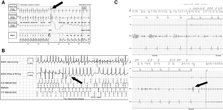
| Interrogation finding | Interpretation | Immediate response |
|---|---|---|
| Ventricular rate faster than atrial rate, atrioventricular dissociation, morphology change from baseline | Appropriate therapy for VT | Storm pathway; ATP optimized; triggers corrected |
| Irregular ventricular intervals with atrial fibrillation on the atrial channel, gradual onset | Inappropriate therapy for atrial fibrillation | Rate control, discriminators, rate cutoff raised; therapy suspended if necessary |
| 1:1 atrioventricular relation with gradual acceleration | Sinus or atrial tachycardia | Treatment of the cause; discriminators and cutoffs reviewed |
| Double counting of R or T waves on the sensing channel | Cardiac oversensing (T-wave oversensing is most common in subcutaneous ICDs) | Sensitivity or sensing vector changed; therapy suspended until corrected |
| Very short, nonphysiologic intervals, impedance change, noise on the near-field channel only | Lead fracture, insulation breach, connection problem | Therapy suspended; lead revision; temporary pacing in pacing-dependent patients |
| Myopotentials with arm or diaphragm movement, interference from external sources | Extracardiac oversensing | Removal of the source; reprogramming |
| Nonsustained VT treated because of short detection duration | Unnecessary therapy | Longer detection duration |
| Tolerated VT terminated by a shock after ATP failure | Appropriate but avoidable shock | More ATP attempts; commanded ATP or drugs for tolerated VT |
Interpretation of ICD therapies in suspected electrical storm, adapted from the 2024 EHRA consensus (Flowcharts 1 and 2 and Section 8.2.3).
When a programmer is not immediately available and inappropriate therapy is evident from telemetry or the ECG, a magnet placed over the generator suspends tachyarrhythmia detection and therapy in most transvenous ICDs while it remains in place. The magnet response is manufacturer- and model-specific and can be programmed. Pacing remains active during magnet application. Device troubleshooting, recalled leads, and long-term programming are discussed in the ventricular tachycardia hub under ICD programming, shocks, complications, and deactivation.
Laboratory testing, echocardiography, coronary angiography, and advanced imaging
Blood tests. Initial testing includes sodium, potassium, magnesium, calcium, and thyroid-stimulating hormone (to detect hyperthyroidism or hypothyroidism, including amiodarone-induced thyrotoxicosis). Kidney and liver function are measured because impairment leads to accumulation of renally cleared drugs such as sotalol and the procainamide metabolite N-acetylprocainamide. Markers of infection and inflammation, serial troponin, natriuretic peptide, and digoxin levels where available are also measured, with arterial blood gas and lactate in unstable patients. Blood is stored for later genetic or toxicologic analysis in unexplained cases.
Echocardiography. Transthoracic echocardiography is performed early after termination to measure ventricular function, chamber size, and wall motion; to detect new structural disease in patients without known heart disease; to identify left ventricular thrombus, which affects the choice of left ventricular access for ablation; and to measure progression in those with known disease.
Coronary angiography. Indications follow the ischemia criteria given with the precipitating factors. Termination of incessant VT by intracoronary contrast injection has been reported but is not a therapy.
Cardiac magnetic resonance and FDG-PET. Late gadolinium enhancement on CMR defines scar location and transmurality before ablation, identifies an epicardial or intramural substrate in nonischemic cardiomyopathy, and suggests myocarditis or sarcoidosis. Device artifacts and an irregular rhythm limit CMR during storm. FDG-PET is useful in suspected arrhythmic myocarditis or cardiac sarcoidosis, particularly when CMR is contraindicated or nondiagnostic, or when CMR and biopsy findings disagree. Endomyocardial biopsy is the diagnostic standard when giant cell myocarditis is possible, because immunosuppression started promptly can be curative.
Hemodynamically unstable storm: defibrillation, cardioversion, and resuscitation
Ongoing hemodynamically unstable VT or ventricular fibrillation is managed according to advanced life support algorithms, with high-quality chest compressions, early defibrillation, airway management, and epinephrine as indicated. The adult cardiac arrest and advanced life support hub covers the algorithm, defibrillation energy and pad position, and the amiodarone and lidocaine doses for shock-refractory ventricular fibrillation. In the Amiodarone, Lidocaine, or Placebo Study (ALPS), survival to discharge in shock-refractory out-of-hospital arrest was 24.4% with amiodarone, 23.7% with lidocaine, and 21.0% with placebo, a difference that was not significant.
In a patient with an ICD, external pads are placed at least 8 cm from the generator, preferably in an anteroposterior position. A conscious patient is sedated before synchronized cardioversion; the EHRA consensus cites an initial energy of 120 to 150 J for VT with a pulse, increased if the first shock fails. Even hemodynamically tolerated VT is terminated promptly, usually by cardioversion under sedation, because deterioration can occur; drug termination is an alternative when the risk of sedation is high. Unsynchronized shocks are used for polymorphic VT, whose changing morphology prevents synchronization.
Instability can persist after termination because of prolonged low output and the cumulative effect of shocks, especially in patients with poor ventricular function. These patients are assessed for cardiogenic shock and early mechanical circulatory support, described below. Extracorporeal cardiopulmonary resuscitation (ECPR) for refractory ventricular fibrillation is covered in the ECPR section of the cardiac arrest hub.
Reprogramming the defibrillator during storm
Reprogramming aims to terminate VT painlessly when possible, to avoid treating arrhythmias that would end spontaneously, and to remove the pauses and pacing patterns that trigger episodes. It is performed by a clinician experienced with the specific device.
| Problem found | Reprogramming |
|---|---|
| Shocks for VT that ATP could terminate | ATP enabled in the VT zone and during charging in the fast VT and VF zones; additional burst sequences before shocks |
| ATP accelerating VT (for example, ramp as the first sequence) | Burst pacing as the first sequence; fewer or less aggressive sequences when acceleration recurs |
| Treatment of nonsustained VT | Longer detection duration (number of intervals or seconds) |
| Slow VT below the detection rate, tolerated | Detection zone extended for monitoring; VT terminated by commanded ATP or drugs; shocks avoided |
| Shocks for hemodynamically tolerated VT | Device therapy disabled under monitoring; termination by commanded ATP, cardioversion under sedation, or drugs |
| Pause-dependent or bradycardia-dependent initiation; post-shock pauses | Lower rate raised, rate hysteresis and sleep rates disabled, post-shock pacing rate reviewed |
| Recently started left ventricular pacing followed by storm | Left ventricular pacing disabled temporarily |
| Low VF detection rate causing shocks for tolerated VT | VF detection rate and VT zones restructured |
ICD reprogramming during electrical storm, from the 2024 EHRA consensus (Section 8.2.3). Programming principles after stabilization follow the 2015 HRS/EHRA/APHRS/SOLAECE consensus and its 2019 focused update.
The 2024 EHRA consensus states that sedation and inactivation of unnecessary shocks, even appropriate ones, may mitigate storm, because recurrent shocks are traumatic events that sustain the hyperadrenergic state. Inactivation is acceptable only in a monitored setting with immediate access to external defibrillation, and therapies are restored before monitoring ends. Antiarrhythmic drugs slow VT and can bring its rate below the detection cutoff; amiodarone also raises the defibrillation threshold, and mexiletine and quinidine do the same. Detection zones are therefore reviewed after each drug change.
Sedation, analgesia, intubation, and general anesthesia
Sedation treats distress and reduces sympathetic tone. The 2024 EHRA consensus advises mild-to-moderate sedation, usually with a benzodiazepine, in all patients with storm and ongoing or recurrent arrhythmia, and states that deep sedation or general anesthesia with mechanical ventilation may be appropriate for drug-refractory storm.
In a 2020 series of 46 conscious patients admitted with recurrent ventricular arrhythmia refractory to initial treatment, 31 were stabilized with usual care; 15 with refractory storm or incessant VT were deeply sedated and intubated, mainly with propofol. Complete resolution of VT or ventricular fibrillation within minutes to hours occurred in 12 (80%) and partial resolution in 2 (13%). Hypotension was the most common adverse effect and was treated with norepinephrine. The 2024 EHRA consensus summarizes a multicenter cohort of 116 patients with storm refractory to antiarrhythmic drugs. In that cohort, an acute response to deep sedation, defined as termination of the storm within 15 minutes without recurrence over 24 hours, occurred in 47% and independently predicted in-hospital survival.
| Agent | Initial dose | Infusion | Notes |
|---|---|---|---|
| Midazolam | 0.01–0.05 mg/kg, repeated every 5–15 minutes | 0.02–0.1 mg/kg/h, titrated by 25%–50% | No negative inotropy; first-line for initial sedation |
| Lorazepam | 0.02–0.04 mg/kg | 0.01–0.1 mg/kg/h (not above 10 mg/h) | Longer acting than midazolam |
| Propofol | 0.8–1.2 mg/kg | 5–50 µg/kg/min | Suppresses sympathetic nerve activity; onset 2–3 minutes; negative inotropy and hypotension; propofol infusion syndrome with prolonged high doses; occasional QT prolongation |
| Fentanyl | 1–2 µg/kg | 1–2 µg/kg/h | Analgesia with mild negative inotropy; onset under 2 minutes |
| Remifentanil | 0.5–1.5 µg/kg | 0.05–2 µg/kg/min | Offset in 3–5 minutes regardless of infusion duration; unaffected by kidney or liver failure |
| Dexmedetomidine | 1 µg/kg over 10 minutes | 0.2–0.7 µg/kg/h | Central sympatholysis without respiratory depression; bradycardia and hypotension |
Sedative and analgesic agents in electrical storm, from the 2024 EHRA consensus (Table 7 and Section 8.2.4.1). Doses are titrated to effect, blood pressure, and ventricular function.
Mechanical ventilation removes the work of breathing, permits deeper sedation, and protects the airway. Its costs are hypotension from sedatives and positive pressure in patients with poor ventricular function, and the need for vasopressor support. Propofol depresses contractility, and norepinephrine used to counter hypotension adds adrenergic stimulation; doses are balanced against each other, and mechanical circulatory support is used when sedation cannot be achieved without circulatory shock. In primary electrical disease and arrhythmogenic cardiomyopathy in which adrenergic stress drives the arrhythmia, deep anesthesia is the final pharmacologic option.
Beta-blockade in electrical storm: propranolol, metoprolol, esmolol, and landiolol
Beta-blockade is the first-line drug treatment of storm in structural heart disease unless contraindicated, and the 2024 EHRA consensus prefers a nonselective agent. Two randomized comparisons support this approach.
Sympathetic blockade versus antiarrhythmic drugs. In a 2000 study of 49 patients with storm (recurrent ventricular fibrillation) after recent MI, 27 received sympathetic blockade (left stellate ganglion block in 6, esmolol in 7, and propranolol in 14). The other 22 received antiarrhythmic drugs according to the advanced life support guidelines of the time (lidocaine, then procainamide or bretylium). Mortality at 1 week was 22% with sympathetic blockade and 82% with antiarrhythmic drugs. The EHRA consensus advises sympathetic blockade with esmolol or propranolol in preference to intravenous lidocaine in storm within 3 months of MI.
Propranolol versus metoprolol. In a randomized trial of 60 ICD recipients with storm, all treated with intravenous amiodarone for 48 hours, oral propranolol 160 mg/day was compared with oral metoprolol 200 mg/day. Propranolol reduced the incidence of ventricular arrhythmic events (incidence rate ratio 0.375) and of ICD shocks (incidence rate ratio 0.428). At 24 hours, 90% of patients taking propranolol were free of arrhythmic events, compared with 53% of those taking metoprolol. Propranolol blocks β2 as well as β1 receptors. The combination of intravenous amiodarone and oral propranolol is preferred to amiodarone with metoprolol (EHRA, advice to do, based on the randomized trial).
| Beta-blocker | Regimen in storm | Comments |
|---|---|---|
| Propranolol | Oral 160 mg/24 h (randomized trial, with intravenous amiodarone); 160–320 mg/24 h in the post-MI series; intravenous 0.15 mg/kg over 10 minutes | Nonselective and lipophilic; bronchospasm; hypotension |
| Esmolol | 0.5 mg/kg over 1 minute, then 25–50 µg/kg/min titrated every 5–10 minutes up to 250 µg/kg/min | Half-life of 5 to 10 minutes, so effects resolve quickly if hypotension occurs; useful when tolerance is uncertain |
| Metoprolol | Intravenous 2–5 mg every 5 minutes up to 3 doses | β1-selective; less effective than propranolol in storm |
| Landiolol | Intravenous infusion titrated to rate | Ultra-short-acting β1-selective blocker; effective in recurrent unstable VT despite amiodarone in 2 small studies; US label limited to supraventricular tachycardia |
| Nadolol | Oral, long-term | Nonselective; preferred in long QT syndrome and CPVT |
Beta-blocker regimens used in electrical storm, from the 2024 EHRA consensus (Table 8), the randomized propranolol-metoprolol trial, and the 2000 post-MI storm series. Landiolol is approved in the United States for short-term reduction of ventricular rate in supraventricular tachycardia, including atrial fibrillation and flutter.
Beta-blockade in a patient with decompensated heart failure or shock requires hemodynamic monitoring, because cardiac output may depend on adrenergic drive. Options in that setting are a short-acting agent such as esmolol titrated from a low dose under arterial pressure monitoring, or beta-blockade started after mechanical circulatory support is in place. Beta-blockers are avoided in Brugada syndrome and early repolarization syndrome, in which slowing of the heart rate is proarrhythmic.
Intravenous antiarrhythmic drugs in structural heart disease
The drug is chosen by the type of arrhythmia, the likely substrate, ventricular function, kidney and liver function, drugs already taken, and local availability.
Amiodarone. Intravenous amiodarone blocks sodium, potassium, and calcium channels and has antiadrenergic effects, and its acute electrophysiologic profile differs from that of chronic oral therapy. The US label indicates it for frequently recurring ventricular fibrillation and hemodynamically unstable VT refractory to other therapy. In a double-blind dose-ranging trial of 342 patients with recurrent, hemodynamically destabilizing VT or ventricular fibrillation, arrhythmia event rates fell with increasing 24-hour doses (125, 500, and 1,000 mg). Supplemental boluses were needed less often at higher doses, and hypotension was the most common adverse effect (26%). In PROCAMIO, amiodarone 5 mg/kg over 20 minutes terminated tolerated wide-complex tachycardia within 40 minutes in 38% of patients. Amiodarone terminates VT slowly and serves mainly to prevent recurrence. Patients already taking oral amiodarone may still benefit from intravenous reloading.
Lidocaine. Lidocaine binds inactivated sodium channels and acts preferentially on depolarized, ischemic tissue and at fast rates. It is only moderately effective for stable monomorphic VT, in which it was inferior to procainamide and to intravenous sotalol in comparative trials. Its main roles in storm are treating ischemic VT or ventricular fibrillation and adding to amiodarone when storm persists. Prophylactic lidocaine in acute MI did not reduce mortality. Clearance falls with reduced hepatic blood flow in heart failure, shock, and older age, and neurotoxicity (confusion, tremor, seizures) limits dosing. Lidocaine combined with amiodarone is common in practice: in the STAR study of stellate ganglion block for refractory storm, 33% of procedures were performed in patients already receiving both drugs. No randomized trial has tested the combination in storm.
Procainamide. Procainamide is effective for termination of hemodynamically tolerated monomorphic VT. In the randomized PROCAMIO trial (62 patients analyzed), procainamide 10 mg/kg over 20 minutes caused major cardiac adverse events within 40 minutes in 9% of patients, compared with 41% with amiodarone. Procainamide terminated the tachycardia within 40 minutes in 67%, compared with 38% with amiodarone. Procainamide is avoided with hypotension, severely reduced ejection fraction, QT prolongation, Brugada syndrome, and severe kidney disease, in which N-acetylprocainamide accumulates.
Sotalol. Intravenous sotalol terminates stable VT and is an alternative in patients with preserved kidney function and without decompensated heart failure; QT monitoring is required, and torsades is the principal risk.
| Drug | Intravenous regimen | Main use in storm | Cautions |
|---|---|---|---|
| Amiodarone | 150 mg over 10 minutes, then 1 mg/min for 6 hours, then 0.5 mg/min; supplemental 150 mg over 10 minutes for breakthrough VT or VF | First-line drug in structural heart disease, combined with a beta-blocker | Hypotension (mean daily doses above 2,100 mg increased risk in trials), bradycardia, QT prolongation; central line for concentrations above 2 mg/mL; raises defibrillation threshold |
| Lidocaine | 1–1.5 mg/kg; repeat 0.5–0.75 mg/kg every 5–10 minutes to 3 mg/kg; infusion 1–4 mg/min | Ischemic VT or VF; addition to amiodarone | Neurotoxicity; dose reduction in heart and liver failure and older adults |
| Procainamide | 10–17 mg/kg at 20–50 mg/min; maintenance 1–4 mg/min; stopped for hypotension or QRS widening above 50% | Termination of tolerated monomorphic VT | Hypotension, QRS and QT prolongation; kidney disease; Brugada syndrome |
| Quinidine | Oral 600–1,600 mg/day in divided doses (EHRA); availability is limited | Polymorphic VT storm after MI or revascularization; Brugada syndrome; early repolarization; idiopathic VF | QT prolongation and torsades, diarrhea, thrombocytopenia; raises defibrillation threshold |
| Mexiletine | Oral only; 400 mg loading, then 600 mg in the first 24 hours (EHRA) | Addition to amiodarone for scar-related VT; long QT syndrome type 3 | Tremor, nausea, ataxia; rapid loading precipitated heart failure in severe left ventricular dysfunction |
| Isoproterenol | Infusion 0.5–10 µg/min | Brugada syndrome, early repolarization, idiopathic VF, short QT; pause-dependent torsades | Contraindicated in ischemia and CPVT; tachycardia, hypotension |
| Magnesium sulfate | 2 g intravenously, repeated as needed | Torsades de pointes | Hypotension, respiratory depression in kidney failure; no benefit in VF without torsades |
Intravenous and rapid oral drugs for electrical storm. Amiodarone regimen from the US prescribing information; lidocaine and procainamide doses as used in the 2017 AHA/ACC/HRS guideline; quinidine and mexiletine from the 2024 EHRA consensus (Table 11) and isoproterenol from its Table 8. Isoproterenol is labeled in the United States for shock and for bronchospasm during anesthesia; its use in storm is off-label.
Ranolazine. In the RAID trial of 1,012 high-risk ICD recipients, ranolazine did not reduce the primary composite of VT, ventricular fibrillation, or death. It reduced recurrent VT or ventricular fibrillation requiring ICD therapy in a prespecified secondary analysis. It has no approved arrhythmia indication in the United States and no defined role in acute storm.
Drugs that worsen storm. Class IC drugs (flecainide and propafenone) are avoided in coronary disease and reduced ejection fraction. Combining several QT-prolonging or conduction-slowing drugs compounds proarrhythmic risk. The pharmacology of each agent is covered in the drug sections of the ventricular tachycardia hub.
Quinidine for polymorphic VT storm after myocardial infarction or revascularization
A distinct form of storm occurs in patients with coronary disease days after an otherwise uncomplicated infarction or revascularization, without ongoing ischemia. Short-coupled PVCs trigger polymorphic VT that degenerates to ventricular fibrillation, and the QT interval is not prolonged. In a 2019 series of 43 such patients, arrhythmic storms of 4 to 16 episodes occurred in 23 (53%); the storms were refractory to conventional drugs, including intravenous amiodarone, and responded to quinidine in every case. In-hospital mortality among patients with storm was 17%, and all patients treated with quinidine survived to discharge. During long-term follow-up, 16% of patients discharged without quinidine had recurrent polymorphic VT, and none taking quinidine did. A 2020 series of 20 patients with coronary disease and out-of-hospital polymorphic VT without acute ischemia reported the same pattern: short-coupled triggers (376 ± 49 ms), storms in 70%, refractoriness to conventional drugs, and response to quinidine.
The EHRA consensus states that quinidine is appropriate for recurrent polymorphic VT resistant to amiodarone and beta-blockers in the early post-infarction phase or after coronary revascularization. When the triggering PVC has a consistent morphology, catheter ablation of the trigger, typically in the Purkinje network of the infarct border zone, is an effective alternative.
Storm in primary electrical disease: long QT, Brugada, early repolarization, idiopathic VF, short QT, and CPVT
In the inherited arrhythmia syndromes, treatment of storm follows the mechanism, and the same drug can terminate storm in one syndrome and provoke it in another. Isoproterenol is the clearest example: it suppresses ventricular fibrillation in Brugada syndrome, early repolarization syndrome, idiopathic ventricular fibrillation, and short QT syndrome. It is harmful in CPVT and in adrenergic congenital long QT syndrome.
| Syndrome | Acute treatment of storm | Avoided | Prevention of recurrence |
|---|---|---|---|
| Acquired long QT (torsades de pointes) | Withdrawal of offending drugs; intravenous magnesium; potassium to 4.0 mEq/L or more; temporary pacing or isoproterenol for pause-dependent torsades | Class IA and III drugs, amiodarone, other QT-prolonging drugs | Avoidance of the causative drug and interacting drugs |
| Congenital long QT syndrome | Nonselective beta-blocker (propranolol or nadolol), magnesium, potassium, sedation; pacing in long QT syndrome types 2 and 3; mexiletine in type 3 | Isoproterenol; class III drugs | Nadolol or propranolol; left cardiac sympathetic denervation for recurrent shocks or storm despite beta-blockade (EHRA, advice to do) |
| Brugada syndrome | Isoproterenol infusion (EHRA, advice to do); quinidine; aggressive treatment of fever | Class I antiarrhythmic drugs, beta-blockers, other sodium-channel blockers | Quinidine (EHRA, may be appropriate); epicardial ablation of the right ventricular outflow tract substrate at experienced centers |
| Early repolarization syndrome | Isoproterenol (EHRA, may be appropriate) | Hypothermia, bradycardia | Quinidine with the ICD; ablation of triggering PVCs |
| Idiopathic ventricular fibrillation | Isoproterenol or quinidine; verapamil in selected patients (EHRA, may be appropriate) | — | Ablation of PVCs of consistent morphology that trigger VF; quinidine |
| Short QT syndrome | Isoproterenol (EHRA, may be appropriate; very limited data) | — | Quinidine or hydroquinidine |
| CPVT | Sedation, removal of every adrenergic trigger, nonselective beta-blockade; reprogramming to avoid shock cascades | Epinephrine (including during resuscitation), isoproterenol, repeated shocks | Nadolol with flecainide; left cardiac sympathetic denervation (EHRA, advice to do for recurrent shocks or storm) |
Electrical storm in primary electrical disease, from the 2024 EHRA consensus (Advice Tables 5 and 8) and the 2019 ablation consensus. The EHRA advice to avoid catecholamines in CPVT storm applies during cardiac arrest as well.
Torsades de pointes storm. Torsades is pause dependent and arises from early afterdepolarizations on a prolonged, heterogeneous repolarization. Magnesium suppresses the triggers without shortening the QT, and pacing or isoproterenol shortens repolarization and removes the pauses. Standard ventricular antiarrhythmic drugs prolong the QT and worsen the storm. The full acute protocol, including potassium targets, pacing rates, and the approach to congenital long QT syndrome, is in the long QT hub section on acute treatment of torsades de pointes and electrical storm.
Brugada syndrome. Storm typically occurs at rest, at night, or with fever. When fibrillation recurs despite drugs, epicardial ablation of the right ventricular outflow tract substrate is performed at experienced centers, because triggering PVCs are rarely present between episodes. Diagnosis and ICD indications are in the Brugada syndrome section of the ventricular tachycardia hub.
CPVT. Storm is rare and almost always adrenergically triggered, and an ICD shock can perpetuate it. The CPVT and early repolarization and idiopathic VF sections of the ventricular tachycardia hub cover these syndromes.
Overdrive pacing and temporary pacing
Pacing suppresses storm through several mechanisms. Overdrive pacing at a rate faster than the native rhythm suppresses abnormal automaticity and can produce exit block from an ectopic focus. In re-entry, faster pacing shortens and homogenizes refractoriness. Pacing at a higher rate removes the pauses and short-long-short sequences that initiate torsades and some polymorphic VT, and shortens repolarization.
Overdrive pacing is delivered through an implanted pacemaker or ICD by raising the lower rate, or through a temporary right ventricular lead, at a rate 10 to 20 beats/min above the native rate. Decremental overdrive pacing (progressively shorter intervals) terminates VT more often than fixed-interval burst pacing. Temporary atrial or ventricular overdrive pacing has prevented drug-resistant ventricular arrhythmia in case reports, mainly with acute or recent ischemia, and bradycardia pacing is useful when polymorphic VT is bradycardia dependent. After cardiac surgery, temporary epicardial wires allow overdrive pacing without new access.
Temporary pacing is also required when antiarrhythmic drugs cause bradycardia or atrioventricular block, and before therapy suspension or lead revision in a pacing-dependent patient. Pacing rate and mode for torsades are set out in the long QT hub, and temporary pacing technique in the bradycardia hub.
Stepwise management of electrical storm
Management proceeds in parallel, not in sequence. Interrogation, trigger correction, sedation, and drug therapy begin within the first minutes. A multidisciplinary team plans escalation to ablation, mechanical support, or autonomic modulation early. The team includes an electrophysiologist, a critical care or anesthesia specialist, and, when needed, a heart failure specialist and cardiac surgeon.
- Hemodynamically unstable VT or ventricular fibrillation: advanced life support, defibrillation or synchronized cardioversion under sedation, external pads at least 8 cm from the generator.
- Rhythm documentation and device interrogation: 12-lead ECG of the arrhythmia when possible; stored electrograms classify every therapy as appropriate, inappropriate, or unnecessary, and the device is reprogrammed or therapy suspended under monitoring.
- Admission to a monitored unit; external defibrillator at the bedside; arterial line in unstable patients.
- Correction of triggers: potassium and magnesium, ischemia (angiography for ST-elevation MI or suspected ongoing ischemia), decompensated heart failure, drug proarrhythmia, thyroid excess, fever, and infection.
- Mild-to-moderate sedation and analgesia in every conscious patient with recurrent episodes.
- Substrate-directed drug therapy: in structural heart disease, intravenous amiodarone with a nonselective beta-blocker (propranolol), or esmolol when tolerance is uncertain; lidocaine added for ischemic or persistent storm; procainamide for tolerated monomorphic VT; quinidine for post-infarction polymorphic VT storm. In primary electrical disease, the syndrome-specific regimen.
- Persistent storm: deep sedation or general anesthesia with mechanical ventilation; overdrive pacing for pause- or bradycardia-dependent arrhythmia.
- Recurrent monomorphic VT or ventricular fibrillation triggered by a consistent PVC despite drugs and sedation: catheter ablation at an experienced center, with transfer when needed.
- Hemodynamic deterioration, or high risk of decompensation on the PAAINESD score before ablation: temporary mechanical circulatory support.
- Refractory storm or ablation not possible: stellate ganglion block or thoracic epidural anesthesia as a bridge; cardiac sympathetic denervation; bailout ablation techniques; evaluation for heart transplantation or durable left ventricular assist device.
- Goals of care discussed throughout, including the option of shock deactivation in end-stage disease.
| Tier | Intervention | Strength of evidence |
|---|---|---|
| Initial (all patients) | Interrogation and reprogramming; trigger correction; mild-to-moderate sedation; beta-blockade; amiodarone in structural heart disease | Randomized trial for propranolol with amiodarone; observational data and consensus for the rest |
| Escalation | Deep sedation and ventilation; lidocaine; procainamide; quinidine for post-MI polymorphic VT; overdrive pacing | Observational series |
| Definitive substrate therapy | Catheter ablation of monomorphic VT or VF-triggering PVCs | Class I recommendations for storm refractory to drugs in ischemic and nonischemic cardiomyopathy (2019 consensus, B-NR); randomized trials of ablation in recurrent VT |
| Hemodynamic support | Temporary mechanical circulatory support | Observational data; EHRA "may be appropriate" |
| Refractory | Stellate ganglion block, thoracic epidural anesthesia, cardiac sympathetic denervation, renal denervation; bailout ablation; transplant or durable LVAD | Prospective observational (STAR) and case series |
Escalation tiers in electrical storm and the evidence behind each, from the 2024 EHRA consensus, the 2019 HRS/EHRA/APHRS/LAHRS ablation consensus, and the randomized propranolol-metoprolol trial.
Catheter ablation in electrical storm: indications and timing
Catheter ablation is the only therapy that modifies the substrate of scar-related monomorphic VT, and in storm it is both an emergency treatment and the main means of preventing recurrence. The 2024 EHRA consensus describes ablation as the treatment of choice for monomorphic VT refractory to drug therapy in the acute phase and advises it in all eligible patients to reduce recurrence of storm.
| Recommendation | Source and strength |
|---|---|
| Ischemic heart disease with VT storm refractory to antiarrhythmic drugs: catheter ablation is recommended | 2019 HRS/EHRA/APHRS/LAHRS, Class I, B-NR |
| Ischemic heart disease with recurrent monomorphic VT despite chronic amiodarone: ablation is recommended in preference to escalation of antiarrhythmic drugs | 2019 consensus, Class I, B-R (VANISH) |
| Nonischemic cardiomyopathy with storm refractory to antiarrhythmic drugs: ablation is useful for reducing recurrent VT and ICD shocks | 2019 consensus, Class I, B-NR |
| Prior MI with recurrent symptomatic sustained VT, or VT storm, after failure or intolerance of amiodarone or other antiarrhythmic drugs: catheter ablation is recommended | 2017 AHA/ACC/HRS, Class 1 (B-R after amiodarone; B-NR after other drugs) |
| Recurrent monomorphic VT despite optimal medical therapy, with an experienced team available: perform ablation | 2024 EHRA, advice to do (observational) |
| Recurrent VF triggered by a similar PVC despite optimal medical therapy: perform ablation | 2024 EHRA, advice to do (observational) |
| Storm due to monomorphic VT recurring on chronic amiodarone in coronary disease: perform ablation in preference to drug escalation | 2024 EHRA, advice to do (randomized evidence) |
| Ablation as first-line treatment in coronary disease with monomorphic VT storm, at experienced centers | 2024 EHRA, may be appropriate |
| Transfer to a specialized center for ablation of recurrent monomorphic VT or PVC-triggered VF | 2024 EHRA, advice to do |
Recommendations for catheter ablation in electrical storm. B-NR, nonrandomized evidence; B-R, randomized evidence.
Timing. The optimal timing is uncertain. Rescue ablation within the first 24 hours of storm in a patient with ongoing cardiogenic shock from sustained VT is associated with higher rates of ablation failure and recurrence. Observational data suggest that ablation after the first 48 hours from admission is associated with lower 30-day mortality than conservative therapy or very early ablation. The EHRA consensus therefore advises attempting hemodynamic stabilization and arrhythmia suppression before ablation, while recognizing that this is not always possible. Early ablation is reasonable for incessant VT, or for hemodynamically significant recurrent PVC-triggered arrhythmia, despite drugs and deep sedation. Acute ablation, defined as ablation for rhythm stabilization within 8 hours of onset, is rare, and few laboratories provide it around the clock. The 2019 consensus describes networks centered on experienced VT centers with 24-hour availability and immediate transfer.
Outcome in storm. In a prospective series of 95 patients with drug-refractory storm (72 with coronary disease, 10 with dilated cardiomyopathy, and 13 with ARVC), storm was acutely suppressed in all after 1 to 3 procedures. Inducibility of clinical VTs was abolished in 89%. At a median of 22 months, 92% were free of storm and 66% free of VT recurrence. Eight of 10 patients with persistent inducibility of clinical VT had recurrent storm, and 4 of them died suddenly despite appropriate ICD therapy; no storm recurred when all clinical VTs were abolished. Persistent inducibility after ablation is consistently associated with recurrence and worse survival.
Randomized trials of catheter ablation versus antiarrhythmic drugs
No randomized trial has enrolled only patients in storm. The evidence comes from trials in recurrent VT and ICD shocks, most of which counted storm as an end point component.
| Trial | Population | Comparison | Principal result |
|---|---|---|---|
| SMASH-VT (2007) | 128 patients with prior MI and an ICD for spontaneous VT or VF, no antiarrhythmic drugs | Substrate ablation plus ICD versus ICD alone | Appropriate ICD therapy 12% versus 33% (hazard ratio 0.35); shocks 9% versus 31% |
| VTACH (2010) | 107 patients with prior MI, stable VT, and LVEF 50% or less | Ablation before ICD versus ICD alone | Survival free of VT or VF at 2 years 47% versus 29% (hazard ratio 0.61); median time to recurrence 18.6 versus 5.9 months |
| VANISH (2016) | 259 patients with ischemic cardiomyopathy, an ICD, and VT despite antiarrhythmic drugs | Ablation versus escalated drug therapy (amiodarone started, increased, or mexiletine added) | Death, VT storm, or appropriate shock 59.1% versus 68.5% (hazard ratio 0.72); no mortality difference |
| BERLIN VT (2020) | 159 patients with ischemic cardiomyopathy, LVEF 30% to 50%, and documented VT | Preventive ablation versus ablation deferred until 3 ICD shocks | No difference in death or hospitalization (hazard ratio 1.09); stopped for futility |
| PARTITA (2022) | 47 patients with ischemic or nonischemic dilated cardiomyopathy after a first appropriate ICD shock | Ablation within 2 months versus standard therapy | Death or worsening heart failure hospitalization 4% versus 42% (hazard ratio 0.11); 0 versus 8 deaths |
| VANISH2 (2025) | 416 patients with prior MI, an ICD, and clinically significant VT, including VT storm | First-line ablation versus sotalol or amiodarone | Death, VT storm, appropriate shock, or treated sustained VT 50.7% versus 60.6% (hazard ratio 0.75) over a median 4.3 years |
Randomized trials comparing catheter ablation with antiarrhythmic drug therapy or ICD therapy alone in scar-related VT. VANISH counted VT storm (3 or more episodes within 24 hours) as a component of its primary end point.
In VANISH, the benefit of ablation was confined to patients already taking amiodarone, for whom escalation meant a higher amiodarone dose or the addition of mexiletine (hazard ratio 0.55). In patients taking sotalol, for whom escalation meant starting amiodarone, the outcome did not differ (hazard ratio 1.14). Two deaths from pulmonary toxicity and one from hepatic dysfunction occurred with escalated drug therapy. In VANISH2, adverse events within 30 days of ablation included death in 1.0% and nonfatal events in 11.3%, while drug therapy caused death from pulmonary toxicity in 0.5% and nonfatal adverse events in 21.6%. PARTITA, stopped early at its first interim analysis under a Bayesian design, was small, and its effect on mortality needs confirmation. A meta-analysis of randomized trials did not show a significant effect of ablation on survival; nonrandomized studies associate successful ablation with better survival. The 2024 EHRA consensus states that data are insufficient to establish a mortality reduction from ablation in patients with storm.
Mapping and ablation in electrical storm
Most VT in storm cannot be tolerated long enough for activation and entrainment mapping, and the procedure relies on substrate mapping in sinus rhythm or paced rhythm. Bipolar voltage maps define dense scar and border zone. Late potentials, local abnormal ventricular activities, and fractionated electrograms identify surviving channels. Pace mapping from the scar border reproduces the morphology of each recorded VT, guided by the 12-lead and device electrogram morphologies archived during the storm. Ablation strategies include elimination of late potentials, scar homogenization, and isolation or transection of channels. When VT is tolerated, or when mechanical support maintains perfusion, activation and entrainment mapping localize the critical isthmus. In post-infarction patients with focally triggered ventricular fibrillation storm, elimination of the triggering Purkinje PVCs provides short- and long-term freedom from storm.
An epicardial approach, through percutaneous subxiphoid pericardial access, is needed more often in nonischemic cardiomyopathy, ARVC, Chagas disease, and Brugada syndrome, and in some patients with coronary disease after failed endocardial ablation. Combined endocardial and epicardial ablation at the index procedure is appropriate in ARVC and Chagas cardiomyopathy. Epicardial access carries a risk of right ventricular puncture, coronary artery injury, and phrenic nerve injury, and major complications occur in about 5% of epicardial procedures in published series.
Mapping techniques, entrainment criteria, and outcomes of ablation outside the storm setting are covered in the catheter ablation section of the ventricular tachycardia hub.
Hemodynamic decompensation during ablation and the PAAINESD score
Acute hemodynamic decompensation, defined as sustained hypotension despite vasopressors that requires mechanical support or discontinuation of the procedure, occurred in 22 of 193 patients (11%) undergoing ablation of scar-related VT in the cohort from which the PAAINESD score was derived. Decompensation was associated with higher in-hospital and long-term mortality, more VT recurrence, and procedure failure. Patients who decompensated were older and had more diabetes, ischemic cardiomyopathy, chronic obstructive pulmonary disease, New York Heart Association (NYHA) class III or IV symptoms, lower ejection fraction, and presentation with storm (77% versus 43%), and general anesthesia was also a predictor.
The PAAINESD score adds points for each factor and ranges from 0 to 35, or from 0 to 31 when the modifiable intraprocedural item, general anesthesia, is excluded (PAINESD). A score of 17 or more with anesthesia, or 15 or more without it, was associated with a 24% risk of decompensation. Without the anesthesia item, a score of 9 to 14 was associated with a 6% risk in one series. The 2024 EHRA consensus classifies the PAINESD score as low risk at 8 or less, intermediate at 9 to 14, and high at 15 or more.
PAAINESD score
Risk of hemodynamic decompensation during scar-related VT ablationNumbers in parentheses are points. PAAINESD = chronic obstructive Pulmonary disease 5 + Age above 60 years 3 + general Anesthesia 4 + Ischemic cardiomyopathy 6 + NYHA class III or IV 6 + Ejection fraction below 25% 3 + VT Storm 5 + Diabetes 3. Point values and thresholds from the 2019 HRS/EHRA/APHRS/LAHRS ablation consensus (Table 4) and the 2024 EHRA consensus (Table 10). The score was derived and validated in single-center and multicenter observational cohorts, some of which used mechanical support routinely.
Measures that reduce the risk of decompensation include optimization of volume status and heart failure therapy before the procedure and close monitoring of fluid balance and urine output. During the procedure, measures include substrate mapping with limited VT induction, avoidance of general anesthesia and myocardial depressants such as propofol where feasible, and prophylactic mechanical support in high-risk patients. In a single-center series of 93 patients, 30-day mortality was 4% when a percutaneous left ventricular assist device was placed before ablation and 3% in patients who did not need support, compared with 58% when the device was inserted as a rescue after hemodynamic collapse. The EHRA consensus states that prophylactic mechanical support before ablation may be appropriate in high-risk patients presenting with storm, citing a systematic review of 19 nonrandomized studies (2,465 adults with life-threatening arrhythmia). In that review, prophylactic support in patients with high PAINESD scores or storm was associated with better survival than rescue support or no support. Support was also associated with more complications and longer procedures.
Temporary mechanical circulatory support in electrical storm
Temporary mechanical circulatory support maintains organ perfusion during refractory, hemodynamically unstable ventricular arrhythmia while its cause is treated. It serves three purposes in storm: rescue of the patient in cardiogenic shock or circulatory collapse from recurrent arrhythmia; support during ablation of hemodynamically unstable VT; and a bridge to recovery, durable left ventricular assist device, or transplantation. Evidence is observational, the devices carry substantial rates of vascular and bleeding complications, and their use is confined to experienced multidisciplinary teams.
| Device | Support provided | Advantages in storm | Limitations |
|---|---|---|---|
| Intra-aortic balloon pump | Counterpulsation; cardiac output increase of 0.5–1.0 L/min | Simple, widely available, few complications; afterload reduction | Very limited support; requires a stable rhythm for timing |
| Impella CP | Microaxial flow pump from left ventricle to aorta; up to 3.7 L/min; 14 Fr | Left ventricular unloading; percutaneous insertion and removal | Partial support; interference with electroanatomic mapping; catheter manipulation hindered; transseptal access may be needed |
| Impella 5.5 | Up to 5.5 L/min; surgical axillary insertion | Full left ventricular support for longer durations; allows mobilization | Surgical insertion and removal; cost |
| TandemHeart | Left atrial to femoral artery bypass; up to 5 L/min | Left ventricular support for longer duration | Transseptal cannula can impede transseptal ablation access; large cannulas |
| Venoarterial extracorporeal membrane oxygenation (ECMO) | Full cardiopulmonary support; up to 8 L/min | Rapid deployment; biventricular support; maintains perfusion during VF | Increases left ventricular afterload and may require venting; limb ischemia; bleeding; complex logistics |
Temporary mechanical circulatory support devices in electrical storm, adapted from the 2024 EHRA consensus (Table 9). Contraindications listed in the consensus include moderate-to-severe aortic regurgitation (balloon pump, microaxial pumps, TandemHeart, and ECMO), a mechanical aortic prosthesis and left ventricular thrombus (microaxial pumps), aortic dissection (balloon pump and ECMO), and severe peripheral artery disease (femoral devices).
Rescue support. Venoarterial ECMO is the preferred rescue device for hemodynamic collapse due to storm, refractory VT, recurrent ventricular fibrillation, or cardiogenic shock persisting after the arrhythmia stops, because it provides full support and can be started rapidly. Rescue support started during ablation for unexpected decompensation has been associated with short-term mortality of 38% to 76% despite successful ablation. In a series of 64 patients (62% with storm) who received ECMO for ablation of unstable VT, at least one VT was terminated in 81% of procedures with baseline inducible VT, noninducibility was achieved in 69%, and 88% of patients were alive at a median 21 months. VT recurrence was 19% at 180 days when VT was noninducible after ablation and 75% when clinical VT remained inducible.
Device selection, hemodynamic targets, left ventricular venting, and complications are covered in the cardiogenic shock hub sections on VA-ECMO and left ventricular unloading and microaxial flow pumps.
Stellate ganglion block in refractory electrical storm
The stellate ganglion, formed by fusion of the inferior cervical and first thoracic sympathetic ganglia, lies anterior to the neck of the first rib and the transverse process of C7. It carries most of the efferent sympathetic supply to the heart. Percutaneous stellate ganglion block injects a local anesthetic (lidocaine, bupivacaine, ropivacaine, or a combination) next to the ganglion, usually on the left side, by an anterior paratracheal landmark technique at the level of C6 or by a lateral ultrasound-guided approach. A catheter left in place permits continuous infusion, which controls arrhythmia better than a single bolus. Ipsilateral Horner syndrome, with miosis and ptosis, indicates that the ganglion has been reached.
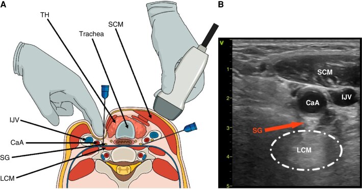
Evidence. In the prospective multicenter STAR study, 131 patients with storm refractory to standard treatment (median age 68 years, mean ejection fraction 25%) underwent 184 percutaneous stellate ganglion blocks at 19 centers, 98% on the left. The primary outcome, a reduction of at least 50% in treated arrhythmic events in the 12 hours after the block compared with the 12 hours before, was achieved in 92% of patients, and the median reduction was 100%. Treated episodes fell from a median of 6 in the 12 hours before the first block to 0 in the 12 hours after the last. One major complication (respiratory depression) occurred among 184 procedures. About one-fifth of blocks were performed in intubated patients. Because anticoagulant or antiplatelet therapy was present in most patients, the percutaneous block was feasible in many patients in whom thoracic epidural anesthesia would have been contraindicated.
In a single-center series of 20 patients given ultrasound-guided bilateral blocks, half of whom were receiving inotropes and nearly half mechanical support, ventricular arrhythmia episodes fell from a median of 5.5 in the 24 hours before the block to 0 in the 24 hours after. About half had complete suppression for 48 hours. In the 2000 post-infarction storm series, left stellate ganglion block was one of the sympathetic blockade strategies that outperformed antiarrhythmic drugs. A randomized sham-controlled trial of noninvasive transcutaneous magnetic stimulation of the left stellate ganglion in 26 patients did not reduce VT recurrence within 24 hours, although it reduced VT burden over 72 hours.
The 2024 EHRA consensus states that stellate ganglion block or thoracic epidural anesthesia may be appropriate in structural heart disease with storm or incessant VT or ventricular fibrillation, to reduce the arrhythmia burden and ICD shocks as a bridge to definitive therapy. Its effect is temporary, and it does not replace ablation or surgical denervation. Reported complications include hematoma, symptoms from anesthetic absorption or intravascular injection, hoarseness, brachial plexus injury, and, rarely, respiratory depression.
Thoracic epidural anesthesia, cardiac sympathetic denervation, and renal denervation
Thoracic epidural anesthesia. A local anesthetic infused into the high thoracic epidural space (C7 to T4; the 2019 consensus describes bupivacaine at T1–T2 or T2–T3) blocks sympathetic outflow along the stellate and upper thoracic ganglia bilaterally. In a 2010 series of 14 patients with structural heart disease and VT refractory to medical therapy and ablation, 12 of them in storm, 8 received thoracic epidural anesthesia. VT burden fell by 80% or more in 6 of those 8, with infusions in the responders lasting 48 to 96 hours. The technique is contraindicated by active infection, dual antiplatelet therapy, and the need for uninterrupted anticoagulation, which limits its use in patients with recent stents, mechanical support, or atrial fibrillation.
Cardiac sympathetic denervation. Surgical or video-assisted thoracoscopic denervation removes the lower third to half of the stellate ganglion and the T2 to T4 (or T5) thoracic ganglia and produces permanent afferent and efferent sympathetic denervation. In the largest multicenter series, 121 patients with structural heart disease (mean ejection fraction 30%, 71% with nonischemic cardiomyopathy, 75% with VT storm) underwent left or bilateral denervation. One-year freedom from sustained VT or ICD shock was 58%, and one-year freedom from ICD shock, transplantation, and death was 50%. Shocks fell from a median of 10 in the year before to 0 after denervation. Advanced heart failure, VT cycle length, and a left-sided procedure alone were independently associated with recurrence or death, and bilateral procedures were more effective in structural heart disease. Complications include Horner syndrome, harlequin syndrome, abnormal sweating, and regional temperature changes.
The 2019 consensus describes denervation as a reasonable option for storm when beta-blockers, antiarrhythmic drugs, and ablation are ineffective or not tolerated, provided the team has expertise. The 2017 AHA/ACC/HRS guideline assigns cardiac sympathetic denervation a Class 2b recommendation in that setting. The EHRA consensus states that left or bilateral denervation may be appropriate in ischemic and nonischemic cardiomyopathy when arrhythmia or storm recurs despite medical therapy and ablation, or when ablation is not possible. The consensus adds that performing it early may lead to better outcomes than urgent or emergency surgery. Left cardiac sympathetic denervation in long QT syndrome and CPVT is described in the long QT hub.
Renal denervation. Ablation of renal artery nerves reduces afferent renal and efferent cardiac sympathetic signaling. In a pooled analysis of 121 patients from case series (mean ejection fraction 30%, 99% taking beta-blockers, 79% taking amiodarone), renal denervation, with or without VT ablation, reduced ICD shocks and ATP. There are no clear procedural end points and no randomized data, and the EHRA consensus lists its role as an adjunct to ablation as an area of uncertainty.
Bailout ablation, radiotherapy, transplantation, and durable LVAD
Catheter ablation fails most often because lesions cannot reach deep intramural or septal circuits. Bailout strategies, described in the 2024 EHRA consensus and reserved for high-volume centers, include sequential ablation from opposite surfaces, prolonged or high-power applications, bipolar or simultaneous unipolar ablation, half-normal saline irrigation, retractable needle ablation, mapping and ablation through septal coronary veins or intracoronary wires, intracoronary ethanol ablation, coil embolization, and stereotactic body radiotherapy. None has been tested in large or randomized studies. Stereotactic radiotherapy, which delivers a single 25-Gy fraction to imaging-defined scar, remains investigational and is summarized in the ablation section of the ventricular tachycardia hub. Its antiarrhythmic effect develops over weeks and does not terminate an active storm.
When storm is refractory to all measures, or recurs in a patient with end-stage ventricular dysfunction, urgent heart transplantation is an option for selected candidates, and a durable left ventricular assist device is a bridge or destination therapy. Temporary mechanical support stabilizes the patient while candidacy is assessed. Early evaluation for advanced heart failure therapy is advised for patients with dilated cardiomyopathy, ejection fraction of 32% or less, and early VT recurrence after ablation for storm, given their high mortality regardless of the arrhythmic outcome. Referral criteria are in the advanced heart failure section of the heart failure with reduced ejection fraction (HFrEF) hub.
Electrical storm in patients with a left ventricular assist device
Ventricular arrhythmias occur in 20% to 50% of patients with a continuous-flow LVAD, and storm in about 9%. Because the device maintains systemic flow, ventricular arrhythmias are usually well tolerated, but short-term mortality after storm is high, and repeated shocks in a conscious patient are a concern. In a 2026 multicenter cohort of 1,151 LVAD recipients, late storm beyond 30 days after implantation occurred in 4.3% at a median of 9.2 months and was independently associated with 5-year all-cause mortality (adjusted hazard ratio 2.87), whereas isolated late ventricular arrhythmias were not.
Suction events, in which an underfilled left ventricle collapses around the inflow cannula, provoke ventricular arrhythmia that is managed by adjusting pump speed. Mapping studies show that most monomorphic VT arises from intrinsic myocardial scar remote from the inflow cannula. Drug therapy, sedation, and autonomic modulation follow the same principles as in patients without an LVAD. Ablation in LVAD recipients is performed by experienced teams and requires adaptations. Nonpulsatile flow complicates arterial access and pressure monitoring, and a closed aortic valve favors transseptal access. The catheter can be entrapped in the inflow cannula. HeartMate 3 pumps cause substantial noise on surface ECG and mapping signals. The hemodynamic support provided by the LVAD usually allows transfer to such a center.
Storm in nonischemic cardiomyopathy, sarcoidosis, myocarditis, ARVC, and hypertrophic cardiomyopathy
Nonischemic cardiomyopathy comprises genetic and acquired dilated cardiomyopathy, hypertrophic cardiomyopathy, ARVC, cardiac sarcoidosis, myocarditis, Chagas disease, and valvular cardiomyopathy. The substrate is more often intramural, epicardial, basal, or septal than in post-infarction VT. Outcomes after ablation are worse, with more recurrence than in coronary disease. They are poorest in hypertrophic cardiomyopathy, valvular heart disease, active sarcoidosis, and inherited dilated cardiomyopathy, reflecting complex substrates and disease progression. Observational data suggest lower in-hospital mortality with ablation than with medical management in selected patients. Preprocedural CMR guides the choice between endocardial, epicardial, and combined approaches, and transfer to a high-volume center is advised.
Cardiac sarcoidosis. Storm may be the first manifestation of the disease. Corticosteroid therapy for patients with active sarcoidosis and ventricular arrhythmia is advised by the European Society of Cardiology (ESC) working group on myocardial and pericardial diseases. Data on the acute effect of steroids in storm are lacking, and antiarrhythmic drugs or ablation are frequently needed. FDG-PET identifies active inflammation, and ablation outcomes are worse during active disease.
Myocarditis. Ventricular arrhythmia in acute myocarditis arises from inflammation, edema, and patchy necrosis, and the substrate changes as the inflammation heals. Giant cell myocarditis requires immunosuppression. Immunosuppression is also appropriate in autoimmune myocarditis without active viral replication. Antiarrhythmic drugs are used in the inflammatory phase, and ablation is reserved for drug-refractory storm; in a small cohort with biopsy-proven chronic active myocarditis, ablation controlled monomorphic VT. Venoarterial ECMO bridges fulminant myocarditis with storm to recovery or transplantation.
ARVC. VT has a left bundle branch block-like morphology and arises from fibrofatty replacement of the right ventricular free wall, inflow, and outflow regions. The substrate is predominantly subepicardial, and patients with ARVC benefit more from a combined endocardial and epicardial ablation strategy. Sotalol and beta-blockers are used for prevention, and flecainide with a beta-blocker or sotalol has prevented recurrent VT in patients refractory to a single drug or ablation, with caution because of possible ventricular dysfunction. ARVC diagnosis and management are covered in the ARVC section of the ventricular tachycardia hub.
Hypertrophic cardiomyopathy. Monomorphic VT arises mainly from septal or apical scar, including apical aneurysms, and storm is more frequent in end-stage disease with reduced ejection fraction. Management follows the principles for structural heart disease, with attention to outflow obstruction, which inotropes and vasodilators aggravate. Advanced heart failure in hypertrophic cardiomyopathy is covered in the hypertrophic cardiomyopathy hub. Other nonischemic substrates are summarized in the nonischemic cardiomyopathy section of the ventricular tachycardia hub.
Prevention of recurrence after storm
Prevention combines heart failure therapy, avoidance of precipitants, long-term beta-blockade and antiarrhythmic drugs, catheter ablation, and, in selected patients, sympathetic denervation.
Heart failure therapy. Beta-blockers, angiotensin-converting enzyme (ACE) inhibitors or angiotensin receptor blockers, mineralocorticoid receptor antagonists, and sacubitril-valsartan reduce arrhythmic events and sudden death in HFrEF, and preliminary data suggest a benefit from sodium-glucose cotransporter 2 (SGLT2) inhibitors. No trial has tested their effect on recurrence of storm specifically, but optimization of heart failure therapy is advised by the EHRA consensus with randomized-trial support, and complete revascularization may be beneficial in coronary disease. Guideline-directed therapy and resynchronization are covered in the HFrEF hub.
| Drug | Oral dose | Evidence and use after storm | Principal limitations |
|---|---|---|---|
| Amiodarone with a beta-blocker | Loading 600–1,200 mg/day for 8–10 days, then 200–400 mg/day | Meta-analysis of 22 randomized trials: VT recurrence hazard ratio 0.34 and ICD shocks 0.33 versus control therapy; in OPTIC, beta-blocker plus amiodarone reduced shocks more than sotalol or beta-blocker alone | Thyroid, pulmonary, hepatic toxicity; an association with higher mortality in a trial meta-analysis (odds ratio 3.36) and in dilated cardiomyopathy after ablation |
| Sotalol | 160–640 mg/day, adjusted for kidney function | Reduced ICD shocks versus placebo (1.43 versus 3.89 per year); in OPTIC, a nonsignificant trend toward fewer shocks than beta-blocker alone (hazard ratio 0.61, P = 0.055); useful in ARVC | Torsades; negative inotropy; renal accumulation |
| Mexiletine | 600–1,200 mg/day | Addition to amiodarone when amiodarone fails or is poorly tolerated; long QT syndrome type 3 | Gastrointestinal and neurologic effects; raises defibrillation threshold |
| Quinidine | 600–1,600 mg/day | Brugada syndrome, early repolarization, idiopathic VF, short QT; post-infarction polymorphic VT | Diarrhea, thrombocytopenia, torsades; limited efficacy for amiodarone-refractory scar VT |
| Procainamide | 500–1,250 mg every 6 hours | VT refractory to other drugs, including amiodarone, in a small series | Drug-induced lupus, agranulocytosis; oral formulation no longer marketed in the United States |
| Propranolol, nadolol | Propranolol 80–320 mg/day; nadolol 40–120 mg/day | Long-term sympathetic blockade after storm; long QT syndrome and CPVT | Bradycardia, bronchospasm |
| Flecainide | 200–400 mg/day | CPVT with a beta-blocker; ARVC refractory to a single drug or ablation | Avoided in coronary disease and HFrEF |
Oral antiarrhythmic drugs to prevent recurrence of electrical storm, from the 2024 EHRA consensus (Table 11 and Section 9.2). OPTIC, Optimal Pharmacological Therapy in Cardioverter Defibrillator Patients.
Ablation after stabilization. The EHRA consensus also advises ablation when antiarrhythmic drugs are contraindicated, not tolerated, or not desired. First-line ablation may be appropriate at experienced centers, supported by VANISH2 and observational data in which early ablation was superior to initial medical therapy for recurrence, complications, and hospital days. In nonischemic cardiomyopathy, ablation may be appropriate when storm recurs despite drug therapy. In primary electrical disease, ablation is not first-line but is performed at experienced centers for Brugada syndrome with recurrent shocks and for VF triggered by a consistent PVC.
ICD considerations. Patients who present with storm without an ICD are evaluated for one once reversible causes are excluded; secondary-prevention indications are summarized in the ventricular tachycardia hub. The ATP burden on remote monitoring identifies patients at risk. In the observational phase of PARTITA, patients with 5 or more ATP episodes in 6 months had the highest risk of subsequent shocks.
Driving. Private driving is restricted after an appropriate shock under the AHA/HRS framework summarized in the ICD programming section of the ventricular tachycardia hub, and commercial driving is precluded with any ICD under US federal rules.
Psychological consequences of repeated shocks
Symptoms of depression and anxiety occur in 24% to 87% of ICD recipients. Both the experience and the anticipation of shocks contribute, and distress is greater in patients younger than 50 years, after frequent shocks, and with poor understanding of the heart condition or the device. In the Danish national DEFIB-WOMEN cohort of 1,503 first-time ICD recipients, shocks during follow-up were associated with depression, anxiety, and device-related concerns. Depression and anxiety are associated with higher mortality in heart failure, and anxiety with a higher risk of ventricular arrhythmia in ICD recipients, which links the psychological consequences of storm to the sympathetic mechanisms that sustain it. Post-traumatic stress symptoms, avoidance of activity, sleep disturbance, and fear of the device are common after storm.
The 2024 EHRA consensus advises psychological counseling as part of multidisciplinary care after storm, yet in an EHRA survey only 46% of European electrophysiology centers referred patients for counseling. Assessment covers fear of further shocks and its effect on sleep, activity, driving, and quality of life. Reducing avoidable therapies through programming and treating the arrhythmia are part of the psychological care, because each prevented shock removes a traumatic event.
Goals of care, palliative care, and ICD deactivation
Storm often occurs in patients with end-stage heart disease or serious comorbidity, in whom invasive treatment may not achieve the patient's goals. The 2024 EHRA consensus advises that the type of treatment, the expected success of invasive procedures, and the goals of the patient and family guide decisions. It also advises that invasive treatment is not applied when a specific directive or do-not-resuscitate order exists. In recurrent storm with intractable end-stage heart failure or major comorbidity, it advises discussing deactivation of the ICD and treatments that relieve distress with the patient and family.
The 2010 HRS expert consensus establishes that a patient with decision-making capacity, or a legitimate surrogate, has the right to refuse or request withdrawal of device therapy. It states that deactivation of shocks is neither physician-assisted suicide nor euthanasia. Clinicians who object may not be compelled to deactivate a device but may not abandon the patient. Deactivation is performed by reprogramming. Pacing, including resynchronization, can remain active when it relieves symptoms. Sedation for comfort and palliative care involvement continue after deactivation. The shared decision-making recommendations of the AHA/ACC/HRS guideline for device deactivation are summarized in the ventricular tachycardia hub.
Electrical storm in pregnancy, kidney disease, and older adults
Pregnancy. Storm in pregnancy is rare and is managed by cardiology, electrophysiology, obstetric, and anesthesia teams together, with fetal monitoring when the fetus is viable. Electrical cardioversion and defibrillation use the same energies as in nonpregnant patients. Beta-blockers, sotalol, and quinidine are preferred. Amiodarone is generally avoided because of the risk of fetal hypothyroidism and is used after a risk-benefit discussion when other therapy fails. Ablation with electroanatomic mapping minimizes fluoroscopy. Ventricular arrhythmia in pregnancy is covered in the pregnancy section of the ventricular tachycardia hub.
Kidney disease. Chronic kidney disease predicts storm and worse survival after it. Dialysis-related potassium and magnesium shifts trigger arrhythmia, and renally cleared drugs accumulate: sotalol and N-acetylprocainamide prolong the QT, and their doses are reduced or the drugs avoided. Amiodarone, lidocaine, and mexiletine are hepatically cleared.
Older adults. Older age is a predictor of storm recurrence and of decompensation during ablation. Lidocaine clearance falls with age, and neurotoxicity occurs at lower doses. Frailty, cognitive function, and goals of care weigh heavily in decisions about ablation, mechanical support, and device therapy.
Sources of error in electrical storm management
- Escalating antiarrhythmic drugs before interrogating the ICD. Shocks for atrial fibrillation, sinus tachycardia, oversensing, or lead noise do not respond to ventricular antiarrhythmic drugs, and a fractured lead can induce true ventricular fibrillation.
- Treating hypokalemia and hypomagnesemia as background tasks. Electrolyte correction is part of the antiarrhythmic regimen and is completed at the start.
- Omitting sedation and beta-blockade. Amiodarone without sympathetic suppression leaves the shock-catecholamine loop intact; when blood pressure is marginal, esmolol or mechanical support permits beta-blockade.
- Choosing a β1-selective agent when a nonselective one is tolerated. Propranolol with amiodarone was superior to metoprolol with amiodarone in a randomized trial of storm.
- Giving amiodarone or other QT-prolonging drugs for torsades. Polymorphic VT with a long QT is treated with magnesium, potassium, drug withdrawal, and pacing or isoproterenol.
- Giving isoproterenol in ischemic storm or CPVT. Isoproterenol is specific to Brugada syndrome, early repolarization, idiopathic VF, short QT, and pause-dependent acquired torsades.
- Using class IC drugs in scar-related VT. Flecainide and propafenone stabilize re-entry in scar and can make VT incessant.
- Treating monomorphic VT storm as ischemia. Monomorphic VT arises from fixed scar; revascularization does not prevent it, and ischemic testing in the absence of an acute coronary syndrome does not improve ablation outcomes.
- Delaying ablation through serial drug trials. Recurrent monomorphic VT despite amiodarone and beta-blockade is referred for ablation, with transfer to an experienced center when needed.
- Ablating a patient in ongoing shock without support planning. Rescue support during ablation carries higher mortality than preemptive support; PAAINESD identifies patients who need a plan in advance.
- Accepting persistent inducibility as an end point. Residual inducible clinical VT after ablation predicts recurrent storm and sudden death.
- Suspending ICD therapy without monitoring. Therapy is disabled only with continuous monitoring and an external defibrillator at the bedside, and restored before monitoring ends.
- Discharging without addressing recurrence and distress. Heart failure therapy, programming review, ablation planning, and psychological support are completed before discharge.
- Overlooking goals of care. In end-stage disease, deactivation of shocks and comfort-directed treatment are discussed with the patient and family.
Self-assessment
A 66-year-old man with a prior anterior myocardial infarction, LVEF 25%, and a secondary-prevention ICD has received seven appropriate shocks for sustained monomorphic VT in 10 hours while taking oral amiodarone 200 mg daily for the past year. Potassium is 4.3 mEq/L, troponin is mildly elevated without new ST changes, and stored electrograms confirm VT with failed antitachycardia pacing. After sedation, reprogramming, and an amiodarone bolus, which beta-blocker regimen was superior in a randomized trial of storm, and what definitive strategy do the 2019 ablation consensus and VANISH support?
Cardiovascular Medicine Board ReviewReferences
- Lenarczyk R, Zeppenfeld K, Tfelt-Hansen J, et al. Management of patients with an electrical storm or clustered ventricular arrhythmias: a clinical consensus statement of the European Heart Rhythm Association of the ESC, endorsed by the Asia-Pacific Heart Rhythm Society, Heart Rhythm Society, and Latin-American Heart Rhythm Society. Europace. 2024;26:euae049. doi:10.1093/europace/euae049
- Al-Khatib SM, Stevenson WG, Ackerman MJ, et al. 2017 AHA/ACC/HRS guideline for management of patients with ventricular arrhythmias and the prevention of sudden cardiac death: a report of the American College of Cardiology/American Heart Association Task Force on Clinical Practice Guidelines and the Heart Rhythm Society. Circulation. 2018;138:e272–e391. doi:10.1161/CIR.0000000000000549
- Cronin EM, Bogun FM, Maury P, et al. 2019 HRS/EHRA/APHRS/LAHRS expert consensus statement on catheter ablation of ventricular arrhythmias. Heart Rhythm. 2020;17:e2–e154. doi:10.1016/j.hrthm.2019.03.002
- Stiles MK, Fauchier L, Morillo CA, et al. 2019 HRS/EHRA/APHRS/LAHRS focused update to 2015 expert consensus statement on optimal implantable cardioverter-defibrillator programming and testing. J Interv Card Electrophysiol. 2020;59:135–144. doi:10.1007/s10840-019-00662-4
- Lampert R, Hayes DL, Annas GJ, et al. HRS expert consensus statement on the management of cardiovascular implantable electronic devices (CIEDs) in patients nearing end of life or requesting withdrawal of therapy. Heart Rhythm. 2010;7:1008–1026. doi:10.1016/j.hrthm.2010.04.033
- Exner DV, Pinski SL, Wyse DG, et al. Electrical storm presages nonsudden death: the Antiarrhythmics Versus Implantable Defibrillators (AVID) trial. Circulation. 2001;103:2066–2071. doi:10.1161/01.cir.103.16.2066
- Sesselberg HW, Moss AJ, McNitt S, et al. Ventricular arrhythmia storms in postinfarction patients with implantable defibrillators for primary prevention indications: a MADIT-II substudy. Heart Rhythm. 2007;4:1395–1402. doi:10.1016/j.hrthm.2007.07.013
- Credner SC, Klingenheben T, Mauss O, et al. Electrical storm in patients with transvenous implantable cardioverter-defibrillators: incidence, management and prognostic implications. J Am Coll Cardiol. 1998;32:1909–1915. doi:10.1016/s0735-1097(98)00495-1
- Guerra F, Shkoza M, Scappini L, et al. Role of electrical storm as a mortality and morbidity risk factor and its clinical predictors: a meta-analysis. Europace. 2014;16:347–353. doi:10.1093/europace/eut304
- Nademanee K, Taylor R, Bailey WE, et al. Treating electrical storm: sympathetic blockade versus advanced cardiac life support-guided therapy. Circulation. 2000;102:742–747. doi:10.1161/01.cir.102.7.742
- Chatzidou S, Kontogiannis C, Tsilimigras DI, et al. Propranolol versus metoprolol for treatment of electrical storm in patients with implantable cardioverter-defibrillator. J Am Coll Cardiol. 2018;71:1897–1906. doi:10.1016/j.jacc.2018.02.056
- Bundgaard JS, Jacobsen PK, Grand J, et al. Deep sedation as temporary bridge to definitive treatment of ventricular arrhythmia storm. Eur Heart J Acute Cardiovasc Care. 2020;9:657–664. doi:10.1177/2048872620903453
- Martins RP, Urien JM, Barbarot N, et al. Effectiveness of deep sedation for patients with intractable electrical storm refractory to antiarrhythmic drugs. Circulation. 2020;142:1599–1601. doi:10.1161/CIRCULATIONAHA.120.047468
- Scheinman MM, Levine JH, Cannom DS, et al. Dose-ranging study of intravenous amiodarone in patients with life-threatening ventricular tachyarrhythmias. Circulation. 1995;92:3264–3272. doi:10.1161/01.cir.92.11.3264
- Ortiz M, Martín A, Arribas F, et al. Randomized comparison of intravenous procainamide vs. intravenous amiodarone for the acute treatment of tolerated wide QRS tachycardia: the PROCAMIO study. Eur Heart J. 2017;38:1329–1335. doi:10.1093/eurheartj/ehw230
- Kudenchuk PJ, Brown SP, Daya MR, et al. Amiodarone, lidocaine, or placebo in out-of-hospital cardiac arrest. N Engl J Med. 2016;374:1711–1722. doi:10.1056/NEJMoa1514204
- Viskin S, Chorin E, Viskin D, et al. Quinidine-responsive polymorphic ventricular tachycardia in patients with coronary heart disease. Circulation. 2019;139:2304–2314. doi:10.1161/CIRCULATIONAHA.118.038036
- Viskin S, Hochstadt A, Chorin E, et al. Quinidine-responsive out-of-hospital polymorphic ventricular tachycardia in patients with coronary heart disease. Europace. 2020;22:265–273. doi:10.1093/europace/euz290
- Zareba W, Daubert JP, Beck CA, et al. Ranolazine in high-risk patients with implanted cardioverter-defibrillators: the RAID trial. J Am Coll Cardiol. 2018;72:636–645. doi:10.1016/j.jacc.2018.04.086
- Sapp JL, Wells GA, Parkash R, et al. Ventricular tachycardia ablation versus escalation of antiarrhythmic drugs. N Engl J Med. 2016;375:111–121. doi:10.1056/NEJMoa1513614
- Sapp JL, Tang ASL, Parkash R, et al. Catheter ablation or antiarrhythmic drugs for ventricular tachycardia. N Engl J Med. 2025;392:737–747. doi:10.1056/NEJMoa2409501
- Della Bella P, Baratto F, Vergara P, et al. Does timing of ventricular tachycardia ablation affect prognosis in patients with an implantable cardioverter defibrillator? Results from the multicenter randomized PARTITA trial. Circulation. 2022;145:1829–1838. doi:10.1161/CIRCULATIONAHA.122.059598
- Reddy VY, Reynolds MR, Neuzil P, et al. Prophylactic catheter ablation for the prevention of defibrillator therapy. N Engl J Med. 2007;357:2657–2665. doi:10.1056/NEJMoa065457
- Kuck KH, Schaumann A, Eckardt L, et al. Catheter ablation of stable ventricular tachycardia before defibrillator implantation in patients with coronary heart disease (VTACH): a multicentre randomised controlled trial. Lancet. 2010;375:31–40. doi:10.1016/S0140-6736(09)61755-4
- Willems S, Tilz RR, Steven D, et al. Preventive or deferred ablation of ventricular tachycardia in patients with ischemic cardiomyopathy and implantable defibrillator (BERLIN VT): a multicenter randomized trial. Circulation. 2020;141:1057–1067. doi:10.1161/CIRCULATIONAHA.119.043400
- Carbucicchio C, Santamaria M, Trevisi N, et al. Catheter ablation for the treatment of electrical storm in patients with implantable cardioverter-defibrillators: short- and long-term outcomes in a prospective single-center study. Circulation. 2008;117:462–469. doi:10.1161/CIRCULATIONAHA.106.686534
- Tung R, Vaseghi M, Frankel DS, et al. Freedom from recurrent ventricular tachycardia after catheter ablation is associated with improved survival in patients with structural heart disease: an International VT Ablation Center Collaborative Group study. Heart Rhythm. 2015;12:1997–2007. doi:10.1016/j.hrthm.2015.05.036
- Jalil A, Rajab F, Li X, et al. Catheter ablation outcomes in electrical storm versus non-electrical storm ventricular tachycardia: a systematic review and meta-analysis. J Arrhythm. 2026;42:e70366. doi:10.1002/joa3.70366
- Santangeli P, Muser D, Zado ES, et al. Acute hemodynamic decompensation during catheter ablation of scar-related ventricular tachycardia: incidence, predictors, and impact on mortality. Circ Arrhythm Electrophysiol. 2015;8:68–75. doi:10.1161/CIRCEP.114.002155
- Baratto F, Pappalardo F, Oloriz T, et al. Extracorporeal membrane oxygenation for hemodynamic support of ventricular tachycardia ablation. Circ Arrhythm Electrophysiol. 2016;9:e004492. doi:10.1161/CIRCEP.116.004492
- Savastano S, Baldi E, Compagnoni S, et al. Electrical storm treatment by percutaneous stellate ganglion block: the STAR study. Eur Heart J. 2024;45:823–833 (correction of an author affiliation in Eur Heart J. 2024;45:4235). doi:10.1093/eurheartj/ehae021
- Fudim M, Qadri YJ, Waldron NH, et al. Stellate ganglion blockade for the treatment of refractory ventricular arrhythmias. JACC Clin Electrophysiol. 2020;6:562–571. doi:10.1016/j.jacep.2019.12.017
- Bourke T, Vaseghi M, Michowitz Y, et al. Neuraxial modulation for refractory ventricular arrhythmias: value of thoracic epidural anesthesia and surgical left cardiac sympathetic denervation. Circulation. 2010;121:2255–2262. doi:10.1161/CIRCULATIONAHA.109.929703
- Vaseghi M, Barwad P, Malavassi Corrales FJ, et al. Cardiac sympathetic denervation for refractory ventricular arrhythmias. J Am Coll Cardiol. 2017;69:3070–3080. doi:10.1016/j.jacc.2017.04.035
- Cherbi M, Groussin P, Galand V, et al. Determinants and outcomes of late electrical storm in patients supported by left ventricular assist device. Heart Rhythm. 2026;23:e2893–e2903. doi:10.1016/j.hrthm.2026.05.046
- Amiodarone hydrochloride injection, US prescribing information. DailyMed, National Library of Medicine. setid 0c9ee15a-5741-4aaa-8d80-c7bd2e45822e
- Isoproterenol hydrochloride injection, US prescribing information. DailyMed, National Library of Medicine. setid 86b2455a-0489-405e-bd0b-9f79d3c5350d
- Rapiblyk (landiolol) for injection, US prescribing information. DailyMed, National Library of Medicine. setid 411b0949-3a5e-2a4b-e063-6394a90abcbf
- Connolly SJ, Dorian P, Roberts RS, et al. Comparison of β-blockers, amiodarone plus β-blockers, or sotalol for prevention of shocks from implantable cardioverter defibrillators: the OPTIC Study: a randomized trial. JAMA. 2006;295:165–171. doi:10.1001/jama.295.2.165
- Kheiri B, Barbarawi M, Zayed Y, et al. Antiarrhythmic drugs or catheter ablation in the management of ventricular tachyarrhythmias in patients with implantable cardioverter-defibrillators: a systematic review and meta-analysis of randomized controlled trials. Circ Arrhythm Electrophysiol. 2019;12:e007600. doi:10.1161/CIRCEP.119.007600
- Mathuria N, Wu G, Rojas-Delgado F, et al. Outcomes of pre-emptive and rescue use of percutaneous left ventricular assist device in patients with structural heart disease undergoing catheter ablation of ventricular tachycardia. J Interv Card Electrophysiol. 2017;48:27–34. doi:10.1007/s10840-016-0168-8
- Mariani S, Napp LC, Lo Coco V, et al. Mechanical circulatory support for life-threatening arrhythmia: a systematic review. Int J Cardiol. 2020;308:42–49. doi:10.1016/j.ijcard.2020.03.045
- Pedersen SS, Wehberg S, Nielsen JC, et al. Patients with an implantable cardioverter defibrillator at risk of poorer psychological health during 24 months of follow-up (results from the Danish national DEFIB-WOMEN study). Gen Hosp Psychiatry. 2023;80:54–61. doi:10.1016/j.genhosppsych.2022.12.006