Library › Arrhythmia › Long QT syndrome
Long QT syndrome and drug-induced QT prolongation
QT measurement and rate correction, congenital long QT syndrome in adults and their families, drug-induced QT prolongation, and the recognition and acute treatment of torsades de pointes.
A corrected QT interval (QTc) of 500 ms or more, or a rise of more than 60 ms after a QT-prolonging drug, marks high risk of torsades de pointes. Bazett correction overstates the QTc at fast heart rates and understates it at slow rates. Fridericia or a linear formula is therefore used outside the mid-range of rate, and a wide QRS is judged by the JT interval. Congenital long QT syndrome is diagnosed by a QTc of 480 ms or more on repeated ECGs, a Schwartz score of 3.5 or more, or a pathogenic variant, and is treated first with a nonselective beta-blocker.
The QT interval as a measure of ventricular repolarization
The QT interval runs from the onset of the QRS complex to the end of the T wave and spans ventricular depolarization and repolarization. Its length is set by the duration of the ventricular action potential, which depends on the balance between inward depolarizing currents (late sodium current and L-type calcium current) and outward repolarizing potassium currents during phases 2 and 3. The rapid delayed rectifier current (IKr, carried by the human ether-à-go-go-related gene (hERG) channel encoded by KCNH2) and the slow delayed rectifier current (IKs, encoded by KCNQ1) dominate phase 3.
Normal myocardium has several overlapping repolarizing currents. This redundancy, called repolarization reserve, explains why partial loss of one current often leaves the QT normal. It also explains why a second insult (a drug, hypokalemia, bradycardia, or the postpartum state) can produce marked prolongation in a person whose reserve is already reduced. Congenital and acquired long QT syndrome therefore form a continuum, and drug-induced torsades sometimes unmasks a latent congenital variant.
The QT shortens as heart rate rises and lengthens as it falls. Comparison of QT values across rates or across serial tracings requires rate correction. The QT also lengthens when depolarization is prolonged, as in bundle branch block or ventricular pacing, a component that does not reflect repolarization risk.
Measuring the QT interval: lead choice, the tangent method, and U waves
The 2009 American Heart Association/American College of Cardiology Foundation/Heart Rhythm Society (AHA/ACCF/HRS) standardization document defines the QT from the earliest QRS onset to the latest T-wave offset. Modern electrocardiographs measure it from a representative complex built from all 12 simultaneous leads, which returns a slightly longer value than any single lead. Manual measurement uses the lead with the longest QT, usually V2 or V3; a value that differs by more than 40 ms from adjacent leads is rechecked, and the same lead is used for serial comparison.
The end of the T wave is the main source of disagreement between observers. In the tangent method, a line is drawn along the steepest part of the T-wave downslope, and the point where it crosses the isoelectric baseline is taken as the end of the T wave. The method is reproducible and excludes low-amplitude terminal tails.
A U wave that is separated from the T wave by a return to baseline is excluded from the QT. When a large U wave merges with the T wave, as in hypokalemia or drug effect, the tangent method usually still identifies the T-wave end. When it cannot, a lead without a prominent U wave is used, or the interval is reported as QU with the limitation stated. A notched T wave, as in long QT syndrome type 2 (LQT2), is part of the T wave and is included.
Several settings degrade the measurement:
- Atrial fibrillation: the QT varies with each preceding RR interval. The QT and preceding RR are averaged over many beats (commonly 10), or the QTc is calculated for the shortest and longest RR intervals and averaged. A single beat after a long pause overstates the QTc.
- Automated measurement: computer QT values are reliable on clean sinus tracings but err with low-amplitude or flat T waves, merged U waves, noise, wide QRS, and atrial fibrillation. A machine QTc of 500 ms or more, or any value used for a drug decision, is confirmed manually.
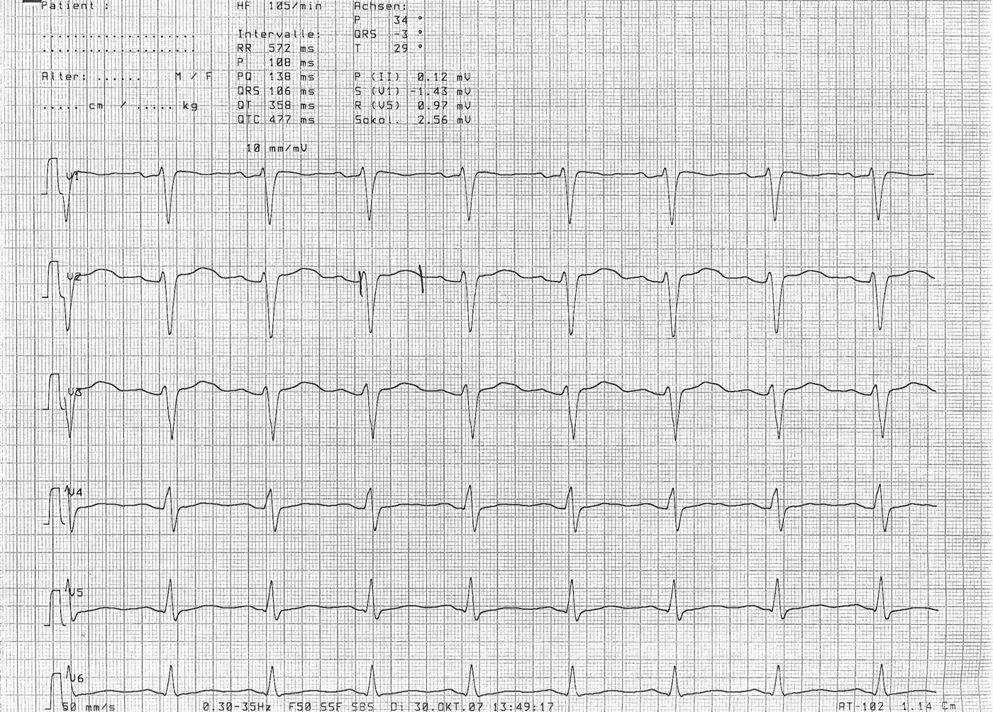
- Tachycardia: above about 100 beats/min the T wave merges with the next P wave, and the end of the T wave is uncertain.
- Telemetry leads: QT values from monitoring leads and from different leads are not interchangeable; trends are followed in one lead.
- Recognition: when 902 physicians classified four ECGs with long and normal QT intervals, 96% of QT experts and 62% of arrhythmia specialists classified all of them correctly, compared with fewer than 25% of cardiologists and non-cardiologists.
QT dispersion (the difference between the longest and shortest QT across the 12 leads) was proposed as a marker of heterogeneous repolarization. Its measurement is not reproducible, its validity as a marker of regional repolarization is unproven, and the 2009 AHA/ACCF/HRS document recommends that it not be included in routine ECG reports.
Rate correction: Bazett, Fridericia, Framingham, and Hodges
Every correction formula expresses the QT at a heart rate of 60 beats/min (RR 1 second). They differ in how they model the QT-RR relation. Bazett divides by the square root of RR, Fridericia by the cube root, and the Framingham and Hodges formulas add a linear term.
| Formula | Equation (QT and RR in seconds unless stated) | Behavior across heart rates |
|---|---|---|
| Bazett, 1920 | QTc = QT ÷ √RR | Overcorrects at fast rates (QTc too long) and undercorrects at slow rates (QTc too short); printed by most electrocardiographs and the basis of most published thresholds |
| Fridericia, 1920 | QTc = QT ÷ ∛RR | Less rate dependent than Bazett; preferred in drug trials and oncology monitoring |
| Framingham (Sagie), 1992 | QTc = QT + 0.154 × (1 − RR) | Linear; derived from 5,018 Framingham participants, with little residual rate dependence |
| Hodges, 1983 | QTc (ms) = QT (ms) + 1.75 × (heart rate − 60) | Linear in heart rate; stable at fast rates |
The 2009 AHA/ACCF/HRS document recommends a linear regression correction over Bazett. All four formulas agree at 60 beats/min; the difference grows as rate departs from 60.
At 105 beats/min, a measured QT of 358 ms gives a Bazett QTc of 474 ms, but a Fridericia QTc of 431 ms, a Framingham QTc of 424 ms, and a Hodges QTc of 437 ms. A hospitalized patient with sinus tachycardia can therefore be labeled as having a long QT, and a QT-prolonging drug withheld, on the basis of the correction formula alone. At 50 beats/min the error reverses: a QT of 440 ms gives a Bazett QTc of 402 ms and a Fridericia QTc of 414 ms, and real prolongation is missed. Drug-induced torsades also occurs mainly during bradycardia.
The International Council for Harmonisation (ICH) E14 guidance governs QT evaluation of new non-antiarrhythmic drugs. A thorough QT study looks for a mean drug effect of about 5 ms, and a drug is judged free of clinically relevant QT effect when the upper bound of the one-sided 95% confidence interval for the mean QTc change excludes 10 ms. Categorical outliers are reported at QTc values above 450, 480, and 500 ms and at increases from baseline of more than 30 and 60 ms. E14 requires uncorrected, Bazett-corrected, and Fridericia-corrected data; Fridericia is usually chosen as the primary correction because Bazett distorts results when a drug changes heart rate.
In poisoning, the Isbister QT nomogram plots the uncorrected QT against heart rate. QT-heart rate pairs above the line are associated with torsades, and the nomogram avoids the overcorrection of Bazett at the fast heart rates common in overdose.
Normal and prolonged QTc values in men and women
After puberty the QTc in women is longer than in men by roughly 10 to 20 ms, in part because androgens shorten repolarization in men. Thresholds are therefore sex-specific.
| Source and purpose | Men | Women |
|---|---|---|
| AHA/ACCF/HRS 2009: prolonged QTc | ≥450 ms | ≥460 ms |
| AHA/ACCF/HRS 2009: short QTc | ≤390 ms | ≤390 ms |
| AHA/ACCF 2010: approximate 99th percentile in healthy adults | 470 ms | 480 ms |
| AHA/ACCF 2010: high risk of torsades with a QT-prolonging drug | >500 ms or increase >60 ms | >500 ms or increase >60 ms |
| Congenital long QT syndrome diagnosis (2013 consensus; 2022 European Society of Cardiology [ESC] guideline) | ≥480 ms on repeated ECGs (≥500 ms in 2013 without other findings) | Same |
Values are Bazett-corrected unless stated. There is no QTc below which torsades cannot occur and none above which it is certain; risk rises continuously; in congenital long QT cohorts cited by the 2010 statement, each 10-ms increase in QTc raises the risk of torsades by about 5% to 7%.
Normal QTc distributions in healthy people and in genotype-positive long QT patients overlap. About 10% to 36% of genotype-positive patients have a resting QTc of 440 ms or less, most often in LQT1, which is why the diagnosis of long QT syndrome in a relative rests on the familial variant and not on the QTc alone.
Short QT. Secondary causes include hypercalcemia, hyperkalemia, acidosis, hyperthermia, and digoxin. Short QT syndrome is an inherited channelopathy with atrial fibrillation and ventricular fibrillation in a structurally normal heart. The 2022 ESC guideline diagnoses it with a QTc of 360 ms or less plus a pathogenic variant, a family history of short QT syndrome, or survival of ventricular tachycardia or fibrillation without heart disease. The diagnosis is also considered at a QTc of 320 ms or less without other findings. Survivors receive an implantable cardioverter-defibrillator (ICD), and quinidine prolongs repolarization in selected patients.
QT measurement with bundle branch block, ventricular pacing, and wide QRS rhythms
When the QRS widens, the QT lengthens by part of the added depolarization time without any change in repolarization risk. Applying narrow-QRS QTc thresholds to the raw QTc in left bundle branch block, right ventricular pacing, or a drug-widened QRS overstates the QTc. The 2009 AHA/ACCF/HRS document states that ventricular conduction defects require adjustment of the QT for QRS duration, best by entering QRS duration and RR interval as covariates in the correction formula or by using the JT interval with JT-specific normal limits.
Three methods are in use:
- JT interval and JTc. JT = QT − QRS, which removes depolarization entirely. The JT is rate corrected with Bazett (JTc) or used to compare serial tracings in the same patient. The JT changes little with the QRS and is the most direct measure of repolarization.
- JT index. The JT index = JT × (heart rate + 100) ÷ 518. It was derived in patients with and without ventricular conduction defects, and a value of 112 or more identifies prolonged repolarization in all conduction-defect categories.
- Bogossian modified QT. In 60 patients with a narrow QRS undergoing electrophysiologic study, right ventricular pacing produced a left bundle branch block pattern that lengthened the QT by 48.5% of the paced QRS duration without changing the JT interval. The modified QT = QT − 0.485 × QRS (rounded in practice to half the QRS), which is then rate corrected and compared with the usual thresholds. The formula was derived in paced, not spontaneous, left bundle branch block.
A simpler bedside approach subtracts the QRS excess over 100 or 120 ms from the QTc before comparing it with the usual cutoffs. It has not been validated against outcomes, and serial JT values in the same patient remain more reliable than any single corrected value.
During a wide QRS tachycardia the QT cannot be measured meaningfully. The baseline QT is measured on the last sinus beats before the arrhythmia, which is the measurement that separates torsades from polymorphic ventricular tachycardia with a normal QT.
QTc and JT calculator
The verdict applies sex-specific thresholds to the Bazett QTc between 60 and 90 beats/min and to the Fridericia QTc outside that range. When the QRS is 120 ms or more, the verdict rests on the JT index.
QTc by four formulas, with JT index for wide QRS
Bazett, Fridericia, Framingham, Hodges, JT index, BogossianRR (s) = 60 ÷ heart rate. Bazett QTc = QT ÷ √RR; Fridericia QTc = QT ÷ ∛RR; Framingham QTc = QT + 154 × (1 − RR); Hodges QTc = QT + 1.75 × (heart rate − 60); JT = QT − QRS; JTc = JT ÷ √RR; JT index = JT × (heart rate + 100) ÷ 518 (abnormal ≥112); Bogossian modified QT = QT − 0.485 × QRS, shown after Bazett correction. All intervals in ms.

Congenital long QT syndrome: prevalence, inheritance, and genes
Congenital long QT syndrome affects about 1 in 2,000 people. In an ECG screening study of 44,596 Italian infants with genetic confirmation, the prevalence was at least 1 in 2,534 live births and probably close to 1 in 2,000, and many affected people are never diagnosed. It is a leading cause of sudden death in young people with structurally normal hearts, and the first event can be sudden death.
The common form, historically called Romano-Ward syndrome, is autosomal dominant with incomplete penetrance and variable expression. Jervell and Lange-Nielsen syndrome is the autosomal recessive form, caused by biallelic variants in KCNQ1 or KCNE1. It combines congenital sensorineural deafness with marked QT prolongation and a high event rate from childhood. Heterozygous parents of these patients usually have a milder long QT phenotype.
KCNQ1, KCNH2, and SCN5A account for about 75% of clinically definite long QT syndrome, and the yield of genetic testing in clinically definite cases is about 75%. In genotyped cohorts long QT syndrome type 1 (LQT1) is the most common subtype, followed by LQT2, with type 3 (LQT3) accounting for about 5% to 10%. In 2020 an international Clinical Genome Resource (ClinGen) curation of 17 genes reported as causing long QT syndrome classified only three (KCNQ1, KCNH2, SCN5A) as definitive for typical disease. Four (CALM1, CALM2, CALM3, TRDN) were definitive or strong for long QT syndrome with atypical features, and CACNA1C had moderate evidence. Nine genes had limited or disputed evidence, including KCNE1 and KCNE2 as causes of isolated long QT syndrome, KCNJ2, ANK2, and CAV3.
| Subtype | Gene and current | Mechanism | Clinical features |
|---|---|---|---|
| LQT1 | KCNQ1; IKs | Loss of function | Broad-based T wave; QT fails to shorten with exercise; events with exertion and swimming; beta-blockers highly effective |
| LQT2 | KCNH2 (hERG); IKr | Loss of function, often by defective channel trafficking | Low-amplitude, notched T waves; events with sudden noise, arousal, and emotion; postpartum risk; higher risk in women after adolescence and with pore-region variants |
| LQT3 | SCN5A; late INa | Gain of function (failure of inactivation) | Long isoelectric ST segment with a late, narrow T wave; events at rest and during sleep; QT prolongs most at slow rates; mexiletine shortens the QT |
| Jervell and Lange-Nielsen | Biallelic KCNQ1 or KCNE1 | Recessive loss of IKs | Congenital deafness; very long QTc; high event rate despite beta-blockers |
| Calmodulinopathy | CALM1, CALM2, CALM3 | Usually de novo dominant variants | Severe, early-onset QT prolongation; overlap with catecholaminergic polymorphic ventricular tachycardia |
| Triadin knockout | TRDN | Recessive | Exercise-induced arrest in the young; T-wave inversion in precordial leads |
| Timothy syndrome | CACNA1C; L-type ICa | Gain of function | Syndactyly, structural heart disease and neurodevelopmental features in the classic form; isolated QT prolongation in adults with some variants |
| Andersen-Tawil syndrome | KCNJ2; IK1 | Loss of function | Periodic paralysis, dysmorphic features, prominent U waves, and bidirectional ventricular ectopy; QTc often only mildly prolonged, and now classified separately from long QT syndrome |
Gene classification follows the 2020 ClinGen reappraisal. SCN5A loss-of-function variants cause Brugada syndrome and conduction disease, and overlap phenotypes with both long QT and Brugada features occur in the same family.
Mechanisms: early afterdepolarizations and dispersion of repolarization
A prolonged action potential plateau allows L-type calcium channels to recover from inactivation and reopen before repolarization is complete. The resulting inward current produces an early afterdepolarization. When it reaches threshold, it triggers a premature ventricular beat. Prolongation is not uniform across the ventricular wall, because midmyocardial (M) cells and Purkinje fibers have less repolarizing reserve than epicardium. The triggered beat therefore meets tissue with widely dispersed refractoriness and initiates functional reentry. The wandering reentrant wavefront produces the twisting QRS axis of torsades de pointes.
Bradycardia and pauses favor early afterdepolarizations because the action potential is longest at long cycle lengths. IKr blockers show reverse use-dependence: their effect is greatest at slow rates. This is why drug-induced torsades classically begins after a pause and at night. In LQT1, by contrast, the deficient IKs is the current that normally shortens the action potential during adrenergic stimulation. Sympathetic surges therefore trigger events, and beta-blockade is most protective.
T-wave morphology and triggers by genotype
Resting T-wave shape and the setting of events point to the genotype before genetic results return, and they guide trigger-specific counseling.
| Feature | LQT1 | LQT2 | LQT3 |
|---|---|---|---|
| Typical T wave | Broad-based, smooth, early onset | Low amplitude, notched or bifid | Long isoelectric ST segment, late-onset narrow or peaked T wave |
| Main trigger | Exercise, especially swimming and diving | Sudden auditory stimuli (alarm clock, telephone), emotion, arousal from sleep | Rest and sleep, slow heart rates |
| Share of events with the main trigger (Schwartz 2001) | 62% during exercise | 43% with emotion | 39% during sleep or rest |
| QT response to exercise | Fails to shorten; prolongs in recovery | Shortens near normally | Shortens, sometimes more than normal |
| Higher-risk groups | Males in childhood | Women after adolescence and postpartum; pore-region variants | Males; events often lethal |
| Beta-blocker protection | High | Intermediate | Lower; mexiletine added |
In the 2001 genotype-phenotype series, only 3% of LQT1 events occurred during sleep or rest, while 13% of LQT3 events occurred during exercise. T-wave patterns overlap, and morphology predicts genotype in most but not all cases.
Diagnosis of congenital long QT syndrome and the Schwartz score
Secondary causes are excluded first: QT-prolonging drugs, electrolyte abnormalities, acute ischemia, intracranial events, hypothermia, and structural heart disease. A single prolonged QTc is confirmed on repeated ECGs.
The 2013 HRS/European Heart Rhythm Association/Asia Pacific Heart Rhythm Society (EHRA/APHRS) consensus diagnoses long QT syndrome in the presence of any of the following: a Schwartz score of 3.5 or more without a secondary cause, an unequivocally pathogenic variant in a long QT gene, or a QTc of 500 ms or more on repeated ECGs without a secondary cause. The diagnosis can also be made with a QTc of 480 to 499 ms on repeated ECGs in a patient with unexplained syncope. The 2022 ESC guideline diagnoses long QT syndrome with a QTc of 480 ms or more on repeated 12-lead ECGs, with or without symptoms, or a long QT diagnostic score above 3 (Class I). It also accepts a pathogenic variant irrespective of the QTc (Class I), and the diagnosis is considered with a QTc of 460 to 479 ms and arrhythmic syncope (Class IIa).
The Schwartz score combines ECG, clinical, and family findings. The 2011 version added QTc prolongation in the fourth minute of exercise recovery. The 2022 ESC modification scores a QTc of 480 ms or more at 3.5 points, adds a pathogenic variant at 3.5 points, and removes congenital deafness, so that either finding alone exceeds the diagnostic threshold of 3.
Schwartz score and the 2022 ESC modification
Off QT-prolonging drugs, secondary causes excludedPoints: QTc ≥480 ms 3 (3.5 in the ESC score), 460–479 ms 2, 450–459 ms in men 1; QTc ≥480 ms in the fourth minute of exercise recovery 1; torsades de pointes 2; T-wave alternans 1; notched T wave in three leads 1; low heart rate for age 0.5; syncope with stress 2, without stress 1 (not scored with torsades); congenital deafness 0.5 (Schwartz only); family member with definite long QT syndrome 1; unexplained sudden death before 30 years in an immediate family member 0.5, with the same relative not counted twice; pathogenic variant 3.5 (ESC only). Schwartz probability: ≤1 low, 1.5–3 intermediate, ≥3.5 high.
Exercise, standing, ambulatory, and epinephrine testing
Provocative tests expose a repolarization defect that the resting ECG conceals. They are used when the resting QTc is borderline, in genotype-negative patients, and in relatives of an affected person whose familial variant is unknown.
Exercise testing. In LQT1, the QT fails to shorten appropriately during exercise, and in recovery the QTc prolongs instead of returning to baseline. A QTc of 480 ms or more in the fourth minute of recovery scores one Schwartz point. The pattern also helps predict an LQT1 genotype before testing. Exercise testing also assesses beta-blockade by the blunting of peak heart rate and documents exercise-induced ventricular ectopy.
Standing test. On brisk standing from the supine position, heart rate rises by about 28 beats/min within seconds, while the QT adapts more slowly. In a 2010 study of 68 patients with long QT syndrome and 82 controls, the QT shortened by a mean of 21 ms in controls but lengthened by 4 ms in patients. The test is read at maximal QT stretching, the beat at which the end of the T wave comes closest to the next P wave because the RR interval has shortened without a matching shortening of the QT. At that point the Bazett QTc rose by 94 ms in patients and by 54 ms in controls, so the QTc rises in healthy people too and the size of the rise discriminates. In patients with a baseline QTc of 390 to 480 ms, a stretched QTc of 487 ms identified long QT syndrome with 90% sensitivity and 86% specificity. The response is most impaired in LQT2, in which standing often exposes notched T waves. The test needs only a continuous ECG recording, but its cutoffs come from small case-control studies.
Ambulatory monitoring. Twelve-lead Holter recording captures QT prolongation at night and after pauses (LQT2 and LQT3), T-wave alternans, and the QT-RR relation across the full rate range.
Epinephrine challenge. A low-dose epinephrine infusion produces paradoxical QT prolongation in LQT1. Sensitivity and specificity are limited, the test can provoke arrhythmia, and it is reserved for selected patients in expert centers.
Genetic testing, variant interpretation, genotype-negative disease, and cascade screening of relatives
Genetic counseling and genetic testing are recommended in patients with clinically diagnosed long QT syndrome by the 2017 American Heart Association/American College of Cardiology/Heart Rhythm Society (AHA/ACC/HRS) guideline and the 2022 ESC guideline (Class I in both). The 2022 international genetic testing consensus supports testing a panel limited to genes with definitive or strong evidence, because adding genes with limited evidence raises the number of variants of uncertain significance without raising the diagnostic yield. The most clearly affected living family member is tested first.
A pathogenic or likely pathogenic variant confirms the diagnosis, identifies the subtype, and permits cascade testing of relatives. A variant of uncertain significance does not establish the diagnosis, is not used to test relatives predictively, and does not by itself justify a defibrillator or activity restriction. Variants are periodically reclassified, so the report is revisited over time.
About a quarter of patients with clinically definite long QT syndrome have no identified variant. A negative test does not exclude the diagnosis in a patient with a clear phenotype. Treatment in these patients follows the phenotype, and their relatives are evaluated clinically with ECGs, exercise testing, and ambulatory monitoring.
After sudden unexplained death in a young person with a structurally normal heart at autopsy, a molecular autopsy on preserved DNA can identify a channelopathy and redirect evaluation to living relatives.
Every first-degree relative of a patient with long QT syndrome undergoes a clinical evaluation and a 12-lead ECG, with exercise testing and ambulatory monitoring when the ECG is borderline. When the familial variant is known, relatives are offered targeted testing for that variant, and newly identified carriers lead to screening of their own relatives. Genotype-positive relatives with a normal QTc have a lower event rate than affected relatives, but their risk is not zero. Beta-blockade for asymptomatic carriers is addressed under beta-blocker therapy, and all carriers follow drug-avoidance and electrolyte precautions.
Natural history and risk stratification
Untreated symptomatic congenital long QT syndrome had a high mortality in historical series, about half of patients dying within 10 years of the first syncope. Beta-blockers, trigger avoidance, and the defibrillator have changed this course. Individual risk is set chiefly by QTc length, genotype, sex, age, and prior events.
In a 2003 study of 647 genotyped patients, the risk of a first cardiac event (syncope, cardiac arrest, or sudden death) before age 40 years and before therapy was 50% or more with a QTc of 500 ms or more in LQT1, in LQT2, and in men with LQT3. The risk was intermediate (30% to 49%) with a QTc below 500 ms in women with LQT2 and in patients of either sex with LQT3, and with a QTc of 500 ms or more in women with LQT3. Patients with LQT1 and men with LQT2 whose QTc was below 500 ms had a risk below 30%. In the 2018 study behind the 1-2-3-LQTS risk calculator, 1,710 patients were followed for a median of 7.1 years. The estimated risk of life-threatening events rose by 15% for each 10-ms increase in QTc in all genotypes. At any QTc, risk was 130% higher in LQT2 and 157% higher in LQT3 than in LQT1. The calculator estimates the five-year risk of life-threatening arrhythmic events in patients not yet taking a beta-blocker from QTc and genotype.
The 2017 AHA/ACC/HRS guideline lists QTc above 500 ms, LQT2 and LQT3 genotypes, female sex with LQT2, age younger than 40 years, onset of symptoms before 10 years of age, and recurrent syncope as high-risk features. Prior cardiac arrest and syncope during beta-blocker therapy carry the highest risk. Arrhythmic syncope (abrupt, exertional or auditory, without prodrome) is separated from reflex syncope, which is common in young people and carries no excess risk.
Beta-blocker therapy: agent choice and adherence
Beta-blockade is the foundation of treatment. The 2017 AHA/ACC/HRS guideline recommends a beta-blocker in long QT syndrome with a resting QTc above 470 ms or with symptoms (Class I), and a beta-blocker is reasonable in asymptomatic patients with a QTc below 470 ms (Class IIa). The 2022 ESC guideline recommends beta-blockers, ideally nonselective (nadolol or propranolol), in patients with documented QT prolongation (Class I).
In a multicenter cohort of patients with LQT1 and LQT2, symptomatic patients taking metoprolol had more breakthrough events than those taking propranolol or nadolol, and propranolol shortened the QTc most. In a second US registry, nadolol was the only beta-blocker that significantly reduced risk in LQT2. The 1-2-3 cohort found that only nadolol reduced arrhythmic risk significantly in all genotypes compared with no therapy (hazard ratio 0.38). Nadolol once daily, or propranolol in divided doses when nadolol is unavailable, is therefore preferred. Atenolol and metoprolol are not first-line choices.
Breakthrough events often follow missed doses, a switch between agents, or a QT-prolonging drug. Adherence, dosing intervals, and drug interactions are therefore reviewed at every visit. Beta-blockers reduce events most in LQT1. In LQT2, beta-blockade is combined with avoidance of hypokalemia and auditory triggers. In LQT3, beta-blockade still reduces events in registry data, and mexiletine is added when the QT is long.
Mexiletine, genotype-directed drug therapy, and gene-specific treatments in development
Mexiletine blocks late sodium current and directly targets the LQT3 defect. In a series of 34 patients with LQT3, mexiletine shortened the QTc by a mean of 63 ms, reduced the proportion of patients with arrhythmic events from 22% to 3%, and reduced the annual event rate by 93%. The 2022 ESC guideline recommends mexiletine in LQT3 patients with a prolonged QT (Class I). Before long-term therapy, an acute oral test confirms that the QTc shortens, generally by 40 ms or more, because some SCN5A variants respond poorly. Mexiletine also shortens the QTc in a subset of patients with LQT2, where its use remains investigational.
Ranolazine and flecainide also block late sodium current and have been studied in LQT3, but neither has outcome data comparable with mexiletine. Potassium supplementation and potassium-sparing drugs raise serum potassium and shorten the QT in LQT2, and normal potassium and magnesium are maintained in every genotype. Routine high-dose potassium supplementation is not a standard LQT2 treatment.
Gene-specific treatments are in development. A dual-component suppression-and-replacement KCNQ1 gene therapy, which silences both normal and variant alleles and supplies a resistant normal copy, shortened the action potential in stem cell-derived cardiomyocytes and engineered heart tissue from patients with LQT1. Similar strategies are under study for LQT2. Drugs that restore hERG trafficking, IKs activators, and allele-specific silencing remain preclinical or early clinical. None has changed guideline management.
Defibrillator therapy, left cardiac sympathetic denervation, and differences between US and European guidance
The ICD protects against death from arrhythmia but does not prevent the arrhythmia. Shocks are painful and are catecholamine stimuli that can provoke further events. In a young patient the device carries decades of lead and generator complications. Left cardiac sympathetic denervation removes the lower half of the left stellate ganglion and the T2 to T4 thoracic ganglia, usually by video-assisted thoracoscopy, and lowers the release of norepinephrine in the heart without causing bradycardia. In a series of 147 high-risk patients, the mean yearly number of cardiac events per patient fell by 91% after denervation. Denervation did not abolish risk: 7% of patients died suddenly during long-term follow-up. Its side effects are a left-sided Horner syndrome in a minority and unilateral loss of sweating and flushing.
| Clinical situation | 2017 AHA/ACC/HRS | 2022 ESC |
|---|---|---|
| QTc above 470 ms or symptoms | Beta-blocker (Class I) | Nonselective beta-blocker for documented QT prolongation (Class I) |
| Asymptomatic, QTc below 470 ms | Beta-blocker reasonable (Class IIa) | Beta-blocker should be considered for genotype-positive patients with a normal QTc (Class IIa) |
| Resuscitated cardiac arrest | Beta-blocker and ICD (Class I) | Beta-blocker and ICD (Class I) |
| Syncope or ventricular arrhythmia despite beta-blocker | Intensified therapy guided by genotype, left cardiac sympathetic denervation, and/or ICD (Class I) | ICD in addition to beta-blocker and genotype-specific drugs (Class I) |
| Recurrent appropriate ICD shocks on maximum tolerated beta-blocker | Intensified therapy with additional drugs or left cardiac sympathetic denervation (Class I) | Left cardiac sympathetic denervation (Class I) |
| Asymptomatic, QTc above 500 ms on beta-blocker | Intensified therapy, left cardiac sympathetic denervation, or ICD may be considered (Class IIb) | ICD may be considered when high risk by the 1-2-3-LQTS risk calculator (Class IIb) |
| Symptomatic, with ICD contraindicated or declined | Not addressed separately | Left cardiac sympathetic denervation (Class I) |
| Beta-blockers and genotype-specific drugs not tolerated or contraindicated | Not addressed separately | ICD or left cardiac sympathetic denervation should be considered (Class IIa) |
| LQT3 with prolonged QT | Intensified therapy guided by genotype | Mexiletine (Class I) |
| QT-prolonging drugs, hypokalemia, hypomagnesemia | Avoided (Class III: Harm) | Avoided (Class I) |
The 2013 HRS/EHRA/APHRS consensus states that, except under special circumstances, an ICD is not indicated in asymptomatic patients who have not had a trial of beta-blocker therapy. Device programming in long QT syndrome uses high detection rates and long detection intervals, and lower-rate pacing in pause-dependent LQT2 and LQT3 prevents post-pause QT prolongation.
A subcutaneous defibrillator avoids transvenous leads in patients who do not need pacing, but large T waves in long QT syndrome cause T-wave oversensing. Sensing screening, including during exercise, precedes implantation. A transvenous system is chosen when pacing is planned to prevent pauses.
The 2017 AHA/ACC/HRS and 2022 ESC guidelines agree on beta-blockers first, the defibrillator after cardiac arrest, genetic testing in diagnosed patients, and avoidance of QT-prolonging drugs. The ESC guideline diagnoses long QT syndrome at a QTc of 480 ms (the 2013 consensus used 500 ms without other findings), names nadolol and propranolol as preferred, recommends mexiletine in LQT3 (Class I), and selects asymptomatic patients for a defibrillator with the 1-2-3-LQTS risk calculator. The US guideline uses a QTc of 470 ms to separate Class I from Class IIa beta-blockade and has no risk calculator.
Lifestyle, drug avoidance, sports, and the differential diagnosis of syncope
Every patient receives a list of drugs to avoid, and CredibleMeds maintains a public list for congenital long QT syndrome. Diarrhea, vomiting, diuretics, and eating disorders deplete potassium and magnesium, and the losses are replaced promptly. Patients with LQT2 limit abrupt auditory stimuli, such as alarm clocks and telephones in the bedroom. Patients with LQT1 do not swim alone. An automated external defibrillator at home and at sports venues and cardiopulmonary resuscitation training for the family are also advised.
The 2025 AHA/ACC scientific statement on competitive sports relies on shared decision-making with a cardiologist experienced in channelopathies. Competitive sports are reasonable for genotype-positive athletes with a resting QTc below 460 ms. They are also reasonable for athletes with a prolonged QTc or prior symptoms who are under expert supervision, after risk assessment, education, and guideline-directed therapy, with an emergency action plan and access to an automated external defibrillator. After symptoms or escalation of therapy, athletes are free of breakthrough arrhythmia for at least 3 months before returning. Competitive swimming and diving, including in LQT1, can be considered with supervision by a person trained in cardiopulmonary resuscitation, a preference for pools over open water, and access to a defibrillator. An ICD is not implanted solely to permit sports participation.
Syncope in long QT syndrome is frequently misdiagnosed. Torsades that does not terminate within seconds causes cerebral hypoperfusion with jerking movements, and patients with long QT syndrome are often first treated for epilepsy. Features that point to arrhythmia are an abrupt onset without prodrome, provocation by exercise, swimming, a loud noise, or the postpartum state, and a family history of sudden death or drowning. Every young patient with unexplained seizure-like episodes or exertional syncope has a 12-lead ECG reviewed for the QT and T-wave morphology.
Pregnancy and the postpartum period
Pregnancy is a period of relatively low risk in long QT syndrome, probably because heart rate rises. The nine months after delivery carry a marked increase in events, especially in LQT2, in which disrupted sleep and sudden infant crying combine with hormonal changes. In a registry cohort of women with long QT syndrome, beta-blockers were associated with a lower event rate after delivery.
The 2018 ESC pregnancy guideline recommends continuing beta-blockers during pregnancy and the postpartum period in long QT syndrome (Class I). Nadolol and propranolol are both used. Fetal growth is followed because beta-blockers are associated with lower birth weight, and the neonate is observed for bradycardia and hypoglycemia. Labor and delivery proceed with telemetry, avoidance of QT-prolonging drugs (some antiemetics, uterotonic agents, and anesthetic drugs), and a defibrillator at hand. The infant of a parent with long QT syndrome undergoes an ECG and, when the familial variant is known, genetic testing.
Drug-induced QT prolongation: hERG block and the multiple-hit model
Almost all drugs that cause torsades block IKr through the hERG channel. The channel's large inner vestibule, with two aromatic residues lining the pore, binds many structurally unrelated drugs. A smaller group prolongs the QT by reducing hERG trafficking to the cell membrane (arsenic trioxide, pentamidine) or by increasing late sodium current. QT prolongation is a surrogate. The strength of hERG block does not fully predict torsades risk, because drugs that also block inward calcium or late sodium current (amiodarone, ranolazine, verapamil) prolong the QT with little torsades.
Torsades usually requires more than one hit: a high drug concentration combined with a second drug or metabolic inhibitor, electrolyte depletion, bradycardia, structural heart disease, or reduced repolarization reserve. Drug-induced long QT syndrome is far more common than the congenital form, and a minority of patients with drug-induced torsades carry a congenital long QT variant. Relatives of a patient with drug-induced torsades are screened when the phenotype or genetic results suggest congenital disease.
Risk factors for drug-induced torsades
The 2010 AHA/ACCF statement lists the factors that make torsades more likely:
- Female sex: about 70% of cases of cardiovascular drug-associated torsades in an analysis of published reports occurred in women.
- Older age, heart failure, left ventricular hypertrophy, and recent myocardial infarction.
- Bradycardia, high-grade atrioventricular block, and recent conversion from atrial fibrillation (with dofetilide, sotalol, and quinidine).
- Hypokalemia, hypomagnesemia, and hypocalcemia; diuretic therapy.
- Rapid intravenous administration and high plasma concentrations from overdose, kidney disease (sotalol, dofetilide, procainamide metabolite), or liver disease.
- Baseline QT prolongation, a history of drug-induced torsades, and congenital long QT variants.
- Two or more QT-prolonging drugs, or a QT-prolonging drug with an inhibitor of its metabolism.
Hypocalcemia lengthens the QT by stretching the isoelectric ST segment while the T wave keeps a normal width. Drugs and congenital long QT syndrome broaden or distort the T wave itself. The component of the interval that has lengthened therefore suggests the mechanism before laboratory results return.
The Tisdale score combines these factors to predict QTc prolongation in hospitalized patients on cardiac units.
| Risk factor | Points |
|---|---|
| Age 68 years or older | 1 |
| Female sex | 1 |
| Loop diuretic | 1 |
| Serum potassium 3.5 mEq/L or less | 2 |
| Admission QTc 450 ms or more | 2 |
| Acute myocardial infarction | 2 |
| Sepsis | 3 |
| Heart failure | 3 |
| One QTc-prolonging drug | 3 |
| Two or more QTc-prolonging drugs (added to the single-drug points) | 3 |
Maximum 21 points. Low risk ≤6, moderate risk 7–10, high risk ≥11. In the validation cohort, QTc prolongation (above 500 ms or an increase above 60 ms) occurred in 15%, 37%, and 73% of patients in the three groups.
QT-prolonging drugs by class and CredibleMeds categories
CredibleMeds (the Arizona Center for Education and Research on Therapeutics) classifies drugs into three groups. Known risk of torsades means that the drug prolongs the QT and is clearly associated with torsades at recommended doses. Possible risk means that it prolongs the QT without convincing evidence of torsades at recommended doses. Conditional risk means that torsades occurs only under conditions such as overdose, interacting drugs, hypokalemia, or congenital long QT syndrome. A separate list names drugs to avoid in congenital long QT syndrome. The lists are updated continuously, so the current entry for each drug applies.
| Drug class | Higher-risk agents | Notes |
|---|---|---|
| Class Ia antiarrhythmics | Quinidine, procainamide, disopyramide | Torsades can occur at low concentrations and early in therapy; procainamide's metabolite N-acetylprocainamide accumulates in kidney disease |
| Class III antiarrhythmics | Dofetilide, sotalol, ibutilide | Dose related and renally cleared (dofetilide, sotalol); started with ECG monitoring. Ibutilide causes torsades in a small percentage of infusions, most often in women and with reduced ejection fraction. Amiodarone and dronedarone prolong the QT with a low rate of torsades |
| Macrolides and fluoroquinolones | Erythromycin (intravenous), clarithromycin, azithromycin; moxifloxacin, levofloxacin | Clarithromycin and erythromycin also inhibit cytochrome P450 3A4 (CYP3A4). In a Medicaid cohort, azithromycin was associated with a small absolute increase in cardiovascular death, highest in patients at high baseline risk |
| Antipsychotics | Haloperidol (intravenous, high dose), thioridazine, pimozide, ziprasidone, quetiapine | The US label of haloperidol warns about torsades with intravenous use and doses above those recommended |
| Antidepressants | Citalopram, escitalopram, tricyclic antidepressants | Citalopram is limited to 40 mg/day, and to 20 mg/day in patients older than 60 years, with hepatic impairment, in CYP2C19 poor metabolizers, and with cimetidine or another CYP2C19 inhibitor |
| Antiemetics | Ondansetron, droperidol, domperidone (not US-marketed) | The 32-mg single intravenous ondansetron dose was withdrawn in the United States in 2012 |
| Opioids and substances | Methadone, cocaine | Methadone blocks hERG in a dose-related manner; cocaine blocks both sodium and potassium channels |
| Antimicrobials and antimalarials | Azole antifungals (ketoconazole, fluconazole, voriconazole), hydroxychloroquine, chloroquine, pentamidine, bedaquiline | Azoles add CYP3A4 inhibition to direct hERG block |
| Anticancer drugs | Arsenic trioxide, vandetanib, nilotinib, ribociclib, osimertinib | Agent-specific ECG schedules and dose-modification rules in the US labels |
| Withdrawn from the US market | Terfenadine, astemizole, cisapride | Torsades occurred when CYP3A4 inhibitors (ketoconazole, erythromycin, grapefruit juice) raised the parent drug concentration; these withdrawals led to routine QT assessment of new drugs |
Examples are not exhaustive. Risk depends on dose, route, and the patient's other risk factors. Cancer-drug QT management follows the agent's labeling and cardio-oncology guidance.
During the early coronavirus disease 2019 (COVID-19) pandemic, hydroxychloroquine with or without azithromycin was given widely to hospitalized patients. Cohort studies reported frequent QTc prolongation, which prompted ECG monitoring protocols for the combination. The US emergency use authorization for hydroxychloroquine in COVID-19 was revoked in June 2020 after trials showed no benefit.
Drug interactions and pharmacokinetic risk
Two mechanisms produce most interactions. Pharmacodynamic interactions add the IKr block of two QT-prolonging drugs, for example a fluoroquinolone with an antipsychotic. Pharmacokinetic interactions raise the concentration of a QT-prolonging drug by inhibiting its metabolism or excretion. CYP3A4 inhibitors (azole antifungals, clarithromycin, erythromycin, protease inhibitors, diltiazem, verapamil, grapefruit juice) raise concentrations of drugs such as quinidine, pimozide, methadone, and dronedarone. Dofetilide is contraindicated with the renal cation-transport inhibitors cimetidine, trimethoprim, ketoconazole, prochlorperazine, megestrol, and dolutegravir; with verapamil, which raises peak dofetilide levels; and with hydrochlorothiazide, which raises dofetilide levels and the QT. Loop and thiazide diuretics add potassium and magnesium loss.
Kidney disease raises sotalol and dofetilide concentrations, and both are dosed by creatinine clearance. Liver disease reduces the clearance of hepatically metabolized drugs and is itself associated with QT prolongation. CYP2D6 and CYP2C19 poor metabolizers accumulate some antidepressants and antipsychotics.
QTc thresholds that change management
The thresholds below come from US labeling, the 2010 AHA/ACCF statement, and opioid treatment consensus. Each assumes that the QT has been confirmed manually in the same lead and corrected appropriately for rate and QRS width.
| Setting | Threshold | Action |
|---|---|---|
| Any QT-prolonging drug (2010 AHA/ACCF) | QTc >500 ms or increase >60 ms from baseline | High risk: drug stopped, reduced, or replaced when possible; potassium and magnesium corrected; continuous monitoring |
| Dofetilide before first dose | QTc >440 ms (>500 ms with ventricular conduction abnormalities) | Contraindicated |
| Dofetilide 2 to 3 hours after first dose | Increase >15% or QTc >500 ms (>550 ms with conduction abnormalities); the uncorrected QT is used when heart rate is below 60 beats/min | Dose reduced; started in a facility with at least 3 days of continuous ECG monitoring |
| Dofetilide after the second dose | QTc >500 ms (>550 ms with conduction abnormalities) | Discontinued |
| Oral sotalol before initiation | Baseline QT >450 ms (atrial fibrillation or flutter indication); serum potassium <4 mEq/L | Not started; initiation with ECG monitoring for at least 3 days (or an approved intravenous loading protocol) and dosing interval set by creatinine clearance |
| Sotalol during therapy | QTc ≥500 ms | Dose reduced, dosing interval lengthened, or drug discontinued |
| Methadone (2009 consensus) | QTc 450–500 ms; QTc >500 ms | 450–500 ms: risks and benefits discussed and monitoring increased. Above 500 ms: dose reduction, discontinuation, or correction of other risk factors |
| Drug development (ICH E14) | Mean effect about 5 ms; categorical >450, >480, >500 ms and increases >30, >60 ms | Regulatory concern and outlier reporting |
Dofetilide and sotalol thresholds are from US prescribing information. Methadone consensus also advises an ECG before treatment, within 30 days, annually, and when the dose exceeds 100 mg/day or unexplained syncope or seizures occur.
A QTc above 500 ms does not always require stopping a needed drug. When the drug has no alternative (for example an antiarrhythmic in a patient with an ICD, or an anticancer agent), the decision weighs the indication against the full set of risk factors. Reversible factors are addressed, interacting drugs are removed, and monitoring is intensified. A rising QTc together with T-wave alternans, new U waves, or pauses followed by distorted T waves signals imminent torsades and requires immediate reassessment of the QT-prolonging drug.
Monitoring the QT in hospital and with high-risk drugs
The 2017 AHA practice standards for in-hospital ECG monitoring recommend QTc monitoring (Class I) for every patient started on an antiarrhythmic drug with known risk of torsades (dofetilide, ibutilide, sotalol, disopyramide, procainamide, quinidine). The same applies to patients with a prolonged QTc or other risk factors started on a non-antiarrhythmic drug with known risk, and after overdose of a drug with known torsades risk or of an unknown drug. Moderate to severe hypokalemia or hypomagnesemia with other risk factors, congenital long QT syndrome with unstable ventricular arrhythmia or superimposed QT prolongation, and targeted temperature management are also Class I indications. QTc monitoring is not recommended for patients without risk factors started on a non-antiarrhythmic QT-prolonging drug (Class III: No Benefit). Patients with torsades or marked QT prolongation are monitored in a unit able to deliver defibrillation and pacing. The 2010 AHA/ACCF statement advises documenting the QTc before a QT-prolonging drug is started or its dose increased and after an overdose, then at least every 8 to 12 hours.
Before a known-risk drug is started in a patient with risk factors, a baseline 12-lead ECG is obtained, potassium and magnesium are measured, and kidney and liver function are reviewed. Potassium and magnesium are kept in the normal range in monitored patients receiving QT-prolonging drugs, and after torsades the 2017 AHA/ACC/HRS guideline recommends potassium repletion to 4.0 mmol/L or more and magnesium repletion to normal values (Class I); the 2010 AHA/ACCF statement adds that repletion to 4.5 to 5.0 mmol/L may be considered, with little supporting evidence (Class IIb). Pharmacist review of the medication list identifies duplicate QT-prolonging drugs and CYP3A4 inhibitors.
Outpatients starting high-risk drugs, such as sotalol (outpatient initiation in selected patients), methadone, and some anticancer agents, follow the drug's ECG schedule.
Torsades de pointes: ECG recognition and initiation
Torsades de pointes is polymorphic ventricular tachycardia (VT) in which the QRS axis rotates around the isoelectric baseline every 5 to 20 beats, so that complexes wax and wane in amplitude. The rate is usually 150 to 250 beats/min. Episodes are often self-terminating, causing palpitations, presyncope, or syncope, but they can degenerate into ventricular fibrillation.
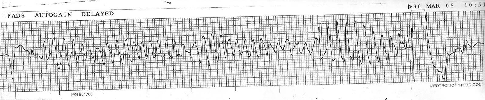

In acquired torsades, the classic initiation is a short-long-short sequence. A premature ventricular beat (short) is followed by a compensatory pause (long). The sinus beat after the pause has a further prolonged, distorted QT and T wave, and a second premature beat arising from its T wave starts the tachycardia (short). Warning signs on the preceding tracing include a QTc above 500 ms, T-wave alternans, prominent and distorted post-pause T-U waves, and runs of ventricular bigeminy. Congenital LQT1 is the exception: its events are adrenergic and often begin without a pause.
| Feature | Torsades de pointes | Polymorphic VT with a normal QT |
|---|---|---|
| QT on the preceding sinus beats | Prolonged, often with distorted T-U waves | Normal |
| Initiation | Pause dependent (short-long-short) in acquired forms; adrenergic in LQT1 and LQT2 | Short-coupled premature beat without a pause |
| Usual cause | Drugs, electrolyte depletion, bradycardia, congenital long QT syndrome | Acute ischemia; also catecholaminergic polymorphic VT, short-coupled torsades variant, cardiomyopathy |
| Treatment | Magnesium, potassium, drug withdrawal, raising the heart rate | Treatment of ischemia (urgent angiography), beta-blockade, amiodarone or lidocaine |
| Harmful treatments | Class Ia and III antiarrhythmics, other QT-prolonging drugs | Isoproterenol in ischemic polymorphic VT |
Rhythm-strip artifact from patient movement or tremor can mimic torsades. Narrow QRS complexes visible within the apparent tachycardia at the underlying sinus rate expose it as artifact.
Acute treatment of torsades de pointes and electrical storm in long QT syndrome
Treatment is directed at the substrate (prolonged, heterogeneous repolarization) and the trigger (pauses and early afterdepolarizations). Suppressing ectopy with standard antiarrhythmic drugs is not the goal and can be harmful.
| Step | Detail |
|---|---|
| Sustained torsades with hemodynamic collapse or ventricular fibrillation | Immediate unsynchronized defibrillation; synchronized cardioversion is not feasible because of the changing QRS morphology |
| Magnesium sulfate | 2 g intravenously over a few minutes, given regardless of the serum magnesium level; repeated if torsades recurs, followed by an infusion in selected patients; monitored for hypotension, loss of reflexes, and respiratory depression, especially in kidney failure |
| Withdrawal of the causative drug | All QT-prolonging drugs stopped; interacting drugs reviewed; drug levels and clearance (kidney function) assessed |
| Potassium | Repleted to 4.0 mmol/L or more, with magnesium restored to normal (2017 AHA/ACC/HRS, Class I); a target of 4.5 to 5.0 mmol/L may be considered (2010 AHA/ACCF, Class IIb); hypocalcemia corrected |
| Raising the heart rate in pause-dependent torsades | Temporary transvenous atrial or ventricular pacing at a rate that suppresses pauses and shortens the QT; isoproterenol infusion titrated to heart rate as a bridge to pacing when there is no ischemia and no congenital adrenergic long QT syndrome |
| Lidocaine | Shortens repolarization and can suppress refractory torsades; useful as an adjunct |
| Drugs avoided | Class Ia (procainamide, quinidine) and class III (sotalol, ibutilide, dofetilide) antiarrhythmics; amiodarone is avoided in torsades caused by QT prolongation |
| After recovery | Causative drug listed as an allergy-type alert; congenital long QT syndrome excluded with off-drug ECGs and genetic testing when the QTc stays long or torsades occurred at a low drug dose |
The 2017 AHA/ACC/HRS and 2022 ESC guidelines both recommend intravenous magnesium, withdrawal of offending drugs, correction of electrolytes, and an increase in heart rate by pacing or isoproterenol for recurrent pause-dependent torsades. In congenital long QT syndrome, beta-blockade is the acute and long-term treatment of adrenergic torsades, and isoproterenol is avoided.
Magnesium suppresses early afterdepolarizations by blocking L-type calcium current and terminates torsades without shortening the QT. Pacing shortens the action potential and removes the pauses that trigger torsades, and it is the most reliable means of preventing recurrence until the drug clears. In a patient with a permanent pacemaker or defibrillator, the lower rate is raised temporarily, and rate hysteresis, sleep rates, and post-shock pauses are reviewed.
Recurrent torsades in congenital long QT syndrome is treated with intravenous beta-blockade (propranolol or esmolol) and magnesium, potassium repletion, and sedation to reduce sympathetic drive. Pacing at a higher rate helps in pause-dependent LQT2 and LQT3, and mexiletine or lidocaine is added in LQT3. The cycle of shocks and recurrence is interrupted by deep sedation, general anesthesia, or urgent left cardiac sympathetic denervation. Ventricular arrhythmia management beyond torsades, including monomorphic ventricular tachycardia, structural heart disease, and defibrillator selection, is covered on the ventricular tachycardia and sudden cardiac death page.
Self-assessment
A 71-year-old woman with heart failure and chronic kidney disease is started on dofetilide for persistent atrial fibrillation. Before the first dose, her QT is 420 ms at a heart rate of 60 beats/min with a QRS of 96 ms. Three hours after the first dose, the QT is 520 ms at a heart rate of 54 beats/min. By Bazett and by Fridericia, what is the post-dose QTc, and what does dofetilide labeling require next?
Cardiovascular Medicine Board ReviewReferences
- Al-Khatib SM, Stevenson WG, Ackerman MJ, et al. 2017 AHA/ACC/HRS guideline for management of patients with ventricular arrhythmias and the prevention of sudden cardiac death: a report of the American College of Cardiology/American Heart Association Task Force on Clinical Practice Guidelines and the Heart Rhythm Society. Circulation. 2018;138:e272–e391. doi:10.1161/CIR.0000000000000549
- Zeppenfeld K, Tfelt-Hansen J, de Riva M, et al. 2022 ESC guidelines for the management of patients with ventricular arrhythmias and the prevention of sudden cardiac death. Eur Heart J. 2022;43:3997–4126. doi:10.1093/eurheartj/ehac262
- Priori SG, Wilde AA, Horie M, et al. HRS/EHRA/APHRS expert consensus statement on the diagnosis and management of patients with inherited primary arrhythmia syndromes. Heart Rhythm. 2013;10:1932–1963. doi:10.1016/j.hrthm.2013.05.014
- Rautaharju PM, Surawicz B, Gettes LS. AHA/ACCF/HRS recommendations for the standardization and interpretation of the electrocardiogram: part IV: the ST segment, T and U waves, and the QT interval. Circulation. 2009;119:e241–e250. doi:10.1161/CIRCULATIONAHA.108.191096
- Drew BJ, Ackerman MJ, Funk M, et al. Prevention of torsade de pointes in hospital settings: a scientific statement from the American Heart Association and the American College of Cardiology Foundation. Circulation. 2010;121:1047–1060. doi:10.1161/CIRCULATIONAHA.109.192704
- Tisdale JE, Chung MK, Campbell KB, et al. Drug-induced arrhythmias: a scientific statement from the American Heart Association. Circulation. 2020;142:e214–e233. doi:10.1161/CIR.0000000000000905
- Sandau KE, Funk M, Auerbach A, et al. Update to practice standards for electrocardiographic monitoring in hospital settings: a scientific statement from the American Heart Association. Circulation. 2017;136:e273–e344. doi:10.1161/CIR.0000000000000527
- Wilde AAM, Semsarian C, Márquez MF, et al. European Heart Rhythm Association (EHRA)/Heart Rhythm Society (HRS)/Asia Pacific Heart Rhythm Society (APHRS)/Latin American Heart Rhythm Society (LAHRS) expert consensus statement on the state of genetic testing for cardiac diseases. Europace. 2022;24:1307–1367. doi:10.1093/europace/euac030
- Kim JH, Baggish AL, Levine BD, et al. Clinical considerations for competitive sports participation for athletes with cardiovascular abnormalities: a scientific statement from the American Heart Association and American College of Cardiology. Circulation. 2025;151:e716–e761. doi:10.1161/CIR.0000000000001297
- Regitz-Zagrosek V, Roos-Hesselink JW, Bauersachs J, et al. 2018 ESC guidelines for the management of cardiovascular diseases during pregnancy. Eur Heart J. 2018;39:3165–3241. doi:10.1093/eurheartj/ehy340
- International Council for Harmonisation. E14: the clinical evaluation of QT/QTc interval prolongation and proarrhythmic potential for non-antiarrhythmic drugs. ICH harmonised tripartite guideline; 2005.
- Bazett HC. An analysis of the time-relations of electrocardiograms. Heart. 1920;7:353–370.
- Fridericia LS. Die Systolendauer im Elektrokardiogramm bei normalen Menschen und bei Herzkranken. Acta Med Scand. 1920;53:469–486.
- Sagie A, Larson MG, Goldberg RJ, Bengtson JR, Levy D. An improved method for adjusting the QT interval for heart rate (the Framingham Heart Study). Am J Cardiol. 1992;70:797–801. doi:10.1016/0002-9149(92)90562-D
- Hodges M, Salerno D, Erlien D. Bazett's QT correction reviewed: evidence that a linear QT correction for heart rate is better. J Am Coll Cardiol. 1983;1:694 (abstract).
- Zhou SH, Wong S, Rautaharju PM, Karnik N, Calhoun HP. Should the JT rather than the QT interval be used to detect prolongation of ventricular repolarization? An assessment in normal conduction and in ventricular conduction defects. J Electrocardiol. 1992;25(suppl):131–136.
- Bogossian H, Frommeyer G, Ninios I, et al. New formula for evaluation of the QT interval in patients with left bundle branch block. Heart Rhythm. 2014;11:2273–2277. doi:10.1016/j.hrthm.2014.08.026
- Postema PG, De Jong JS, Van der Bilt IA, Wilde AA. Accurate electrocardiographic assessment of the QT interval: teach the tangent. Heart Rhythm. 2008;5:1015–1018. doi:10.1016/j.hrthm.2008.03.037
- Viskin S, Rosovski U, Sands AJ, et al. Inaccurate electrocardiographic interpretation of long QT: the majority of physicians cannot recognize a long QT when they see one. Heart Rhythm. 2005;2:569–574. doi:10.1016/j.hrthm.2005.02.011
- Chan A, Isbister GK, Kirkpatrick CM, Duffull SB. Drug-induced QT prolongation and torsades de pointes: evaluation of a QT nomogram. QJM. 2007;100:609–615. doi:10.1093/qjmed/hcm072
- Schwartz PJ, Moss AJ, Vincent GM, Crampton RS. Diagnostic criteria for the long QT syndrome: an update. Circulation. 1993;88:782–784. doi:10.1161/01.CIR.88.2.782
- Schwartz PJ, Crotti L. QTc behavior during exercise and genetic testing for the long-QT syndrome. Circulation. 2011;124:2181–2184. doi:10.1161/CIRCULATIONAHA.111.062182
- Schwartz PJ, Stramba-Badiale M, Crotti L, et al. Prevalence of the congenital long-QT syndrome. Circulation. 2009;120:1761–1767. doi:10.1161/CIRCULATIONAHA.109.863209
- Adler A, Novelli V, Amin AS, et al. An international, multicentered, evidence-based reappraisal of genes reported to cause congenital long QT syndrome. Circulation. 2020;141:418–428. doi:10.1161/CIRCULATIONAHA.119.043132
- Ponce-Balbuena D, Deschênes I. Long QT syndrome: bench to bedside. Heart Rhythm O2. 2021;2:89–106. doi:10.1016/j.hroo.2021.01.006
- Schwartz PJ, Priori SG, Spazzolini C, et al. Genotype-phenotype correlation in the long-QT syndrome: gene-specific triggers for life-threatening arrhythmias. Circulation. 2001;103:89–95. doi:10.1161/01.CIR.103.1.89
- Priori SG, Schwartz PJ, Napolitano C, et al. Risk stratification in the long-QT syndrome. N Engl J Med. 2003;348:1866–1874. doi:10.1056/NEJMoa022147
- Mazzanti A, Maragna R, Vacanti G, et al. Interplay between genetic substrate, QTc duration, and arrhythmia risk in patients with long QT syndrome. J Am Coll Cardiol. 2018;71:1663–1671. doi:10.1016/j.jacc.2018.01.078
- Chockalingam P, Crotti L, Girardengo G, et al. Not all beta-blockers are equal in the management of long QT syndrome types 1 and 2: higher recurrence of events under metoprolol. J Am Coll Cardiol. 2012;60:2092–2099. doi:10.1016/j.jacc.2012.07.046
- Abu-Zeitone A, Peterson DR, Polonsky B, McNitt S, Moss AJ. Efficacy of different beta-blockers in the treatment of long QT syndrome. J Am Coll Cardiol. 2014;64:1352–1358. doi:10.1016/j.jacc.2014.05.068
- Mazzanti A, Maragna R, Faragli A, et al. Gene-specific therapy with mexiletine reduces arrhythmic events in patients with long QT syndrome type 3. J Am Coll Cardiol. 2016;67:1053–1058. doi:10.1016/j.jacc.2015.12.033
- Schwartz PJ, Priori SG, Cerrone M, et al. Left cardiac sympathetic denervation in the management of high-risk patients affected by the long-QT syndrome. Circulation. 2004;109:1826–1833. doi:10.1161/01.CIR.0000125523.14403.1E
- Seth R, Moss AJ, McNitt S, et al. Long QT syndrome and pregnancy. J Am Coll Cardiol. 2007;49:1092–1098. doi:10.1016/j.jacc.2006.09.054
- Roden DM. Drug-induced prolongation of the QT interval. N Engl J Med. 2004;350:1013–1022. doi:10.1056/NEJMra032426
- Makkar RR, Fromm BS, Steinman RT, Meissner MD, Lehmann MH. Female gender as a risk factor for torsades de pointes associated with cardiovascular drugs. JAMA. 1993;270:2590–2597. doi:10.1001/jama.1993.03510210076031
- Tisdale JE, Jaynes HA, Kingery JR, et al. Development and validation of a risk score to predict QT interval prolongation in hospitalized patients. Circ Cardiovasc Qual Outcomes. 2013;6:479–487. doi:10.1161/CIRCOUTCOMES.113.000152
- Krantz MJ, Martin J, Stimmel B, Mehta D, Haigney MC. QTc interval screening in methadone treatment. Ann Intern Med. 2009;150:387–395. doi:10.7326/0003-4819-150-6-200903170-00103
- Ray WA, Murray KT, Hall K, Arbogast PG, Stein CM. Azithromycin and the risk of cardiovascular death. N Engl J Med. 2012;366:1881–1890. doi:10.1056/NEJMoa1003833
- Dotzler SM, Kim CSJ, Gendron WAC, et al. Suppression-replacement KCNQ1 gene therapy for type 1 long QT syndrome. Circulation. 2021;143:1411–1425. doi:10.1161/CIRCULATIONAHA.120.051836
- Viskin S, Postema PG, Bhuiyan ZA, et al. The response of the QT interval to the brief tachycardia provoked by standing: a bedside test for diagnosing long QT syndrome. J Am Coll Cardiol. 2010;55:1955–1961. doi:10.1016/j.jacc.2009.12.015