Library › Arrhythmia › Bradycardia and AV block
Bradycardia and atrioventricular block
Sinus node dysfunction, atrioventricular block, and conduction disease in adults, from causes and localization through acute treatment, pacing indications, pacing strategy, device problems, and special populations.
Bradycardia is treated when it causes symptoms or when its anatomy predicts sudden failure of the escape rhythm. Sinus node dysfunction is paced only for symptoms that correlate with the rhythm. Acquired Mobitz II, high-grade, and third-degree atrioventricular (AV) block that is neither reversible nor physiologic is paced regardless of symptoms (Class 1). The level of block, nodal or infranodal, sets prognosis, and the expected ventricular pacing burden and left ventricular ejection fraction (LVEF) set the pacing strategy.
Definitions and classification of bradycardia and conduction disorders
Bradyarrhythmias arise from failure of impulse formation (sinus node dysfunction), failure of impulse conduction (AV or intraventricular block), or both. The 2018 American College of Cardiology/American Heart Association/Heart Rhythm Society (ACC/AHA/HRS) bradycardia guideline uses a sinus rate below 50/min and a sinus pause longer than 3 seconds as working thresholds. Neither number is an indication for treatment; healthy older adults and athletes record pauses of 2 to 3 seconds.
| Term | Definition |
|---|---|
| Sinus bradycardia | Sinus rate <50/min |
| Sinus pause | >3 s between the last atrial depolarization and the next sinus depolarization |
| Sinoatrial exit block | Pause that is a multiple of the preceding P–P interval; the node fires but the impulse fails to reach the atrium |
| Tachy-brady syndrome | Sinus bradycardia, ectopic atrial rhythm, or pauses alternating with atrial tachyarrhythmia, often with a long pause at termination |
| Chronotropic incompetence | Inability to raise heart rate in proportion to demand; in many studies, failure to reach 80% of the expected heart rate reserve |
| First-degree AV block | PR >200 ms with 1:1 conduction; severe when PR >300 ms |
| Mobitz type I (Wenckebach) | Progressive PR prolongation before a single nonconducted P wave; the PR after the dropped beat is the shortest |
| Mobitz type II | Constant PR before and after a single nonconducted P wave at a constant sinus rate |
| 2:1 AV block | Every second P wave nonconducted; cannot be classified as type I or type II from the surface ECG |
| High-grade (advanced) AV block | Two or more consecutive P waves at a constant physiologic rate fail to conduct, with evidence of some conduction |
| Third-degree (complete) AV block | No conduction; atrial rate faster than ventricular rate with an independent escape rhythm |
| Bundle branch block | QRS ≥120 ms (complete) or 110–119 ms (incomplete) with characteristic morphology; nonspecific intraventricular delay >110 ms |
Definitions from the 2018 ACC/AHA/HRS bradycardia guideline. AV, atrioventricular.
Epidemiology and natural history of bradycardia and conduction disease
Conduction disease becomes more common with age. In the Atherosclerosis Risk in Communities (ARIC) and Cardiovascular Health Study cohorts, the incidence of sick sinus syndrome was 0.8 per 1,000 person-years and rose about 1.7-fold with each 5 years of age. It was associated with higher body mass index, longer QRS duration, lower heart rate, right bundle branch block, hypertension, and prior cardiovascular disease. The projected number of new US cases rises from 78,000 in 2012 to 172,000 in 2060. Sinus node dysfunction peaks in the eighth and ninth decades, in parallel with pacemaker implantation for AV nodal disease.
First-degree AV block is not uniformly benign. In 7,575 Framingham participants, a PR interval above 200 ms carried an adjusted 2-fold risk of atrial fibrillation, a 3-fold risk of pacemaker implantation, and a 1.4-fold risk of death. Each 20-ms increment in PR raised the hazard of pacemaker implantation by 22%.
Natural history follows the level of disease. Sinus node dysfunction rarely threatens life, because the junction and ventricles provide a dependable escape, and it progresses slowly toward AV block or atrial fibrillation in a share of patients. Nodal block is often vagal or reversible. Infranodal disease progresses unpredictably to complete block with an unreliable ventricular escape; the high mortality of acquired complete heart block before pacing is the basis for pacing high-grade block regardless of symptoms.
Conduction system anatomy and levels of block
The sinus node lies at the junction of the superior vena cava and right atrium. Its artery arises from the right coronary artery in about 60% of people and from the circumflex in the rest. The compact AV node sits at the apex of the triangle of Koch. It is supplied by the AV nodal artery, usually from the right coronary artery, which explains nodal block in inferior infarction. The His bundle penetrates the central fibrous body beneath the membranous septum, next to the noncoronary and right coronary aortic cusps. This position explains block after transcatheter aortic valve replacement (TAVR), aortic valve surgery, and calcific annular disease. Septal perforators of the left anterior descending artery supply the bundle branches.
AV nodal tissue depends on the L-type calcium current, conducts decrementally, slows with vagal tone, and accelerates with sympathetic tone and atropine. His-Purkinje tissue depends on the fast sodium current, conducts all-or-none, and is little affected by autonomic tone. Nodal block therefore lengthens the PR before it fails, and infranodal block fails without warning. Junctional escape rhythms arise at 40 to 60/min with a narrow QRS; ventricular escape rhythms arise at 20 to 40/min with a wide QRS and may fail altogether.
Causes of bradycardia and atrioventricular block
Bradycardia is intrinsic when conduction tissue is diseased and extrinsic when a normal system is suppressed; the two often coexist, as when an AV nodal blocker unmasks intrinsic disease.
| Category | Causes | Typical level and course |
|---|---|---|
| Degenerative | Idiopathic fibrosis of the sinus node; progressive cardiac conduction disease (Lenègre); fibrosis and calcification extending from the aortic and mitral annuli (Lev) | Sinus node or infranodal; progressive |
| Ischemic | Inferior infarction (AV nodal artery); anterior infarction (septal perforators); chronic ischemic cardiomyopathy | Inferior: nodal, usually transient. Anterior: infranodal, marks extensive necrosis |
| Infiltrative and inflammatory | Cardiac sarcoidosis, amyloidosis, hemochromatosis, myocarditis, rheumatologic disease | Any level; sarcoidosis often in younger adults |
| Infectious | Lyme carditis; Chagas cardiomyopathy; aortic valve endocarditis with root abscess; diphtheria | Lyme: nodal, reversible. Chagas: RBBB with LAFB, progressive. Abscess: infranodal |
| Iatrogenic | TAVR; aortic, mitral, and tricuspid surgery; septal myectomy and alcohol septal ablation; catheter ablation, including deliberate AV node ablation | Infranodal or nodal; may be delayed |
| Genetic and neuromuscular | SCN5A and TRPM4 variants; LMNA cardiomyopathy; myotonic dystrophy type 1; Kearns-Sayre syndrome; Emery-Dreifuss and limb-girdle dystrophies | Infranodal; progressive, may be abrupt |
| Congenital | Maternal anti-Ro/SSA and anti-La/SSB antibodies; congenitally corrected transposition | Nodal; narrow escape in most |
| Drugs and toxins | Beta-blockers, nondihydropyridine calcium-channel blockers, digoxin, antiarrhythmic drugs, ivabradine, lithium, clonidine, and others | Nodal or sinus; reversible unless disease is unmasked |
| Metabolic and environmental | Hyperkalemia, hypothyroidism, hypothermia, hypoxemia, acidosis, anorexia nervosa | Any level; reversible |
| Autonomic | Vasovagal reflex, carotid sinus hypersensitivity, sleep apnea, vomiting, intubation, raised intracranial pressure, athletic training | Sinus and nodal; paroxysmal |
Causes of bradycardia and AV block in adults, after the 2018 ACC/AHA/HRS guideline. RBBB, right bundle branch block; LAFB, left anterior fascicular block.
Lyme carditis. Early studies found carditis in 4% to 10% of untreated patients with Lyme disease, and more recent data suggest a lower rate. AV block is the most common manifestation, and only about 40% of patients with Lyme carditis recall erythema migrans. Block is usually nodal and can fluctuate within hours from first-degree to complete block, typically in a young adult from an endemic area. The 2020 Infectious Diseases Society of America/American Academy of Neurology/American College of Rheumatology (IDSA/AAN/ACR) guideline recommends hospital admission with continuous ECG monitoring for patients with or at risk for severe cardiac complications. These include a PR interval longer than 300 ms, other arrhythmias, or myopericarditis. Hospitalized patients receive intravenous ceftriaxone until clinical improvement and then oral doxycycline, amoxicillin, cefuroxime axetil, or azithromycin, for a total of 14 to 21 days; outpatients receive oral therapy. For symptomatic bradycardia that cannot be managed medically, the guideline recommends temporary pacing rather than a permanent pacemaker. Virtually all patients recover conduction within 3 to 7 days, and permanent pacing is reserved for block that does not resolve.
Infiltrative and inflammatory disease. New high-grade block in an adult too young for degenerative disease suggests cardiac sarcoidosis and leads to cardiac magnetic resonance (CMR) and fluorodeoxyglucose positron emission tomography (FDG-PET) before a device is chosen. In small series, corticosteroids restored AV conduction in 13% to 60% of patients, and the block often recurred. The 2018 guideline therefore considers implantation without a further period of observation reasonable (Class 2a). A device with defibrillator capability is often selected because of the ventricular arrhythmic substrate. Amyloidosis causes infranodal disease and sinus node dysfunction, as covered on the cardiac amyloidosis page. Chagas disease, which affects at least 300,000 US residents, typically produces right bundle branch block with left anterior fascicular block, sinus node dysfunction, and progressive AV block.
Endocarditis. New PR prolongation or AV block in aortic valve endocarditis indicates perivalvular extension toward the His bundle and is an indication for surgical evaluation, as covered on the infective endocarditis page.
Genetic and neuromuscular disease. Myotonic dystrophy type 1 and Kearns-Sayre syndrome (external ophthalmoplegia, pigmentary retinopathy, onset before age 20) cause progressive infranodal disease with risk of sudden death. LMNA cardiomyopathy presents with conduction disease and atrial arrhythmias years before ventricular dysfunction. Familial conduction disease, skeletal myopathy, or premature sudden death in relatives leads to genetic counseling and testing. When a pathogenic variant is found, mutation-specific testing of first-degree relatives is recommended (Class 1). In inherited conduction disease, genetic counseling and targeted testing may be considered to facilitate cascade screening of relatives (Class 2b).
Metabolic and environmental causes. Hyperkalemia slows sinus and AV conduction and widens the QRS toward a sine-wave rhythm that responds to intravenous calcium within minutes. Hypothermia produces sinus bradycardia with J (Osborn) waves that resolves with rewarming. In symptomatic second-degree or third-degree block with abnormal thyroid tests but no clinical myxedema, permanent pacing without a period of observation may be considered (Class 2b).
Drug-induced bradycardia and atrioventricular block
Drugs are the most common reversible cause, usually in combination (a beta-blocker with a nondihydropyridine calcium-channel blocker, digoxin, or amiodarone) in an older patient with declining kidney function. A QT-prolonging drug given during bradycardia predisposes to pause-dependent torsades de pointes.
| Drug class | Examples | Effect and specific treatment |
|---|---|---|
| Beta-blockers | Metoprolol, carvedilol, atenolol, sotalol, timolol eye drops | Sinus and nodal slowing; glucagon and high-dose insulin in overdose |
| Nondihydropyridine calcium-channel blockers | Verapamil, diltiazem | Nodal block, hypotension; intravenous calcium, high-dose insulin, glucagon |
| Cardiac glycosides | Digoxin | Vagal and direct nodal effect; nodal block with atrial tachycardia suggests toxicity; digoxin-specific antibody fragments |
| Class I and III antiarrhythmic drugs | Amiodarone, dronedarone, flecainide, propafenone, procainamide | Sinus node suppression, nodal and infranodal block, QRS widening with sodium-channel blockers |
| If-channel inhibitor | Ivabradine | Sinus bradycardia; no effect on AV conduction |
| Central sympatholytics and others | Clonidine, dexmedetomidine, lithium, donepezil and other cholinesterase inhibitors, opioids, propofol | Sinus bradycardia and nodal block |
| Antineoplastic and immune therapies | Immune checkpoint inhibitors (myocarditis), thalidomide, crizotinib | Myocarditis with AV block requires urgent recognition |
Drugs that cause bradycardia or AV block. Antidote doses are listed in the acute management section.
New AV block on long-term stable doses of nodal blockers is often intrinsic disease unmasked by the drug, and it frequently persists or recurs after withdrawal. The guideline therefore considers it reasonable to proceed to permanent pacing without a washout period in a patient with symptomatic second-degree or third-degree block on a stable, medically necessary dose of an antiarrhythmic drug or beta-blocker (Class 2a). When the drug is required by guideline-directed therapy and no alternative exists, symptomatic bradycardia is itself a Class 1 indication for pacing.
Physiologic bradycardia in athletes and during sleep
Endurance training raises vagal tone and remodels the sinus node, producing resting rates in the 30s and 40s, junctional rhythms, first-degree AV block, and Mobitz I block that resolve with exercise. The 2017 international recommendations for ECG interpretation in athletes classify these as training-related findings that need no evaluation in an athlete without symptoms. A resting rate of 30/min or less, a sinus pause of 3 seconds or more, or a PR interval of 400 ms or more prompts evaluation. Evaluation begins with a brief bout of aerobic activity to confirm an appropriate rise in rate and shortening of the PR interval. Mobitz II and third-degree block are never attributed to training. Symptoms during exertion, failure of the block to resolve with exercise, and a family history of conduction disease or sudden death distinguish disease from adaptation. Detraining for several weeks reverses physiologic bradycardia.
During sleep, sinus bradycardia, pauses, and Mobitz I block are common in healthy adults; bradycardia clustered during apneas suggests obstructive sleep apnea. Screening for sleep apnea is recommended in documented or suspected sleep-related bradycardia (Class 1), and documented apnea is treated directly with positive airway pressure and weight loss (Class 1). Sleep-related bradycardia is not paced without another pacing indication (Class 3: Harm). Screening is also reasonable in patients who have, or are being considered for, a permanent pacemaker (Class 2a).
Sinus node dysfunction
Sinus node dysfunction, historically sick sinus syndrome, is usually age-related fibrosis of the sinus node and surrounding atrium. The same atrial disease that slows the node also generates atrial fibrillation, so the two coexist in many patients. Its electrocardiographic forms are persistent inappropriate sinus bradycardia, sinus pauses and arrest, sinoatrial exit block, tachy-brady syndrome, and chronotropic incompetence. A pause that is an exact multiple of the preceding P–P interval indicates sinoatrial exit block; a pause unrelated to the cycle indicates sinus arrest. Second-degree sinoatrial block of the Wenckebach type shows progressive shortening of P–P intervals before the pause.
In tachy-brady syndrome, the pause at termination of atrial fibrillation or flutter causes presyncope or syncope, while drugs that control the tachycardia aggravate the bradycardia. Pacing permits rate control, and catheter ablation of atrial fibrillation resolves the pauses in selected patients.
Chronotropic incompetence is failure to reach 80% of the heart rate reserve (age-predicted maximum of 220 minus age, less the resting rate) on exercise testing without beta-blockade. It causes exertional fatigue with a normal resting rate and is the principal indication for a rate-response sensor.
Intrinsic disease is separated from autonomic suppression by the intrinsic heart rate after combined autonomic blockade with intravenous propranolol 0.1 mg/kg and atropine 0.02 mg/kg (expected rate 118.1 − [0.57 × age]). The sinus node recovery time at electrophysiology study serves the same purpose. Both are reserved for cases in which noninvasive testing is nondiagnostic and the result will change management. Electrophysiology study may be considered in symptomatic patients with suspected sinus node dysfunction after a nondiagnostic noninvasive evaluation (Class 2b). It is not performed in asymptomatic sinus bradycardia (Class 3: No Benefit).
Atrioventricular block by degree: ECG recognition
First-degree AV block prolongs the PR interval beyond 200 ms with every P wave conducted. The delay is usually nodal, but a long PR with a wide QRS can reflect His-Purkinje delay. A PR interval above 300 ms can cause symptoms resembling pacemaker syndrome, because atrial contraction occurs against closed AV valves.
Mobitz type I (Wenckebach) block lengthens the PR until a P wave fails to conduct. The PR increment is largest on the second beat of the cycle, so the R–R intervals shorten before the pause, and the pause is less than twice the preceding R–R interval. With a narrow QRS, Mobitz I is nodal in nearly all cases.
Mobitz type II block shows a constant PR before and after a single nonconducted P wave at a stable sinus rate. Mobitz II is infranodal, usually accompanies bundle branch block, and often progresses to complete block. Vagal surges imitate it, but the P–P interval slows before the blocked beat.
2:1 AV block has no consecutive conducted beats, so it cannot be classified by PR behavior. The level is localized by QRS width, response to exercise or atropine, adjacent Wenckebach cycles on a longer strip, and, when needed, electrophysiology study.
High-grade (advanced) block is failure of two or more consecutive P waves to conduct while some beats still conduct; the conducted beats show a constant PR interval.
Third-degree (complete) block dissociates atria and ventricles with an atrial rate faster than the ventricular rate. The escape rhythm sets prognosis: a narrow junctional escape at 40 to 60/min suggests nodal block, and a wide escape at 20 to 40/min suggests infranodal block. Complete block in atrial fibrillation appears as a slow, regular ventricular rhythm.
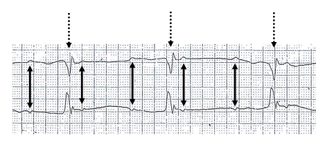

AV dissociation without complete block. AV dissociation means only that atria and ventricles beat independently. It occurs without block when a junctional or ventricular rhythm runs faster than the sinus rate (accelerated junctional rhythm, accelerated idioventricular rhythm, ventricular tachycardia) or when sinus slowing allows a junctional escape to take over (isorhythmic dissociation). Complete block is diagnosed only when P waves that fall at times when conduction should occur still fail to conduct, with an atrial rate faster than the ventricular rate. At the bedside, AV dissociation produces intermittent cannon a waves in the jugular venous pulse, when the atrium contracts against a closed tricuspid valve, and variable intensity of the first heart sound.
Paroxysmal AV block is a sudden transition from 1:1 conduction to prolonged ventricular asystole. Intrinsic paroxysmal block is phase 4 (bradycardia-dependent) block in diseased His-Purkinje tissue, typically triggered by a premature beat or a pause, and is an indication for pacing even when the baseline ECG shows only bundle branch block. Extrinsic vagal paroxysmal block occurs with simultaneous sinus slowing, often with prodromal symptoms, and is managed as reflex syncope.
Localizing atrioventricular block: nodal versus infranodal
Localization decides prognosis and often the indication. A narrow QRS with Wenckebach behavior is nodal; Mobitz II with bundle branch block is infranodal. The difficult rhythms are 2:1 block, high-grade block, and complete block, in which bedside maneuvers exploit the opposite autonomic responses of the two tissues. Atropine and exercise accelerate the sinus rate and improve nodal conduction; in infranodal block, the faster atrial rate meets a fixed refractory His-Purkinje system and the conduction ratio worsens. Vagal maneuvers do the reverse.
| Feature | AV nodal block | Infranodal (His-Purkinje) block |
|---|---|---|
| Second-degree pattern | Mobitz I | Mobitz II |
| Conducted QRS | Usually narrow | Usually wide (bundle branch block) |
| PR of conducted beats | Often long | Often normal |
| Escape rhythm | Junctional, narrow, 40–60/min, reliable | Ventricular, wide, 20–40/min, unreliable |
| Atropine | Conduction improves | No change or worse (faster atrial rate) |
| Exercise or isoproterenol | Conduction improves | Conduction ratio worsens |
| Carotid sinus massage | Conduction worsens | No change or better (slower atrial rate) |
| His bundle electrogram | Prolonged AH interval; block proximal to His deflection | HV >55 ms, split His, or block distal to His deflection |
| Typical settings | Vagal tone, drugs, inferior infarction, Lyme carditis, congenital block | Degenerative disease, anterior infarction, TAVR, neuromuscular disease, infiltration |
| Progression and prognosis | Usually stable or reversible | Progressive; abrupt asystole and Stokes-Adams syncope |
Clues to the level of AV block. These are probabilities: a narrow-QRS block can be intra-Hisian, and wide-QRS Mobitz I occasionally arises in the bundle branches. AH, atrium-to-His interval; HV, His-to-ventricle interval.
In second-degree block, carotid sinus massage or pharmacologic challenge may be considered (Class 2b), and electrophysiology study may be considered to localize the block (Class 2b). The normal HV interval is 35 to 55 ms. In a patient with syncope and bundle branch block, an HV interval of 70 ms or more, or infranodal block provoked by atrial pacing or procainamide, identifies clinically important His-Purkinje disease. Either finding is a Class 1 indication for pacing. Stokes-Adams syncope (abrupt loss of consciousness without prodrome, sometimes with brief myoclonic jerks, followed by rapid recovery and flushing) is the clinical signature of infranodal block.
Bundle branch and fascicular block
Intraventricular conduction disease is common, usually asymptomatic, and slowly progressive. Its importance lies in the substrate it signals (structural heart disease, cardiomyopathy) and in the rare patient whose syncope reflects intermittent complete block.
| Pattern | ECG criteria | Significance |
|---|---|---|
| Right bundle branch block | QRS ≥120 ms; rsR′ in V1–V2; wide S in I and V6 | Common and often benign; new RBBB in acute illness suggests right ventricular strain or septal infarction |
| Left bundle branch block | QRS ≥120 ms; broad notched R in I, aVL, V5–V6; absent septal q waves; QS or rS in V1 | Marker of structural disease; can cause dyssynchrony-mediated cardiomyopathy; echocardiography recommended (Class 1) |
| Left anterior fascicular block | Axis −45° to −90°; qR in aVL; rS in II, III, aVF; QRS <120 ms | Common; low risk alone |
| Left posterior fascicular block | Axis +90° to +180°; rS in I and aVL; qR in III and aVF; right ventricular hypertrophy excluded | Uncommon; implies more extensive disease |
| Bifascicular block | RBBB with LAFB or LPFB, or LBBB | Annual progression to complete block is low; syncope changes the evaluation |
| Bifascicular block with first-degree AV block | Bifascicular pattern with PR >200 ms (imprecisely called trifascicular) | PR delay may be nodal; HV interval defines infranodal risk |
| Alternating bundle branch block | RBBB and LBBB on successive ECGs or beats | Disease in both bundles; pacing recommended (Class 1) |
Intraventricular conduction disorders in adults, after the 2018 ACC/AHA/HRS guideline. LPFB, left posterior fascicular block.
When structural disease is suspected in newly detected left bundle branch block and echocardiography is unrevealing, CMR, CT, or nuclear imaging is reasonable (Class 2a). In patients with symptoms suggestive of intermittent bradycardia and conduction disease without documented AV block, ambulatory monitoring is useful (Class 1) and electrophysiology study is reasonable (Class 2a). In patients with chronic left bundle branch block, the 2023 Heart Rhythm Society/Asia Pacific Heart Rhythm Society/Latin American Heart Rhythm Society (HRS/APHRS/LAHRS) physiologic pacing guideline considers periodic assessment of LVEF reasonable (Class 2a), for example every 1 to 2 years. In heart failure with LVEF 36% to 50% and left bundle branch block with QRS duration of 150 ms or more, cardiac resynchronization therapy may be considered (Class 2b). The 2023 guideline extends this to cardiac physiologic pacing in patients with New York Heart Association (NYHA) class II to IV symptoms on guideline-directed therapy (Class 2b). In selected asymptomatic patients with bifascicular or trifascicular block, ambulatory monitoring may be considered to document higher-degree block (Class 2b). In asymptomatic left bundle branch block, stress testing with imaging may be considered when ischemic heart disease is suspected (Class 2b).
Diagnostic evaluation of suspected bradycardia
The evaluation establishes the rhythm, its correlation with symptoms, and any reversible or disease-defining cause. A 12-lead ECG is recommended (Class 1), and targeted laboratory tests (thyroid function, Lyme serology, potassium, pH) are reasonable when the clinical picture suggests them (Class 2a). Routine imaging is not indicated for asymptomatic sinus bradycardia or first-degree AV block without signs of structural disease (Class 3: No Benefit).
| Monitor | Duration | Best suited to |
|---|---|---|
| Holter monitor | 24–72 h | Daily symptoms; heart rate profile; nocturnal rhythm |
| Patch monitor | Up to 14 days | Symptoms every few days; continuous recording without leads |
| External event or loop recorder | 2–6 weeks | Weekly or monthly symptoms with time to activate the device |
| Mobile cardiac telemetry | Up to 30 days | Symptoms that need real-time transmission and response |
| Implantable cardiac monitor | Up to about 3 years | Infrequent symptoms (more than 30 days apart) after nondiagnostic noninvasive testing (Class 2a) |
Choice of ambulatory monitor according to symptom frequency. The 2018 guideline recommends selecting the monitor by frequency and nature of symptoms and patient preference (Class 1).
Exercise testing is reasonable for suspected chronotropic incompetence (Class 2a) and for exertional symptoms or 2:1 block of unknown level (Class 2a).
Imaging. Transthoracic echocardiography is recommended for new left bundle branch block, Mobitz II, high-grade, or third-degree block (Class 1). CMR detects sarcoidosis, myocarditis, amyloidosis, and hemochromatosis; FDG-PET identifies active sarcoid inflammation; and bone-avid tracer scintigraphy identifies transthyretin amyloidosis.
Electrophysiology study measures AH and HV intervals, provokes infranodal block by incremental atrial pacing or procainamide challenge, and assesses sinus node recovery. It is most useful in syncope with bundle branch block and in 2:1 block of uncertain level.
Tilt-table testing and carotid sinus massage. Tilt testing reproduces reflex syncope and identifies a cardioinhibitory response. Carotid sinus massage is performed for 5 to 10 seconds on each side, supine and upright, with ECG and blood pressure monitoring, after a carotid bruit is excluded. Asystole longer than 3 seconds or a systolic fall of 50 mm Hg or more with reproduction of symptoms is diagnostic of carotid sinus syndrome.
Acute management of symptomatic bradycardia
The first judgment is whether bradycardia is causing hypoperfusion: hypotension, altered mentation, ischemic chest pain, acute heart failure, or shock. Reversible causes are corrected at the same time as the rate is supported (Class 1): hypoxemia, hyperkalemia, acidosis, hypothermia, drug toxicity, and ischemia. Atropine is reasonable for sinus bradycardia and AV nodal block with symptoms or hemodynamic compromise (Class 2a). It is often ineffective in infranodal block, and it is avoided in heart transplant recipients without autonomic reinnervation, in whom it can provoke paradoxical block (Class 3: Harm).
| Drug | Dose | Comment |
|---|---|---|
| Atropine | 0.5–1 mg IV every 3–5 min, maximum 3 mg | The 2020 AHA advanced life support guideline uses 1 mg |
| Dopamine | 5–20 µg/kg/min IV | Start at 5 µg/kg/min and increase by 5 µg/kg/min every 2 min; higher doses cause tachyarrhythmia and vasoconstriction |
| Epinephrine | 2–10 µg/min or 0.1–0.5 µg/kg/min IV | Titrated to rate and pressure |
| Isoproterenol | 20–60 µg IV bolus, then 10–20 µg doses, or 1–20 µg/min infusion | Pure beta-agonist; ischemia and hypotension |
| Aminophylline | 250 mg IV bolus in acute inferior infarction; 6 mg/kg IV in 100–200 mL over 20–30 min after heart transplant or spinal cord injury | After heart transplant and in acute spinal cord injury (Class 2a); second-degree or third-degree block in acute inferior infarction (Class 2b) |
| Theophylline | 300 mg IV, then 5–10 mg/kg/day orally, target level 10–20 µg/mL | After heart transplant (Class 2a). In acute spinal cord injury (Class 2a), oral 5–10 mg/kg/day is effective, often at serum levels below 10–20 µg/mL |
| Calcium chloride 10% | 1–2 g IV every 10–20 min, or 0.2–0.4 mL/kg/h | Calcium-channel blocker overdose (Class 2a); hyperkalemia |
| Calcium gluconate 10% | 3–6 g IV every 10–20 min, or 0.6–1.2 mL/kg/h | Alternative to calcium chloride |
| Glucagon | 3–10 mg IV, then 3–5 mg/h | Beta-blocker or calcium-channel blocker overdose (Class 2a) |
| High-dose insulin | 1 unit/kg IV bolus, then 0.5 units/kg/h | With dextrose and potassium monitoring (Class 2a) |
| Digoxin-specific antibody fragments | Dose by level or amount ingested; one vial binds about 0.5 mg digoxin | Digoxin toxicity with symptomatic bradycardia (Class 2a); dialysis does not remove digoxin (Class 3: No Benefit) |
Drug therapy for acute bradycardia, from the 2018 ACC/AHA/HRS guideline (Table 8). In patients at low likelihood of coronary ischemia, isoproterenol, dopamine, dobutamine, or epinephrine may be considered for sinus node dysfunction (Class 2b) and for second-degree or third-degree AV block (Class 2b).
Temporary pacing. Transcutaneous pacing is available within seconds and serves as a bridge (Class 2b) until transvenous pacing is established or the rhythm recovers. Capture is confirmed by a palpable pulse or arterial waveform, because the stimulus artifact can imitate a QRS complex. Temporary transvenous pacing is reasonable for persistent hemodynamically unstable bradycardia refractory to medical therapy (Class 2a); perforation, tamponade, dislodgement, infection, and venous thrombosis are its complications. For prolonged temporary pacing, an externalized active-fixation permanent lead is reasonable over a passive-fixation temporary lead (Class 2a). Temporary pacing is not used for minimal or infrequent symptoms without hemodynamic compromise (Class 3: Harm).
Bradycardia and atrioventricular block in acute myocardial infarction
Inferior infarction produces sinus bradycardia and nodal block through ischemia of the AV nodal artery and the Bezold-Jarisch reflex. The escape is junctional, atropine often restores conduction, and the block usually resolves within days of reperfusion. Anterior infarction produces infranodal block through septal necrosis; new bundle branch block, Mobitz II, or complete block with a wide escape signals a large infarct and high mortality.
The 2018 guideline recommends temporary pacing for medically refractory symptomatic or hemodynamically significant bradycardia (Class 1) and a waiting period of several days before deciding on permanent pacing (Class 1). Permanent pacing is indicated after the waiting period for Mobitz II, high-grade, alternating bundle branch, or persistent or infranodal third-degree block (Class 1). Transient block that resolves, and new bundle branch or fascicular block without second-degree or third-degree block, are not paced (Class 3: Harm). Reperfusion, conduction in specific infarct territories, and in-hospital management are covered on the ST-elevation myocardial infarction page.
Conduction disease after cardiac surgery, TAVR, and septal reduction
Postoperative block arises from edema, hemorrhage, suture injury, or ischemia near the conduction tissue, most often after aortic and tricuspid valve surgery. Routine temporary epicardial wires are recommended for surgical aortic valve replacement or repair, tricuspid valve surgery, and surgery for atrial fibrillation (Class 1), and they are reasonable for isolated coronary bypass and mitral valve surgery (Class 2a). Permanent epicardial leads are reasonable during tricuspid surgery in patients at high risk of postoperative block (Class 2a), because later transvenous access across a repaired or replaced tricuspid valve is difficult. When future cardiac resynchronization therapy is likely, intraoperative placement of a permanent epicardial left ventricular lead may be considered in the 2018 guideline (Class 2b) and can be useful in the 2023 physiologic pacing guideline (Class 2a). Persistent new sinus node dysfunction or AV block that causes symptoms or instability after isolated coronary bypass, valve surgery, or surgical ablation of atrial fibrillation is paced before discharge (Class 1).
After TAVR, new AV block with symptoms or hemodynamic instability that does not resolve is paced before discharge (Class 1). New persistent bundle branch block is managed with careful surveillance (Class 2a), and pacing may be considered for new persistent left bundle branch block (Class 2b). Device depth, preexisting right bundle branch block, and delayed block after discharge are discussed on the aortic stenosis page. After alcohol septal ablation or septal myectomy for hypertrophic cardiomyopathy, Mobitz II, high-grade, or persistent complete block is paced before discharge (Class 1). A device with defibrillator capability is used when sudden death risk is high (Class 2a). After alcohol septal ablation, prolonged ambulatory monitoring for late AV block may be considered (Class 2b), as may evaluation of ventriculoatrial conduction at electrophysiology study during the ablation (Class 2b). Further detail is on the hypertrophic cardiomyopathy page. Ablation near the compact node carries a small risk of AV block, and AV node ablation for refractory atrial fibrillation creates pacing dependence. For AV junction ablation, the 2023 physiologic pacing guideline considers biventricular pacing reasonable when LVEF is 50% or less (Class 2a). His bundle pacing, with or without a backup ventricular lead, or a left bundle branch area lead may be reasonable (Class 2b).
Indications for permanent pacing in sinus node dysfunction
Pacing in sinus node dysfunction relieves symptoms; it has not been shown to prolong life. The central requirement is correlation of symptoms with the rhythm.
| Clinical situation | Recommendation | Class |
|---|---|---|
| Symptoms directly attributable to sinus node dysfunction | Permanent pacing to increase rate and improve symptoms | 1 |
| Symptomatic bradycardia caused by necessary guideline-directed therapy with no alternative | Permanent pacing | 1 |
| Symptomatic sinus node dysfunction secondary to a reversible cause | Treatment of the cause first | 1 |
| Tachy-brady syndrome with symptoms attributable to bradycardia | Permanent pacing | 2a |
| Symptomatic chronotropic incompetence | Pacing with rate response | 2a |
| Symptoms likely due to sinus node dysfunction | Oral theophylline trial | 2b |
| Asymptomatic sinus node dysfunction, or symptoms shown to occur without bradycardia | No pacing | 3: Harm |
| Sleep-related bradycardia or pauses without another indication | No pacing | 3: Harm |
| Asymptomatic physiologic vagal bradycardia or pauses | No pacing | 3: Harm |
Permanent pacing in sinus node dysfunction, from the 2018 ACC/AHA/HRS guideline.
The 2018 guideline frames every pacing decision as shared decision-making (Class 1): the patient receives information on benefits, risks, alternatives, and long-term consequences, including lead and generator replacement and the possibility of extraction, before implantation, replacement, or lead revision (Class 1). Implantation or replacement is not performed when comorbidities or goals of care make benefit unlikely (Class 3: No Benefit). Before implantation, the risk of future ventricular arrhythmia and the need for a defibrillator are assessed (Class 1).
Indications for permanent pacing in atrioventricular block and conduction disease
In AV block, pacing prevents syncope and death from asystole, so the indication follows the level and type of block as much as the symptoms.
| Clinical situation | Recommendation | Class |
|---|---|---|
| Acquired Mobitz II, high-grade, or third-degree AV block not due to a reversible or physiologic cause | Permanent pacing regardless of symptoms | 1 |
| Neuromuscular disease (myotonic dystrophy type 1, Kearns-Sayre) with second-degree or third-degree block or HV ≥70 ms | Pacing regardless of symptoms, with defibrillator capability if needed | 1 |
| Permanent atrial fibrillation with symptomatic bradycardia | Permanent pacing | 1 |
| Symptomatic AV block from necessary guideline-directed therapy with no alternative | Permanent pacing | 1 |
| Symptomatic AV block that persists after treatment of a known reversible cause | Permanent pacing | 1 |
| Syncope with bundle branch block and HV ≥70 ms or infranodal block at electrophysiology study | Permanent pacing | 1 |
| Alternating bundle branch block | Permanent pacing | 1 |
| Infiltrative cardiomyopathy (sarcoidosis, amyloidosis) with Mobitz II, high-grade, or third-degree block | Pacing with defibrillator capability if needed | 2a |
| Lamin A/C variant with PR >240 ms and left bundle branch block | Pacing with defibrillator capability if needed | 2a |
| Marked first-degree or Mobitz I block with symptoms clearly attributable to the block | Permanent pacing | 2a |
| Kearns-Sayre syndrome with any conduction disorder | Permanent pacing | 2a |
| Myotonic dystrophy type 1 with PR >240 ms, QRS >120 ms, or fascicular block | Pacing with defibrillator capability if needed | 2b |
| Anderson-Fabry disease with QRS >110 ms | Pacing with defibrillator capability if needed | 2b |
| Asymptomatic first-degree, Mobitz I, or nodal 2:1 block; asymptomatic vagal block; reversible block that has resolved | No pacing | 3: Harm |
| Asymptomatic isolated conduction disease with 1:1 AV conduction | No pacing | 3: Harm |
Permanent pacing in AV block and conduction disease, from the 2018 ACC/AHA/HRS guideline. "With defibrillator capability if needed" applies when meaningful survival beyond 1 year is expected. Adults with congenital complete heart block are covered under adult congenital heart disease below.
The 2021 European Society of Cardiology (ESC) pacing guideline additionally allows empiric pacing in selected older, frail patients with bifascicular block and unexplained syncope without electrophysiology study (Class 2b). The same guideline recommends pacing for unexplained syncope with bifascicular block when the baseline HV interval is 70 ms or more, second-degree or third-degree intra-Hisian or infra-Hisian block appears during incremental atrial pacing, or the response to pharmacologic challenge is abnormal (Class 1). It places the annual progression of bundle branch block to AV block at about 1% to 2%.
Pacing indication finder
The finder combines the rhythm, symptom correlation, reversibility, and clinical setting into the 2018 ACC/AHA/HRS class of recommendation, and adds the pacing strategy suggested by LVEF and the expected ventricular pacing burden from the 2023 HRS/APHRS/LAHRS physiologic pacing guideline.
Pacing indication finder
2018 ACC/AHA/HRS and 2023 HRS/APHRS/LAHRSClasses from the 2018 ACC/AHA/HRS bradycardia, 2017 ACC/AHA/HRS syncope, and 2023 HRS/APHRS/LAHRS physiologic pacing guidelines. Pregnancy and adult congenital heart disease are covered separately below.
Reflex syncope and carotid sinus syndrome
Reflex bradycardia occurs on a structurally normal conduction system. In vasovagal syncope, the cardioinhibitory component (sinus arrest or paroxysmal vagal AV block) usually coexists with a vasodepressor component that pacing cannot correct. In ISSUE-3, 77 patients aged 40 years or older with recurrent reflex syncope were randomized to dual-chamber pacing with rate-drop response switched on or off. All had asystole documented on an implantable loop recorder (3 seconds or more with syncope, or 6 seconds or more without). Two-year syncope recurrence fell from 57% to 25%. The 2017 ACC/AHA/HRS syncope guideline states that dual-chamber pacing might be reasonable in this selected population (Class 2b). Pacing is not used for vasovagal syncope without documented asystole or when tilt testing shows a dominant vasodepressor response.
Carotid sinus syndrome affects older adults, often men, with syncope during head turning, shaving, or tight collars. The US syncope guideline considers permanent pacing reasonable for cardioinhibitory or mixed carotid sinus syndrome (Class 2a) and a dual-chamber device possibly reasonable (Class 2b), because ventricular pacing during retained sinus activity can aggravate hypotension. The 2021 ESC pacing guideline recommends dual-chamber pacing for patients older than 40 years with severe, unpredictable, recurrent syncope and any of the following: cardioinhibitory carotid sinus syndrome, asystolic syncope during tilt testing, documented symptomatic pauses longer than 3 seconds, or asymptomatic pauses longer than 6 seconds (Class 1). It does not recommend pacing without a documented cardioinhibitory reflex (Class 3).
Pacemaker code and pacing modes
Pacing modes are described by the revised North American Society of Pacing and Electrophysiology/British Pacing and Electrophysiology Group (NASPE/BPEG) generic (NBG) code. Most programmed modes use only the first four positions.
| Position | Meaning | Letters |
|---|---|---|
| I | Chamber paced | O none, A atrium, V ventricle, D dual (A + V) |
| II | Chamber sensed | O none, A atrium, V ventricle, D dual |
| III | Response to a sensed event | O none, T triggered, I inhibited, D dual (T + I) |
| IV | Rate modulation | O none, R rate modulation by a sensor |
| V | Multisite pacing | O none, A atrium, V ventricle, D dual |
The revised NASPE/BPEG generic pacemaker code (2002).
| Mode | Behavior | Typical use and limitation |
|---|---|---|
| AAI(R) | Paces and senses the atrium; inhibited by atrial events | Sinus node dysfunction with intact AV conduction; no protection if AV block develops |
| VVI(R) | Paces and senses the ventricle; inhibited by ventricular events | Permanent atrial fibrillation; infrequent pacing need; no AV synchrony |
| VDD | Senses atrium and ventricle, paces the ventricle, tracks the atrium | AV block with normal sinus function; single-lead or leadless designs |
| DDD(R) | Paces and senses both chambers; atrial tracking with inhibition | AV block and sinus node dysfunction; preserves AV synchrony |
| DDI(R) | Paces and senses both chambers without atrial tracking | Mode-switch target during atrial tachyarrhythmia |
| AOO, VOO, DOO | Asynchronous pacing at a fixed rate without sensing | Magnet response and electrocautery protection; competitive pacing risk |
| Atrial-based minimal ventricular pacing algorithms | Function as AAI(R) and switch to DDD(R) when AV conduction fails | Sinus node dysfunction with intermittent AV block; reduce right ventricular pacing |
Common pacing modes. Rate response (R) uses an accelerometer or minute-ventilation sensor and is programmed for chronotropic incompetence.
Dual-chamber timing rests on the lower rate limit, AV delay, postventricular atrial refractory period (PVARP), and upper tracking rate. When the atrial rate exceeds the upper tracking rate, the device produces pacemaker Wenckebach behavior, a progressive lengthening of the sensed AV interval with a dropped ventricular beat. At still faster atrial rates, when the atrial cycle falls within the total atrial refractory period (AV delay plus PVARP), the device conducts 2:1. Both are normal programmed behaviors. During atrial fibrillation, mode switching changes a tracking mode to a nontracking mode to prevent rapid ventricular pacing.
Choice of pacing mode: randomized evidence
Preserving AV synchrony reduces atrial fibrillation and pacemaker syndrome and improves quality of life, without reducing mortality or stroke.
| Trial | Population | Comparison | Principal result |
|---|---|---|---|
| CTOPP (2000) | 2,568 patients with symptomatic bradycardia | Physiologic (atrial or dual-chamber) versus ventricular | No difference in stroke or cardiovascular death (4.9% versus 5.5% per year); atrial fibrillation reduced by 18%; more perioperative complications (9.0% versus 3.8%) |
| MOST (2002) | 2,010 patients with sinus node dysfunction | DDDR versus VVIR | No difference in death or stroke (21.5% versus 23.0%); atrial fibrillation HR 0.79; better heart failure scores and quality of life |
| UKPACE (2005) | 2,021 patients aged 70 years or older with high-grade AV block | Dual-chamber versus single-chamber ventricular | Annual mortality 7.4% versus 7.2% (HR 0.96); no difference in atrial fibrillation, heart failure, or thromboembolism |
| DANPACE (2011) | 1,415 patients with sick sinus syndrome | AAIR versus DDDR | No mortality difference (HR 1.06); paroxysmal atrial fibrillation HR 1.27 and reoperation HR 1.99 with AAIR |
| BLOCK HF (2013) | 691 patients with AV block, LVEF ≤50%, NYHA class I–III | Biventricular versus right ventricular pacing | Death, urgent heart failure care, or ≥15% rise in end-systolic volume index: 45.8% versus 55.6% (HR 0.74) |
Randomized trials of pacing mode. HR, hazard ratio; NYHA, New York Heart Association.
The 2018 guideline therefore recommends atrial-based pacing over single-chamber ventricular pacing in symptomatic sinus node dysfunction (Class 1), and programming of dual-chamber devices to minimize ventricular pacing when AV conduction is intact (Class 2a). In patients with sinus node dysfunction and AV block, dual-chamber pacing is recommended over single-chamber ventricular pacing (Class 1). Single-chamber ventricular pacing is effective in selected patients with AV block in whom frequent ventricular pacing is not expected or in whom comorbidities determine outcome (Class 1). A patient in sinus rhythm with a ventricular pacemaker who develops pacemaker syndrome is revised to a dual-chamber system (Class 1). An atrial lead is not implanted in permanent or persistent atrial fibrillation when rhythm control is not planned (Class 3: Harm). After DANPACE, dual-chamber devices with minimal ventricular pacing algorithms replaced AAI pacing in sinus node dysfunction.
Pacemaker syndrome is the symptom complex of AV dissociation during ventricular pacing: fatigue, dizziness, dyspnea, neck pulsation, and hypotension, caused by atrial contraction against closed AV valves, retrograde ventriculoatrial conduction, loss of the atrial contribution to filling, and reflex vasodilation. It was common in the ventricular-paced group of MOST and resolves with restoration of AV synchrony.
Right ventricular pacing, pacing-induced cardiomyopathy, and physiologic pacing
Right ventricular pacing activates the ventricles from the apex or septum through slow myocardial conduction, producing a left bundle branch block pattern with mechanical dyssynchrony. In MOST patients with a normal baseline QRS, cumulative ventricular pacing above 40% in DDDR mode tripled the risk of heart failure hospitalization (HR 2.99), and the risk of atrial fibrillation rose with each increment of ventricular pacing. Pacing-induced cardiomyopathy is usually defined as an LVEF decline of 10% or more from a baseline above 50%, with ventricular pacing of 20% or more and no alternative explanation. Among 823 patients paced for complete heart block with preserved LVEF, 12.3% developed it over 4.3 years; right ventricular pacing of 20% or more carried an HR of 6.76, and 84% of patients upgraded to biventricular pacing responded.
Cardiac physiologic pacing restores or preserves ventricular synchrony by biventricular pacing, His bundle pacing, or left bundle branch area pacing. His bundle pacing produces a normal QRS but has higher thresholds and more lead revisions, and cannot bypass block distal to the His bundle. In a 5-year comparison, His bundle pacing succeeded in 80% of attempts and reduced pacing-induced cardiomyopathy from 22% to 2%. Left bundle branch area pacing places a lumenless lead deep in the septum to capture the left bundle beyond the block, with low, stable thresholds. In the MELOS registry of 2,533 patients, implant success was 92.4% for bradycardia indications, with a learning curve that plateaued after about 250 cases. In a 703-patient registry, left bundle branch area pacing was associated with less death, heart failure hospitalization, or biventricular upgrade than right ventricular pacing (10.0% versus 23.3%). The difference was larger when ventricular pacing exceeded 20%.
| Patient with a pacing indication | Recommendation | Class |
|---|---|---|
| LVEF 36%–50%, substantial ventricular pacing expected | Cardiac physiologic pacing (biventricular, His bundle, or left bundle branch area) to reduce pacing-induced cardiomyopathy | 2a |
| Normal LVEF, substantial ventricular pacing expected | Cardiac physiologic pacing may be reasonable | 2b |
| Pacing-dependent patient receiving a His bundle lead | Additional backup ventricular lead may be reasonable | 2b |
| LVEF 36%–50%, less than substantial pacing expected | Right ventricular lead with minimized ventricular pacing reasonable; conduction system pacing may be considered | 2a / 2b |
| Normal LVEF, less than substantial pacing expected | Left bundle branch area lead may be considered; biventricular pacing not indicated | 2b / 3: No Benefit |
| Substantial right ventricular pacing that cannot be minimized | Periodic assessment of ventricular function | 1 |
| Decline in left ventricular function or heart failure attributed to ventricular pacing | Upgrade to biventricular pacing; revision to conduction system pacing | 1 / 2a |
Pacing strategy by LVEF and expected ventricular pacing, from the 2023 HRS/APHRS/LAHRS cardiac physiologic pacing guideline. Substantial pacing is an anticipated or actual ventricular pacing burden of 20% to 40% or more. The 2018 ACC/AHA/HRS guideline used more than 40% for its Class 2a recommendation in LVEF 36% to 50%.
Complete heart block and AV node ablation predict near-continuous ventricular pacing; sinus node dysfunction with intact conduction predicts little. Cardiac resynchronization therapy for heart failure with reduced ejection fraction and LVEF 35% or less is covered on the heart failure with reduced ejection fraction page.
Leadless pacemakers
Leadless pacemakers are self-contained capsules delivered through the femoral vein and fixed in the right ventricle or atrium, removing the pocket and transvenous lead. In the Micra Transcatheter Pacing Study, implantation succeeded in 719 of 725 patients (99.2%) and freedom from major complications at 6 months was 96.0%. There were no dislodgements, and major complications were 51% lower than in a historical transvenous cohort (HR 0.49). Early complications include cardiac perforation and tamponade, groin vascular injury, and device dislodgement.
| System type | Function | Principal results | Limitations |
|---|---|---|---|
| Single-chamber ventricular | VVI(R) | Micra TPS: 99.2% implant success; 96.0% free of major complications at 6 months | No AV synchrony; pacemaker syndrome possible in sinus rhythm |
| Ventricular with accelerometer atrial sensing | VDD from mechanical atrial contraction | MARVEL 2: AV synchrony 89.2% at rest versus 26.8% with VVI pacing | Synchrony falls at higher sinus rates and with weak atrial contraction; no atrial pacing |
| Dual-chamber (atrial and ventricular capsules) | DDD(R) with implant-to-implant communication | Aveir DR i2i: 98.3% implant success; 90.3% free of complications at 90 days; ≥70% AV synchrony in 97.3% | Two femoral implants; shorter atrial battery longevity; long-term data limited |
Leadless pacing systems. TPS, transcatheter pacing system.
Leadless pacing is favored with venous occlusion, prior device infection, hemodialysis access needs, high infection risk, or lead-related tricuspid regurgitation. The 2021 ESC pacing guideline considers a leadless pacemaker an alternative to a transvenous system when no upper-extremity venous access exists or when the risk of pocket infection is particularly high, as after previous infection and in patients on hemodialysis (Class 2a). It also considers a leadless pacemaker an alternative to standard single-lead ventricular pacing after shared decision-making that weighs life expectancy (Class 2b). Leadless devices provide neither defibrillation nor biventricular pacing, and implantation planning includes end-of-service management (retrieval or a second device) and later upgrades.
Paced electrocardiogram and pacemaker malfunction
Right ventricular apical pacing produces a left bundle branch block pattern with a superior axis. A right bundle branch block pattern during apparent right ventricular pacing suggests perforation or left-sided lead placement and requires imaging. His bundle pacing reproduces the native QRS, and left bundle branch area pacing produces a narrow right bundle branch delay pattern. Bipolar stimuli may be nearly invisible on the surface ECG.
Malfunction is classified by four elements: output, capture, sensing, and timing. Failure to capture is a pacing stimulus not followed by depolarization.
| Problem | ECG appearance | Causes |
|---|---|---|
| Failure to capture | Stimulus artifact without a following P wave or QRS complex at the expected time | Early lead dislodgement or microdislodgement; exit block from fibrosis at the electrode; rising threshold; hyperkalemia, acidosis, hypoxemia; class IC antiarrhythmic drugs; infarction at the lead tip; perforation; conductor fracture or insulation breach; inadequate programmed output; battery depletion |
| Failure to output | No stimulus when pacing is due; pauses longer than the lower rate interval | Oversensing that inhibits output; conductor fracture or loose set screw; battery depletion; generator failure |
| Oversensing | Pauses (inhibition) or inappropriate tracking; ICD shocks | T waves, myopotentials, electromagnetic interference, lead noise from fracture, far-field signals, crosstalk |
| Undersensing | Stimuli delivered after intrinsic beats; competitive pacing | Low-amplitude intrinsic signals (infarction, fibrosis, bundle branch block), dislodgement, sensitivity programmed too high in value, magnet application |
| Pacemaker-mediated tachycardia | Regular paced tachycardia at or near the upper tracking rate | Retrograde ventriculoatrial conduction sensed and tracked; terminated by a magnet or longer PVARP |
| Crosstalk | Ventricular output inhibited by the atrial stimulus | Ventricular channel senses the atrial stimulus; prevented by blanking and safety pacing |
| Expected behaviors mistaken for malfunction | Pacemaker Wenckebach; 2:1 upper rate behavior; hysteresis; rate response; mode switching; fusion and pseudofusion beats | Normal programmed function |
Pacemaker malfunction and its mimics. ICD, implantable cardioverter-defibrillator.
Impedance is read as a trend: an abrupt rise suggests conductor fracture or a loose connection, and an abrupt fall suggests insulation breach. Failure to capture in a pacing-dependent patient is an emergency: output is increased to maximum, metabolic causes are corrected, and transcutaneous or transvenous pacing is prepared. Fusion and pseudofusion beats, in which a stimulus coincides with intrinsic activation, are normal and are not loss of capture.
Magnet response. Over most pacemakers, a magnet produces asynchronous pacing (VOO or DOO) at a manufacturer- and battery-dependent rate, which protects against oversensing of electrocautery and terminates pacemaker-mediated tachycardia; some devices are programmed to have no magnet response. Over most implantable cardioverter-defibrillators, a magnet suspends tachyarrhythmia detection and therapy without changing bradycardia pacing, so a pacing-dependent patient with an ICD requires reprogramming before electrocautery.
Implantation, complications, tricuspid regurgitation, and lead extraction
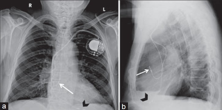
Transvenous pacemakers are implanted through the cephalic, axillary, or subclavian vein with a pectoral pocket. Extrathoracic axillary and cephalic access avoid the costoclavicular space and the subclavian crush that damages leads after medial subclavian puncture.

| Complication | Recognition | Management and prevention |
|---|---|---|
| Pneumothorax | Dyspnea, pleuritic pain after subclavian puncture | Chest radiograph after implantation; axillary or cephalic access |
| Cardiac perforation and tamponade | Chest pain, hypotension, new pericardial effusion, change in pacing parameters or right bundle branch pattern | Echocardiography; pericardiocentesis; lead repositioning |
| Lead dislodgement | Loss of capture or sensing, usually in the first weeks | Radiograph and interrogation; repositioning |
| Pocket hematoma | Tense, painful pocket | Continued warfarin had fewer clinically significant hematomas than heparin bridging (3.5% versus 16.0%, BRUISE CONTROL) |
| Device infection | Pocket erythema, erosion, drainage; bacteremia; lead or valve vegetation | Complete system extraction with pathogen-directed antibiotics; antibacterial envelope in higher-risk procedures (WRAP-IT) |
| Lead-related tricuspid regurgitation | New or worse regurgitation, right heart failure years after implantation | Three-dimensional echocardiography to define lead–leaflet interaction; lead repositioning or extraction, tricuspid intervention |
| Venous occlusion | Arm swelling, superior vena cava syndrome, failed access at upgrade | Venography; venoplasty or extraction for access |
| Twiddler and reel syndromes | Loss of capture with leads coiled around the generator on radiograph | Lead revision; pocket fixation |
| Phrenic or diaphragmatic stimulation | Hiccup-like twitching with pacing | Reprogramming of output or vector; lead revision |
Complications of transvenous pacemaker implantation.
In WRAP-IT, an absorbable antibiotic-eluting envelope reduced major infection at 12 months from 1.2% to 0.7% (HR 0.60) without more complications. The 6,983 patients underwent generator replacement, revision, upgrade, or initial cardiac resynchronization therapy defibrillator implantation.
Lead extraction. The 2017 HRS expert consensus statement recommends complete removal of the system, including all leads, for definite device infection: pocket infection, erosion, lead or valve endocarditis, and occult Staphylococcus aureus bacteremia. Noninfectious extraction (malfunction, venous occlusion blocking an upgrade, lead-related tricuspid regurgitation) is individualized against procedural risk. In the ELECTRa registry of 3,510 transvenous extractions, procedure-related major complications occurred in 1.7% and procedure-related death in 0.5%, with complete clinical success in 96.7%. Extraction is performed in centers with immediate cardiac surgical backup.
Device follow-up, remote monitoring, MRI, surgery, and driving
Follow-up. Each visit records pacing dependence, capture thresholds, sensed amplitudes, impedance trends, battery status, pacing percentages, and stored arrhythmias, including atrial high-rate episodes. The 2023 HRS/European Heart Rhythm Association/APHRS/LAHRS consensus statement maintains remote monitoring as the standard of care for patients with implanted devices. It advises prompt enrollment after implantation and alert-based monitoring, which allows longer intervals between office visits. Patients with substantial right ventricular pacing undergo periodic assessment of LVEF (Class 1). The 2023 physiologic pacing guideline suggests LVEF assessment every 1 to 2 years when high-risk features are present, such as a baseline QRS duration above 115 ms or a paced QRS duration above 150 ms, and less often when left ventricular function has been stable.
Magnetic resonance imaging. MRI-conditional systems are scanned under their labeled conditions. In the MagnaSafe registry of 1,500 nonthoracic 1.5-tesla examinations with non-MRI-conditional pacemakers or defibrillators, no deaths, lead failures, or losses of capture occurred when the interrogation, reprogramming, and monitoring protocol was followed. The 2017 HRS consensus statement supports MRI in non-MRI-conditional systems at centers with a defined protocol, with pacing-dependent patients programmed to asynchronous pacing and tachyarrhythmia therapy suspended during the scan. Abandoned and epicardial leads require individual risk assessment.
Surgery and procedures. The 2011 HRS/American Society of Anesthesiologists consensus statement bases perioperative management on device type, pacing dependence, magnet response, and the site of electromagnetic interference. Electrocautery below the umbilicus rarely affects a pectoral device; above it, a pacing-dependent patient's device is reprogrammed to asynchronous pacing or a magnet is applied to a pacemaker with a known response. Transcutaneous pacing pads are reasonable in patients at high risk of perioperative bradycardia (Class 2a). Routine prophylactic temporary transvenous pacing is not performed in patients with left bundle branch block who need pulmonary artery catheterization (Class 3: Harm). Devices are interrogated afterward when reprogramming or substantial interference occurred.
Driving and activity. The Canadian Cardiovascular Society 2023 fitness-to-drive guideline restricts both private and commercial driving for 1 week after transvenous or leadless pacemaker implantation in patients with previous impaired consciousness or high-grade AV block. It places no restriction on patients without either, and local law governs. Arm elevation and heavy lifting on the device side are limited for several weeks while leads mature, and strong magnets, arc welding, and industrial equipment can cause interference.
Pregnancy and conduction disease
Pregnancy raises the resting heart rate by 10 to 20/min, so sinus bradycardia is uncommon, and Mobitz I block rarely indicates progressive disease. Most advanced block in pregnancy is congenital, with a stable narrow junctional escape, and is well tolerated. Women with pacemakers usually have uneventful pregnancies. Women who are considering pregnancy and meet indications for a pacemaker, defibrillator, or cardiac resynchronization device undergo implantation before pregnancy (Class 1).
| Situation | Recommendation | Class |
|---|---|---|
| Advanced heart block or conduction system disease in pregnancy | Transthoracic echocardiography; cardiac MRI for selected suspected myocardial or infiltrative disease, done postpartum | 1 |
| Irreversible symptomatic bradycardia from third-degree or Mobitz II block or severe sinus node dysfunction, with syncope or presyncope endangering mother or fetus | Permanent pacemaker, with radiation kept as low as reasonably achievable | 1 |
| Symptomatic bradycardia refractory to, or with contraindication to, drug therapy, at risk of peripartum instability or syncope | Temporary ventricular pacing | 1 |
| Asymptomatic sinus bradycardia or Mobitz I block without structural heart disease | Reassurance without intervention | 1 |
| Stable, asymptomatic congenital heart block in a patient considered for pacing before pregnancy | Device decision deferred until after delivery | 2a |
| Stable, asymptomatic congenital heart block with acceptable rate, narrow QRS, and normal ventricular function | No prophylactic temporary pacing at delivery | 3: No Benefit |
Bradycardia and heart block in pregnancy, from the 2023 HRS expert consensus statement on arrhythmias during pregnancy.
When implantation is needed during pregnancy, fluoroscopy is minimized with ultrasound-guided access, echocardiographic guidance, electroanatomic mapping, and single-lead systems. Gadolinium is avoided. Delivery is generally tolerated, and surveillance continues postpartum because late syncope and sudden death have been reported in women with unpaced complete block.
Adult congenital heart disease, heart transplant, spinal cord injury, epilepsy, and older age
Adults with congenital heart disease. In adults with congenital complete AV block, permanent pacing is recommended for symptomatic bradycardia, a wide QRS escape rhythm, mean daytime heart rate below 50/min, complex ventricular ectopy, or ventricular dysfunction (Class 1). Pacing is reasonable even without these features (Class 2a). Atrial-based pacing is recommended for symptomatic sinus node dysfunction or chronotropic incompetence (Class 1). Postoperative Mobitz II, high-grade, or third-degree block not expected to resolve is paced (Class 1). Endocardial leads are potentially harmful in patients with intracardiac shunts that allow paradoxical embolism (Class 3: Harm). In adults with repaired congenital heart disease who need bradycardia pacing, a device with atrial antitachycardia pacing is reasonable (Class 2a). Intraoperative epicardial leads are reasonable when preexisting sinus node or AV conduction disease is present at cardiac surgery (Class 2a), and atrial-based pacing to prevent atrial arrhythmias may be considered (Class 2b).
Heart transplant recipients. Denervation, surgical trauma, ischemia, rejection, and prior amiodarone cause sinus node dysfunction after transplantation, and rates below 70 to 80/min may be inadequate for postoperative demand. Theophylline or aminophylline is reasonable (Class 2a), atropine is avoided (Class 3: Harm), and many patients recover sinus function, so permanent pacing is deferred unless bradycardia persists. Late new AV block raises the question of rejection or cardiac allograft vasculopathy.
Acute spinal cord injury. Loss of sympathetic outflow after severe cervical injury causes sinus bradycardia and asystole, most often in the first 2 to 4 weeks; cardiac arrest occurred in 16% of patients with severe cervical injury in one series. Aminophylline or theophylline is reasonable (Class 2a), and most patients recover without permanent pacing.
Epilepsy. Ictal bradycardia and asystole occur during temporal lobe seizures; permanent pacing is reasonable when severe symptomatic ictal bradycardia persists despite antiepileptic therapy (Class 2a).
Older adults and end of life. Age alone is not a contraindication to pacing; frailty, cognition, and goals of care determine the device. Discontinuing pacing at the request of a patient with decision-making capacity, or a legal surrogate, is ethically and legally permissible; in a pacing-dependent patient, withdrawal is expected to cause death, and palliative care support is planned. When the original indication has resolved, discontinuation of pacing after monitoring with pacing off is reasonable (Class 2a).
Prognosis of bradycardia and atrioventricular block
With pacing, survival in sinus node dysfunction and AV block is determined chiefly by age and comorbid disease, not by the bradyarrhythmia; pacing removes most of the excess early mortality of untreated acquired complete heart block. The substrate then sets prognosis: block from anterior infarction, sarcoidosis, LMNA disease, or myotonic dystrophy carries ventricular arrhythmic and heart failure risk that pacing alone does not address. The pacing strategy shapes long-term outcome through atrial fibrillation, pacing-induced cardiomyopathy, and lead complications over decades of device life.
Self-assessment
A 74-year-old man has had two episodes of abrupt syncope without prodrome. The ECG shows sinus rhythm at 72/min, PR interval 210 ms, right bundle branch block, and left anterior fascicular block. LVEF is 60%. Fourteen days of ambulatory monitoring show no AV block. At electrophysiology study the HV interval is 78 ms. What does the 2018 ACC/AHA/HRS guideline recommend, and with what class?
Cardiovascular Medicine Board ReviewReferences
- Kusumoto FM, Schoenfeld MH, Barrett C, et al. 2018 ACC/AHA/HRS guideline on the evaluation and management of patients with bradycardia and cardiac conduction delay: a report of the American College of Cardiology/American Heart Association Task Force on Clinical Practice Guidelines and the Heart Rhythm Society. Circulation. 2019;140:e382–e482. doi:10.1161/CIR.0000000000000628
- Chung MK, Patton KK, Lau CP, et al. 2023 HRS/APHRS/LAHRS guideline on cardiac physiologic pacing for the avoidance and mitigation of heart failure. Heart Rhythm. 2023;20:e17–e91. doi:10.1016/j.hrthm.2023.03.1538
- Glikson M, Nielsen JC, Kronborg MB, et al. 2021 ESC guidelines on cardiac pacing and cardiac resynchronization therapy. Eur Heart J. 2021;42:3427–3520. doi:10.1093/eurheartj/ehab364
- Shen WK, Sheldon RS, Benditt DG, et al. 2017 ACC/AHA/HRS guideline for the evaluation and management of patients with syncope. Circulation. 2017;136:e60–e122. doi:10.1161/CIR.0000000000000499
- Joglar JA, Kapa S, Saarel EV, et al. 2023 HRS expert consensus statement on the management of arrhythmias during pregnancy. Heart Rhythm. 2023;20:e175–e264. doi:10.1016/j.hrthm.2023.05.017
- Panchal AR, Bartos JA, Cabañas JG, et al. Part 3: adult basic and advanced life support: 2020 American Heart Association guidelines for cardiopulmonary resuscitation and emergency cardiovascular care. Circulation. 2020;142(16 Suppl 2):S366–S468. doi:10.1161/CIR.0000000000000916
- Bernstein AD, Daubert JC, Fletcher RD, et al. The revised NASPE/BPEG generic code for antibradycardia, adaptive-rate, and multisite pacing. Pacing Clin Electrophysiol. 2002;25:260–264. doi:10.1046/j.1460-9592.2002.00260.x
- Jensen PN, Gronroos NN, Chen LY, et al. Incidence of and risk factors for sick sinus syndrome in the general population. J Am Coll Cardiol. 2014;64:531–538. doi:10.1016/j.jacc.2014.03.056
- Cheng S, Keyes MJ, Larson MG, et al. Long-term outcomes in individuals with prolonged PR interval or first-degree atrioventricular block. JAMA. 2009;301:2571–2577. doi:10.1001/jama.2009.888
- Sharma S, Drezner JA, Baggish A, et al. International recommendations for electrocardiographic interpretation in athletes. J Am Coll Cardiol. 2017;69:1057–1075. doi:10.1016/j.jacc.2017.01.015
- Lantos PM, Rumbaugh J, Bockenstedt LK, et al. Clinical practice guidelines by the Infectious Diseases Society of America, American Academy of Neurology, and American College of Rheumatology: 2020 guidelines for the prevention, diagnosis, and treatment of Lyme disease. Clin Infect Dis. 2021;72:e1–e48. doi:10.1093/cid/ciaa1215
- Birnie DH, Sauer WH, Bogun F, et al. HRS expert consensus statement on the diagnosis and management of arrhythmias associated with cardiac sarcoidosis. Heart Rhythm. 2014;11:1305–1323. doi:10.1016/j.hrthm.2014.03.043
- Nunes MCP, Beaton A, Acquatella H, et al. Chagas cardiomyopathy: an update of current clinical knowledge and management: a scientific statement from the American Heart Association. Circulation. 2018;138:e169–e209. doi:10.1161/CIR.0000000000000599
- Connolly SJ, Kerr CR, Gent M, et al. Effects of physiologic pacing versus ventricular pacing on the risk of stroke and death due to cardiovascular causes (CTOPP). N Engl J Med. 2000;342:1385–1391. doi:10.1056/NEJM200005113421902
- Lamas GA, Lee KL, Sweeney MO, et al. Ventricular pacing or dual-chamber pacing for sinus-node dysfunction (MOST). N Engl J Med. 2002;346:1854–1862. doi:10.1056/NEJMoa013040
- Sweeney MO, Hellkamp AS, Ellenbogen KA, et al. Adverse effect of ventricular pacing on heart failure and atrial fibrillation among patients with normal baseline QRS duration in a clinical trial of pacemaker therapy for sinus node dysfunction. Circulation. 2003;107:2932–2937. doi:10.1161/01.CIR.0000072769.17295.B1
- Toff WD, Camm AJ, Skehan JD, et al. Single-chamber versus dual-chamber pacing for high-grade atrioventricular block (UKPACE). N Engl J Med. 2005;353:145–155. doi:10.1056/NEJMoa042283
- Nielsen JC, Thomsen PE, Højberg S, et al. A comparison of single-lead atrial pacing with dual-chamber pacing in sick sinus syndrome (DANPACE). Eur Heart J. 2011;32:686–696. doi:10.1093/eurheartj/ehr022
- Curtis AB, Worley SJ, Adamson PB, et al. Biventricular pacing for atrioventricular block and systolic dysfunction (BLOCK HF). N Engl J Med. 2013;368:1585–1593. doi:10.1056/NEJMoa1210356
- Kiehl EL, Makki T, Kumar R, et al. Incidence and predictors of right ventricular pacing-induced cardiomyopathy in patients with complete atrioventricular block and preserved left ventricular systolic function. Heart Rhythm. 2016;13:2272–2278. doi:10.1016/j.hrthm.2016.09.027
- Vijayaraman P, Naperkowski A, Subzposh FA, et al. Permanent His-bundle pacing: long-term lead performance and clinical outcomes. Heart Rhythm. 2018;15:696–702. doi:10.1016/j.hrthm.2017.12.022
- Sharma PS, Patel NR, Ravi V, et al. Clinical outcomes of left bundle branch area pacing compared to right ventricular pacing: results from the Geisinger-Rush Conduction System Pacing Registry. Heart Rhythm. 2022;19:3–11. doi:10.1016/j.hrthm.2021.08.033
- Jastrzębski M, Kiełbasa G, Cano O, et al. Left bundle branch area pacing outcomes: the multicentre European MELOS study. Eur Heart J. 2022;43:4161–4173. doi:10.1093/eurheartj/ehac445
- Reynolds D, Duray GZ, Omar R, et al. A leadless intracardiac transcatheter pacing system. N Engl J Med. 2016;374:533–541. doi:10.1056/NEJMoa1511643
- Steinwender C, Khelae SK, Garweg C, et al. Atrioventricular synchronous pacing using a leadless ventricular pacemaker: results from the MARVEL 2 study. JACC Clin Electrophysiol. 2020;6:94–106. doi:10.1016/j.jacep.2019.10.017
- Knops RE, Reddy VY, Ip JE, et al. A dual-chamber leadless pacemaker. N Engl J Med. 2023;388:2360–2370. doi:10.1056/NEJMoa2300080
- Brignole M, Menozzi C, Moya A, et al. Pacemaker therapy in patients with neurally mediated syncope and documented asystole: Third International Study on Syncope of Uncertain Etiology (ISSUE-3). Circulation. 2012;125:2566–2571. doi:10.1161/CIRCULATIONAHA.111.082313
- Birnie DH, Healey JS, Wells GA, et al. Pacemaker or defibrillator surgery without interruption of anticoagulation (BRUISE CONTROL). N Engl J Med. 2013;368:2084–2093. doi:10.1056/NEJMoa1302946
- Tarakji KG, Mittal S, Kennergren C, et al. Antibacterial envelope to prevent cardiac implantable device infection (WRAP-IT). N Engl J Med. 2019;380:1895–1905. doi:10.1056/NEJMoa1901111
- Kusumoto FM, Schoenfeld MH, Wilkoff BL, et al. 2017 HRS expert consensus statement on cardiovascular implantable electronic device lead management and extraction. Heart Rhythm. 2017;14:e503–e551. doi:10.1016/j.hrthm.2017.09.001
- Bongiorni MG, Kennergren C, Butter C, et al. The European Lead Extraction ConTRolled (ELECTRa) study: a European Heart Rhythm Association (EHRA) registry of transvenous lead extraction outcomes. Eur Heart J. 2017;38:2995–3005. doi:10.1093/eurheartj/ehx080
- Indik JH, Gimbel JR, Abe H, et al. 2017 HRS expert consensus statement on magnetic resonance imaging and radiation exposure in patients with cardiovascular implantable electronic devices. Heart Rhythm. 2017;14:e97–e153. doi:10.1016/j.hrthm.2017.04.025
- Russo RJ, Costa HS, Silva PD, et al. Assessing the risks associated with MRI in patients with a pacemaker or defibrillator (MagnaSafe). N Engl J Med. 2017;376:755–764. doi:10.1056/NEJMoa1603265
- Crossley GH, Poole JE, Rozner MA, et al. The Heart Rhythm Society (HRS)/American Society of Anesthesiologists (ASA) expert consensus statement on the perioperative management of patients with implantable defibrillators, pacemakers and arrhythmia monitors: facilities and patient management. Heart Rhythm. 2011;8:1114–1154. doi:10.1016/j.hrthm.2010.12.023
- Ferrick AM, Raj SR, Deneke T, et al. 2023 HRS/EHRA/APHRS/LAHRS expert consensus statement on practical management of the remote device clinic. J Arrhythm. 2023;39:250–302. doi:10.1002/joa3.12851
- Guerra PG, Simpson CS, Van Spall HGC, et al. Canadian Cardiovascular Society 2023 guidelines on the fitness to drive. Can J Cardiol. 2024;40:500–523. doi:10.1016/j.cjca.2023.09.033
- Heidenreich PA, Bozkurt B, Aguilar D, et al. 2022 AHA/ACC/HFSA guideline for the management of heart failure: a report of the American College of Cardiology/American Heart Association Joint Committee on Clinical Practice Guidelines. Circulation. 2022;145:e895–e1032. doi:10.1161/CIR.0000000000001063