Library › Pericardial disease › Effusion and tamponade
Pericardial effusion and cardiac tamponade
Pericardial effusion and cardiac tamponade in adults, from causes, physiology, and imaging through triage, pericardiocentesis, surgical drainage, and management in cancer, surgery, trauma, and kidney failure.
Pericardial effusion is an abnormal amount of fluid in the pericardial space. Cardiac tamponade is the hemodynamic state in which pericardial contents, whether fluid, pus, blood, clot, or gas, raise intrapericardial pressure enough to impair cardiac filling. Management therefore rests on three separate judgments: whether the effusion is compromising the circulation, what is causing it, and whether drainage is needed for treatment, diagnosis, or both. No American College of Cardiology/American Heart Association (ACC/AHA) clinical practice guideline covers pericardial disease. US practice draws on the 2013 American Society of Echocardiography (ASE) recommendations for multimodality imaging, the 2024 international imaging position statement, and the 2025 ACC expert consensus on pericarditis. The 2015 European Society of Cardiology (ESC) pericardial guideline remains the most detailed recommendation set for effusion and tamponade. The 2025 ESC guideline on myocarditis and pericarditis places pericardial syndromes within a broader framework of inflammatory myopericardial disease. Pericardial inflammation and its anti-inflammatory treatment are covered on the acute and recurrent pericarditis page, and chronic and effusive-constrictive physiology on the constrictive pericarditis page.
Definitions and classification of pericardial effusion and tamponade
The 2015 ESC guideline classifies a pericardial effusion by five attributes, each of which carries a different clinical implication. Onset separates acute and subacute effusions from chronic effusions, defined as present for more than 3 months. Size is graded semiquantitatively from the end-diastolic echo-free space. Distribution separates circumferential from loculated collections. Hemodynamic impact ranges from none, through tamponade, to effusive-constrictive physiology. Composition separates transudate, exudate, blood, and, rarely, air or gas from bacterial infection.
| Attribute | Categories | Clinical implication |
|---|---|---|
| Onset | Acute, subacute, chronic (more than 3 months) | Rapid accumulation produces tamponade at small volume; a chronic effusion can reach 1 L or more with normal hemodynamics |
| Size (end-diastolic echo-free space) | Trivial (systole only), small (below 10 mm), moderate (10–20 mm), large (above 20 mm); the ASE adds very large (above 25 mm) | Larger effusions carry a higher likelihood of a specific cause and of progression; size alone does not define tamponade |
| Distribution | Circumferential, loculated | Loculated or posterior collections cause regional tamponade, escape transthoracic detection, and are poorly suited to needle drainage |
| Hemodynamic impact | None, cardiac tamponade, effusive-constrictive | Tamponade requires drainage; persistent venous hypertension after drainage indicates effusive-constrictive disease |
| Composition | Transudate, exudate, blood, pus, chyle, gas | Hemopericardium from aortic dissection, rupture, or trauma is a surgical problem; pus requires complete drainage; chyle points to thoracic duct injury or obstruction |
Classification of pericardial effusion. Size categories follow the 2015 ESC guideline; the 2013 ASE recommendations add the very large category.
Tamponade is a continuum. In the ASE description, mild tamponade, with intrapericardial pressure typically below 10 mm Hg, is often asymptomatic, whereas severe tamponade, typically above 15 mm Hg, produces tachycardia and marked dyspnea. Hypotension is a late sign. Low-pressure and regional tamponade depart from the classic picture. The ASE also describes tamponade caused by a large pleural effusion with an insignificant pericardial effusion.
Pericardial anatomy, normal fluid, and the pressure-volume relation
The pericardium consists of an outer fibrous layer lined by the parietal serous layer, and a visceral serous layer, the epicardium, that covers the heart. The pericardial space lies between the two serous layers. The normal sac contains 10 to 50 mL of fluid, a plasma ultrafiltrate that lubricates the layers. The normal pericardium measures 0.7 to 2.0 mm on computed tomography (CT) and 1.2 to 1.7 mm on cardiovascular magnetic resonance (CMR). The space extends into the transverse and oblique sinuses and their recesses, and small effusions often collect there first. Because fluid collects unevenly, a single linear measurement only approximates volume.
Pathologic fluid accumulates by two mechanisms. Inflammation of the pericardium increases production of an exudate. Raised systemic venous pressure, as in heart failure or pulmonary hypertension, reduces reabsorption and produces a transudate. Hemopericardium results from bleeding into the space, and chylopericardium from leakage of lymph.
The pericardial pressure-volume curve has an initial flat segment followed by an almost vertical rise. Because the fibrous pericardium stretches slowly, the rate of accumulation matters more than the volume. After a ventricular perforation or type A aortic dissection, a small volume reaches the steep segment within minutes, and the ASE notes tamponade with volumes as small as 250 mL. A slowly accumulating effusion allows the sac to enlarge over days to weeks, and a liter or more can collect before pressure rises. The ESC guideline describes tamponade as a "last-drop" phenomenon. On the steep segment, the final increment of fluid produces critical compression, and the first increment removed during drainage produces the largest relative decompression. Pericardial anatomy and its relation to inflammation are covered in more detail on the acute and recurrent pericarditis page.
Epidemiology and causes of pericardial effusion
Effusions are frequently found on echocardiograms and CT scans performed for other reasons. The ASE recommendations cite a 5% prevalence of pericardial effusion among emergency department patients who underwent CT to exclude pulmonary embolism. According to the surveys summarized in the 2015 ESC guideline, many moderate to large effusions in developed countries remain idiopathic (up to 50%). Other common causes are cancer (10% to 25%), infection (15% to 30%), iatrogenic injury (15% to 20%), and connective tissue disease (5% to 15%). In developing countries where tuberculosis is endemic, it is the dominant cause, accounting for more than 60% of effusions. An effusion is associated with a known or previously unknown condition in up to 60% of cases, so the search for an underlying disease is part of every evaluation.
The setting changes the spectrum. Among 1,127 therapeutic pericardiocenteses at the Mayo Clinic, cardiothoracic surgery replaced malignancy as the leading cause in the most recent period, and surgery, malignancy, and perforation during catheter procedures together accounted for nearly 70% of procedures. In a prospective series of 71 patients with tamponade at a European referral center, 59.2% had cancer, most often lung cancer, breast cancer, or lymphoproliferative disease.
| Mechanism | Causes | Clinical and fluid clues |
|---|---|---|
| Inflammatory, idiopathic or viral | Idiopathic pericarditis, viral infection | Pleuritic pain, rub, raised C-reactive protein (CRP); effusion usually small and regresses with anti-inflammatory therapy |
| Bacterial | Tuberculosis; purulent infection from pneumonia, empyema, bacteremia, or surgery | Tuberculosis: subacute course, endemic exposure, human immunodeficiency virus (HIV) infection, lymphocytic exudate. Purulent: sepsis, neutrophilic fluid with low glucose |
| Neoplastic | Metastatic lung and breast cancer, melanoma, lymphoma, leukemia; primary mesothelioma and angiosarcoma (rare) | Large, often hemorrhagic effusion; tamponade at presentation; recurrence after drainage |
| Cancer therapy | Mediastinal radiation, anthracyclines and other chemotherapy, immune checkpoint inhibitors | In almost two thirds of patients with cancer and an effusion, the effusion is not malignant |
| Post-cardiac injury | Postpericardiotomy syndrome, post-infarction (Dressler) syndrome, after percutaneous intervention or device implantation | Onset weeks after injury, fever, pleural effusion, raised CRP |
| Hemopericardium | Type A aortic dissection, free-wall rupture, chest trauma, perforation during catheter procedures, anticoagulation after procedures | Rapid accumulation, small volume, shock; clot on imaging |
| Systemic autoimmune disease | Systemic lupus erythematosus, rheumatoid arthritis, scleroderma, Sjögren syndrome, vasculitis, sarcoidosis, inflammatory bowel disease | Reflects activity of the underlying disease; rarely the first manifestation |
| Metabolic and endocrine | Hypothyroidism, kidney failure (uremic and dialysis-associated) | Hypothyroidism: bradycardia, low QRS voltage, slow accumulation. Uremia: often hemorrhagic, few electrocardiogram (ECG) changes |
| Hemodynamic transudate | Heart failure, pulmonary arterial hypertension, cirrhosis, nephrotic syndrome, pregnancy | Small effusion; tamponade unusual except when right-sided pressures mask the usual signs |
| Drug-related | Hydralazine and procainamide (drug-induced lupus), minoxidil, chemotherapy | Temporal relation to the drug; resolution after withdrawal |
| Lymphatic | Thoracic duct injury after surgery, mediastinal tumor, lymphangiomatosis, subclavian vein thrombosis | Milky fluid with high triglycerides (chylopericardium) |
Causes of pericardial effusion in adults grouped by mechanism, adapted from the 2015 ESC guideline. The differential diagnosis of pericarditis itself, including specific infectious and autoimmune causes, is covered on the acute and recurrent pericarditis page.
Two associations from the ESC guideline help triage an effusion without obvious cause. Tamponade without inflammatory signs is associated with a neoplastic cause (likelihood ratio 2.9). A large effusion without tamponade or inflammatory signs is usually chronic idiopathic (likelihood ratio 20).
Causes of cardiac tamponade and their urgency
The ESC guideline lists pericarditis, tuberculosis, iatrogenic injury (procedure-related and after cardiac surgery), trauma, and neoplasm as the common causes of tamponade. Less common causes are systemic autoimmune disease, radiation, myocardial infarction, uremia, aortic dissection, bacterial infection, and pneumopericardium. In practice, the cause determines both the tempo and the method of drainage.
| Setting | Typical tempo | Usual drainage |
|---|---|---|
| Perforation during coronary, electrophysiology, or structural procedures | Minutes to hours, often on the table | Immediate percutaneous drainage, anticoagulant reversal, autotransfusion; surgery for continued bleeding |
| Type A aortic dissection with hemopericardium | Minutes to hours | Emergency aortic surgery; controlled drainage of small volumes only as a bridge |
| Left ventricular free-wall rupture | Sudden collapse or subacute leak | Emergency surgical repair; needle drainage only as a bridge |
| Penetrating chest trauma | Minutes | Thoracotomy; pericardiocentesis only as a bridge |
| Early after cardiac surgery (first 24 hours) | Hours; surgical bleeding or coagulopathy | Surgical re-exploration |
| Late after cardiac surgery (beyond 5–7 days) | Days; often loculated or posterior | Percutaneous drainage when accessible; surgery for clot or loculation |
| Malignant effusion | Days to weeks | Pericardiocentesis with extended drainage; window or intrapericardial therapy for recurrence |
| Purulent pericarditis | Days; sepsis | Urgent complete drainage, often surgical, with intravenous antibiotics |
| Idiopathic, viral, tuberculous, uremic, hypothyroid | Days to months | Pericardiocentesis; treatment of the cause |
Causes of cardiac tamponade grouped by setting. Timing after cardiac surgery follows the 2013 ASE recommendations; the surgical indications follow the 2015 ESC guideline.
Pathophysiology: ventricular interdependence, equalization, and loss of the y descent
As fluid accumulates, intrapericardial pressure rises, and systemic and pulmonary venous pressures must rise with it to maintain filling. The four cardiac chambers compete with the fluid for a fixed total volume. Intrapericardial pressure is transmitted to all chambers throughout the cardiac cycle, so right atrial, right ventricular diastolic, pulmonary artery diastolic, and pulmonary capillary wedge pressures rise toward the intrapericardial pressure and toward one another. In the ESC description, diastolic pressures throughout the heart usually lie between 15 and 30 mm Hg, except in low-pressure tamponade.
Because total cardiac volume is fixed, filling of one ventricle occurs at the expense of the other. During inspiration, the fall in intrathoracic pressure still augments systemic venous return, the right ventricle fills, the interventricular septum bulges toward the left ventricle, and left ventricular filling and stroke volume fall. During expiration, the changes reverse. This exaggerated ventricular interdependence is the principal mechanism of pulsus paradoxus. A second mechanism contributes: the inspiratory fall in the pulmonary venous to left atrial pressure gradient is larger than normal, which reduces left heart filling further.
Atrial filling occurs mainly during ventricular systole, when ejection lowers total cardiac volume and intrapericardial pressure falls. The systolic x descent of the right atrial and jugular venous waveforms is therefore preserved. In early diastole, the right atrium and ventricle are already at the same compressed pressure, and there is no period of unimpeded ventricular filling, so the y descent is attenuated or absent. That loss of the y descent separates tamponade from constriction, in which early filling is rapid and the y descent is steep.
Cardiac output is maintained at first by tachycardia and vasoconstriction. As the filling gradients narrow, stroke volume falls, and blood pressure is sustained by sympathetic tone until compensation fails. The ASE recommendations describe an abrupt fall in heart rate and blood pressure as the usual terminal event.
Symptoms and physical examination in pericardial effusion and tamponade
The presentation depends on the rate of accumulation. A slowly accumulating effusion produces exertional dyspnea that progresses to orthopnea, chest pain or fullness, and fatigue. Compression of neighboring structures causes nausea (diaphragm), dysphagia (esophagus), hoarseness (recurrent laryngeal nerve), and hiccups (phrenic nerve). Cough, anorexia, and palpitations are nonspecific. Fever suggests infectious or immune-mediated pericarditis. A rapidly accumulating hemopericardium presents as sudden hypotension, syncope, or pulseless electrical activity.
Physical examination is normal in many patients without hemodynamic compromise. In tamponade, the classic signs are elevated jugular venous pressure, pulsus paradoxus, and diminished heart sounds with a moderate or large effusion. The Beck triad (hypotension, raised venous pressure, and muffled heart sounds) describes acute surgical tamponade and is incomplete in most medical tamponade. A friction rub is heard mainly when pericarditis coexists and can persist despite a large effusion.
A systematic review of the clinical examination in patients with effusion found five features in most patients with tamponade, with pooled sensitivities of 87% to 89% for dyspnea, 77% for tachycardia, 82% for pulsus paradoxus, 76% for elevated jugular venous pressure, and 89% for cardiomegaly on chest radiograph. The review concluded that a minority of patients with tamponade lack each of these findings, and that diagnostic certainty requires additional testing.
Kussmaul sign, the failure of jugular pressure to fall with inspiration, is not a feature of uncomplicated tamponade, and its presence suggests a constrictive component. Hepatomegaly, ascites, and edema indicate subacute or chronic venous congestion and are more typical of constriction.
A patient with tamponade and a normal or slow heart rate raises three possibilities: severe hypothyroidism, uremia, or atrioventricular nodal blocking drugs. Myxedema can produce a large effusion with relative bradycardia and low QRS voltage, which masks the expected tachycardia.
Pulsus paradoxus: mechanism, measurement, and false results
Pulsus paradoxus is an inspiratory decrease in systolic arterial pressure of more than 10 mm Hg during normal breathing. A small inspiratory fall is normal; in tamponade, ventricular interdependence exaggerates it. In severe tamponade, the radial pulse can disappear during inspiration.
Pulsus paradoxus is measured with a manual sphygmomanometer while the patient breathes quietly. The cuff is inflated above systolic pressure and deflated slowly. The pressure at which Korotkoff sounds are first heard, and then only during expiration, is recorded. Deflation continues until sounds are heard with every beat, throughout both phases of respiration, and that pressure is recorded. The difference between the two pressures is the pulsus paradoxus. Automated oscillometric cuffs cannot measure it. With an arterial line, it is the difference between the highest expiratory and lowest inspiratory systolic pressure, and the respiratory variation of the waveform can be seen directly.
| Situation | Effect on pulsus paradoxus | Mechanism |
|---|---|---|
| Aortic regurgitation | Absent despite tamponade | Regurgitant filling of the left ventricle blunts the inspiratory fall in left ventricular volume |
| Atrial septal defect | Absent despite tamponade | Shunting equalizes the respiratory changes in right and left atrial filling |
| Severe left ventricular hypertrophy or dysfunction, raised left ventricular diastolic pressure | Absent or reduced | The stiff, high-pressure left ventricle is less affected by septal shift |
| Severe pulmonary hypertension, right ventricular hypertrophy | Reduced | The right ventricle resists inspiratory expansion |
| Regional or loculated tamponade | Often absent | Compression is confined to one chamber |
| Positive-pressure ventilation | Abolished or reversed | Mechanical inspiration raises intrathoracic pressure and reverses the normal respiratory pattern |
| Severe hypotension or shock | Not measurable | Korotkoff sounds are inaudible; an arterial line is required |
| Severe asthma or obstructive lung disease | Present without tamponade | Large swings in intrathoracic pressure |
| Massive pulmonary embolism, right ventricular infarction | Present without tamponade | Right ventricular dilation within the pericardium increases interdependence |
| Tension pneumothorax, severe hypovolemia | Present without tamponade | External compression or exaggerated respiratory effort |
Conditions that mask or mimic pulsus paradoxus. The masking conditions are those listed in the 2015 ESC guideline and the 2013 ASE recommendations for absent chamber collapse or absent respiratory variation.
In a patient with a known effusion, a pulsus paradoxus above 10 mm Hg increased the likelihood of tamponade (likelihood ratio 3.3). A value of 10 mm Hg or less made tamponade unlikely (likelihood ratio 0.03). Both figures come from a single study within the systematic review. The finding is most useful at the bedside in a spontaneously breathing patient in sinus rhythm without the masking conditions listed above. Respiratory variation in mitral inflow and aortic outflow velocities is its echocardiographic counterpart.
ECG and chest radiograph in pericardial effusion and tamponade
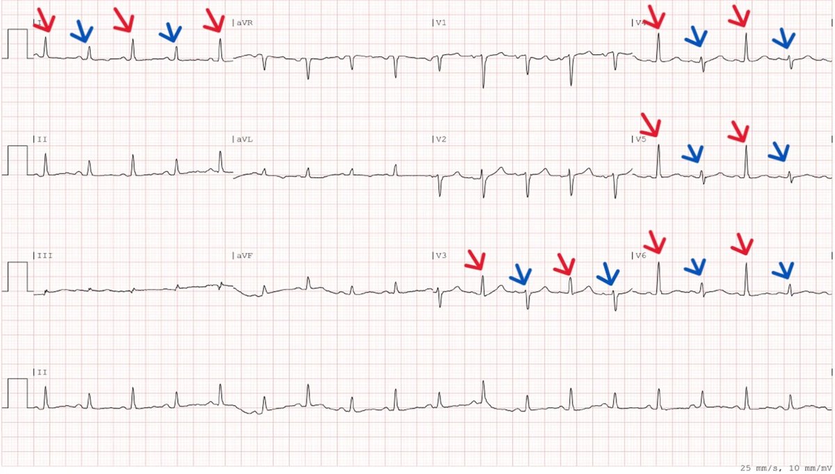
The ECG in tamponade typically shows sinus tachycardia, low QRS voltage, and sometimes electrical alternans. Low voltage is defined as QRS amplitude below 5 mm in all limb leads or below 10 mm in all precordial leads. It reflects damping by fluid and is also caused by obesity, emphysema, infiltrative cardiomyopathy, and hypothyroidism. Electrical alternans is a beat-to-beat alternation of QRS amplitude or axis produced by the heart swinging within a large effusion, the electrical counterpart of the swinging heart on echocardiography. It is specific but insensitive. Alternation of the P, QRS, and T waves together is particularly suggestive of a large effusion. Diffuse ST elevation and PR depression indicate coexisting pericarditis. A new effusion with focal ST elevation after infarction raises concern for subacute free-wall rupture.
The chest radiograph is normal with a small effusion. A slowly accumulated moderate or large effusion enlarges the cardiac silhouette into a symmetrical globular or "water bottle" shape with clear lung fields. In the systematic review, cardiomegaly on chest radiograph was present in 89% of patients with tamponade. Acute hemopericardium can produce tamponade with a normal silhouette, because the volume is small. The 2015 ESC guideline recommends a chest radiograph in suspected pericardial effusion or pleuropulmonary involvement (Class 1, Level C). Its main value lies in the lungs, pleura, and mediastinum, where it shows pneumonia, tuberculosis, lung cancer, pleural effusion, or hilar and mediastinal enlargement.
Echocardiographic detection and sizing of pericardial effusion
Transthoracic echocardiography is recommended in all patients with suspected pericardial effusion (ESC Class 1, Level C). It detects an effusion with an accuracy approaching 100%, can be performed at the bedside without delay, and assesses hemodynamic impact in the same study. On M-mode, separation of the pericardial layers seen only in systole represents a normal or insignificant amount of fluid, and separation throughout the cardiac cycle corresponds to an effusion of more than about 50 mL.
Size is graded from the largest end-diastolic echo-free space between the visceral and parietal pericardium, using the categories in the classification table. The grades derive from M-mode criteria and describe loculated collections poorly. Seeing an evenly distributed effusion from several windows increases confidence in the grade, and small and very large effusions are the most likely to be unevenly distributed. The 2015 ESC guideline recommends digital storage of images and a report that states the size and the location of each measurement, so that serial studies are comparable.
The report also describes distribution (circumferential or loculated, and where the largest pocket lies), the appearance of the fluid, and its effect on the heart. A simple transudate is echolucent. Fibrin strands, adhesions, and uneven distribution suggest an exudate. Echodense material or clot suggests hemorrhage, particularly after cardiac surgery or procedures, and nonclotted blood can show slowly swirling spontaneous contrast.
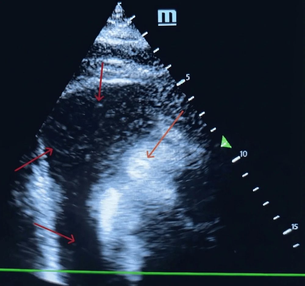
Several structures mimic an effusion. Epicardial fat lies anteriorly, is often brighter than myocardium, has granular texture and uniform thickness, and moves with the heart, whereas a transudative effusion is echolucent and motionless. An isolated anterior echo-free space without posterior fluid is often epicardial fat. A left pleural effusion is separated from a posterior pericardial effusion in the parasternal long-axis view: pericardial fluid extends between the heart and the descending thoracic aorta, whereas pleural fluid lies posterior to the aorta. Fluid tracking into the oblique sinus behind the left atrium is pericardial. A pericardial cyst, usually at the right cardiophrenic angle, appears as a loculated echo-free space adjacent to the right atrium. Ascites seen from the subcostal window lies below the diaphragm. CT or CMR resolves equivocal cases, because CT identifies fat by its negative attenuation and both show the whole pericardium.
Transesophageal echocardiography identifies posterior and loculated collections and clot after cardiac surgery when transthoracic windows are poor. Point-of-care ultrasound detects a large effusion and gross chamber collapse quickly, and a formal Doppler study follows when the patient's condition allows.
Echocardiographic signs of cardiac tamponade
In suspected tamponade, the 2015 ESC guideline recommends echocardiography as the first imaging technique to evaluate the size, location, and hemodynamic impact of the effusion (Class 1, Level C). The ASE recommendations state that the diagnosis rests on the combination of an effusion, reduced stroke volume and cardiac output, elevated central venous pressure, and corroborating chamber collapse and Doppler changes. No single sign is diagnostic, and the findings are interpreted with the clinical state.
Chamber collapse. A cardiac chamber indents when intrapericardial pressure exceeds its intracavitary pressure. Each chamber collapses during its relaxation phase, when its pressure is lowest. Right atrial collapse begins near the peak of the R wave, in late ventricular diastole and early systole, and the wall stays inverted until filling during ventricular systole raises atrial pressure. A brief right atrial inversion is common without tamponade because the wall is thin. In the original series of 127 patients with moderate or large effusions, inversion lasting more than 0.34 of the cardiac cycle raised specificity for tamponade from 82% to 100%, with a sensitivity of 94%. Right ventricular diastolic collapse occurs in early diastole after the end of the T wave. In the ASE description, it develops with moderate elevation of pericardial pressure, when cardiac output has fallen by about 20% but systemic pressure has not. It appears first during inspiration only and later persists throughout respiration. M-mode through the affected wall shows the timing and duration of collapse. The absence of any chamber collapse has a negative predictive value above 90% for clinical tamponade.
Collapse behaves differently when baseline chamber pressures are abnormal. Right ventricular hypertrophy, severe pulmonary hypertension, and severe left ventricular dysfunction delay or prevent right-sided collapse. Hypovolemia allows collapse to appear earlier. Left atrial and left ventricular diastolic collapse occur with posterior loculated effusions after surgery and in severe pulmonary arterial hypertension, in which left atrial pressure is lower than right-sided pressure.
Inferior vena cava plethora. A dilated inferior vena cava, above 21 mm, with less than 50% inspiratory reduction in diameter reflects raised right atrial pressure. In a series of 115 patients with moderate or large effusions, plethora was present in 92% of the 63 who underwent a drainage procedure. Plethora is sensitive but not specific, because it accompanies any cause of raised right atrial pressure. When the vena cava is hard to image, dilated hepatic veins confirm raised venous pressure. A small, collapsing vena cava with an effusion and chamber collapse indicates low-pressure tamponade.
Respiratory variation in inflow and outflow. In tamponade, tricuspid and pulmonary velocities increase with inspiration while mitral and aortic velocities fall. Normal respiratory variation in peak mitral E velocity is about 5%. The 2015 ESC guideline lists respiratory variation of more than 25% in mitral inflow as a sign of tamponade. The ASE recommendations report that the mitral E velocity usually varies by more than 30% and the tricuspid E velocity by more than 60%. Variation is greatest on the first beat of inspiration and the first beat of expiration. The ASE consensus formula for both valves is (expiratory E − inspiratory E) ÷ expiratory E, which gives a negative value for the tricuspid valve, where inspiration increases velocity. Variation is recorded with a respirometer trace, is not interpretable in atrial fibrillation, and is much smaller during positive-pressure ventilation even when tamponade is present. The ASE recommendations state that respiratory variation is not a stand-alone criterion without chamber collapse, inferior vena cava dilation, or abnormal hepatic vein flow.
Hepatic vein flow. Normal hepatic venous flow is biphasic, with systolic forward velocity greater than diastolic. In tamponade, forward velocities fall, systolic flow predominates because intrapericardial pressure falls mainly during ejection, and diastolic forward flow becomes blunted and then absent. On the first beat of expiration, diastolic flow falls further or reverses. Hepatic vein recordings had positive and negative predictive values of 82% and 88% for tamponade but could not be evaluated in about one third of patients. Forward flow confined to inspiration indicates equalized systemic venous and intracardiac pressures and imminent arrest.
Ventricular size, septal motion, and swinging. The compressed left ventricle appears small and thick-walled, and the septum bulges toward it with inspiration, a sign also seen in obstructive lung disease and pulmonary embolism. In a large effusion, the heart swings within the sac, the imaging counterpart of electrical alternans. Swinging indicates a large effusion but not tamponade by itself.
| Sign | Criterion and timing | Accuracy | Limitations |
|---|---|---|---|
| Right atrial collapse | Inversion from late diastole into systole; duration above one third of the cycle | Any duration: sensitivity 100%, specificity 82%. Index above 0.34: sensitivity 94%, specificity 100% | Brief inversion occurs without tamponade; raised right atrial pressure delays collapse |
| Right ventricular diastolic collapse | Early diastole, after the T wave; inspiratory only at first | More specific than atrial collapse; appears when output has fallen by about 20% | Absent with right ventricular hypertrophy, severe pulmonary hypertension, or adherent pericardium |
| Left atrial or left ventricular collapse | Left atrial relaxation phase; left ventricular early diastole | Characteristic of regional tamponade | Requires off-axis views, transesophageal imaging, or CT |
| Absence of any chamber collapse | No collapse in any chamber | Negative predictive value above 90% | Does not exclude regional tamponade |
| Inferior vena cava plethora | Diameter above 21 mm with less than 50% inspiratory collapse | Present in 92% of patients needing drainage in one series | Nonspecific; absent in low-pressure tamponade |
| Mitral E variation | Inspiratory fall above 25% (ESC); usually above 30% (ASE) | Doppler counterpart of pulsus paradoxus | Invalid in atrial fibrillation and during positive-pressure ventilation; not a stand-alone criterion |
| Tricuspid E variation | Inspiratory rise usually above 60% (ASE) | Reciprocal right-sided change | Same as mitral; obstructive lung disease produces a different beat-to-beat pattern |
| Hepatic vein flow | Blunted diastolic forward flow, expiratory diastolic reversal | Positive predictive value 82%, negative predictive value 88% | Not evaluable in about one third of patients |
| Septal shift, small thick-walled left ventricle | Inspiratory septal bulge toward the left ventricle | Supportive | Also seen in obstructive lung disease and pulmonary embolism |
| Swinging heart | Anteroposterior swinging, often alternate beats | Indicates a large effusion | Does not by itself indicate tamponade |
Echocardiographic signs of cardiac tamponade. Right atrial collapse data are from the original 127-patient series (Circulation 1983). Inferior vena cava data are from a 115-patient series (J Am Coll Cardiol 1988). The remaining criteria are from the 2013 ASE recommendations and the 2015 ESC guideline.
Obstructive lung disease produces respiratory variation that can resemble tamponade. In tamponade, the tricuspid velocity is highest on the first beat of inspiration, intermediate on the second, and lowest with expiration. In obstructive lung disease, the largest tricuspid velocity occurs on the second inspiratory beat, and the expiratory beat is not the lowest. Superior vena cava flow changes little with respiration in tamponade.
Tamponade Doppler and pulsus paradoxus calculator
The calculator grades effusion size and computes mitral and tricuspid respiratory variation, inferior vena cava collapse, and pulsus paradoxus, then counts the findings that support tamponade physiology. Chamber collapse and the clinical state are assessed separately.
Mitral variation = (expiratory E − inspiratory E) ÷ expiratory E. Tricuspid variation is shown as the inspiratory rise, (inspiratory E − expiratory E) ÷ expiratory E; the ASE formula gives the same magnitude with a negative sign. Thresholds: mitral fall above 25% (2015 ESC guideline), tricuspid rise above 60% (2013 ASE recommendations), inferior vena cava above 21 mm with less than 50% inspiratory collapse (ASE), and pulsus paradoxus above 10 mm Hg. Inflow variation is not interpretable in atrial fibrillation and is attenuated during positive-pressure ventilation.
CT and cardiac MRI of pericardial effusion and tamponade
The 2015 ESC guideline recommends CT or CMR for suspected loculated effusion, pericardial thickening and masses, and associated chest abnormalities (Class 2a, Level C). Both modalities show the whole pericardium, including anterior, superior, and loculated collections that echocardiography misses, and measure the volume of fluid. On CMR, a fluid-filled pericardial space wider than 5 mm anterior to the right ventricle corresponds to a moderate or larger effusion; a moderate effusion is about 100 to 500 mL. CT and CMR have no role in acute tamponade requiring urgent drainage, and the long acquisition of CMR is unsuitable for unstable patients.
CT characterizes fluid by attenuation. A 2026 multimodality imaging review gives 0 to 20 Hounsfield units (HU) for a simple transudate, with higher values in exudative, hemorrhagic, or purulent fluid. The ASE recommendations give more detailed ranges, shown below. Very high attenuation suggests leakage of contrast into the pericardium, as from a ruptured aortic dissection. CT also shows pericardial thickening and enhancement, calcification, mediastinal and hilar lymphadenopathy, lung and pleural disease, and tumor, and it plans the route of surgical or percutaneous drainage of a complex collection.
| Fluid or tissue | CT attenuation | CMR appearance |
|---|---|---|
| Simple transudate | Close to water, below 10 HU (ASE); 0–20 HU (2026 review) | Low signal on dark-blood images, high signal on bright-blood cine |
| Exudate, purulent, malignant, or myxedematous fluid | 20–60 HU | High signal on T1- and T2-weighted images; septations or debris in complex effusions |
| Hemorrhage | Above 60 HU | Varies with age of blood; acute hemorrhage low on gradient-echo images |
| Organized hematoma | Heterogeneous, high | Low signal with dark foci and a dark rim from fibrosis, calcification, or hemosiderin |
| Chylous fluid | Negative, close to fat (−60 to −80 HU) | Fat signal |
| Gas (pneumopericardium) | Air attenuation | Signal void |
| Epicardial fat (mimic) | Negative, about −50 to −100 HU | Fat signal, suppressed on fat-saturated sequences |
Characterization of pericardial contents by CT attenuation and CMR signal. Ranges are from the 2013 ASE recommendations; the 0–20 HU transudate range and the epicardial fat range (about −50 to −100 HU) are from a 2026 multimodality imaging review.
CT signs of tamponade include a flattened heart from compression of its anterior surface, compression of the coronary sinus, and bowing of the interventricular septum. Indirect signs include a superior vena cava as wide as or wider than the adjacent thoracic aorta and an inferior vena cava more than twice the width of the adjacent abdominal aorta. Reflux of contrast into the inferior vena cava or azygos vein, periportal edema, and dilated hepatic and renal veins also occur. These findings are often the first evidence of tamponade in a patient scanned for dyspnea or suspected aortic dissection.
CMR detects effusions as small as 30 mL, quantifies volume, and separates thickened pericardium from a small effusion. Pericardial edema and late gadolinium enhancement identify active inflammation, and real-time cine and phase-contrast imaging show septal motion, chamber collapse, and inflow variation.
Fluorodeoxyglucose positron emission tomography (PET) with CT shows intense pericardial uptake with metastatic disease and lymphoma, often with a focal soft-tissue mass. Tuberculous pericarditis shows higher uptake than idiopathic pericarditis. PET is used for staging and when the cause of an effusion remains undefined after standard evaluation.
Right heart catheterization and intrapericardial pressure in tamponade
Catheterization is rarely needed to diagnose tamponade, but pressures are recorded during pericardiocentesis in the catheterization laboratory, when tamponade develops during a procedure, and when effusive-constrictive disease is suspected.
The characteristic findings are elevated right atrial pressure with a preserved x descent and an attenuated or absent y descent, and intrapericardial pressure close to right atrial pressure. Mean right atrial, right ventricular diastolic, pulmonary artery diastolic, and pulmonary capillary wedge pressures equalize. Right and left ventricular peak systolic pressures move out of phase with respiration, aortic systolic pressure varies by more than 10 to 12 mm Hg, and cardiac output falls. The right ventricular tracing shows no early diastolic dip and plateau.
Transmural pressure, the difference between intracardiac and intrapericardial pressure, is the distending pressure of the chamber. In tamponade, it approaches zero. In the prospective series of large idiopathic chronic effusions, catheterization before drainage showed an intrapericardial pressure of 4.75 ± 3.79 mm Hg and a right atrial transmural pressure of only 1.0 ± 2.50 mm Hg. Both improved after pericardiocentesis.
Drainage of a pure tamponade lowers intrapericardial pressure to near zero or below, right atrial pressure falls in parallel, the y descent returns, and cardiac output and arterial pressure rise. When intrapericardial pressure falls to near zero but right atrial pressure fails to fall by at least 50% or to below 10 mm Hg, and the waveform takes on a prominent y descent, the diagnosis is effusive-constrictive pericarditis. That entity is described on the constrictive pericarditis page. Normal pressure waveforms and the patterns of constriction and restriction are described on the right heart catheterization page.
Low-pressure tamponade
Low-pressure tamponade is cardiac compression at a comparatively low intrapericardial pressure in a patient whose filling pressures are low. It occurs with hemorrhage, hemodialysis or ultrafiltration, overdiuresis, and poor oral intake and vomiting, as in patients with cancer. In the ASE description, equalized pericardial and end-diastolic intracardiac pressures are below 10 mm Hg.
Among 1,429 patients with pericarditis evaluated at one referral center from 1986 to 2004, 279 underwent combined pericardiocentesis and catheterization. Twenty-nine met catheterization criteria for low-pressure tamponade and 114 for classic tamponade, so low-pressure tamponade made up 20% of tamponade defined by catheterization. These patients less often had clinical signs of tamponade, but echocardiographic signs were as frequent as in classic tamponade. Most were clinically stable, cardiac output rose significantly after pericardiocentesis, and prognosis depended on the underlying disease.
Recognition depends on echocardiography: the jugular veins are not distended and the inferior vena cava is small and collapsing, but right-sided chamber collapse and respiratory Doppler variation are present. Volume expansion raises filling pressure, and drainage relieves the compression.
Regional and loculated tamponade
A loculated effusion or a localized hematoma compresses only selected chambers. Regional tamponade is most common after cardiac surgery and pericardiotomy, and after myocardial infarction, and it occurs with purulent and malignant effusions that become loculated. Compression is frequently left-sided or confined to the right atrium. The physical, hemodynamic, and transthoracic echocardiographic signs of tamponade are often absent, and pulsus paradoxus is not reliable.
After cardiac surgery, the pericardium is often left open, posterior collections are common, and a clot behind the left atrium or along the right atrium can compress the heart without circumferential fluid. Transthoracic windows are limited by dressings, drains, and ventilation. Transesophageal echocardiography, CT, or CMR identifies the collection and the compressed chamber. Unexplained hypotension, falling cardiac output, rising filling pressures, or declining urine output in the days after cardiac surgery prompt imaging for regional tamponade. Loculated and posterior collections are often inaccessible to the needle, and surgical evacuation is usually required.
Differential diagnosis of hypotension with raised venous pressure
Tamponade belongs to a group of conditions that combine low output with high systemic venous pressure and differ in treatment. Tamponade and tension pneumothorax are relieved by decompression, constriction by pericardiectomy, and the other conditions by specific medical, catheter, or surgical treatment.
| Condition | Distinguishing features | Key imaging or hemodynamic finding |
|---|---|---|
| Cardiac tamponade | Effusion, pulsus paradoxus, clear lungs | Chamber collapse; equalized diastolic pressures with absent y descent |
| Constrictive pericarditis | Chronic venous congestion, Kussmaul sign, pericardial knock; pulsus paradoxus uncommon | Septal bounce, annulus reversus, expiratory hepatic vein reversal; dip and plateau with steep y descent |
| Effusive-constrictive pericarditis | Presents as tamponade | Right atrial pressure remains elevated after intrapericardial pressure falls to near zero |
| Restrictive cardiomyopathy | Heart failure with thick walls or infiltration | Reduced e′, high natriuretic peptides, pulmonary hypertension; no effusion-driven collapse |
| Right ventricular infarction | Inferior infarction, clear lungs, Kussmaul sign | Dilated, hypokinetic right ventricle; ST elevation in V4R |
| Massive pulmonary embolism | Sudden dyspnea, hypoxemia, pulsus paradoxus | Dilated right ventricle, septal flattening, no significant effusion |
| Tension pneumothorax | Absent breath sounds, tracheal shift, high airway pressures | Absent lung sliding; relieved by decompression |
Differential diagnosis of hypotension with raised systemic venous pressure. The full comparison of constriction and restriction is on the constrictive pericarditis page, and obstructive shock is discussed on the cardiogenic shock page.
Triage of pericardial effusion and timing of drainage
The 2015 ESC guideline recommends assessment of size, hemodynamic importance, and associated disease when an effusion is found, admission for high-risk patients (Class 1, Level C), and triage of every patient with an effusion according to the simplified algorithm (Class 1, Level C). The high-risk features are those used for pericarditis: fever above 38°C, subacute onset, large effusion or tamponade, and failure of anti-inflammatory therapy, with myocarditis, immunosuppression, trauma, and oral anticoagulation as minor features. The algorithm proceeds in four steps.
- Tamponade or suspected bacterial or neoplastic cause: pericardiocentesis and a search for the cause.
- Elevated inflammatory markers: empiric anti-inflammatory therapy, with the effusion managed as pericarditis.
- A known disease associated with effusion, such as hypothyroidism, kidney failure, or cancer: the effusion is probably related, and the disease is treated.
- No associated disease: a large effusion (above 20 mm) that has persisted beyond 3 months is considered for pericardiocentesis and drainage; smaller or more recent effusions are followed.
Measurement of CRP is recommended in every patient with an effusion (Class 1, Level C), because an elevated value identifies the inflammatory effusions that respond to anti-inflammatory therapy. Recent cohort data have narrowed the indication for routine drainage of large chronic idiopathic effusions without inflammation, as described in the section on large chronic idiopathic effusion.
| Recommendation | Class | Level |
|---|---|---|
| Transthoracic echocardiography in all patients with suspected pericardial effusion | 1 | C |
| Chest radiograph with suspected effusion or pleuropulmonary involvement | 1 | C |
| Markers of inflammation (CRP) in patients with effusion | 1 | C |
| Admission of high-risk patients with pericardial effusion | 1 | C |
| Triage of patients with pericardial effusion according to the simplified algorithm | 1 | C |
| CT or CMR for suspected loculated effusion, thickening, masses, or associated chest abnormalities | 2a | C |
| Echocardiography as the first imaging technique in suspected tamponade | 1 | C |
| Urgent pericardiocentesis or cardiac surgery to treat tamponade | 1 | C |
| Clinical evaluation including echocardiographic findings to guide the timing of pericardiocentesis | 1 | C |
| A triage system to guide the timing of pericardiocentesis | 2b | C |
| Vasodilators and diuretics in the presence of tamponade | 3 (not recommended) | C |
| Therapy targeted at the cause of the effusion | 1 | C |
| Aspirin or nonsteroidal anti-inflammatory drugs (NSAIDs), colchicine, and treatment of pericarditis when the effusion is associated with systemic inflammation | 1 | C |
| Pericardiocentesis or surgery for tamponade, for symptomatic moderate to large effusions not responding to medical therapy, and for suspected unknown bacterial or neoplastic cause | 1 | C |
2015 ESC recommendations for the diagnosis, triage, and treatment of pericardial effusion and cardiac tamponade. The 2025 ESC guideline on myocarditis and pericarditis presents an algorithm for the management of cardiac tamponade and a separate algorithm for the triage of pericardial effusion.
The ESC Working Group on Myocardial and Pericardial Diseases proposed a triage score in 2014 for tamponade without hemodynamic shock. It assigns points for etiology (such as malignancy and tuberculosis), clinical presentation (such as orthopnea, pulsus paradoxus, and rapid worsening), and imaging (such as a circumferential effusion above 2 cm and chamber collapse). In the 2014 statement, a total of 6 or more points indicates urgent pericardiocentesis in the absence of contraindications, and the statement advises that a hemodynamically stable patient with tamponade undergo drainage within 12 to 24 hours of diagnosis. In the 2025 ESC guideline, immediate pericardiocentesis is considered at more than 6 points from at least two categories, and a lower score allows drainage to be delayed while the cause is evaluated. Tamponade from aortic dissection, free-wall rupture, or penetrating trauma is managed surgically, as described in the section on hemopericardium. The 2015 ESC guideline graded this tamponade score, as a guide to the timing of pericardiocentesis, Class 2b, Level C, and noted that it requires additional validation. This recommendation is separate from the Class 1 algorithm for effusion triage. In a prospective series of 71 patients with tamponade, pericardiocentesis was postponed in 32.4% of patients with a score of 6 or more and performed early in 9.9% with a lower score. These deviations did not affect complications or short-term mortality, and mortality reflected progression of the underlying, usually malignant, disease.
Initial stabilization before pericardial drainage
Drainage is the only definitive treatment of tamponade, and supportive measures serve only as a bridge to it. In a hemodynamically unstable patient, pericardiocentesis is performed without delay, and the ESC guideline recommends urgent pericardiocentesis or cardiac surgery (Class 1, Level C).
Volume expansion. A fluid bolus raises venous pressure and the filling gradient into the compressed heart. In 49 patients with large effusions and hemodynamic tamponade who received 500 mL of normal saline over 10 minutes, mean arterial pressure rose from 88 to 94 mm Hg and cardiac index from 2.46 to 2.64 L/min/m². Intrapericardial, right atrial, and left ventricular end-diastolic pressures all rose. Cardiac index increased by more than 10% in 47% of patients, was unchanged in 22%, and fell in 31%. A systolic pressure below 100 mm Hg and a low cardiac index predicted a favorable response. Volume is therefore most useful in hypotensive and hypovolemic patients, including those with low-pressure tamponade, and repeated large boluses are not a substitute for drainage.
Vasoactive drugs. Inotropes and vasopressors are used only to bridge to drainage. Sympathetic tone is already high in tamponade, and no drug restores filling of a compressed heart. Agents, doses, and their hemodynamic effects are covered on the vasopressors and inotropes page.
Drugs that lower preload. Vasodilators and diuretics are not recommended in tamponade (ESC Class 3, Level C), because they lower venous pressure and the filling gradient. Nitrates and diuretics given for presumed heart failure in a patient with an unrecognized effusion can precipitate collapse.
Positive-pressure ventilation. Mechanical inspiration raises intrathoracic pressure and reduces venous return, and induction drugs add vasodilation and loss of sympathetic tone, so intubation before drainage can precipitate collapse. Drainage under local anesthesia with the patient breathing spontaneously and sitting semi-upright is preferred. When airway control cannot wait, drainage is prepared in parallel, and intubation is performed with volume and vasopressor support, the lowest feasible airway pressures, and immediate drainage afterward.
Anticoagulation. Anticoagulants are withheld, and in hemopericardium after procedures or with active bleeding, anticoagulation is reversed when the thrombotic risk allows. Heparin given during a procedure is reversed with protamine.
Pericardiocentesis: indications, contraindications, and setting
Pericardiocentesis is indicated for cardiac tamponade, for symptomatic moderate to large effusions that do not respond to medical therapy, and for suspected bacterial or neoplastic effusion of unknown cause (ESC Class 1, Level C). In suspected tuberculous pericarditis, the 2015 ESC guideline recommends that diagnostic pericardiocentesis be considered in all patients (Class 2a, Level C). Routine drainage of a small idiopathic effusion for diagnosis has a low yield.
The 2015 ESC guideline states that pericardiocentesis is guided by fluoroscopy or echocardiography and that blind procedures are not used, except in rare, immediately life-threatening situations. An experienced operator and staff perform the procedure in a facility equipped for radiographic, echocardiographic, hemodynamic, and ECG monitoring. Pericardiocentesis is a core competency for every laboratory that performs intracardiac interventions, because procedural perforation requires immediate drainage.
| Category | Situation | Approach |
|---|---|---|
| Indication | Cardiac tamponade of medical or procedural cause | Urgent percutaneous drainage |
| Indication | Suspected purulent, tuberculous, or neoplastic effusion | Diagnostic and therapeutic drainage with fluid analysis |
| Indication | Symptomatic moderate or large effusion not responding to treatment of the cause or anti-inflammatory therapy | Drainage, with extended catheter drainage to reduce recurrence |
| Contraindication to unrestricted drainage | Hemopericardium from type A aortic dissection | Emergency surgery; controlled drainage of small volumes only as a bridge |
| Contraindication to unrestricted drainage | Left ventricular free-wall rupture | Emergency surgical repair |
| Surgical preference | Penetrating cardiac trauma with tamponade | Thoracotomy; pericardiocentesis as a bridge only |
| Surgical preference | Clotted hemopericardium, early postoperative bleeding, loculated or purulent collections needing lysis of adhesions, need for pericardial biopsy | Surgical drainage |
| Relative contraindication | Uncorrected coagulopathy, therapeutic anticoagulation, severe thrombocytopenia | Correction when time allows; drainage in extremis regardless |
| Relative contraindication | Small (below 10 mm), posterior, lateral, or loculated effusion without a safe window | Surgical approach or CT-guided drainage depending on expertise |
Indications for and contraindications to pericardiocentesis, from the 2015 ESC guideline. In a series of 212 patients with cancer, major bleeding after pericardiocentesis did not differ between platelet counts below and above 50,000/µL.
Pericardiocentesis technique: echocardiographic and fluoroscopic guidance
Echocardiographic guidance identifies the entry site where the effusion is closest to the transducer and deepest, and the angle of the transducer defines the trajectory. The path avoids the liver, the myocardium, the lung, the internal thoracic (mammary) artery, which runs about 3 to 5 cm from the parasternal border, and the neurovascular bundle at the inferior margin of each rib. Fluoroscopic guidance in the catheterization laboratory uses the subxiphoid route and allows simultaneous hemodynamic measurement.
| Approach | Entry and trajectory | Advantages | Risks |
|---|---|---|---|
| Subxiphoid (subcostal) | Between the xiphoid and the left costal margin, directed toward the left shoulder at a shallow angle to the skin | Extrapleural; standard for fluoroscopic guidance and for emergency drainage | Liver and right ventricular puncture; longer path through tissue |
| Apical or para-apical | Intercostal space near the apex where the effusion is closest to the skin | Shortest path in many patients; most common echocardiography-guided route in some series | Pleura and left ventricle; requires a lateral or apical pocket |
| Parasternal | Left parasternal intercostal space, lateral to the internal thoracic artery | Useful for anterior collections | Internal thoracic artery and pneumothorax |
Approaches to pericardiocentesis. In a prospective series of 71 patients, subcostal access was used in 93.8% of fluoroscopy-guided procedures and in about half of echocardiography-guided procedures, with apical intercostal access in the remainder.
Pericardiocentesis proceeds in seven steps.
- Preparation: the indication, coagulation tests, platelet count, and anticoagulant therapy are reviewed. Monitoring includes ECG, pulse oximetry, and arterial pressure. Resuscitation equipment and surgical backup are available. The patient lies semi-upright at 30 to 45 degrees, which brings the fluid anteriorly and inferiorly.
- Imaging: echocardiography identifies the largest pocket nearest the skin with no interposed liver, lung, or vessel, and the skin-to-pericardium distance and fluid depth are measured. The entry point is marked and the beam direction noted.
- Puncture: after local anesthesia, a needle with or without a polytetrafluoroethylene sheath is advanced along the planned path under continuous aspiration. At intercostal sites, it passes over the upper border of the rib. In the fluoroscopic technique described in the ESC guideline, a 17-gauge Tuohy blunt-tip needle is advanced from the subxiphoid space toward the left shoulder at a 30-degree angle to the skin. The lateral projection and small injections of diluted contrast identify the epicardial halo.
- Confirmation: agitated saline injected through the needle opacifies the pericardial space on echocardiography; bubbles within a cardiac chamber indicate intracardiac entry. Under fluoroscopy, contrast layering in the pericardial space confirms the position. Blood that has been in the pericardium for some time does not clot, so rapidly clotting aspirate suggests cardiac puncture.
- Catheter placement: a soft J-tipped guidewire is passed and its position in the pericardial space confirmed, the tract is dilated, and a multihole pigtail catheter is advanced over the wire.
- Pressure and drainage: intrapericardial and, when available, right atrial pressures are recorded before and after drainage. Fluid is removed in aliquots. The 2014 ESC Working Group statement advises against rapid evacuation of more than 1 L. The 2025 ESC guideline advises that, whenever possible, the initial drainage remove less than about 500 mL to prevent pericardial decompression syndrome. The remainder drains through the indwelling catheter, which is removed when output falls to 30 mL or less per 24 hours.
- Reassessment and samples: repeat echocardiography confirms resolution of collapse and records residual fluid. Samples are sent for the tests directed by the clinical question.
With imaging guidance, success rates were 97% in 1,127 procedures at the Mayo Clinic, 99.3% in 149 patients at an Asian center, and 99% in 212 patients with cancer. In a prospective European series of 71 patients, the success rate was 97.1%, with no difference between echocardiographic and fluoroscopic guidance.
Complications of pericardiocentesis and pericardial decompression syndrome
The 2015 ESC guideline gives a complication rate of 4% to 10%, depending on monitoring, operator skill, and the setting (emergency, urgent, or elective). In the Mayo Clinic series, the total complication rate was 4.7%: 1.2% major and 3.5% minor. In the European series without additional confirmation of needle position, minor complications occurred in 14% of patients, including pleural puncture in 5.6% and pneumothorax in 4.2%, with no major complication requiring surgery. Malignancy and diabetes predicted complications regardless of route or guidance.
Major complications are laceration of a cardiac chamber or coronary vessel, injury to the internal thoracic artery, hemothorax, pneumothorax, hepatic or peritoneal injury, and sustained arrhythmia. Minor complications include vagal reactions, transient arrhythmia, pneumopericardium, small pneumothorax, and catheter infection. Imaging of the needle path and confirmation of the wire position before dilation prevent most chamber injuries.
Pericardial decompression syndrome is paradoxical hemodynamic deterioration or pulmonary edema after drainage of tamponade, usually with new left, right, or biventricular dysfunction. Proposed mechanisms include a sudden mismatch between restored venous return and afterload, coronary microvascular ischemia, and the withdrawal of a high adrenergic state. In an analysis of 35 published cases, tamponade was the indication in 94%, the mean volume drained was 888 mL, and the smallest volume was 450 mL. Onset ranged from immediate to 48 hours. Ten patients (29%) died, and mortality was associated only with surgical drainage. Ventricular dysfunction normalized in survivors. A later review identified 72 published cases with similar heterogeneity. The syndrome is rare, and its true incidence is unknown because cases are reported individually.
Extended catheter drainage, monitoring after drainage, and catheter removal
Leaving a catheter in the pericardial space after the initial drainage reduces recurrence. In the Mayo Clinic series, extended drainage rose from 23% to 75% of procedures across the study periods, and recurrence and the need for pericardial surgery fell significantly. At a US cancer center, catheter drainage for 3 to 5 days was associated with the lowest recurrence, 10%. The 2015 ESC guideline describes prolonged drainage until output is 30 mL or less per 24 hours, to promote adherence of the pericardial layers.
Monitoring after drainage confirms hemodynamic recovery and detects reaccumulation, bleeding, constriction, and pericardial decompression syndrome. When tamponade is relieved, heart rate falls, arterial pressure rises, and pulsus paradoxus resolves. Persistent tachycardia, hypotension, or high venous pressure after adequate drainage points to a loculated pocket, continued bleeding, a constricting visceral pericardium, or ventricular failure. Bloody catheter output that does not fall, or a falling hematocrit, indicates ongoing bleeding.
Echocardiography is repeated after drainage and before catheter removal. It documents residual fluid, resolution of chamber collapse and respiratory variation, ventricular function, and septal motion. The ESC imaging table lists re-evaluation for timing of catheter removal as an indication for echocardiography in tamponade. Persistent septal shift, respiratory inflow variation, and expiratory hepatic vein reversal after drainage suggest effusive-constrictive pericarditis. Where an invasive line is in place, right atrial pressure that remains elevated after intrapericardial pressure has fallen to near zero confirms the diagnosis. A new fall in blood pressure or a rise in venous pressure after the catheter is removed requires repeat imaging for reaccumulation.
The catheter is kept patent by intermittent aspiration or flushing according to local protocol and is removed once daily output reaches the removal threshold, usually within a few days. Prolonged dwell increases the risk of infection. When intrapericardial therapy is planned for a malignant effusion, the catheter stays until instillation is complete.
Pericardial fluid analysis
The 2015 ESC guideline lists the main analyses: general chemistry, cytology, polymerase chain reaction (PCR) for tuberculosis, and mycobacterial, aerobic, and anaerobic cultures. A sterile sample is stored for further tests when the cause remains uncertain.
Biochemistry is less useful than in pleural fluid. In 120 patients who underwent pericardiocentesis, 118 effusions met Light's pleural criteria for an exudate, whatever the cause, and no biochemical or cell-count parameter distinguished among causes with reasonable accuracy. Fluid characteristics therefore support, without establishing, a diagnosis. The cause is established by cytology, culture, molecular tests, biopsy, and the clinical context.
| Test | Threshold or finding | Interpretation |
|---|---|---|
| Protein, fluid-to-serum protein ratio | Protein above 3 g/dL; ratio above 0.5 | Exudate by pleural criteria; met by almost all pericardial effusions, so it does not identify the cause |
| Lactate dehydrogenase (LDH), fluid-to-serum ratio | LDH above 200 U/L; ratio above 0.6 | Exudate; same limitation |
| Appearance and hematocrit | Serous, serosanguineous, hemorrhagic, purulent, milky | Hemorrhagic fluid occurs in malignant, tuberculous, uremic, traumatic, post-injury, iatrogenic, and idiopathic effusions; a fluid hematocrit close to blood indicates active bleeding |
| Cell count and differential | Neutrophil or lymphocyte predominance | Neutrophils: purulent or acute inflammatory. Lymphocytes: tuberculosis, malignancy, lymphoma |
| Glucose, fluid-to-serum ratio | Low ratio (mean 0.3 in purulent pericarditis) | Purulent effusion; ratios of about 0.7 in tuberculous and 0.8 in neoplastic effusions in the cited series |
| Gram stain and cultures | Aerobic, anaerobic, fungal, and mycobacterial cultures; blood cultures in parallel | Purulent and tuberculous pericarditis |
| Mycobacterial tests | Xpert MTB/RIF PCR, adenosine deaminase (ADA), unstimulated interferon-gamma | Xpert: sensitivity 63.8%, specificity 100%. ADA 35 IU/L or more: sensitivity 95.7%, specificity 84%. Unstimulated interferon-gamma 44 pg/mL or more: sensitivity 95.7%, specificity 96.3% |
| Cytology | Malignant cells | Confirms neoplastic effusion (ESC Class 1, Level B); larger volumes, centrifugation, and rapid processing improve yield |
| Tumor markers | Carcinoembryonic antigen and others | Testing considered to distinguish malignant from benign effusion (ESC Class 2a, Level B); no marker is accurate enough alone |
| Triglycerides and cholesterol | Milky fluid with high triglycerides | Chylopericardium; clear fluid with cholesterol equal to or above serum indicates cholesterol pericarditis |
| Molecular and genetic tests | Viral PCR; epidermal growth factor receptor (EGFR) mutation in lung adenocarcinoma | Viral PCR rarely alters management; EGFR status guides therapy in malignant effusion from lung adenocarcinoma |
Pericardial fluid analysis. Chemistry thresholds are from the 2015 ESC guideline. Glucose ratios are from the purulent pericarditis section of the same guideline. Mycobacterial test accuracy is from a prospective study of 151 patients in a high-burden setting (BMC Med 2014), with culture or histology as the reference standard.
Cytology identifies malignant effusion with high specificity, but its sensitivity is limited. In the European tamponade series, cytology diagnosed new malignancy with a sensitivity of 38%. When cytology is negative and malignancy remains likely, pericardial or epicardial biopsy is considered (ESC Class 2a, Level B). Surgical drainage through a subxiphoid incision provides fluid and biopsy together, and the ESC guideline describes it as the gold standard for pericardial drainage and biopsy. Pericardioscopy with targeted biopsy of 7 to 10 samples is performed in a few specialized centers.
Surgical drainage, pericardial window, and percutaneous balloon pericardiotomy
Surgical drainage is preferred when needle drainage is unsafe or insufficient: purulent pericarditis with loculation, clotted hemopericardium, early postoperative bleeding, posterior or loculated effusions without a percutaneous window, recurrent effusions, and need for pericardial tissue. Hemopericardium from aortic dissection, free-wall rupture, and penetrating trauma requires surgical repair of the source.
A subxiphoid pericardiostomy drains the pericardium through a small incision below the xiphoid, provides tissue, and leaves a drain. It can be performed under local anesthesia, which matters in a patient with tamponade, because induction of general anesthesia can precipitate collapse. A pericardial window creates a communication between the pericardial space and the pleural cavity, so fluid drains into the chest. It is created by thoracotomy, left minithoracotomy, or video-assisted thoracoscopic surgery. The ESC guideline describes the main indication as recurrent large effusions or tamponade in a patient for whom pericardiectomy is too risky or life expectancy is limited, as in neoplastic disease. The window can close, and loculated recurrence occurs. Pericardiectomy is a larger but complete operation, reserved for constriction, recurrent effusion after other measures, and complications of earlier procedures.
In 74 patients with chronic, large, idiopathic, CRP-negative effusions without hemodynamic compromise, 39 underwent pericardiocentesis, 13 a surgical window, and 22 conservative management. Over a median follow-up of 24 months, fluid reaccumulated in 61.5% of the 52 drained patients: in 76.9% after pericardiocentesis and in 15.4% after a window. Large reaccumulation occurred in 41% and 7.7%, respectively, and major complications requiring treatment occurred in 12.8% and 15.4%. The window was the only independent predictor of freedom from reaccumulation, but neither procedure established new diagnoses.
Percutaneous balloon pericardiotomy dilates the parietal pericardium with a balloon catheter introduced over a wire, creating a communication with the pleural space without surgery. In the first 50 cases reported in a multicenter registry, 36 patients had tamponade and 14 a moderate or large effusion. The procedure was successful in 46 patients over a mean follow-up of 3.6 months. Two required early surgery and two late surgery for recurrent tamponade. Minor complications included fever, pleural drainage in 8, and small pneumothorax in 2. The 44 patients with malignant disease had a mean survival of 3.3 months. The ESC guideline gives balloon pericardiotomy a Class 2b recommendation for prevention of recurrence of neoplastic effusions and describes success rates of 90% to 97% in large malignant effusions and recurrent tamponade.
Malignant pericardial effusion and pericardial disease in cancer
Pericardial involvement in cancer arises from metastasis or direct invasion, from radiation, from chemotherapy and immunotherapy, and from infection in immunosuppressed patients. The most common secondary tumors are lung cancer, breast cancer, melanoma, lymphoma, and leukemia. Primary tumors of the pericardium, benign (lipoma, fibroma) or malignant (mesothelioma, angiosarcoma, fibrosarcoma), are very rare, and mesothelioma is almost always incurable. A malignant effusion may be the first sign of cancer. The ESC guideline emphasizes that in almost two thirds of patients with documented malignancy, the pericardial effusion has a nonmalignant cause, such as radiation, other cancer therapy, or opportunistic infection. Management therefore depends on whether the effusion is malignant and on the patient's prognosis and treatment plan, which requires joint decisions with oncology.
Radiation causes acute pericarditis soon after treatment and late effusion, effusive-constrictive disease, or constriction, with a latency of up to 15 to 20 years. Anthracyclines, cyclophosphamide, cytarabine, 5-fluorouracil, and immune checkpoint inhibitors also cause pericarditis and effusion, sometimes with tamponade. The inflammatory and cardio-oncology aspects of these syndromes are covered on the acute and recurrent pericarditis page.
Recurrence after simple aspiration is the dominant problem. The ESC guideline reports recurrence in 40% to 70% of large malignant effusions and recommends extended pericardial drainage in suspected or definite neoplastic effusion (Class 1, Level B). In large suspected neoplastic effusion without tamponade, it sets out three steps: systemic antineoplastic therapy, pericardiocentesis to relieve symptoms and establish the diagnosis, and intrapericardial instillation of cytostatic or sclerosing agents to prevent recurrence.
| 2015 ESC recommendations for neoplastic pericardial disease | Class | Level |
|---|---|---|
| Pericardiocentesis for tamponade, to relieve symptoms and establish the diagnosis | 1 | B |
| Cytology to confirm malignant pericardial disease | 1 | B |
| Pericardial or epicardial biopsy to confirm malignant pericardial disease | 2a | B |
| Tumor marker testing to distinguish malignant from benign effusion | 2a | B |
| Systemic antineoplastic treatment in confirmed neoplastic disease | 1 | B |
| Extended pericardial drainage in suspected or definite neoplastic effusion | 1 | B |
| Intrapericardial cytostatic or sclerosing agents to prevent recurrence | 2a | B |
| Intrapericardial cisplatin for lung cancer and thiotepa for breast cancer | 2a | B |
| Radiation therapy for radiosensitive tumors such as lymphoma and leukemia | 2a | B |
| Pericardiotomy when pericardiocentesis cannot be performed | 2a | B |
| Percutaneous balloon pericardiotomy to prevent recurrence | 2b | B |
| Pericardial window by left minithoracotomy for malignant tamponade | 2b | B |
| Interventional choices weighing seeding of neoplastic cells, prognosis, and quality of life | 2a | C |
2015 ESC recommendations for the diagnosis and management of neoplastic pericardial involvement.
The evidence for intrapericardial therapy is limited. Among 42 patients with neoplastic effusion treated with a single instillation of cisplatin (30 mg/m²) in addition to systemic chemotherapy, the effusion did not recur at 3 months in 92.8% and at 6 months in 83.3%. Relapse at 6 months was less frequent with lung cancer (4.5%) than with breast cancer (37.5%). In the JCOG9811 randomized trial, 79 eligible patients with lung cancer and drained malignant effusion were assigned to observation or intrapericardial bleomycin. Effusion failure-free survival at 2 months was 29% with observation and 46% with bleomycin (one-sided P = 0.086), median survival was 79 and 119 days, and toxicity did not differ. The trial did not meet its primary end point, and the authors judged the advantage modest. The ESC guideline reports control in about 85% of cases with tetracyclines as sclerosing agents, at the cost of fever (19%), chest pain (20%), and atrial arrhythmias (10%). It reports control in 93% with radiation for radiosensitive tumors.
Outcomes reflect the cancer. At a US cancer center, 212 of 1,645 patients with cancer referred for effusion underwent pericardiocentesis. Success was 99%, there were no procedure-related deaths, and major bleeding did not differ by platelet count. Median overall survival was 143 days. Age above 65 years, lung cancer, a platelet count below 20,000/µL, and malignant fluid predicted shorter survival. Among patients with lung cancer, 1-year survival was 16.2% with a malignant effusion and 49.0% with a nonmalignant effusion, whereas in breast cancer it was about 40% in both groups. In an Asian academic center, 24.6% of malignant effusions recurred, and 12-month survival after drainage of a malignant effusion was 45.0%. In patients with a short life expectancy, the least invasive procedure that controls symptoms is chosen, and extended catheter drainage often suffices.
Hemopericardium: aortic dissection, free-wall rupture, trauma, and procedures
Hemopericardium accumulates rapidly, tamponades at small volume, and often clots. In each of its main causes, the source of bleeding determines treatment.
Type A aortic dissection. Rupture into the pericardium is a leading cause of early death, and the treatment is emergency aortic repair. Unrestricted drainage raises aortic pressure and can extend the rupture. The 2015 ESC guideline recommends that, in aortic dissection with hemopericardium, controlled drainage of very small amounts be considered to stabilize the patient temporarily, maintaining blood pressure at about 90 mm Hg (Class 2a, Level C). In a series of 18 patients with critical tamponade who did not respond to volume, intermittent drainage through an 8F catheter raised systolic pressure from 64 to 95 mm Hg. The mean aspiration was 40 mL. Ten patients needed 30 mL or less, and all reached aortic repair. Emergency transthoracic echocardiography or CT confirms the diagnosis. Surgery for type A dissection is described on the acute aortic syndromes page.
Free-wall rupture after myocardial infarction. Acute rupture causes hemopericardium, electromechanical dissociation, and death within minutes. Subacute rupture, sealed by clot or adhesions, presents with recurrent pain, hypotension, and an enlarging effusion with echodense clot. After infarction, an effusion thicker than 10 mm is investigated for subacute rupture. Treatment is emergency surgical repair, with pericardiocentesis only as a bridge. Mechanical complications are covered on the ST-elevation myocardial infarction page.
Chest trauma. The 2015 ESC guideline recommends urgent transthoracic echocardiography or CT in patients with chest trauma and systemic hypotension (Class 1, Level B). It recommends immediate thoracotomy for tamponade from penetrating trauma to the heart and chest (Class 1, Level B), and pericardiocentesis only as a bridge to thoracotomy (Class 2b, Level B). The 2015 Eastern Association for the Surgery of Trauma guideline strongly recommends emergency department thoracotomy for patients who arrive pulseless with signs of life after penetrating thoracic injury. In its pooled data, survival was 21.3% in that group and 8.3% without signs of life. In blunt injury without signs of life, survival was 0.7%, and the guideline recommends against the procedure.
Perforation during procedures. Perforation can complicate coronary intervention, transseptal puncture, ablation, lead implantation, endomyocardial biopsy, and structural procedures, and anticoagulation increases the risk that an effusion progresses to tamponade. Tamponade is suspected with sudden hypotension, a change in heart rate, loss of cardiac border motion on fluoroscopy, or a new effusion on intracardiac or transthoracic echocardiography. Management is immediate pericardiocentesis, reversal of heparin with protamine when the procedure allows, and treatment of the source. A covered stent or embolization treats coronary perforation, and surgical repair is used when bleeding continues despite drainage or the tear is large. Aspirated blood can be returned to the patient through a venous sheath (autotransfusion) while bleeding continues. Presentation can also be delayed by hours to days after the procedure.
Cardiac arrest. Tamponade is one of the reversible causes of pulseless electrical activity. Chest compressions have limited effect on a compressed heart, and the circulation returns only when the pericardium is decompressed. Focused ultrasound during pauses in compressions shows an effusion with a small, compressed, or swinging heart. Ultrasound-guided pericardiocentesis is the immediate treatment of medical tamponade during arrest. After cardiac surgery, the chest is reopened. After penetrating trauma, resuscitative thoracotomy is performed in selected patients. In type A dissection and free-wall rupture, drainage serves only as a bridge to surgery. Arrest algorithms and the role of point-of-care ultrasound are covered on the adult cardiac arrest page.
Postoperative pericardial effusion and tamponade after cardiac surgery
Pericardial effusion is common after cardiac surgery. In the ESC summary, postoperative effusions usually disappear within 7 to 10 days, and asymptomatic effusions are present in 22% of patients 2 weeks after surgery. About two thirds are mild and carry a good prognosis. The remaining third are moderate to large and progress to tamponade in about 10% of cases at 1 month. In a survey of more than 4,500 patients cited in the ASE recommendations, 48 had moderate or large effusions on echocardiography and 36 of these met criteria for tamponade. Preoperative anticoagulants, valve surgery, and female sex were associated with tamponade.
Postoperative tamponade has two forms. Early tamponade, within 24 hours, results from surgical bleeding or coagulopathy after cardiopulmonary bypass, and surgical re-exploration is required. Late tamponade, beyond 5 to 7 days, is multifactorial, with anticoagulation, excessive mediastinal drainage, and postpericardiotomy syndrome implicated. The compression is often caused by a hematoma or a posterior loculated collection that compresses the right atrium or the left-sided chambers, so the presentation and imaging are those of regional tamponade. Late tamponade can appear after discharge, with fatigue, dyspnea, or hypotension.
Drug therapy does not reduce asymptomatic postoperative effusions. The POPE trial assigned 196 patients with moderate to large effusions more than 7 days after surgery to diclofenac 50 mg twice daily for 14 days or placebo. Diclofenac did not reduce effusion grade or late tamponade (9 patients versus 11 with placebo). In POPE-2, colchicine 1 mg daily in 197 similar patients also did not reduce effusion grade or tamponade (6 versus 7 patients). The 2015 ESC guideline does not recommend colchicine or NSAIDs for postoperative effusions without systemic inflammation. Perioperative colchicine reduces postpericardiotomy syndrome, the inflammatory form of post-cardiac injury, as described on the acute and recurrent pericarditis page.
A surgical technique reduces postoperative effusion. In the PALACS trial, 420 patients undergoing coronary, aortic valve, or ascending aortic surgery were randomly assigned to posterior left pericardiotomy, an incision that drains the pericardial space into the left pleural cavity, or no intervention. Postoperative atrial fibrillation, the primary outcome, occurred in 17% and 32% (P = 0.0007). Postoperative pericardial effusion occurred in 12% and 21% (relative risk 0.58), and no complication related to the pericardiotomy was observed.
Effusion in kidney failure, hypothyroidism, and pulmonary arterial hypertension
Kidney failure. The ESC guideline describes three pericardial syndromes in end-stage kidney disease. Uremic pericarditis develops before kidney replacement therapy or within 8 weeks of its start. Dialysis pericarditis develops after stabilization on dialysis, usually 8 weeks or more after its start. Constriction is very rare. The incidence of pericarditis has fallen to about 5% among patients starting dialysis. Up to 30% of patients have no pleuritic pain, and most have no ECG changes because the myocardium is not inflamed. Chronic effusion is more likely because of continuous volume overload, and effusions are often bloody, so anticoagulation during dialysis is used cautiously or avoided. Low-pressure tamponade can appear during ultrafiltration.
| 2015 ESC recommendations in kidney failure | Class | Level |
|---|---|---|
| Dialysis for uremic pericarditis | 2a | C |
| Intensified dialysis when pericarditis develops despite adequate dialysis | 2a | C |
| Pericardial aspiration or drainage when the effusion does not respond to dialysis | 2b | C |
| NSAIDs and corticosteroids, systemic or intrapericardial, when intensive dialysis is ineffective | 2b | C |
| Colchicine in pericarditis with severe kidney impairment | 3 (contraindicated) | C |
2015 ESC recommendations for pericardial disease in kidney failure. Tamponade at any stage requires drainage.
Hypothyroidism. Pericardial effusion occurs in about 5% to 30% of patients with hypothyroidism, according to older data cited in the ESC guideline. It can be large, but tamponade is rare because the effusion accumulates slowly. The diagnosis rests on a high thyroid-stimulating hormone level, and relative bradycardia and low QRS voltage are typical. Thyroid replacement leads to gradual resolution. Drainage is reserved for tamponade.
Pulmonary arterial hypertension. Effusion is present in 25% to 30% of patients with pulmonary arterial hypertension and is usually small. It reflects right ventricular failure, high right atrial pressure, raised coronary sinus and thebesian venous pressure, and impaired lymphatic drainage. Even a small effusion carries an adverse prognosis, as a marker of right ventricular failure or associated connective tissue disease. High right-sided pressures mask right-sided collapse, and left atrial collapse is more common. Pericardiocentesis carries a higher risk in this setting. A 2025 systematic review of 35 published patients with pulmonary arterial hypertension and large or hemodynamically significant effusions found an overall mortality of 20% and pericardial decompression syndrome in 14% after pericardiocentesis. Drainage is therefore reserved for clear tamponade and performed with echocardiographic guidance, gradual decompression, and continuous hemodynamic monitoring. Pulmonary hypertension is covered on the pulmonary hypertension page.
Tuberculous and purulent pericardial effusion
Tuberculous pericarditis. Tuberculosis accounts for 4% or less of pericardial disease in the developed world. In endemic areas, it causes more than 90% of clinically significant effusions in people with HIV infection and 50% to 70% in people without HIV infection. The 2015 ESC guideline reports a 6-month mortality of 17% to 40% after diagnosis. A definite diagnosis requires tubercle bacilli in pericardial fluid or tissue by culture or PCR. A probable diagnosis rests on tuberculosis elsewhere, a lymphocytic exudate with elevated unstimulated interferon-gamma or ADA, or response to therapy in endemic areas. Therapeutic pericardiocentesis is indicated for tamponade, and diagnostic pericardiocentesis is considered in all patients with suspected tuberculous pericarditis.
Treatment is a standard 6-month four-drug regimen. In endemic areas, empiric therapy is recommended for an exudative effusion after other causes are excluded (Class 1, Level C). In nonendemic areas, it is not recommended when systematic investigation fails to establish tuberculosis (Class 3, Level C). In the IMPI trial, 6 weeks of adjunctive prednisolone did not reduce the composite of death, tamponade requiring pericardiocentesis, or constriction, but it reduced constriction and hospitalization and increased HIV-associated cancers. The ESC guideline allows adjunctive corticosteroids in tuberculous pericarditis without HIV infection and recommends that they be avoided with HIV infection (Class 2b, Level C). Intrapericardial urokinase may be considered to reduce constriction in tuberculous effusive pericarditis (Class 2b, Level C). Constriction after tuberculosis is covered on the constrictive pericarditis page.
Purulent pericarditis. Bacterial infection of the pericardium accounts for less than 1% of pericarditis. Staphylococci, streptococci, and pneumococci predominate in Western series, usually with empyema or pneumonia. After thoracic surgery and in immunosuppressed patients, Staphylococcus aureus and fungi are more common. The disease presents as a serious febrile illness with sepsis and frequently tamponade. Suspicion is an indication for urgent pericardiocentesis, which is diagnostic. The fluid shows a low fluid-to-serum glucose ratio and a high neutrophil proportion. Death is inevitable without treatment, whereas about 85% of patients survive with comprehensive therapy.
| 2015 ESC recommendations for purulent pericarditis | Class | Level |
|---|---|---|
| Effective pericardial drainage | 1 | C |
| Intravenous antibiotics | 1 | C |
| Subxiphoid pericardiotomy and rinsing of the pericardial cavity | 2a | C |
| Intrapericardial thrombolysis for loculated effusion | 2a | C |
| Pericardiectomy for dense adhesions, loculated or thick purulent effusion, recurrent tamponade, persistent infection, or progression to constriction | 2a | C |
2015 ESC recommendations for the therapy of purulent pericarditis.
Large chronic idiopathic effusion: observation or drainage
A large effusion that persists beyond 3 months without inflammation or an identifiable cause is a common referral problem. The 2015 ESC guideline cited a 30% to 35% risk of progression to tamponade and suggested that pericardiocentesis and drainage be considered for chronic effusions above 20 mm. That recommendation was based on a prospective series of 28 patients evaluated from 1977 to 1992. Overt tamponade was present in 8 (29%) at initial evaluation. Pericardiocentesis resolved or markedly reduced the effusion in 8 of 24 patients, the effusion recurred in 11, and pericardiectomy, performed in 20, gave excellent long-term results. No patient died of pericardial disease over a median follow-up of 7 years. The authors concluded that severe tamponade can develop unexpectedly at any time.
Larger contemporary cohorts support observation for most patients. In a prospective cohort of 100 patients with idiopathic chronic large effusions at three Italian referral centers, followed for a mean of 50 months, the median effusion fell from 25 mm to 7 mm. The effusion regressed completely in 39 patients. Tamponade occurred in 8 patients, a risk the authors reported as 2.2% per year. Pericardiocentesis was performed in 30, a pericardial window in 12, and pericardiectomy in 3, and recurrence-free and complication-free survival was better in patients managed without intervention. No new causes were identified. In a second cohort of 74 such patients, 52 underwent drainage. Fluid reaccumulated in 61.5% of them, and drainage established no new diagnoses, as described in the section on surgical drainage.
Current expert reviews therefore favor watchful waiting for asymptomatic, CRP-negative, large chronic idiopathic effusions, with clinical and echocardiographic follow-up every 3 to 6 months and drainage for symptoms, tamponade, or a change in the clinical picture. When drainage is required for recurrence, a pericardial window or pericardiectomy offers more durable control than repeated pericardiocentesis.
Colchicine, NSAIDs, and corticosteroids. When an effusion is associated with pericarditis or systemic inflammation, management follows that of pericarditis: aspirin or an NSAID with colchicine (ESC Class 1, Level C), and treatment of the specific cause. In chronic effusion without a definite cause, the ESC guideline notes that no data support NSAIDs, colchicine, or corticosteroids, and that a trial may be considered only if inflammatory markers are elevated. Without inflammation, these drugs are generally ineffective, as the postoperative trials of diclofenac and colchicine also show. Corticosteroids are the treatment when the effusion reflects systemic autoimmune disease or immune checkpoint inhibitor toxicity. Drug choice, dosing, duration, and the CRP-guided taper are described on the acute and recurrent pericarditis page, and colchicine on its dedicated page.
Pregnancy, older adults, and patients receiving anticoagulation
Pregnancy. A small, clinically silent effusion is common late in normal pregnancy. In a study of 123 pregnant women examined by echocardiography, 19 of 46 women between the 32nd and 38th weeks (41.3%) had an effusion, small in 13, moderate in 4, and large in 2. The effusions were associated with greater weight gain and resolved within a month of delivery. A physiologic effusion needs no treatment. A large, symptomatic, or hemodynamically significant effusion in pregnancy is evaluated for the same causes as in other adults, and tamponade is drained with echocardiographic guidance, which avoids fluoroscopic radiation. Treatment of pericarditis in pregnancy is covered on the acute and recurrent pericarditis page.
Older adults. The mean age of patients undergoing pericardiocentesis rose from 49 to 57 years over 21 years in the Mayo Clinic series, as surgery and catheter procedures became the leading causes. Older adults more often have malignant, post-procedural, and uremic effusions, take anticoagulants and antiplatelet drugs, and have coexisting ventricular hypertrophy or dysfunction that blunts the classic signs. Age above 65 years predicted shorter survival after pericardiocentesis in patients with cancer.
Anticoagulation. Anticoagulants are often considered a risk factor for hemorrhagic effusion and tamponade. The ESC guideline notes that, in nearly 500 consecutive patients with acute pericarditis and in another 274 patients with pericarditis or myopericarditis, heparin or other anticoagulants were not associated with tamponade. After procedures, however, full anticoagulation is a risk factor for tamponade and complications. In a patient with a mechanical valve, recent venous thromboembolism, or atrial fibrillation at high stroke risk, anticoagulation is continued when essential, with serial echocardiography, and it is interrupted or reversed for tamponade or hemopericardium. Oral anticoagulation is a minor high-risk feature in the ESC triage of pericarditis.
Follow-up, recurrence, and prognosis of pericardial effusion
Follow-up is based on symptoms, echocardiographic size, and inflammatory markers. The 2015 ESC guideline does not require specific monitoring of a small idiopathic effusion below 10 mm. It suggests echocardiography every 6 months for a moderate idiopathic effusion and every 3 to 6 months for a large one, with tailored timing according to whether the size is stable or changing. New symptoms, a fall in blood pressure, or an increase in size prompt earlier imaging. After treatment of a specific cause, such as thyroid replacement, intensified dialysis, or cancer therapy, the effusion is reassessed. After drainage of tamponade, echocardiography before discharge and at early follow-up detects reaccumulation and constrictive physiology, and the follow-up of constriction is described on the constrictive pericarditis page.
Recurrent effusion is managed by cause. Inflammatory recurrence is managed as recurrent pericarditis. Malignant recurrence is managed by extended drainage, intrapericardial therapy, a window, or balloon pericardiotomy. Recurrent large idiopathic effusion is managed by a window or pericardiectomy.
Prognosis. The prognosis of an effusion depends mainly on its cause. Idiopathic effusions have a good prognosis with a very low risk of complications, especially when mild or moderate, whereas larger effusions are more often bacterial or neoplastic. Tuberculous pericarditis carries a 6-month mortality of 17% to 40%, purulent pericarditis is fatal without drainage, and malignant effusion marks advanced cancer, with the survival figures given in the section on malignant effusion. After pericardiocentesis in an Asian series, short-term mortality was highest with tamponade from acute aortic syndromes and myocardial rupture. In the European tamponade series, mortality at 3 months was about 15% and reflected progression of the underlying disease.
Sources of error in pericardial effusion and tamponade
- Excluding tamponade because the jugular veins are flat: low-pressure tamponade occurs with hemorrhage, dialysis, diuresis, and poor intake.
- Excluding tamponade because pulsus paradoxus is absent: aortic regurgitation, atrial septal defect, severe left ventricular disease, pulmonary hypertension, regional tamponade, and positive-pressure ventilation abolish it.
- Excluding tamponade after cardiac surgery because transthoracic imaging shows no circumferential effusion: a posterior clot or loculated collection causes regional tamponade, and transesophageal echocardiography or CT identifies it.
- Diagnosing tamponade from respiratory Doppler variation alone: variation without chamber collapse, inferior vena cava dilation, or abnormal hepatic vein flow is not diagnostic, and obstructive lung disease produces a different beat-to-beat pattern.
- Mistaking epicardial fat or a left pleural effusion for a pericardial effusion: fat is anterior, bright, and moves with the heart, and pleural fluid lies posterior to the descending aorta.
- Intubating before drainage: positive-pressure ventilation and induction drugs reduce venous return and can cause cardiovascular collapse.
- Performing unrestricted needle drainage of hemopericardium from type A dissection: the treatment is surgery, and drainage is limited to small controlled volumes as a bridge.
- Attributing persistent venous congestion after successful drainage to residual fluid alone: effusive-constrictive pericarditis, myocardial disease, and pulmonary hypertension are also considered.
- Assuming an effusion in a patient with cancer is malignant: almost two thirds have a nonmalignant cause, such as radiation, other cancer therapy, or opportunistic infection, which changes management and prognosis.
- Performing routine drainage of every large chronic idiopathic effusion: contemporary cohorts show a tamponade rate of about 2% per year and frequent reaccumulation after pericardiocentesis.
- Giving NSAIDs or colchicine for an asymptomatic postoperative effusion: randomized trials showed no reduction in effusion size or tamponade.
Self-assessment
A 58-year-old woman receiving hemodialysis becomes lightheaded at the end of a session with 3 L of ultrafiltration. Blood pressure is 88/56 mm Hg, heart rate 112 beats/min, and the jugular veins are not visible. Echocardiography shows a 16-mm circumferential effusion, right atrial inversion lasting about 40% of the cardiac cycle, early diastolic right ventricular collapse, and an inferior vena cava of 13 mm that collapses by 60% with inspiration. Which diagnosis explains these findings, and what treatment follows?
Echocardiography Board ReviewReferences
- Adler Y, Charron P, Imazio M, et al. 2015 ESC guidelines for the diagnosis and management of pericardial diseases. Eur Heart J. 2015;36:2921–2964. doi:10.1093/eurheartj/ehv318
- Schulz-Menger J, Collini V, Gröschel J, et al. 2025 ESC guidelines for the management of myocarditis and pericarditis. Eur Heart J. 2025;46:3952–4041. doi:10.1093/eurheartj/ehaf192
- Wang TKM, Klein AL, Cremer PC, et al. 2025 concise clinical guidance: an ACC expert consensus statement on the diagnosis and management of pericarditis: a report of the American College of Cardiology Solution Set Oversight Committee. J Am Coll Cardiol. 2025;86:2691–2719. doi:10.1016/j.jacc.2025.05.023
- Klein AL, Abbara S, Agler DA, et al. American Society of Echocardiography clinical recommendations for multimodality cardiovascular imaging of patients with pericardial disease: endorsed by the Society for Cardiovascular Magnetic Resonance and Society of Cardiovascular Computed Tomography. J Am Soc Echocardiogr. 2013;26:965–1012. doi:10.1016/j.echo.2013.06.023
- Klein AL, Wang TKM, Cremer PC, et al. Pericardial diseases: international position statement on new concepts and advances in multimodality cardiac imaging. JACC Cardiovasc Imaging. 2024;17:937–988. doi:10.1016/j.jcmg.2024.04.010
- Ristić AD, Imazio M, Adler Y, et al. Triage strategy for urgent management of cardiac tamponade: a position statement of the European Society of Cardiology Working Group on Myocardial and Pericardial Diseases. Eur Heart J. 2014;35:2279–2284. doi:10.1093/eurheartj/ehu217
- Imazio M, Collini V, Aimo A, et al. Update on the diagnosis and treatment of pericardial diseases: a position paper of the Italian Society of Cardiology in collaboration with the study group on cardiomyopathies and pericardial diseases. J Cardiovasc Med (Hagerstown). 2025;26:29–37. doi:10.2459/JCM.0000000000001684
- Gajjar RA, Kafil TS, Luis SA. Role of multimodal cardiac imaging in pericardial effusions and tamponade. Methodist Debakey Cardiovasc J. 2026;22:60–73. doi:10.14797/mdcvj.1784
- Roy CL, Minor MA, Brookhart MA, Choudhry NK. Does this patient with a pericardial effusion have cardiac tamponade? JAMA. 2007;297:1810–1818. doi:10.1001/jama.297.16.1810
- Gillam LD, Guyer DE, Gibson TC, King ME, Marshall JE, Weyman AE. Hydrodynamic compression of the right atrium: a new echocardiographic sign of cardiac tamponade. Circulation. 1983;68:294–301. doi:10.1161/01.cir.68.2.294
- Himelman RB, Kircher B, Rockey DC, Schiller NB. Inferior vena cava plethora with blunted respiratory response: a sensitive echocardiographic sign of cardiac tamponade. J Am Coll Cardiol. 1988;12:1470–1477. doi:10.1016/s0735-1097(88)80011-1
- Sagristà-Sauleda J, Angel J, Sambola A, Alguersuari J, Permanyer-Miralda G, Soler-Soler J. Low-pressure cardiac tamponade: clinical and hemodynamic profile. Circulation. 2006;114:945–952. doi:10.1161/CIRCULATIONAHA.106.634584
- Sagristà-Sauleda J, Angel J, Sambola A, Permanyer-Miralda G. Hemodynamic effects of volume expansion in patients with cardiac tamponade. Circulation. 2008;117:1545–1549. doi:10.1161/CIRCULATIONAHA.107.737841
- Sagristà-Sauleda J, Angel J, Permanyer-Miralda G, Soler-Soler J. Long-term follow-up of idiopathic chronic pericardial effusion. N Engl J Med. 1999;341:2054–2059. doi:10.1056/NEJM199912303412704
- Imazio M, Lazaros G, Valenti A, et al. Outcomes of idiopathic chronic large pericardial effusion. Heart. 2019;105:477–481. doi:10.1136/heartjnl-2018-313532
- Lazaros G, Antonopoulos AS, Lazarou E, et al. Long-term outcome of pericardial drainage in cases of chronic, large, hemodynamically insignificant, C-reactive protein negative, idiopathic pericardial effusions. Am J Cardiol. 2020;126:89–93. doi:10.1016/j.amjcard.2020.03.035
- Lazaros G, Imazio M, Tsioufis P, Lazarou E, Vlachopoulos C, Tsioufis C. Chronic pericardial effusion: causes and management. Can J Cardiol. 2023;39:1121–1131. doi:10.1016/j.cjca.2023.02.003
- Tsang TS, Enriquez-Sarano M, Freeman WK, et al. Consecutive 1127 therapeutic echocardiographically guided pericardiocenteses: clinical profile, practice patterns, and outcomes spanning 21 years. Mayo Clin Proc. 2002;77:429–436. doi:10.4065/77.5.429
- Simeunović DS, Milinković I, Polovina M, et al. Safety and efficacy of echo- vs. fluoroscopy-guided pericardiocentesis in cardiac tamponade. Medicina (Kaunas). 2025;61:265. doi:10.3390/medicina61020265
- El Haddad D, Iliescu C, Yusuf SW, et al. Outcomes of cancer patients undergoing percutaneous pericardiocentesis for pericardial effusion. J Am Coll Cardiol. 2015;66:1119–1128. doi:10.1016/j.jacc.2015.06.1332
- Cheong XP, Law LKP, Seow SC, et al. Causes and prognosis of symptomatic pericardial effusions treated by pericardiocentesis in an Asian academic medical centre. Singapore Med J. 2020;61:137–141. doi:10.11622/smedj.2019065
- Pradhan R, Okabe T, Yoshida K, Angouras DC, DeCaro MV, Marhefka GD. Patient characteristics and predictors of mortality associated with pericardial decompression syndrome: a comprehensive analysis of published cases. Eur Heart J Acute Cardiovasc Care. 2015;4:113–120. doi:10.1177/2048872614547975
- Sarode K, Patel A, Arrington K, Makhija R, Mukherjee D. Pericardial decompression syndrome: a comprehensive review of a controversial entity. Int J Angiol. 2024;33:139–147. doi:10.1055/s-0044-1780536
- Ben-Horin S, Bank I, Shinfeld A, Kachel E, Guetta V, Livneh A. Diagnostic value of the biochemical composition of pericardial effusions in patients undergoing pericardiocentesis. Am J Cardiol. 2007;99:1294–1297. doi:10.1016/j.amjcard.2006.12.048
- Pandie S, Peter JG, Kerbelker ZS, et al. Diagnostic accuracy of quantitative PCR (Xpert MTB/RIF) for tuberculous pericarditis compared to adenosine deaminase and unstimulated interferon-γ in a high burden setting: a prospective study. BMC Med. 2014;12:101. doi:10.1186/1741-7015-12-101
- Mayosi BM, Ntsekhe M, Bosch J, et al. Prednisolone and Mycobacterium indicus pranii in tuberculous pericarditis. N Engl J Med. 2014;371:1121–1130. doi:10.1056/NEJMoa1407380
- Maisch B, Ristić AD, Pankuweit S, Neubauer A, Moll R. Neoplastic pericardial effusion: efficacy and safety of intrapericardial treatment with cisplatin. Eur Heart J. 2002;23:1625–1631. doi:10.1053/euhj.2002.3328
- Kunitoh H, Tamura T, Shibata T, et al. A randomised trial of intrapericardial bleomycin for malignant pericardial effusion with lung cancer (JCOG9811). Br J Cancer. 2009;100:464–469. doi:10.1038/sj.bjc.6604866
- Ziskind AA, Pearce AC, Lemmon CC, et al. Percutaneous balloon pericardiotomy for the treatment of cardiac tamponade and large pericardial effusions: description of technique and report of the first 50 cases. J Am Coll Cardiol. 1993;21:1–5. doi:10.1016/0735-1097(93)90710-i
- Meurin P, Tabet JY, Thabut G, et al. Nonsteroidal anti-inflammatory drug treatment for postoperative pericardial effusion: a multicenter randomized, double-blind trial. Ann Intern Med. 2010;152:137–143. doi:10.7326/0003-4819-152-3-201002020-00004
- Meurin P, Lelay-Kubas S, Pierre B, et al. Colchicine for postoperative pericardial effusion: a multicentre, double-blind, randomised controlled trial. Heart. 2015;101:1711–1716. doi:10.1136/heartjnl-2015-307827
- Gaudino M, Sanna T, Ballman KV, et al. Posterior left pericardiotomy for the prevention of atrial fibrillation after cardiac surgery: an adaptive, single-centre, single-blind, randomised, controlled trial. Lancet. 2021;398:2075–2083. doi:10.1016/S0140-6736(21)02490-9
- Hayashi T, Tsukube T, Yamashita T, et al. Impact of controlled pericardial drainage on critical cardiac tamponade with acute type A aortic dissection. Circulation. 2012;126(11 Suppl 1):S97–S101. doi:10.1161/CIRCULATIONAHA.111.082685
- Seamon MJ, Haut ER, Van Arendonk K, et al. An evidence-based approach to patient selection for emergency department thoracotomy: a practice management guideline from the Eastern Association for the Surgery of Trauma. J Trauma Acute Care Surg. 2015;79:159–173. doi:10.1097/TA.0000000000000648
- Holmes DR Jr, Nishimura R, Fountain R, Turi ZG. Iatrogenic pericardial effusion and tamponade in the percutaneous intracardiac intervention era. JACC Cardiovasc Interv. 2009;2:705–717. doi:10.1016/j.jcin.2009.04.019
- Cabada-Garcia MJ, Rodriguez-Rivera J, Jerjes-Sanchez C, et al. Safety of pericardiocentesis in pulmonary arterial hypertension: a systematic review. Front Cardiovasc Med. 2025;12:1610419. doi:10.3389/fcvm.2025.1610419
- Haiat R, Halphen C. Silent pericardial effusion in late pregnancy: a new entity. Cardiovasc Intervent Radiol. 1984;7:267–269. doi:10.1007/BF02625109
- Aldweib N, Farah V, Biederman RWW. Clinical utility of cardiac magnetic resonance imaging in pericardial diseases. Curr Cardiol Rev. 2018;14:200–212. doi:10.2174/1573403X14666180619104515