Library › Pericardial disease › Pericarditis
Acute and recurrent pericarditis
Acute, incessant, and recurrent pericarditis in adults, with pericardial effusion, cardiac tamponade, and pericardial drainage, from cause and diagnosis through anti-inflammatory therapy, IL-1 blockade, and special settings.
Acute pericarditis is inflammation of the pericardial layers that presents with chest pain and objective evidence of pericardial inflammation. Most cases in high-income countries are idiopathic or presumed viral and resolve with anti-inflammatory therapy. Management turns on four decisions: confirming the diagnosis and excluding acute coronary syndrome, identifying the minority with a specific cause or a high-risk course, preventing recurrence, and recognizing when pericardial fluid has begun to limit cardiac filling. US practice is framed by the 2025 American College of Cardiology (ACC) Concise Clinical Guidance. European practice is framed by the 2025 European Society of Cardiology (ESC) guideline on myocarditis and pericarditis, which introduced the umbrella term inflammatory myopericardial syndrome.
Pericardial anatomy and the pressure-volume relation
The pericardium is a two-layered sac. The outer fibrous pericardium attaches to the great vessels, the sternum, and the central tendon of the diaphragm. The inner serous pericardium has a parietal layer lining the fibrous sac and a visceral layer, the epicardium, on the heart surface. Between the serous layers lies 15 to 50 mL of plasma ultrafiltrate. The normal pericardium is 2 mm or less thick on CT and cardiac magnetic resonance (CMR). The phrenic nerves descend on the outer surface of the fibrous pericardium and supply the parietal pericardium, which explains pain referred to the trapezius ridge.
The pericardial pressure-volume curve is flat at low volumes and then rises steeply. A slowly accumulating effusion stretches the sac and shifts the curve to the right, so a liter or more can collect at near-normal pressure. Rapid accumulation of 100 to 200 mL, as from a ventricular perforation or type A aortic dissection, reaches the steep segment before the sac can adapt and causes tamponade. Once on the steep segment, a small additional volume raises intrapericardial pressure sharply, and removal of a small volume lowers it sharply. That nonlinearity explains both sudden decompensation and the immediate hemodynamic response to drainage of the first 50 to 100 mL.
The pericardium also couples the two ventricles. When total cardiac volume is constrained, inspiratory augmentation of right ventricular filling shifts the septum toward the left ventricle and reduces left ventricular filling. Exaggerated ventricular interdependence is the mechanism of pulsus paradoxus in tamponade and of the respiratory septal shift in constriction.
Classification by course, epidemiology, and natural history of acute pericarditis
| Term | Definition | Clinical implication |
|---|---|---|
| Acute | New-onset pericarditis meeting diagnostic criteria | Most episodes resolve within 4 to 6 weeks on treatment |
| Incessant | Symptoms persisting beyond 4 to 6 weeks without a remission | Often reflects undertreatment, early taper, or corticosteroid exposure; cause reassessed |
| Recurrent | New episode after a documented symptom-free interval of 4 to 6 weeks or longer | Colchicine for at least 6 months; inflammatory phenotype assessed |
| Chronic | Pericarditis lasting more than 3 months | Effusive or constrictive forms; specific causes sought |
Temporal classification of the 2015 ESC guideline. The 2025 ESC guideline defines acute pericarditis as symptoms for 4 weeks or less, subacute or incessant pericarditis as symptoms for more than 4 weeks up to 3 months, and recurrence as new disease activity after clinical remission and complete withdrawal of therapy. It also classifies by form: dry, effusive, effusive-constrictive, transient constrictive, constrictive, and pericarditis with tamponade or polyserositis.
Acute pericarditis accounts for about 5% of emergency department visits for chest pain without myocardial infarction and about 0.1% of hospital admissions in European data. In a Finnish national registry, the standardized incidence of hospitalization for acute pericarditis was 3.32 per 100,000 person-years. Men aged 16 to 65 years had twice the risk of women, and the share of cardiovascular admissions due to pericarditis fell by about half with each decade of age. In-hospital mortality was 1.1%, concentrated in older patients and those with pneumonia or sepsis. Because most low-risk patients are treated as outpatients, admission data underestimate the true incidence.
The course of idiopathic or viral pericarditis is usually benign. In a prospective cohort of 453 patients, 21% had a complication over 31 months: recurrence in 18.3%, tamponade in 3.1%, and constriction in 1.5%. Recurrence after a first episode occurs in 15% to 30% of patients not given colchicine and is the dominant long-term problem. The risk of constriction depends on cause: under 0.5% after idiopathic or viral pericarditis, 2% to 5% after autoimmune, post-injury, or neoplastic pericarditis, and 20% to 30% after purulent pericarditis.
Causes of pericarditis and pericardial effusion
In high-income countries, 80% to 90% of acute pericarditis is labeled idiopathic after a basic evaluation, and most such cases are presumed viral. A systematic search for viral agents does not change management and is not performed. In the Italian cohort that defined high-risk features, a specific cause was established in 16.8%: autoimmune disease in 7.3%, cancer in 5.1%, tuberculosis in 3.8%, and purulent infection in 0.7%. Tuberculosis remains the leading cause in endemic regions and among people with HIV.
| Category | Examples | Features that point to the cause | Management consequence |
|---|---|---|---|
| Idiopathic or viral | Coxsackievirus, echovirus, adenovirus, influenza, SARS-CoV-2, Epstein-Barr virus, cytomegalovirus, HIV | Preceding respiratory or gastrointestinal illness; acute onset; young adult | Aspirin or a nonsteroidal anti-inflammatory drug (NSAID) with colchicine |
| Tuberculous | Mycobacterium tuberculosis | Endemic exposure, HIV, subacute course, large effusion, lymphocytic exudate | Four-drug antituberculous therapy; highest constriction risk after purulent disease |
| Purulent bacterial | Staphylococcus, Streptococcus, gram-negative bacilli, anaerobes | High fever, sepsis, contiguous pneumonia or empyema, recent cardiac surgery, bacteremia | Urgent drainage, intravenous antibiotics, often surgical drainage |
| Systemic autoimmune | Lupus, rheumatoid arthritis, Sjögren syndrome, systemic sclerosis, vasculitis, sarcoidosis, inflammatory bowel disease | Polyserositis, arthritis, rash, cytopenias, known diagnosis | Treatment of the underlying disease, often with corticosteroids |
| Autoinflammatory | Familial Mediterranean fever, tumor necrosis factor (TNF) receptor-associated periodic syndrome, idiopathic recurrent pericarditis with an autoinflammatory phenotype | Fever, high C-reactive protein (CRP), neutrophilia, pleural effusion, family history, short attack-free intervals | Colchicine; interleukin-1 (IL-1) blockade for refractory recurrence |
| Post-cardiac injury | Postpericardiotomy syndrome, post-myocardial infarction (Dressler) syndrome, after percutaneous coronary intervention (PCI), ablation, pacemaker implantation, or chest trauma | Onset days to weeks after injury, fever, pleural effusion | Aspirin or NSAID with colchicine; aspirin after infarction |
| Neoplastic | Lung and breast cancer, lymphoma, melanoma, leukemia, primary mesothelioma | Known cancer, large hemorrhagic effusion, tamponade at presentation | Cytology, drainage, prevention of reaccumulation, oncologic therapy |
| Radiation | Mediastinal radiotherapy | Early pericarditis or late effusion and constriction years later | Anti-inflammatory therapy; pericardiectomy carries high risk |
| Metabolic | Uremia, dialysis, hypothyroidism | Kidney failure with elevated urea; bradycardia and myxedema | Intensified dialysis; thyroid replacement |
| Drug-related | Hydralazine, procainamide, isoniazid (drug-induced lupus); immune checkpoint inhibitors; anthracyclines, cyclophosphamide; mRNA COVID-19 vaccines | Temporal relation to exposure | Withdrawal of the agent where possible; anti-inflammatory therapy |
Causes of acute pericarditis and pericardial effusion in adults. Effusion without inflammation also occurs in heart failure, pulmonary hypertension, nephrotic syndrome, cirrhosis, and pregnancy, and as hemopericardium after trauma, aortic dissection, or myocardial rupture.
Vaccine-associated myopericarditis after mRNA COVID-19 vaccination clusters in young men within days of the second dose. In US surveillance data, myocarditis reporting rates were highest in males aged 16 to 17 years (105.9 per million second doses of BNT162b2) and 18 to 24 years (52.4 to 56.3 per million). Most cases resolved by discharge. Pericarditis without myocardial involvement after vaccination is treated in the same way as idiopathic pericarditis.
Pathophysiology of recurrent pericarditis: autoinflammatory and autoimmune pathways
Two immune mechanisms sustain recurrence. In the autoinflammatory pathway, damage to pericardial cells activates the NLRP3 inflammasome in innate immune cells. Interleukin-1 beta and interleukin-1 alpha are released, recruit neutrophils, and induce further interleukin-1 production in a self-amplifying loop. Clinically, this phenotype shows fever, high CRP, neutrophilia, pleural effusion, and pericardial enhancement on CMR. The phenotype resembles the monogenic periodic fever syndromes, and a minority of patients with recurrent pericarditis carry variants in MEFV, the familial Mediterranean fever gene, or TNFRSF1A. Colchicine, which inhibits neutrophil microtubule function and inflammasome assembly, and IL-1 blockade target this pathway.
Familial Mediterranean fever is the model. It is an autosomal recessive disease of MEFV, which encodes pyrin, most common in populations of eastern Mediterranean ancestry. Attacks last 1 to 3 days and combine fever with peritonitis, pleuritis, arthritis, or, less often, pericarditis. Lifelong colchicine prevents attacks and amyloidosis. Recurrent pericarditis with short attack-free intervals, high fever, and polyserositis resembles this pattern even when no pathogenic variant is found.
In the autoimmune pathway, antiheart and anti-intercalated disc antibodies, organ-specific autoantibodies, and T-cell responses to pericardial or myocardial antigens released by viral or surgical injury sustain inflammation. Systemic autoimmune disease, post-cardiac injury syndromes, and drug-induced lupus belong to this group. Corticosteroids suppress the autoimmune pathway but impair viral clearance, and in idiopathic pericarditis they are associated with recurrence. The distinction is clinical: an inflammatory phenotype with elevated CRP predicts response to IL-1 blockade, and persistent pain without objective inflammation predicts that escalating immunosuppression will not help.
Chest pain, friction rub, and examination in acute pericarditis
The pain of pericarditis is sharp and pleuritic, worse with inspiration, cough, and lying supine, and relieved by sitting up and leaning forward. It is retrosternal or left precordial, and radiation to the trapezius ridge is characteristic because the phrenic nerve supplies both. Fever, myalgia, and a preceding viral illness are common. Dyspnea suggests a large effusion, pleural involvement, or myocardial dysfunction. Older patients, those with uremia, and those with tuberculous or neoplastic disease can present with effusion and few symptoms.
The pericardial friction rub is a high-pitched scratching sound produced by inflamed pericardial surfaces. In sinus rhythm it has up to three components: atrial systole, ventricular systole, and rapid early diastolic filling. The ventricular systolic component is present in almost all rubs. A one-component rub can be mistaken for a murmur. The rub is best heard with the diaphragm at the left lower sternal border with the patient leaning forward at end-expiration. It is highly specific but heard in fewer than one third of patients, and it varies from hour to hour. A rub persisting through breath-holding is pericardial and not pleural. A rub can persist even with a large effusion.
The examination also screens for complications and alternative diagnoses: tachycardia, hypotension, elevated jugular venous pressure, and pulsus paradoxus suggest tamponade; a new murmur or heart failure suggests myocarditis or endocarditis; asymmetric pulses suggest dissection; and pleural rub, arthritis, rash, or lymphadenopathy point to systemic disease.
Diagnostic criteria for acute pericarditis
| Element | 2025 ACC guidance | 2015 ESC guideline |
|---|---|---|
| Entry criterion | Pleuritic chest pain or an equivalent suggestive presentation (required) | Pericarditic chest pain (one of four criteria) |
| Pericardial friction rub | Supporting criterion | Criterion |
| ECG | Widespread ST elevation or PR depression | New widespread ST elevation or PR depression |
| Pericardial effusion | New or worsening effusion | New or worsening effusion |
| Inflammatory markers | Elevated CRP or erythrocyte sedimentation rate | Supportive only |
| Imaging evidence of inflammation | Pericardial edema or late gadolinium enhancement (LGE) on CMR; CT as alternative | Supportive only |
| Rule | Suggestive presentation plus at least one supporting finding | At least two of four criteria |
Diagnostic criteria for acute pericarditis. The 2025 ACC guidance grades the diagnosis as definite, possible, or unlikely. The 2025 ESC guideline uses the same supporting findings and gives CRP and CMR formal diagnostic status, but a definite diagnosis requires a clinical presentation with more than one additional criterion; a single additional criterion makes the diagnosis possible. Its clinical presentations include atypical forms without classic pain.
The older ESC rule required two of four clinical criteria and treated CRP and imaging as supportive. Under it, a patient with typical pain, a normal ECG, no effusion, and an elevated CRP did not meet criteria. Both 2025 documents admit inflammatory markers and CMR evidence of pericardial edema or LGE as diagnostic findings. A compatible presentation with no supporting finding makes pericarditis unlikely and directs evaluation elsewhere.
The differential diagnosis of pleuritic chest pain includes acute coronary syndrome, pulmonary embolism, aortic dissection, pneumothorax, pneumonia with pleurisy, esophageal rupture, and costochondritis. Troponin elevation with focal ST elevation, regional wall-motion abnormality, or reciprocal ST depression prompts coronary angiography. Pulmonary embolism produces pleuritic pain, tachycardia, and sometimes a small effusion and is excluded when the clinical probability is not low.
ECG in acute pericarditis: stages, PR changes, and distinction from ST-elevation myocardial infarction (STEMI)
Typical ECG changes appear in about 60% of patients. The pericardium is electrically inert, so the ST changes reflect inflammation of the subepicardial myocardium. ST elevation is concave upward and widespread, usually in I, II, aVL, aVF, and V2 through V6. Lead aVR, and often V1, shows reciprocal ST depression. PR depression reflects atrial subepicardial injury and appears in the same leads, with reciprocal PR elevation in aVR. PR changes can be the only abnormality and are the most specific early sign.
The classic four-stage evolution is seen in a minority of patients. In stage 1, during the first hours to days, ST elevation and PR depression appear. In stage 2, the ST and PR segments return to baseline and T waves flatten. In stage 3, T waves invert diffusely, after the ST segment has returned to baseline. In stage 4, the ECG normalizes over weeks, although T-wave inversion can persist for months. Early anti-inflammatory therapy can halt progression at any stage. Sinus tachycardia is common. Atrial fibrillation or flutter is uncommon in idiopathic pericarditis and suggests coexisting structural heart disease or myocardial involvement. Low voltage and electrical alternans indicate a large effusion.
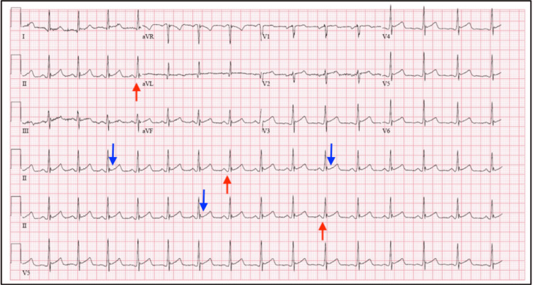
| Feature | Acute pericarditis | STEMI | Early repolarization |
|---|---|---|---|
| Distribution of ST elevation | Widespread, beyond one coronary territory | Coronary territory | Precordial V2 to V5, sometimes inferior |
| ST morphology | Concave upward | Convex or straight; hyperacute T waves | Concave, often with J-point notch or slur |
| Reciprocal ST depression | Only in aVR and V1 | In the opposite territory, such as aVL in inferior STEMI | Absent |
| PR segment | Depressed, with PR elevation in aVR | Usually unchanged; atrial infarction rare | Unchanged |
| ST elevation in III greater than in II | Uncommon | Favors right coronary occlusion | Uncommon |
| Q waves | Absent | Develop | Absent |
| T-wave inversion | After ST normalization | While ST still elevated | Absent |
| ST/T ratio in V6 | 0.25 or more | Not applicable | Less than 0.25 |
| Evolution on serial ECG | Over days | Over minutes to hours | Stable over years |
| Wall motion on echocardiography | Normal unless myocarditis | Regional abnormality | Normal |
ECG distinction of pericarditis from STEMI and early repolarization. The ST/T ratio is the ST elevation at the J point divided by the T-wave amplitude, both measured from the PR segment as baseline; a ratio of 0.25 or more in V6 identified every patient with pericarditis in the original series (positive and negative predictive values 1.0). No single ECG feature excludes STEMI. Persistent uncertainty is resolved by emergency echocardiography and coronary angiography. ECG criteria for STEMI are covered on the STEMI page.
Troponin, CRP, and initial laboratory tests
The ACC and ESC documents recommend the same baseline evaluation for every patient: a 12-lead ECG, chest radiograph, transthoracic echocardiography, complete blood count, CRP or erythrocyte sedimentation rate, troponin, renal function, and liver tests. CRP is elevated in most patients with acute pericarditis. It supports the diagnosis, identifies the inflammatory phenotype, and guides the duration of therapy. Treatment continues at full dose until CRP normalizes, and normalization precedes each step of tapering. A normal CRP in a patient already taking an NSAID or corticosteroid does not exclude active inflammation. A high-sensitivity assay detects low-grade residual activity.
Troponin is elevated in up to about one third of patients and signals myocardial involvement. Troponin elevation does not alter the prognosis of myopericarditis with preserved ventricular function, but it prompts echocardiographic assessment of ventricular function, rhythm monitoring, and often CMR. A white cell count with neutrophilia supports the autoinflammatory phenotype, and lymphocytosis or anemia raises concern for tuberculosis, lymphoma, or leukemia. Additional tests depend on the clinical setting. Blood cultures are obtained for fever above 38°C. An interferon-gamma release assay or tuberculin skin test is performed with endemic exposure or HIV, and HIV serology in at-risk patients. Antinuclear antibody and other serologies are indicated for systemic features, thyroid-stimulating hormone for an effusion without inflammation, and urea for kidney disease. Routine viral serology is not recommended.
Echocardiography, CMR, and CT in pericarditis
Transthoracic echocardiography is recommended for every patient with suspected pericarditis. Its purposes are to detect and size an effusion, assess tamponade physiology, identify constrictive physiology, and measure ventricular function. Most patients with idiopathic pericarditis have no effusion or a small one, so a normal echocardiogram does not exclude the diagnosis. An echo-bright, thickened pericardium supports inflammation but is nonspecific.
CMR is recommended in the ACC guidance when the diagnosis is uncertain, when disease is recurrent, incessant, or complicated, and when myocardial involvement is suspected. Pericardial edema on T2-weighted short-tau inversion recovery imaging and pericardial LGE indicate active inflammation. In recurrent pericarditis, quantitative pericardial LGE added to clinical criteria improved identification of an ongoing recurrence (sensitivity 70%, specificity 74%). In a RHAPSODY substudy, patients with moderate or severe LGE had more recurrences per year and higher CRP than patients with trace or mild LGE. CMR also detects myocardial LGE and edema, measures pericardial thickness, and shows the real-time septal shift of constriction.
CT shows pericardial thickening, calcification, effusion density, loculation, and mass. Attenuation separates simple fluid from hemorrhage, pus, or chyle, and CT plans pericardiectomy and percutaneous access. Enhancement of a thickened pericardium after contrast indicates inflammation. Fluorodeoxyglucose (FDG) positron emission tomography (PET) with CT detects pericardial uptake in tuberculous, neoplastic, and autoimmune disease when the cause remains undefined. A chest radiograph is normal in most patients with acute pericarditis. A globular "water bottle" silhouette appears only once an effusion exceeds about 200 mL.
| Modality | Findings in pericarditis | Principal indications |
|---|---|---|
| Transthoracic echocardiography | Effusion size and distribution, chamber collapse, respiratory Doppler variation, inferior vena cava (IVC) size, ventricular function, septal motion | All patients at presentation; serial follow-up of effusion; tamponade; guidance of pericardiocentesis |
| Transesophageal echocardiography | Posterior and loculated collections, thrombus after surgery | Suspected regional tamponade with inconclusive transthoracic imaging |
| CMR | Pericardial edema (T2 STIR), pericardial LGE, myocardial edema and LGE, thickness, real-time septal shift | Uncertain diagnosis; recurrent or incessant disease; suspected myocarditis; phenotype and duration of therapy |
| CT | Thickening, calcification, fluid attenuation, loculation, masses, contrast enhancement | Loculated or complex effusion, suspected neoplasm, calcification, planning of drainage or pericardiectomy |
| FDG PET/CT | Pericardial and nodal metabolic activity | Suspected tuberculous, neoplastic, or systemic inflammatory disease |
Imaging in acute and recurrent pericarditis, following the 2013 American Society of Echocardiography (ASE) multimodality recommendations and the 2025 ACC guidance.
High-risk features and admission
Most patients with idiopathic pericarditis are treated as outpatients. In a triage program of 300 consecutive patients, 85% were classified as low risk and treated at home, therapy was effective in 87% of them, and no outpatient developed tamponade over a mean of 38 months. Patients without response to aspirin had more recurrences (60.6% versus 10.4%) and more constriction (9.1% versus 0.5%). The ESC major predictors come from a cohort of 453 patients. Fever above 38°C, subacute onset, large effusion or tamponade, and failure of aspirin or an NSAID independently predicted a specific cause or complications.
| Category | Feature | Reason |
|---|---|---|
| Major | Fever above 38°C | Bacterial, tuberculous, or neoplastic cause (hazard ratio 3.56 for a specific cause) |
| Subacute onset over days to weeks | Nonidiopathic cause (hazard ratio 3.97) | |
| Large effusion (diastolic separation above 20 mm) or tamponade | Hemodynamic risk and specific cause | |
| No response to aspirin or NSAID after at least 1 week | Recurrence, constriction, and specific cause (hazard ratio 5.50 for complications) | |
| Minor | Myopericarditis | Arrhythmia and ventricular dysfunction |
| Immunosuppression | Opportunistic and bacterial infection | |
| Trauma | Hemopericardium | |
| Oral anticoagulation | Hemorrhagic effusion and tamponade |
High-risk features in acute pericarditis from the 2015 ESC guideline. Any feature warrants admission and a search for a specific cause. Patients without high-risk features are treated as outpatients and reassessed for response after about 1 week.
Anticoagulant therapy during active pericarditis increases the risk of hemorrhagic effusion. When anticoagulation is not essential, it is withheld during the acute illness. When it is essential, as with a mechanical valve, recent venous thromboembolism, or atrial fibrillation with a high stroke risk, the patient is admitted and the effusion is followed by serial echocardiography. Pericarditis and pericardial effusion after STEMI are not by themselves reasons to stop indicated antithrombotic therapy, but a growing effusion after infarction raises concern for subacute free-wall rupture.
First-line therapy: aspirin or an NSAID with colchicine
Aspirin or an NSAID combined with colchicine is first-line therapy for acute and first recurrent pericarditis in both the 2025 ACC guidance and the 2025 ESC guideline (Class 1 in the ESC document, with Level of Evidence B for aspirin or an NSAID with a proton pump inhibitor and Level of Evidence A for colchicine). The anti-inflammatory drug is given at full dose, every 8 hours, until symptoms resolve and CRP normalizes. In a first episode this is usually 1 to 2 weeks. The drug is then tapered, one class at a time. Aspirin is preferred when the patient already needs antiplatelet therapy and after myocardial infarction. From 20 weeks of pregnancy, full-dose aspirin is avoided along with other NSAIDs, and prednisone at the lowest effective dose is used. Ibuprofen is the usual NSAID because of its tolerability. Indomethacin is an alternative, but it reduces coronary blood flow and is avoided in coronary disease. A proton pump inhibitor accompanies full-dose aspirin or an NSAID.
| Drug | Dose | Duration and taper | Notes |
|---|---|---|---|
| Aspirin | 500–1000 mg every 8 h | Full dose until symptoms resolve and CRP normalizes; then reduced by 250–500 mg every 1–2 weeks | Preferred with coronary disease or after myocardial infarction |
| Ibuprofen | 600–800 mg every 8 h | Same principle; reduced by 200–400 mg every 1–2 weeks | Avoided at 20 weeks of pregnancy or later |
| Indomethacin | 25–50 mg every 8 h | Same principle; stepwise taper every 1–2 weeks | Avoided in coronary disease |
| Colchicine | 0.6 mg twice daily, or 0.6 mg once daily at 70 kg or less, in older adults, or with intolerance | 3 months after a first episode; at least 6 months after a recurrence; colchicine stopped after the other drugs | No loading dose; trials used 0.5 mg twice daily (once daily at 70 kg or less) |
| Prednisone | 0.2–0.5 mg/kg/day | Held until remission; slow taper, with steps of 1.25–2.5 mg every 2–6 weeks below 15 mg/day | Second line; with colchicine |
| Anakinra | 1–2 mg/kg/day subcutaneously, up to 100 mg/day | Months, then gradual withdrawal | Daily injection; off-label in the United States |
| Rilonacept | 320 mg subcutaneously once, then 160 mg weekly | Commonly 12 to 18 months or longer | Approved by the US Food and Drug Administration for recurrent pericarditis |
| Goflikicept | 80 mg subcutaneously every 2 weeks | Not established | Not available in the United States |
| Azathioprine | 1 mg/kg/day, increased to 2–3 mg/kg/day | Months; slow onset | Corticosteroid-sparing; thiopurine methyltransferase (TPMT) activity checked before use |
| Intravenous immunoglobulin | 400–500 mg/kg/day for 5 days | Single course, repeated as needed | Refractory disease; safe in infection-related disease |
Anti-inflammatory therapy for acute and recurrent pericarditis. Doses follow the 2025 ACC guidance; taper steps follow the 2015 ESC guideline. In the United States, colchicine is available as a 0.6-mg tablet.
Tapering is guided by symptoms and CRP, not by calendar. A recurrence during taper returns the patient to the last effective dose. Colchicine continues through the taper of the other drugs and is stopped last. An optional final step of every-other-day or once-daily colchicine for the last weeks is used to reduce rebound.
Colchicine in pericarditis: trials, dosing, interactions, and kidney function
Colchicine is the only drug shown in randomized trials to prevent recurrence after a first episode, and the only first-line agent shown to prevent further recurrence after a first or later recurrence. It roughly halves the risk of recurrence in every population studied.
| Trial | Population | Colchicine regimen | Primary result |
|---|---|---|---|
| COPE, 2005 | 120 patients, first episode, open label | 1.0–2.0 mg on day 1, then 0.5–1.0 mg/day for 3 months | Recurrence at 18 months 10.7% versus 32.3%; corticosteroid use an independent risk factor for recurrence (odds ratio 4.30) |
| ICAP, 2013 | 240 patients, first episode, double blind | 0.5 mg twice daily (above 70 kg) or once daily (70 kg or less) for 3 months | Incessant or recurrent pericarditis 16.7% versus 37.5%; number needed to treat 4; hospitalization 5.0% versus 14.2% |
| CORP, 2011 | 120 patients, first recurrence | 1.0–2.0 mg on day 1, then 0.5–1.0 mg/day for 6 months | Recurrence at 18 months 24% versus 55%; number needed to treat 3 |
| CORP-2, 2014 | 240 patients, two or more recurrences | 0.5 mg twice daily (above 70 kg) or once daily for 6 months | Recurrence 21.6% versus 42.5%; number needed to treat 5 |
| COPPS, 2010 | 360 patients after cardiac surgery | Loading dose, then 0.5 mg twice daily for 1 month (halved below 70 kg) | Postpericardiotomy syndrome at 12 months 8.9% versus 21.1% |
| COPPS-2, 2014 | 360 patients, started before cardiac surgery | 0.5 mg twice daily (once daily below 70 kg) for 1 month, no loading | Postpericardiotomy syndrome 19.4% versus 29.4%; no reduction in postoperative effusion or atrial fibrillation; gastrointestinal adverse effects increased |
Randomized trials of colchicine in pericarditis. Recurrence rates are colchicine versus placebo or conventional therapy. All trials excluded tuberculous, purulent, and neoplastic disease.
Diarrhea and nausea are the main adverse effects. Loading doses and higher doses increased gastrointestinal intolerance in early trials, so weight-adjusted dosing without a loading dose became standard. Serious toxicity, including myopathy, neuropathy, rhabdomyolysis, and marrow suppression, results from accumulation. Colchicine is metabolized by CYP3A4 and exported by P-glycoprotein. Clarithromycin, erythromycin, azole antifungals, HIV protease inhibitors, cyclosporine, verapamil, and diltiazem raise colchicine levels. Coadministration requires dose reduction or a different drug, and the combination is avoided in kidney or liver impairment. Statins and fibrates add a risk of myopathy. Mild to moderate kidney impairment calls for closer monitoring. With creatinine clearance below 30 mL/min, the dose is reduced, usually to once daily as in the 2025 ESC guideline, and toxicity is monitored. Colchicine is not removed by dialysis. In older adults, the 2015 ESC guideline halves the dose, and the 2025 guideline halves it in selected patients.
Anti-inflammatory regimen calculator for pericarditis
The regimen depends on body weight, the episode, the clinical setting, age, and kidney function. The calculator applies the 2025 ACC doses and the ESC taper principles to these inputs.
Doses from the 2025 ACC guidance (aspirin 500–1000 mg, ibuprofen 600–800 mg, or indomethacin 25–50 mg every 8 hours; colchicine 0.6 mg once or twice daily; prednisone 0.2–0.5 mg/kg/day, rounded to 2.5 mg). Colchicine is given for 3 months after a first episode and at least 6 months after a recurrence. The 70-kg threshold for once-daily colchicine is that of the ICAP and CORP-2 trials. Specific causes (tuberculous, purulent, neoplastic, uremic, systemic autoimmune) are treated by cause.
Incessant and recurrent pericarditis: inflammatory and noninflammatory phenotypes
Without colchicine, the risk of a further recurrence after a first recurrence approaches 50%. Recurrence follows inadequate dose or duration of first-line therapy, early or rapid tapering, corticosteroid exposure, and an autoinflammatory predisposition. Female sex, failure of aspirin or NSAID, and corticosteroid use predict recurrence. Each recurrence is confirmed objectively before therapy is escalated. Pain alone after months of illness has many causes, including central sensitization and musculoskeletal pain.
The 2025 ACC guidance separates recurrent disease into two phenotypes. The inflammatory phenotype has elevated CRP, fever, neutrophilia, pericardial or pleural effusion, or pericardial edema and LGE on CMR. It responds to escalation, and it is the indication for IL-1 blockade. The noninflammatory phenotype has recurrent pain with normal CRP and no imaging evidence of inflammation. Escalating immunosuppression does not help, and the diagnosis is reconsidered.
Incessant pericarditis, in which symptoms never fully remit, usually reflects first-line therapy at too low a dose, tapering while CRP is still elevated, or early corticosteroid exposure. It is managed by restoring full-dose aspirin or NSAID with colchicine until remission and then tapering slowly, and by reassessing the cause. Chronic pericarditis, lasting beyond 3 months, is usually effusive and calls for fluid analysis or pericardial biopsy when the cause remains unknown and the diagnosis would change treatment.
Evaluation of recurrent pericarditis repeats the search for a cause: tuberculosis, systemic autoimmune and autoinflammatory disease, cancer, hypothyroidism, and post-injury syndromes. A family history of periodic fever and attacks with fever, high CRP, and polyserositis prompt genetic testing for periodic fever syndromes. Patients with multiple recurrences, corticosteroid dependence, or complications are referred to a center with a pericardial disease program, as recommended by both the ACC and ESC documents.
Corticosteroids in pericarditis: indications and low-dose regimens
Corticosteroids relieve symptoms quickly but promote recurrence and dependence in idiopathic pericarditis. In COPE, corticosteroid use was an independent risk factor for recurrence (odds ratio 4.30). The 2025 ESC guideline gives low-to-moderate dose corticosteroids a Class 2a recommendation after failure of first-line therapy. The ACC guidance places them for patients with a contraindication to aspirin and NSAIDs, a systemic autoimmune indication, pregnancy when NSAIDs are contraindicated, or the noninflammatory phenotype. Colchicine is continued with them. Infection, particularly tuberculosis and bacterial disease, is excluded before they are started.
In a comparison of 100 patients with recurrent pericarditis, prednisone 1.0 mg/kg/day was associated with more severe adverse effects, recurrences, and hospitalizations than 0.2 to 0.5 mg/kg/day (hazard ratio 3.61). The starting dose of 0.2 to 0.5 mg/kg/day is held until symptoms resolve and CRP normalizes, then tapered slowly. Recurrences cluster below 10 to 15 mg/day of prednisone, where each reduction is 1.25 to 2.5 mg every 2 to 6 weeks. When symptoms return during taper, the dose is not raised. Aspirin or an NSAID is added at full dose, colchicine is continued, and the taper resumes. Bone protection with calcium, vitamin D, and a bisphosphonate where indicated accompanies prolonged therapy.
IL-1 blockade: anakinra, rilonacept, and goflikicept
Interleukin-1 blockade has changed the management of recurrent pericarditis with an inflammatory phenotype. The 2025 ACC guidance places anakinra or rilonacept after failure of, or intolerance to, aspirin or NSAID with colchicine in patients with elevated CRP or pericardial inflammation on CMR. It does not require a trial of corticosteroids first. The 2025 ESC guideline retains a stepwise sequence. Anti-IL-1 agents receive a Class 1 recommendation (Level of Evidence A) for recurrence after failure of first-line therapy and corticosteroids when CRP is elevated, and a Class 2a recommendation (Level of Evidence C) when CMR shows pericardial inflammation regardless of CRP.
| Agent and trial | Mechanism and dose | Population | Result |
|---|---|---|---|
| Anakinra: AIRTRIP, 2016 | IL-1 receptor antagonist; 2 mg/kg/day up to 100 mg, daily subcutaneous injection | 21 patients with three or more recurrences, elevated CRP, colchicine resistance, and corticosteroid dependence; randomized withdrawal | Recurrence 18.2% with anakinra versus 90% with placebo; corticosteroids withdrawn in responders |
| Rilonacept: RHAPSODY, 2021 | Soluble receptor fusion protein binding IL-1 alpha and beta; 320 mg loading, then 160 mg weekly | 86 patients with recurrent pericarditis, pain, and elevated CRP; 61 randomized after a 12-week run-in | Recurrence 7% (2 of 30) versus 74% (23 of 31); hazard ratio 0.04; median time to CRP normalization 7 days |
| Rilonacept: long-term extension, 2024 | Continued weekly therapy, median 22 months | 74 patients from RHAPSODY | Recurrence 0.04 events per patient-year on treatment versus 4.4 before; 75% recurred after suspension at 18 months |
| Goflikicept: phase 2/3, 2023 | Soluble receptor fusion protein binding IL-1 alpha and beta; 80 mg every 2 weeks | 22 patients with idiopathic recurrent pericarditis; 20 randomized | No recurrence with goflikicept versus 9 of 10 with placebo over 24 weeks |
Trials of IL-1 blockade in recurrent pericarditis. The three randomized trials used a randomized-withdrawal design after an open-label response phase; the 2024 rilonacept study is an open-label extension.
Response is rapid. In RHAPSODY, pain resolved in a median of 5 days and CRP normalized in a median of 7 days, which allowed taper of corticosteroids and NSAIDs. Injection-site reactions and upper respiratory infections were the most common adverse effects. Before treatment, active infection is excluded, latent tuberculosis is screened, and vaccinations are updated. Live vaccines are avoided during therapy. Rilonacept was approved by the US Food and Drug Administration in 2021 for recurrent pericarditis in patients 12 years and older. Anakinra is used off-label.
Duration is the unresolved question. Recurrence after withdrawal is common: three quarters of patients who suspended rilonacept at 18 months had a recurrence. In the RHAPSODY CMR substudy, recurrence after withdrawal to placebo was as frequent with trace or mild pericardial LGE (5 of 7 patients) as with moderate or severe LGE (5 of 7), although it came sooner with more extensive enhancement (median 4.2 versus 10.7 weeks). Current practice continues therapy for 12 to 18 months or longer and then withdraws it gradually, guided by symptoms, CRP, and CMR.
An inadequate response to IL-1 blockade prompts a review of adherence and dose, of the evidence for active inflammation, and of the diagnosis itself. Switching between anakinra and rilonacept, the addition of corticosteroid-sparing agents, and referral for pericardiectomy are the remaining options. A recurrence during or after IL-1 therapy is confirmed by CRP or CMR before treatment is changed.
Azathioprine, IVIG, methotrexate, and pericardiectomy for refractory recurrence
Azathioprine, intravenous immunoglobulin (IVIG), and methotrexate are corticosteroid-sparing options for patients who remain corticosteroid dependent and cannot receive or do not respond to IL-1 blockade. Azathioprine takes weeks to months to act and allows gradual corticosteroid withdrawal. IVIG acts faster and is useful when infection is a concern, but its effect is often transient. Methotrexate is used mainly in systemic autoimmune disease. Evidence for these agents comes from observational series.
Radical pericardiectomy is recommended in the ACC guidance for medically refractory pericarditis. Experienced centers report lower recurrence after surgery, but chest pain can persist. The procedure is reserved for patients who have failed medical therapy, including IL-1 blockade, and is performed at a high-volume pericardial surgical center. A limited pericardiectomy leaves inflamed pericardium and does not reliably prevent recurrence.
Myopericarditis and perimyocarditis
Myocardial involvement accompanies pericarditis in about 15% of patients in contemporary series. Myopericarditis is predominantly pericarditis with troponin elevation and preserved ventricular function. Perimyocarditis is predominantly myocarditis with new focal or global ventricular dysfunction. The 2025 ESC guideline places both within the inflammatory myopericardial syndrome and classifies the patient by the dominant process.
In a prospective cohort of 486 patients, no deaths occurred among those with myopericarditis or perimyocarditis, and ventricular function normalized in more than 90%. Recurrence was less frequent with myocardial involvement (11% to 12%) than with isolated pericarditis (32%). The patient is admitted for rhythm monitoring when troponin is elevated. Ventricular function is measured, and CMR defines the extent of myocardial edema and LGE. In myopericarditis, the doses of aspirin and NSAIDs are often lower than in isolated pericarditis, because animal data suggest NSAIDs can worsen myocarditis. Colchicine is used for the pericarditic component. Perimyocarditis with ventricular dysfunction is managed on the myocarditis pathway, including heart failure therapy, and the 2025 ESC guideline advises consideration of a beta-blocker for at least 6 months in acute myocarditis, especially with troponin elevation (Class 2a).
Exercise restriction and return to sport after pericarditis
The 2025 ACC guidance recommends restriction for at least 1 month after diagnosis or a flare, with heart rate kept below 100 beats/min, until clinical remission. The 2015 ESC guideline set a minimum of 3 months for competitive athletes and allowed a shorter period for nonathletes. The 2025 ESC guideline recommends restriction until remission, for at least 1 month, in athletes and nonathletes alike (Class 1), with return individualized by symptoms, biomarkers, and imaging. Return requires resolution of symptoms and normalization of CRP, the ECG, and the echocardiogram. In the 2015 American Heart Association and ACC eligibility recommendations, athletes with pericarditis do not compete during active disease and return only when no evidence of active inflammation remains. When the myocardium is involved, the myocarditis recommendations apply: no competitive sport for 3 to 6 months, followed by assessment of ventricular function, biomarkers, and arrhythmia on ambulatory and exercise testing. In recurrent pericarditis, exertion is a common trigger of flares, and each flare restarts the restriction.
Pericardial effusion: size, cause, and evaluation
A pericardial effusion is classified by onset (acute, subacute, or chronic beyond 3 months), size, distribution (circumferential or loculated), hemodynamic effect, and composition. Size is the end-diastolic echo-free separation between the pericardial layers at the widest point.
| Size | End-diastolic separation | Typical significance |
|---|---|---|
| Trivial | Seen only in systole | Physiologic |
| Small | Below 10 mm | Common in acute pericarditis; rarely affects hemodynamics |
| Moderate | 10–20 mm | Tamponade with rapid accumulation or low filling pressures |
| Large | Above 20 mm | High-risk feature; higher likelihood of a specific cause |
Echocardiographic grading of pericardial effusion from the 2015 ESC guideline and the 2013 ASE recommendations. Epicardial fat, which lies anteriorly, moves with the heart, and has granular texture, is distinguished from fluid. A posterior echo-free space anterior to the descending aorta is pericardial, whereas a pleural effusion lies posterior to the aorta.
Evaluation establishes whether the effusion is causing tamponade and whether it is inflammatory. Inflammatory effusions associated with acute pericarditis are treated as pericarditis and usually regress. Without inflammation and without a known cause, the 2015 ESC guideline lists the common causes: hypothyroidism, cancer, kidney failure, heart failure, pulmonary hypertension, cirrhosis, nephrotic syndrome, and medications. A large effusion without inflammatory signs is more often neoplastic or idiopathic chronic. Myxedema can cause a large effusion with bradycardia, which masks the tachycardia expected in tamponade. Effusion in hypothyroidism resolves with thyroid replacement.
Large idiopathic chronic effusion is usually well tolerated but can tamponade without warning. In a series of 28 patients with large idiopathic effusions lasting more than 3 months, overt tamponade occurred in 29%, and pericardiocentesis alone was followed by reaccumulation in about half. Pericardiocentesis is performed for tamponade, for suspected bacterial or neoplastic disease, and for a large symptomatic effusion that does not respond to medical therapy. Pericardiectomy is reserved for recurrent large effusions.
Pericardial fluid analysis
Fluid is analyzed when drainage is performed for diagnosis, or when therapeutic drainage provides the opportunity. Light criteria from pleural fluid do not identify the cause, and most pericardial effusions meet exudative thresholds regardless of cause.
| Test | Finding | Interpretation |
|---|---|---|
| Appearance | Serous, serosanguineous, hemorrhagic, purulent, milky | Hemorrhagic: cancer, tuberculosis, uremia, trauma, iatrogenic. Purulent: bacterial. Milky: chylopericardium |
| Cell count and differential | Neutrophil predominance; lymphocyte predominance | Neutrophils: bacterial or acute inflammation. Lymphocytes: tuberculosis, cancer, lymphoma |
| Glucose | Low | Bacterial, tuberculous, or rheumatoid effusion |
| Gram stain and culture | Positive | Purulent pericarditis; blood cultures obtained in parallel |
| Mycobacterial tests | Acid-fast stain, culture, nucleic acid amplification, adenosine deaminase, unstimulated interferon-gamma | Adenosine deaminase above 40 U/L and elevated interferon-gamma support tuberculosis; culture and pericardial biopsy confirm it |
| Cytology | Malignant cells | Neoplastic effusion; biopsy raises yield when cytology is negative |
| Tumor markers | Carcinoembryonic antigen and others | Support, without proving, malignancy |
| Triglycerides | Above 110 mg/dL, cholesterol lower than serum | Chylopericardium from thoracic duct injury or lymphatic obstruction |
Pericardial fluid analysis. A hematocrit of the fluid compared with blood distinguishes a hemorrhagic effusion from active bleeding into the pericardium.
Cardiac tamponade: physiology and bedside signs
Cardiac tamponade is compression of the heart by pericardial contents that raises intrapericardial pressure above chamber filling pressure and limits filling throughout diastole. Diastolic pressures in the right atrium, right ventricle, pulmonary artery, and left ventricle converge on the intrapericardial pressure. Stroke volume falls, and output is maintained by tachycardia and vasoconstriction until they are exhausted, after which hypotension and shock develop abruptly. Because the compression persists throughout diastole, the y descent of the jugular and right atrial waveform is blunted or absent, the reverse of the prominent y descent in constriction.
Tamponade is a clinical and hemodynamic diagnosis, and echocardiography confirms and supports it. In a systematic review, dyspnea (87% to 89%), tachycardia (77%), pulsus paradoxus (82%), elevated jugular venous pressure (76%), and cardiomegaly on chest radiograph (89%) were present in most patients. The Beck triad of hypotension, elevated venous pressure, and muffled heart sounds describes rapid surgical tamponade and is present in a minority of medical cases. Its absence does not exclude tamponade.
Pulsus paradoxus is an inspiratory fall in systolic pressure of more than 10 mm Hg. It is measured by deflating the cuff slowly and noting the pressure at which Korotkoff sounds are first heard only in expiration and then the pressure at which they are heard throughout respiration. A pulsus above 10 mm Hg in a patient with an effusion increased the likelihood of tamponade (likelihood ratio 3.3), and 10 mm Hg or less made it unlikely (likelihood ratio 0.03). Pulsus paradoxus can be absent despite tamponade with aortic regurgitation, atrial septal defect, severe left ventricular hypertrophy or dysfunction, positive-pressure ventilation, regional tamponade, and severe hypotension. It occurs without tamponade in severe obstructive lung disease, massive pulmonary embolism, and right ventricular infarction.
The ECG shows sinus tachycardia, low QRS voltage, and electrical alternans, a beat-to-beat alternation of QRS amplitude caused by the heart swinging within a large effusion. Alternans is specific but insensitive. Hemodynamic shock from tamponade is an indication for immediate drainage. A cautious fluid bolus of 250 to 500 mL can raise filling pressure transiently in a hypovolemic patient. Vasopressors and inotropes are temporizing only. Diuretics and vasodilators lower filling pressure and worsen tamponade.
At catheterization, right atrial pressure and intrapericardial pressure are elevated and nearly equal, and the right ventricular and pulmonary artery diastolic pressures and the pulmonary capillary wedge pressure converge on the same value. The right atrial waveform preserves the systolic x descent and loses the y descent. Drainage lowers intrapericardial pressure to near zero, right atrial pressure falls, and cardiac output and systemic pressure rise. Simultaneous measurement of right atrial and intrapericardial pressure before and after drainage separates pure tamponade from effusive-constrictive pericarditis, in which right atrial pressure remains elevated after the intrapericardial pressure has fallen.
Positive-pressure ventilation raises intrathoracic pressure, lowers venous return, and can cause cardiovascular collapse at induction. Drainage precedes intubation whenever possible. When airway control cannot wait, drainage is prepared in parallel, and the induction strategy preserves spontaneous respiration and venous return as far as feasible.
Low-pressure tamponade. Hypovolemia from hemorrhage, dialysis, or diuresis lowers filling pressures, so a comparatively low intrapericardial pressure compresses the heart. Jugular venous pressure is not elevated, and most patients lack the classic physical signs. Among 279 patients who underwent combined pericardiocentesis and catheterization at one referral center, low-pressure tamponade made up 20% of tamponade cases. Echocardiographic findings were as frequent as in classic tamponade, and cardiac output rose after drainage.
Regional tamponade. After cardiac surgery, a loculated collection or clot compresses one chamber, often the right or left atrium or the posterior left ventricle, without circumferential fluid. Classic signs and transthoracic findings are absent. Transesophageal echocardiography or CT identifies the collection, and surgical evacuation is usually required. Unexplained hypotension, falling output, or rising filling pressures in the days after cardiac surgery raise this diagnosis.
Echocardiographic signs of tamponade
Echocardiography confirms the effusion and demonstrates its hemodynamic effect. A chamber collapses when intrapericardial pressure exceeds the pressure inside it, so the low-pressure right-sided chambers collapse first, each in the phase of the cycle when its pressure is lowest.
| Sign | Criterion | Performance and limitations |
|---|---|---|
| Right atrial collapse | Systolic inversion of the right atrial wall lasting more than one third of the cardiac cycle | Inversion of any duration: sensitivity 100%, specificity 82%; duration above 0.34 of the cycle: specificity 100% |
| Right ventricular diastolic collapse | Early diastolic inward motion of the right ventricular free wall or outflow tract | More specific than atrial collapse; absent with right ventricular hypertrophy or pulmonary hypertension |
| Left atrial or ventricular collapse | Collapse of a left-sided chamber | Regional or loculated tamponade after surgery; severe pulmonary hypertension |
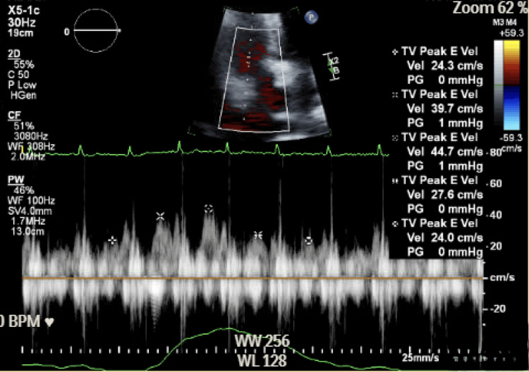
| Respiratory variation in mitral inflow | Inspiratory fall in mitral E velocity of more than 25% to 30% | Doppler counterpart of pulsus paradoxus; not valid during positive-pressure ventilation or atrial fibrillation |
| Respiratory variation in tricuspid inflow | Inspiratory rise in tricuspid E velocity of more than 40% to 60% | Reciprocal right-sided change |
| Inferior vena cava plethora | Diameter above 21 mm with less than 50% inspiratory collapse | Present in most patients with tamponade (high sensitivity); low specificity; absent in low-pressure tamponade |
| Hepatic vein flow | Blunted or absent diastolic forward flow; expiratory diastolic reversal | Supports elevated right atrial pressure and interdependence |
| Respiratory septal shift and swinging heart | Leftward septal motion in inspiration; anteroposterior swinging in a large effusion | Swinging is the image counterpart of electrical alternans |
Echocardiographic signs of cardiac tamponade. Right atrial collapse data are from the original 127-patient series; inferior vena cava criteria from the 2013 ASE recommendations. Mitral variation is (expiratory E − inspiratory E) ÷ expiratory E; tricuspid variation is (inspiratory E − expiratory E) ÷ expiratory E. The absence of any chamber collapse makes clinical tamponade unlikely, except in regional tamponade.
The 2014 ESC Working Group triage statement combines etiology, clinical presentation, and imaging into a score. A score of 6 or more warrants urgent pericardiocentesis. In the 2025 ESC guideline, immediate pericardiocentesis is considered at more than 6 points from at least two categories. A lower score allows drainage to be delayed while the cause is evaluated. The 2014 statement advises that a hemodynamically stable patient with tamponade undergo drainage within 12 to 24 hours of diagnosis. The score does not apply to surgical tamponade from dissection, rupture, or trauma, which goes directly to surgery.
Pericardiocentesis and surgical drainage
Pericardiocentesis is recommended for tamponade and for suspected bacterial or neoplastic effusion. It is performed under echocardiographic or fluoroscopic guidance. The apical approach is the most common in contemporary echocardiography-guided practice. The subxiphoid approach, with the needle directed toward the left shoulder, avoids the pleura and is the route used without imaging in extremis. The 2014 ESC Working Group statement advises against rapid evacuation of more than 1 L. The 2025 ESC guideline advises that, whenever possible, the initial drainage remove less than about 500 mL to prevent the rare pericardial decompression syndrome, and the remainder drains through the indwelling catheter.
Echocardiography-guided pericardiocentesis proceeds as follows.
- Preparation: coagulation tests and the platelet count are reviewed, anticoagulants are held where feasible, and the procedure is performed with ECG and arterial pressure monitoring, resuscitation equipment, and surgical backup. The patient lies semi-upright at 30 to 45 degrees, which brings fluid anteriorly and inferiorly.
- Site and trajectory: the transducer identifies the point where the collection is deepest and closest to the skin, with no interposed liver, lung, or internal thoracic vessel, and the transducer angle defines the needle path. The skin-to-fluid distance and the fluid depth are measured.
- Puncture: after local anesthesia, the needle is advanced along the planned path with continuous aspiration. At chest wall sites the needle passes over the upper border of the rib, away from the intercostal bundle, and lateral to the course of the internal thoracic artery.
- Confirmation: agitated saline injected through the needle opacifies the pericardial space on imaging. Bubbles in a cardiac chamber indicate puncture of the heart. Bloody aspirate that clots rapidly raises the same concern, because blood that has been in the pericardium for some time is defibrinated.
- Catheter placement: a J-tipped guidewire is passed and its position in the pericardial space confirmed by imaging or fluoroscopy, and a pigtail catheter is advanced over a dilator.
- Drainage and documentation: intrapericardial pressure is recorded when a transducer is available, fluid is drained in aliquots, a repeat echocardiogram documents residual fluid and chamber re-expansion, and samples are sent for the tests directed by the clinical question.
Extended catheter drainage reduces reaccumulation. The catheter remains until output falls to 30 mL or less per 24 hours (the 2015 ESC guideline threshold), typically after 2 to 5 days, and is flushed to prevent occlusion. In 1,127 echocardiography-guided procedures at the Mayo Clinic, the success rate was 97%, with major complications in 1.2% and minor complications in 3.5%. The use of extended drainage rose from 23% to 75% across the study periods, and recurrence and the need for surgery fell. Cardiac surgery, cancer, and perforation during catheter procedures accounted for nearly 70% of the drained effusions in the latest period. Major complications include laceration of a coronary artery or cardiac chamber, pneumothorax, ventricular arrhythmia, injury to the liver or peritoneum, and infection. Relative contraindications include uncorrected coagulopathy, anticoagulation, thrombocytopenia below 50,000/µL, and small, posterior, or loculated effusions.
Surgical drainage is preferred for tamponade from aortic dissection, free-wall rupture, or trauma, where needle drainage can raise arterial pressure and extend the bleeding. It is also preferred for purulent pericarditis requiring washout and lysis of loculations, for loculated or posterior collections, for clotted hemopericardium, and when biopsy is needed. A subxiphoid pericardial window drains the pericardium and provides tissue. A thoracoscopic or thoracotomy window drains into the pleural space and suits recurrent effusions.
Malignant effusion recurs in a large proportion of patients after simple aspiration. The options are extended catheter drainage, intrapericardial instillation of sclerosing or cytotoxic agents (cisplatin for lung cancer, thiotepa for breast cancer), percutaneous balloon pericardiotomy, and a surgical window. The choice reflects the expected survival, the risk of the procedure, the treatment plan for the cancer, and the patient's goals.
Pericarditis after myocardial infarction, cardiac surgery, and procedures
Post-cardiac injury syndrome describes immune-mediated pericarditis after injury to the pericardium or myocardium. It includes the postpericardiotomy syndrome after cardiac surgery, the post-myocardial infarction (Dressler) syndrome, and pericarditis after PCI, ablation, pacemaker or defibrillator lead perforation, and chest trauma. The 2015 ESC criteria require at least two of five findings after the injury: fever without another cause, pericarditic or pleuritic chest pain, a friction rub, a pericardial effusion, and a pleural effusion with elevated CRP. Onset is usually weeks after the injury, sometimes days. Treatment is aspirin or an NSAID with colchicine. Perioperative colchicine reduced postpericardiotomy syndrome in COPPS (8.9% versus 21.1%) and COPPS-2 (19.4% versus 29.4%), but it increased gastrointestinal adverse effects and did not reduce postoperative effusion.
Postoperative pericardial effusion is common after cardiac surgery and is usually small and asymptomatic. A moderate or large effusion in the first weeks, especially posterior and with anticoagulation, can cause late tamponade.
Procedural tamponade after coronary perforation, ablation, lead placement, or transcatheter structural intervention develops within minutes to hours and often while the patient is still on the table. Fluoroscopy shows a still cardiac border and echocardiography a new effusion. Management is immediate pericardiocentesis, reversal of anticoagulation where the perforation allows, and autotransfusion of aspirated blood when bleeding continues. Coronary perforation is sealed with a covered stent or embolization. Persistent bleeding despite drainage, or a ventricular tear, requires surgical repair.
After myocardial infarction, two forms occur. Early infarct-associated pericarditis appears in the first days after a transmural infarction. Late post-infarction pericarditis (Dressler syndrome) appears weeks later. Both are less common in the era of rapid reperfusion. Aspirin at anti-inflammatory doses is the treatment. Other NSAIDs and corticosteroids are avoided because they impair infarct healing and increase the risk of rupture. An effusion larger than 10 mm after infarction raises concern for subacute free-wall rupture. Infarct-associated pericarditis is covered on the STEMI page.
Pericardial disease in cancer: malignant effusion, radiation, and immunotherapy
Pericardial involvement in cancer arises from metastasis, direct invasion, radiation, chemotherapy, immunotherapy, and infection in immunosuppressed patients. Lung cancer, breast cancer, lymphoma, leukemia, and melanoma account for most metastatic involvement. Primary pericardial tumors, such as mesothelioma and angiosarcoma, are rare. Malignant effusion presents as a large, often hemorrhagic effusion and frequently as tamponade. In up to two thirds of patients with cancer and an effusion, the effusion is not malignant but results from radiation, drugs, infection, or hypothyroidism, so cytology and fluid analysis determine management.
Radiation causes acute pericarditis during or soon after treatment, chronic effusion, and constriction years to decades later. Anthracyclines, cyclophosphamide, cytarabine, and arsenic trioxide cause pericarditis or effusion. Immune checkpoint inhibitors cause pericarditis and effusion, sometimes with tamponade and sometimes with myocarditis. Checkpoint inhibitor pericarditis is treated with corticosteroids and suspension of the agent, under cardio-oncology direction. Immunotherapy for cancer is restarted only after multidisciplinary review.
Tuberculous, purulent, and uremic pericarditis
Tuberculous pericarditis. Tuberculosis is the most common cause of pericardial effusion in sub-Saharan Africa and parts of Asia, and it is the leading cause of constriction worldwide. The presentation is subacute, with fever, night sweats, weight loss, and a large effusion. Diagnosis combines exposure, fluid studies, and culture or biopsy. Treatment is a standard four-drug regimen for 6 months. In the IMPI trial of 1,400 patients, adjunctive prednisolone did not reduce the composite of death, tamponade requiring pericardiocentesis, or constriction (23.8% versus 24.5%). Prednisolone did reduce constriction (4.4% versus 7.8%) and hospitalization, and it increased HIV-associated cancers.
Purulent pericarditis. Bacterial infection of the pericardium spreads from pneumonia or empyema, follows cardiac surgery or chest trauma, or arises from bacteremia. It presents with high fever, sepsis, and often tamponade, and it is fatal without drainage. Treatment is urgent drainage with intravenous antibiotics, and surgical drainage is often required for loculated fluid. Constriction followed at an incidence of 52.7 per 1,000 person-years in one cohort, the highest of any cause.
Intrapericardial fibrinolysis is used to break down loculations when drainage is incomplete, and early surgical drainage with pericardiectomy is required for dense adhesions or recurrent loculated collections. Tuberculosis and fungal infection are sought when cultures are negative in an immunosuppressed patient.
Uremic and dialysis pericarditis. Uremic pericarditis develops before or soon after dialysis begins. Dialysis pericarditis develops in patients on established dialysis and reflects inadequate dialysis or volume overload. ECG changes are often absent because the myocardium is not inflamed. Intensified dialysis is the treatment. NSAIDs and colchicine have limited roles in kidney failure, and pericardiocentesis is required for tamponade or for a large effusion that persists despite dialysis.
Pericarditis in pregnancy and in older adults
Pericarditis in pregnancy is usually idiopathic, and the outcome is good with planned treatment. Women with recurrent pericarditis are counseled before conception, and the 2025 ESC guideline recommends multidisciplinary preconception planning (Class 1). Aspirin or an NSAID is used in the first and second trimesters. NSAIDs are avoided from 20 weeks of gestation because of fetal renal dysfunction and oligohydramnios, and later because of premature closure of the ductus arteriosus. Prednisone at the lowest effective dose is the main option in late pregnancy, because the placenta inactivates most of it. Colchicine was considered contraindicated in pregnancy in the 2015 ESC guideline. In a meta-analysis that included women with familial Mediterranean fever, colchicine during pregnancy was not associated with miscarriage or major malformations. The 2025 ESC guideline allows it for prevention of recurrence (Class 2b). Anakinra has been used in pregnancy and breastfeeding for refractory recurrence without fetal harm in a case series of 14 women. A small physiologic pericardial effusion is common in the third trimester and needs no treatment.
Older adults have a higher prevalence of specific causes, including cancer, uremia, and post-procedural disease, and higher in-hospital mortality. NSAIDs carry greater gastrointestinal, renal, and cardiovascular risk. Aspirin is often preferred. The 2015 ESC guideline halves the colchicine dose in older adults, the 2025 guideline does so in selected patients, and drug interactions with colchicine are more frequent because of polypharmacy.
Follow-up after acute pericarditis and surveillance of effusion
Patients treated as outpatients are reassessed after about 1 week for response. Persistent pain, fever, or CRP elevation at that point is a high-risk feature and prompts admission or a search for a specific cause. CRP is measured before each tapering step, and therapy is not reduced while it is elevated. An echocardiogram is repeated when an effusion was present at diagnosis, when symptoms change, and before return to competitive sport. Long-term colchicine therapy is monitored with blood counts, creatine kinase, and renal and liver tests, especially when interacting drugs are added. Recurrence presents as a return of the original pain, and an early visit with CRP measurement allows treatment before the episode becomes established.
Effusion surveillance follows size and cause. The 2015 ESC guideline does not require routine follow-up of a small idiopathic asymptomatic effusion. It recommends echocardiography every 6 months for a moderate effusion and every 3 to 6 months for a large one. Any increase in size, new symptoms, or a fall in blood pressure prompts earlier imaging. An effusion in a patient with cancer, kidney failure, or hypothyroidism is reassessed after treatment of the cause.
Prognosis of acute and recurrent pericarditis
The long-term prognosis of idiopathic pericarditis is good. Mortality from the pericardial disease itself is rare outside specific causes, and constriction after idiopathic disease is uncommon. Recurrent pericarditis impairs quality of life through pain, repeated care, exercise restriction, and corticosteroid toxicity, but recurrent idiopathic pericarditis rarely leads to tamponade or constriction. The interval between recurrences lengthens and attacks become milder over years in most patients. Outcomes after IL-1 blockade are favorable, although recurrence after withdrawal remains common. The prognosis of neoplastic, purulent, and tuberculous pericarditis is that of the underlying disease.
A transient constrictive physiology appears during active inflammation in some patients and resolves within 2 to 3 months of anti-inflammatory therapy. Effusive-constrictive pericarditis is recognized when right atrial pressure remains elevated after drainage of a tamponading effusion. Diagnosis, imaging, hemodynamics, and pericardiectomy are covered on the constrictive pericarditis page.
Sources of error in the diagnosis and treatment of pericarditis
- Attribution of diffuse ST elevation to pericarditis before infarction is excluded: ST elevation confined to a coronary territory, reciprocal depression outside aVR and V1, and regional wall-motion abnormality indicate coronary occlusion.
- Exclusion of pericarditis because the ECG is normal or no effusion is present: under the 2025 ACC criteria, a suggestive presentation with elevated CRP or CMR evidence of inflammation is sufficient, and the 2025 ESC guideline classifies it as possible pericarditis.
- Omission of colchicine at the first episode: it halves recurrence and is first-line in both US and European guidance.
- Low-dose or short-course NSAID therapy, or tapering before CRP normalizes: incessant and recurrent disease follow.
- Corticosteroids as first-line therapy for idiopathic pericarditis, or at 1 mg/kg/day: both increase recurrence and dependence.
- Escalation of immunosuppression for recurrent pain without objective inflammation: the noninflammatory phenotype does not respond, and the diagnosis needs to be reconsidered.
- Abrupt withdrawal of IL-1 blockade after a short course: recurrence is frequent, and withdrawal is gradual after a sustained remission.
- Colchicine with clarithromycin, an azole, verapamil, diltiazem, or cyclosporine at full dose, or in severe kidney disease: accumulation causes myopathy and marrow suppression.
- Reliance on effusion size to diagnose tamponade: a small, rapidly accumulating effusion can tamponade, and a large chronic effusion can be hemodynamically silent.
- Exclusion of tamponade because jugular pressure is normal or pulsus paradoxus is absent: low-pressure tamponade, regional tamponade, and coexisting aortic regurgitation, atrial septal defect, or left ventricular dysfunction alter the signs.
- Intubation or diuresis before drainage in tamponade: both reduce preload and can precipitate arrest.
- Needle drainage of hemopericardium from type A dissection or free-wall rupture: surgical repair is the treatment.
- Ibuprofen or corticosteroids for pericarditis after myocardial infarction: aspirin is the anti-inflammatory agent.
- Continued anticoagulation without monitoring in active pericarditis: hemorrhagic effusion and tamponade follow.
- Return to competitive sport on symptom resolution alone: CRP, the ECG, and the echocardiogram must also normalize, and myocardial involvement extends restriction to 3 to 6 months.
Self-assessment
A 36-year-old man has his third episode of pericarditis in 14 months while taking colchicine 0.6 mg twice daily. Pain recurred each time ibuprofen was tapered. CRP is 6.2 mg/dL, and CMR shows moderate pericardial late gadolinium enhancement without myocardial involvement. Tuberculosis testing is negative. Which treatment does the 2025 ACC guidance place next, and how does the 2025 ESC guideline sequence differ?
Cardiovascular Medicine Board ReviewReferences
- Wang TKM, Klein AL, Cremer PC, et al. 2025 concise clinical guidance: an ACC expert consensus statement on the diagnosis and management of pericarditis: a report of the American College of Cardiology Solution Set Oversight Committee. J Am Coll Cardiol. 2025;86:2691–2719. doi:10.1016/j.jacc.2025.05.023
- Schulz-Menger J, Collini V, Gröschel J, et al. 2025 ESC guidelines for the management of myocarditis and pericarditis. Eur Heart J. 2025;46:3952–4041. doi:10.1093/eurheartj/ehaf192
- Adler Y, Charron P, Imazio M, et al. 2015 ESC guidelines for the diagnosis and management of pericardial diseases. Eur Heart J. 2015;36:2921–2964. doi:10.1093/eurheartj/ehv318
- Klein AL, Abbara S, Agler DA, et al. American Society of Echocardiography clinical recommendations for multimodality cardiovascular imaging of patients with pericardial disease: endorsed by the Society for Cardiovascular Magnetic Resonance and Society of Cardiovascular Computed Tomography. J Am Soc Echocardiogr. 2013;26:965–1012. doi:10.1016/j.echo.2013.06.023
- Ehsan M, Ikram J, El Roumi J, et al. Evolving guidelines in pericarditis: aligning practice with pathophysiology. JACC Adv. 2026;5:102509. doi:10.1016/j.jacadv.2025.102509
- Imazio M, Cecchi E, Demichelis B, et al. Indicators of poor prognosis of acute pericarditis. Circulation. 2007;115:2739–2744. doi:10.1161/CIRCULATIONAHA.106.662114
- Imazio M, Demichelis B, Parrini I, et al. Day-hospital treatment of acute pericarditis: a management program for outpatient therapy. J Am Coll Cardiol. 2004;43:1042–1046. doi:10.1016/j.jacc.2003.09.055
- Kytö V, Sipilä J, Rautava P. Clinical profile and influences on outcomes in patients hospitalized for acute pericarditis. Circulation. 2014;130:1601–1606. doi:10.1161/CIRCULATIONAHA.114.010376
- Imazio M, Brucato A, Maestroni S, et al. Risk of constrictive pericarditis after acute pericarditis. Circulation. 2011;124:1270–1275. doi:10.1161/CIRCULATIONAHA.111.018580
- Ginzton LE, Laks MM. The differential diagnosis of acute pericarditis from the normal variant: new electrocardiographic criteria. Circulation. 1982;65:1004–1009. doi:10.1161/01.cir.65.5.1004
- Imazio M, Brucato A, Barbieri A, et al. Good prognosis for pericarditis with and without myocardial involvement: results from a multicenter, prospective cohort study. Circulation. 2013;128:42–49. doi:10.1161/CIRCULATIONAHA.113.001531
- Kumar A, Sato K, Verma BR, et al. Quantitative assessment of pericardial delayed hyperenhancement helps identify patients with ongoing recurrences of pericarditis. Open Heart. 2018;5:e000944. doi:10.1136/openhrt-2018-000944
- Cremer PC, Lin D, Luis SA, et al. Pericardial late gadolinium enhancement and time to recurrence: a substudy from RHAPSODY, a phase 3 clinical trial of rilonacept in recurrent pericarditis. Eur Heart J Imaging Methods Pract. 2023;1:qyad003. doi:10.1093/ehjimp/qyad003
- Imazio M, Bobbio M, Cecchi E, et al. Colchicine in addition to conventional therapy for acute pericarditis: results of the COlchicine for acute PEricarditis (COPE) trial. Circulation. 2005;112:2012–2016. doi:10.1161/CIRCULATIONAHA.105.542738
- Imazio M, Brucato A, Cemin R, et al. A randomized trial of colchicine for acute pericarditis. N Engl J Med. 2013;369:1522–1528. doi:10.1056/NEJMoa1208536
- Imazio M, Brucato A, Cemin R, et al. Colchicine for recurrent pericarditis (CORP): a randomized trial. Ann Intern Med. 2011;155:409–414. doi:10.7326/0003-4819-155-7-201110040-00359
- Imazio M, Belli R, Brucato A, et al. Efficacy and safety of colchicine for treatment of multiple recurrences of pericarditis (CORP-2): a multicentre, double-blind, placebo-controlled, randomised trial. Lancet. 2014;383:2232–2237. doi:10.1016/S0140-6736(13)62709-9
- Imazio M, Brucato A, Cumetti D, et al. Corticosteroids for recurrent pericarditis: high versus low doses: a nonrandomized observation. Circulation. 2008;118:667–671. doi:10.1161/CIRCULATIONAHA.107.761064
- Brucato A, Imazio M, Gattorno M, et al. Effect of anakinra on recurrent pericarditis among patients with colchicine resistance and corticosteroid dependence: the AIRTRIP randomized clinical trial. JAMA. 2016;316:1906–1912. doi:10.1001/jama.2016.15826
- Klein AL, Imazio M, Cremer P, et al. Phase 3 trial of interleukin-1 trap rilonacept in recurrent pericarditis. N Engl J Med. 2021;384:31–41. doi:10.1056/NEJMoa2027892
- Imazio M, Klein AL, Brucato A, et al. Sustained pericarditis recurrence risk reduction with long-term rilonacept. J Am Heart Assoc. 2024;13:e032516. doi:10.1161/JAHA.123.032516
- Myachikova VY, Maslyanskiy AL, Moiseeva OM, et al. Treatment of idiopathic recurrent pericarditis with goflikicept: phase II/III study results. J Am Coll Cardiol. 2023;82:30–40. doi:10.1016/j.jacc.2023.04.046
- Maron BJ, Udelson JE, Bonow RO, et al. Eligibility and disqualification recommendations for competitive athletes with cardiovascular abnormalities: Task Force 3: hypertrophic cardiomyopathy, arrhythmogenic right ventricular cardiomyopathy and other cardiomyopathies, and myocarditis: a scientific statement from the American Heart Association and American College of Cardiology. Circulation. 2015;132:e273–e280. doi:10.1161/CIR.0000000000000239
- Sagristà-Sauleda J, Angel J, Permanyer-Miralda G, Soler-Soler J. Long-term follow-up of idiopathic chronic pericardial effusion. N Engl J Med. 1999;341:2054–2059. doi:10.1056/NEJM199912303412704
- Spodick DH. Acute cardiac tamponade. N Engl J Med. 2003;349:684–690. doi:10.1056/NEJMra022643
- Roy CL, Minor MA, Brookhart MA, Choudhry NK. Does this patient with a pericardial effusion have cardiac tamponade? JAMA. 2007;297:1810–1818. doi:10.1001/jama.297.16.1810
- Sagristà-Sauleda J, Angel J, Sambola A, Alguersuari J, Permanyer-Miralda G, Soler-Soler J. Low-pressure cardiac tamponade: clinical and hemodynamic profile. Circulation. 2006;114:945–952. doi:10.1161/CIRCULATIONAHA.106.634584
- Gillam LD, Guyer DE, Gibson TC, King ME, Marshall JE, Weyman AE. Hydrodynamic compression of the right atrium: a new echocardiographic sign of cardiac tamponade. Circulation. 1983;68:294–301. doi:10.1161/01.cir.68.2.294
- Himelman RB, Kircher B, Rockey DC, Schiller NB. Inferior vena cava plethora with blunted respiratory response: a sensitive echocardiographic sign of cardiac tamponade. J Am Coll Cardiol. 1988;12:1470–1477. doi:10.1016/s0735-1097(88)80011-1
- Ristić AD, Imazio M, Adler Y, et al. Triage strategy for urgent management of cardiac tamponade: a position statement of the European Society of Cardiology Working Group on Myocardial and Pericardial Diseases. Eur Heart J. 2014;35:2279–2284. doi:10.1093/eurheartj/ehu217
- Tsang TS, Enriquez-Sarano M, Freeman WK, et al. Consecutive 1127 therapeutic echocardiographically guided pericardiocenteses: clinical profile, practice patterns, and outcomes spanning 21 years. Mayo Clin Proc. 2002;77:429–436. doi:10.4065/77.5.429
- Imazio M, Trinchero R, Brucato A, et al. COlchicine for the Prevention of the Post-pericardiotomy Syndrome (COPPS): a multicentre, randomized, double-blind, placebo-controlled trial. Eur Heart J. 2010;31:2749–2754. doi:10.1093/eurheartj/ehq319
- Imazio M, Brucato A, Ferrazzi P, et al. Colchicine for prevention of postpericardiotomy syndrome and postoperative atrial fibrillation: the COPPS-2 randomized clinical trial. JAMA. 2014;312:1016–1023. doi:10.1001/jama.2014.11026
- Mayosi BM, Ntsekhe M, Bosch J, et al. Prednisolone and Mycobacterium indicus pranii in tuberculous pericarditis. N Engl J Med. 2014;371:1121–1130. doi:10.1056/NEJMoa1407380
- Oster ME, Shay DK, Su JR, et al. Myocarditis cases reported after mRNA-based COVID-19 vaccination in the US from December 2020 to August 2021. JAMA. 2022;327:331–340. doi:10.1001/jama.2021.24110
- Indraratna PL, Virk S, Gurram D, Day RO. Use of colchicine in pregnancy: a systematic review and meta-analysis. Rheumatology (Oxford). 2018;57:382–387. doi:10.1093/rheumatology/kex353
- Trotta L, Lazaros G, Vecchié A, et al. Use of anakinra during pregnancy and breastfeeding in women with recurrent pericarditis: a case series. Eur J Obstet Gynecol Reprod Biol. 2025;312:114553. doi:10.1016/j.ejogrb.2025.114553