Library › Vascular disease › Acute aortic syndromes
Acute aortic syndromes: dissection, intramural hematoma, and penetrating ulcer
Acute aortic dissection, intramural hematoma, penetrating atherosclerotic ulcer, and blunt traumatic aortic injury in adults, from classification and bedside risk assessment through imaging, emergency treatment, surveillance, and family screening.
An acute aortic syndrome is a breach of the aortic media: classic dissection, intramural hematoma, or penetrating atherosclerotic ulcer. Any involvement of the ascending aorta is a surgical emergency, with untreated mortality of about 1% to 2% per hour early after onset. Acute type B dissection is treated first with intravenous beta blockade to a systolic pressure below 120 mm Hg and a heart rate of 60 to 80 beats/min. Endovascular repair is reserved for complicated disease and selected high-risk anatomy.
The aortic wall and the three acute aortic syndromes
The aortic wall has three layers. The intima is a thin endothelial lining. The media is a laminated sheet of elastic lamellae and smooth muscle cells that gives the aorta its strength and its recoil. The adventitia is a collagen layer that carries the vasa vasorum, which nourish the outer media. An acute aortic syndrome is defined by blood that has entered the media, through an intimal tear, through a ruptured vasa vasorum, or through an ulcerated plaque. The three conditions share symptoms, risk of rupture, and the first steps of treatment. They differ in mechanism, location, and natural history.
Classic aortic dissection. An intimal tear allows blood to enter the media and to split it longitudinally. The separated inner layer is the intimal flap (more precisely, the intimomedial flap), and it divides the aorta into a true lumen, bounded by intima, and a false lumen within the media. Blood can re-enter the true lumen through distal tears. The outer wall of the false lumen consists of the outer media and adventitia only and is the layer that ruptures.
Intramural hematoma. The 2022 American College of Cardiology/American Heart Association (ACC/AHA) aortic disease guideline defines intramural hematoma (IMH) as blood within the media without an overt intimal tear. It is diagnosed by circular or crescentic wall thickening of more than 5 mm without detectable flow. IMH accounts for 5% to 25% of acute aortic syndromes in Western series and 30% to 40% in Asian series. In the guideline's summary, fewer than 10% resolve spontaneously, and 16% to 47% progress to classic dissection.
Penetrating atherosclerotic ulcer. A penetrating atherosclerotic ulcer (PAU) begins as ulceration of an atherosclerotic plaque that breaks through the internal elastic lamina into the media. PAU occurs most often in the mid and distal descending thoracic aorta of older patients with diffuse atherosclerosis. It accounts for an estimated 2% to 7% of acute aortic syndromes, and it can lead to IMH, dissection, pseudoaneurysm, or rupture.
Blunt traumatic aortic injury, a tear produced by sudden deceleration, is managed with the same anti-impulse principles. It is covered below, as are iatrogenic dissection after cardiac surgery or catheter manipulation and dissection during pregnancy.
Epidemiology, sex differences, and in-hospital mortality
Acute aortic dissection is uncommon. The 2022 ACC/AHA guideline cites an incidence of 5 to 30 cases per million people per year, and a systematic review estimated 3.5 to 6.0 acute aortic syndromes per 100,000 patient-years. The prospective population-based Oxford Vascular Study recorded an incidence of 6 per 100,000 per year, with a mean age of 72 years. In that study, 48.6% of patients with type A dissection died before hospital assessment, so registry data from referral centers underestimate the true lethality of the disease. Most dissections occur between ages 50 and 70 years, and men are affected more often. Patients with Marfan syndrome, Loeys-Dietz syndrome, vascular Ehlers-Danlos syndrome, or a bicuspid aortic valve present younger.
The International Registry of Acute Aortic Dissection (IRAD) has defined the clinical picture since 1996. In its first report of 464 patients, the mean age was 63 years, 65.3% were men, and 62.3% had type A dissection. In-hospital mortality was 26% for type A dissection treated surgically, 58% for type A treated medically (usually because of age or comorbidity), and 10.7% for type B treated medically. In the 17-year IRAD trend analysis of 4,428 patients, surgical management of type A increased from 79% to 90%, type A in-hospital mortality fell from 31% to 22%, and surgical mortality fell from 25% to 18%. The use of CT as the diagnostic test in type A dissection rose from 46% to 73%. In type B dissection, endovascular treatment increased from 7% to 31%, and in-hospital mortality did not change (12% to 14%).
Women account for about one-third of IRAD patients. In the IRAD analysis of sex differences, women were older, presented later, more often had altered mental status, and less often had a pulse deficit. Periaortic hematoma, pleural or pericardial effusion, hypotension, and tamponade were more frequent, and the surgical mortality of type A dissection was 32% in women and 22% in men. After adjustment for age and hypertension, female sex remained associated with in-hospital death (odds ratio 1.4).
Risk factors for acute aortic syndrome
Two processes underlie most dissections: chronic mechanical stress on the wall and an intrinsically weak media. Hypertension is the dominant acquired risk factor. In the Oxford Vascular Study, 67.3% of patients were taking antihypertensive drugs, yet 46.0% had at least one systolic pressure of 180 mm Hg or more in primary care records over the preceding 5 years. Atherosclerosis and older age predominate in PAU and IMH. Heritable thoracic aortic disease predominates in young patients.
| Category | Condition | Features relevant to acute aortic syndrome |
|---|---|---|
| Acquired wall stress | Hypertension | Present in most patients with acute dissection; poorly controlled pressure precedes many events |
| Acquired wall stress | Cocaine and amphetamines; heavy isometric exertion | Abrupt surges in pressure and shear; cocaine-related dissection affects young men, mostly type A, within hours of use |
| Syndromic heritable disease | Marfan syndrome (FBN1) | Root aneurysm and type A dissection; distal dissection after root replacement |
| Syndromic heritable disease | Loeys-Dietz syndrome (TGFBR1, TGFBR2, SMAD3, TGFB2, TGFB3) | Dissection at small diameters; arterial tortuosity and branch aneurysms |
| Syndromic heritable disease | Vascular Ehlers-Danlos syndrome (COL3A1) | Spontaneous arterial dissection and rupture; fragile tissue complicates any repair |
| Nonsyndromic heritable disease | ACTA2, MYH11, MYLK, PRKG1, LOX | Type A dissection below conventional size thresholds (below 4.5 cm with ACTA2; after little prior enlargement with MYLK and PRKG1) |
| Congenital and genetic | Bicuspid aortic valve; coarctation; Turner syndrome | Ascending aortopathy; dissection risk in Turner syndrome is judged by aortic size index |
| Hemodynamic state | Pregnancy and early postpartum | Most pregnancy-related dissections occur in women with an aortopathy, often unrecognized beforehand |
| Iatrogenic | Cardiac surgery, aortic cannulation or cross-clamp sites, catheterization, TEVAR, TAVR | Often painless; recognized during or after the procedure |
| Inflammatory and infectious | Giant cell arteritis, Takayasu arteritis, other aortitis | Weakened media, aneurysm, and occasional dissection; aortitis also mimics IMH on imaging |
| Drug exposure | Fluoroquinolones | Observational association with aneurysm or dissection in the 60 days after treatment |
| Atherosclerosis | Older age, smoking, diffuse aortic plaque | Substrate for PAU and, through it, IMH and rupture |
Risk factors and predisposing conditions. Heritable gene list from the 2022 ACC/AHA guideline, which names 11 genes with highly penetrant risk of thoracic aortic disease (FBN1, LOX, COL3A1, TGFBR1, TGFBR2, SMAD3, TGFB2, ACTA2, MYH11, MYLK, PRKG1). TEVAR, thoracic endovascular aortic repair; TAVR, transcatheter aortic valve replacement.
In a series of 45 cocaine-related dissections, the mean age was 41 years, 88.9% were men, 75% were type A, and the median interval from last use to symptom onset was 1 hour. Iatrogenic dissection after cardiac surgery is often painless and presents as new hemodynamic instability, a new effusion, or an abnormal intraoperative transesophageal study.
Catheter-induced dissection begins at a coronary ostium during angiography or percutaneous coronary intervention and can extend retrograde into the sinus and ascending aorta. In a series of 43,143 catheterizations, it occurred in 0.02% of procedures. Limited aortic involvement was treated by stenting the coronary entry point, and extension more than 40 mm up the aorta from the coronary ostium required surgery.
Fluoroquinolones were associated with a higher rate of aortic aneurysm or dissection within 60 days of treatment in a Swedish nationwide cohort (hazard ratio 1.66 compared with amoxicillin), an estimated 82 additional cases per 1 million treatment courses. In 2018 the US Food and Drug Administration advised against fluoroquinolone use in patients with an aortic aneurysm or at risk for one when other treatment options are available. The 2022 ACC/AHA guideline lists the association as an evidence gap and makes no recommendation.
Aortitis from giant cell arteritis or Takayasu arteritis thickens the aortic wall concentrically and can mimic IMH on CT, and wall enhancement or fluorodeoxyglucose uptake on positron emission tomography (PET) favors inflammation. Infection of the aortic wall causes saccular pseudoaneurysm and is treated as a separate surgical problem.
Pathophysiology: medial degeneration, intimal tear, and false lumen
Medial degeneration is the common substrate: fragmentation and loss of elastic fibers, loss of smooth muscle cells, and accumulation of proteoglycans in the media. The heritable conditions produce it through defects in extracellular matrix (fibrillin-1, type III collagen, lysyl oxidase), in transforming growth factor beta signaling, or in the contractile apparatus of smooth muscle cells. Age and hypertension produce a similar but slower degeneration.
The primary entry tear forms where wall stress and flexion are greatest: in the ascending aorta within a few centimeters of the sinotubular junction, along the right lateral wall, and in the proximal descending aorta just distal to the left subclavian artery at the ligamentum arteriosum. The dissection propagates with each systolic pulse. The rate of rise of aortic pressure (dP/dt) drives propagation, which is why heart rate control is the first goal of medical therapy. The false lumen typically spirals along the outer curvature and is often larger than the true lumen. Flow in the false lumen can be patent, partially thrombosed, or completely thrombosed. In an IRAD analysis of patients with type B dissection who survived to discharge, partial thrombosis of the false lumen independently predicted death after discharge (relative risk 2.69), with a 3-year mortality of 31.6%. A partially thrombosed lumen with persistent inflow and impaired outflow is pressurized and prone to expand.
In IMH, the hematoma is thought to arise from rupture of the vasa vasorum within the outer media, or from a small intimal defect not resolved by imaging. CT now shows focal intimal disruptions, ulcer-like projections, and intramural blood pools in many patients once labeled as having IMH alone, and these features carry prognostic weight. In PAU, the lesion begins in an atherosclerotic plaque, and the media beneath it is often scarred, which may explain why PAU tends to produce localized hematoma or saccular aneurysm and less often a long dissection.
Malperfusion results from two mechanisms with different treatments. In dynamic obstruction, the flap prolapses across a branch ostium, or a compressed true lumen fails to supply the branch, usually during systole. Restoring flow in the true lumen by covering the entry tear, or by fenestrating the flap, relieves it. In static obstruction, the dissection extends into the branch itself and narrows or thromboses it. Static obstruction requires stenting or bypass of the branch. Both mechanisms can coexist in one patient and in one vascular bed.
Stanford, DeBakey, SVS/STS, and TEM classification
Classification by anatomy directs initial treatment, and classification by time since onset predicts complications and the response to endovascular repair. The Stanford system classifies by one criterion: involvement of the ascending aorta. The DeBakey system adds the extent of the dissection. Both were designed for open surgery and leave the aortic arch ambiguous. Dissections with an entry tear in the arch that spare the ascending aorta were formerly grouped with type B and are now often reported as non-A non-B dissection.
| System | Category | Definition | Initial management |
|---|---|---|---|
| Stanford | Type A | Involves the ascending aorta, irrespective of the site of the entry tear or the distal extent | Emergency open repair |
| Type B | Spares the ascending aorta; may involve the arch | Medical therapy unless complicated | |
| DeBakey | Type I | Originates in the ascending aorta and extends at least to the arch, usually beyond | Emergency open repair |
| Type II | Confined to the ascending aorta | Emergency open repair | |
| Type III | Originates in the descending thoracic aorta; IIIa limited to the thorax, IIIb extends below the diaphragm | Medical therapy unless complicated | |
| SVS/STS (2020) | Type A, with subscript | Entry tear in zone 0; subscript gives the distal extent (for example, A9 reaches the infrarenal aorta) | Emergency open repair |
| Type B, with subscripts | Entry tear in zone 1 or beyond; subscripts give proximal and distal extent (for example, B39 runs from zone 3 to zone 9) | Medical therapy unless complicated | |
| Indeterminate (I) | Entry tear not identified, with involvement of zone 0 | Managed as type A | |
| TEM (2020) | Type: A, B, or non-A non-B | Stanford type extended by a category for arch dissections that spare the ascending aorta | Records the entry tear and malperfusion that direct the extent of repair |
| Entry: E0 to E3 | E0 not visible; E1 ascending aorta; E2 arch; E3 descending aorta | ||
| Malperfusion: M0 to M3 | M0 none; M1 coronary; M2 supra-aortic vessels; M3 visceral, renal, or lower extremity; + if clinical, − if only radiographic |
Anatomic classification of aortic dissection. Zone 0 is the ascending aorta to the distal edge of the innominate artery origin; zone 1 lies between the innominate and left common carotid arteries; zone 2 between the left common carotid and left subclavian arteries; zone 3 is the proximal descending thoracic aorta to the level of T4; zones 4 and 5 run to the celiac artery; zones 6 to 9 run from the celiac artery to the aortic bifurcation; zones 10 and 11 are the iliac arteries. SVS/STS, Society for Vascular Surgery/Society of Thoracic Surgeons; TEM, type, entry, malperfusion.
The SVS/STS reporting standards describe a dissection by the zone of its primary entry tear and by its proximal and distal extent, which is the vocabulary used to plan endovascular landing zones. The 2022 ACC/AHA guideline uses the Stanford terms and endorses SVS/STS zones for reporting. In the original TEM series of 357 patients, in-hospital mortality was 16% for type A, 5% for type B, and 8% for non-A non-B dissection, and within type A it ranged from 0% with a descending entry tear (E3) to 40% with an arch entry tear (E2).
| Phase | Time from symptom onset | Clinical significance |
|---|---|---|
| Hyperacute | <24 hours | Highest risk of rupture, tamponade, and malperfusion; type A mortality accrues by the hour |
| Acute | 1–14 days | Most complications of type B dissection declare themselves; highest risk of retrograde type A dissection after TEVAR |
| Subacute | 15–90 days | Flap still mobile and responsive to remodeling; window for elective TEVAR in high-risk uncomplicated type B |
| Chronic | >90 days | Flap thick and fixed; late aneurysmal degeneration of the false lumen governs management |
Chronicity of aortic dissection from the SVS/STS reporting standards, adopted in the 2022 ACC/AHA guideline (Table 3). The traditional definition called a dissection acute in the first 2 weeks and chronic thereafter.
Clinical presentation, complications, and malperfusion
Pain is the cardinal symptom. In IRAD, severe or worst-ever pain was reported by 93% of patients with type A and 94% with type B dissection, and chest pain by 83% and 71%. The pain is typically abrupt and maximal at onset. Patients describe it as sharp or stabbing more often than tearing or ripping. Anterior chest pain is typical of ascending involvement, and interscapular back pain or abdominal pain is typical of descending involvement. Pain that migrates as the dissection propagates is characteristic but uncommon. Pain can resolve spontaneously, and resolution does not exclude the diagnosis.
Classic physical findings are frequently absent. In the first IRAD report, an aortic regurgitation murmur was noted in 31.6% of patients and a pulse deficit in 15.1%. Blood pressure is measured in both arms and both legs, and a difference of more than 20 mm Hg between limbs is a high-risk finding. Hypertension at presentation is common in type B dissection. Hypotension or shock is more common in type A and indicates tamponade, acute severe aortic regurgitation, coronary malperfusion, or rupture.
| Presentation | Mechanism | Implication |
|---|---|---|
| Abrupt severe chest, back, or abdominal pain | Medial tear and propagation | Cardinal symptom; recurrence or persistence in type B indicates progression |
| Syncope | Tamponade, acute aortic regurgitation, carotid involvement, or a vagal reflex | More frequent in type A dissection; prompts immediate imaging of the root and pericardium |
| Stroke, transient ischemic attack, or altered consciousness | Carotid or innominate involvement; hypotension | Can dominate the picture and lead to fibrinolysis in error; surgery remains reasonable in nonhemorrhagic stroke |
| Paraplegia or paraparesis | Spinal cord ischemia from intercostal or lumbar artery involvement | Malperfusion; can follow TEVAR |
| Horner syndrome, hoarseness, dysphagia | Compression of the sympathetic chain, recurrent laryngeal nerve, or esophagus | Uncommon; more often with large or chronic aneurysms |
| Diastolic murmur and heart failure | Acute aortic regurgitation from root dilation, cusp prolapse, or flap prolapse into the valve | Type A; adds to the urgency of surgery |
| Hypotension, elevated jugular pressure, pulsus paradoxus | Hemopericardium and tamponade | Leading cause of early death in type A |
| Inferior or global ischemia on ECG | Dissection into a coronary ostium, usually the right | Presents as STEMI; anticoagulation and fibrinolysis are hazardous |
| Abdominal pain, rising lactate, acidosis | Mesenteric malperfusion | Highest-mortality malperfusion syndrome |
| Oliguria, rising creatinine, refractory hypertension | Renal malperfusion | Complicated dissection |
| Cold, pulseless limb | Iliac or femoral malperfusion | Complicated dissection; often dynamic |
| Left pleural effusion, hemothorax, shock | Periaortic bleeding or rupture | Emergency repair |
Presentations of acute aortic dissection by mechanism. STEMI, ST-elevation myocardial infarction.
Malperfusion changes both prognosis and the order of treatment. In data cited by the 2022 ACC/AHA guideline, operative mortality for type A dissection was 30.5% with a malperfusion syndrome and 6.2% without, and 63.2% with mesenteric malperfusion. Mesenteric ischemia is difficult to detect early because abdominal pain is common in dissection, the examination is unreliable, and lactate rises late.
Painless dissection occurs in a minority of patients and is more common in older patients, in those with diabetes or neurologic presentations, and after cardiac surgery. Syncope, stroke, acute limb ischemia, or heart failure without pain can be the presenting event, and the diagnosis is made only when the aorta is imaged.
Differential diagnosis of acute aortic syndrome
Acute aortic syndrome is missed when another diagnosis explains the pain and the aorta is never imaged. The consequences of error are asymmetric: anticoagulation or fibrinolysis given for a presumed acute coronary syndrome or pulmonary embolism can be fatal in dissection.
| Condition | Overlap with acute aortic syndrome | Features that separate them |
|---|---|---|
| Acute coronary syndrome | Chest pain, troponin elevation, ST elevation with coronary malperfusion | Pain maximal at onset, back pain, pulse or pressure differential, new aortic regurgitation, mediastinal widening; bedside echocardiography of the root before antithrombotic loading |
| Pulmonary embolism | Chest pain, dyspnea, syncope, elevated D-dimer | Right ventricular strain, hypoxemia, deep venous thrombosis; a combined CT protocol can examine both |
| Acute pericarditis and tamponade | Pleuritic chest pain and effusion | Effusion in a patient with severe pain and hypertension or hypotension is treated as hemopericardium until the aorta is imaged |
| Ruptured or symptomatic aneurysm without dissection | Pain and shock | Known aneurysm; CT shows contained or free rupture without a flap |
| Aortitis | Wall thickening on CT | Concentric thickening with enhancement, systemic inflammation, FDG uptake on PET |
| Esophageal rupture, perforated viscus, pancreatitis, renal colic | Chest, back, or abdominal pain | Mediastinal air, free air under the diaphragm, elevated lipase, hematuria; none excludes coexisting dissection |
| Stroke without chest pain | Hemiplegia or altered consciousness | Pulse deficit, hypotension, carotid flap on CT angiography of the neck before fibrinolysis |
| Musculoskeletal pain | Back or chest wall pain | Diagnosis of exclusion when any high-risk feature is present |
Differential diagnosis of suspected acute aortic syndrome. FDG, fluorodeoxyglucose.
Aortic dissection detection risk score (ADD-RS) and D-dimer
The aortic dissection detection risk score is built on the high-risk features defined in the 2010 thoracic aortic disease guideline and was formalized and tested in IRAD in 2011. It counts three categories of high-risk features, each scoring 1 point when at least one feature in the category is present, for a total of 0 to 3. The score is a pretest probability tool. It is not a diagnostic test, and it is applied to patients in whom acute aortic syndrome is a realistic possibility.
| Category (1 point if any feature is present) | Features |
|---|---|
| High-risk conditions | Marfan syndrome or other connective tissue disease; family history of aortic disease; known aortic valve disease; recent aortic manipulation; known thoracic aortic aneurysm |
| High-risk pain features | Chest, back, or abdominal pain that is abrupt in onset, severe in intensity, or ripping or tearing in quality |
| High-risk examination features | Perfusion deficit (pulse deficit, systolic blood pressure differential, or focal neurologic deficit with pain); new aortic regurgitation murmur with pain; hypotension or shock |
ADD-RS from the 2010 ACCF/AHA thoracic aortic disease guideline, reproduced in the 2022 ACC/AHA guideline (Table 24). A score of 0 is low risk, 1 intermediate, and 2 or 3 high.
Applied retrospectively to 2,538 IRAD patients with confirmed dissection, the score identified 95.7% as having at least one risk marker. Only 4.3% scored 0, and almost half of those with a recorded chest radiograph had a widened mediastinum. The score is therefore sensitive but not specific, and a score of 0 does not exclude dissection on its own.
D-dimer rises in acute dissection because the false lumen activates coagulation and fibrinolysis. In the IRAD substudy on biomarkers, a D-dimer below 500 ng/mL had a negative likelihood ratio of 0.07 for dissection during the first 24 hours after onset. A thrombosed false lumen, a small IMH, and late presentation can produce a normal value. The prospective ADvISED study combined the two tools in 1,850 patients with suspected acute aortic syndrome at six hospitals, of whom 13% had an acute aortic syndrome. D-dimer of 500 ng/mL or more had a sensitivity of 96.7% and a specificity of 64%. An ADD-RS of 1 or less with D-dimer below 500 ng/mL occurred in 924 patients, with 3 missed cases: a failure rate of 0.3% and an efficiency of 49.9%.
The 2022 ACC/AHA guideline states that a D-dimer below 500 ng/mL in a patient with low prior probability makes acute aortic syndrome unlikely, and it assigns no class of recommendation to the strategy. The 2024 European Society of Cardiology (ESC) guideline builds its diagnostic algorithm on the same thresholds: an ADD-RS of 2 or more leads directly to imaging, and a lower score leads to D-dimer testing.
ADD-RS with D-dimer
Suspected acute aortic syndrome in adultsADD-RS categories from the 2010 and 2022 ACC/AHA guidelines; D-dimer threshold and failure rate from ADvISED (fibrinogen-equivalent units, 500 ng/mL). The strategy was validated in patients evaluated in the emergency department and does not apply to patients with known dissection, recent aortic surgery under follow-up, or presentation days after onset.
The AORTAs score, reproduced in the 2022 guideline as an alternative (Table 25), assigns 2 points for hypotension or shock and 1 point each for known aneurysm, pulse deficit, neurologic deficit, severe pain, and abrupt pain, with 2 or more points indicating high probability. Neither score replaces clinical judgment in a patient with unexplained pain and hemodynamic instability, who proceeds directly to imaging.
ECG, chest radiograph, and laboratory findings
The electrocardiogram is normal in about one-third of patients and nonspecific in many others. Left ventricular hypertrophy reflects long-standing hypertension. ST elevation from coronary malperfusion, low voltage or electrical alternans from a pericardial effusion, and diffuse ST depression from hypotension all occur.
The chest radiograph showed no abnormality in 12.4% of patients in the first IRAD report, so a normal film does not exclude dissection. Findings listed in the 2022 guideline are mediastinal widening, loss of the normal aortic knob contour, a "calcium sign" (intimal calcium displaced more than 5 mm from the outer aortic wall), a double density within the aorta, and rightward deviation of the trachea or a nasogastric tube. A left pleural effusion can represent hemothorax or a sympathetic exudate.
Troponin is elevated with coronary malperfusion, hypotension, or demand ischemia and is associated with higher mortality. It does not distinguish dissection from acute coronary syndrome. Lactate, creatinine, liver enzymes, and lipase identify visceral malperfusion. A complete blood count, coagulation studies, type and crossmatch, and a pregnancy test in women of childbearing potential complete the emergency evaluation. Aorta-specific biomarkers such as smooth muscle myosin heavy chain have not entered practice.
CT angiography, echocardiography, and MRI in acute aortic syndrome
CT angiography is the Class 1 initial imaging test for suspected acute aortic syndrome in the 2022 ACC/AHA guideline because it is widely available, fast, and accurate, and it shows the whole aorta and its branches. Transesophageal echocardiography (TEE) and MRI are reasonable alternatives (Class 2a). In a systematic review, the sensitivity was 100% for CT, 95% to 100% for MRI, and 86% to 100% for TEE.
The CT protocol for dissection differs from a coronary or pulmonary embolism protocol. It covers the chest, abdomen, and pelvis to the femoral arteries. A noncontrast acquisition precedes contrast to show the hyperattenuating crescent of IMH and displaced intimal calcium. Electrocardiographic synchronization of the thoracic acquisition removes pulsation artifact at the root and ascending aorta, where motion can create a linear artifact that mimics a flap or hides a small one. A real flap appears on adjacent slices and orthogonal planes and separates two lumens with different opacification. A delayed phase distinguishes slow false-lumen filling from thrombosis and shows active extravasation.
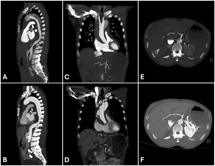
The CT report states the items that determine treatment: the Stanford type and SVS/STS zones; the location and size of the primary entry tear and of re-entry tears; and the maximal aortic diameter. It also gives the true and false lumen diameters and false-lumen status (patent, partially thrombosed, or thrombosed); involvement of the root, coronary ostia, arch branches, and visceral, renal, and iliac arteries, with the mechanism of any branch compromise; and pericardial, pleural, and mediastinal blood. The access vessels for endovascular repair are assessed as well. The true lumen is usually smaller, is continuous with the undissected aorta, and opacifies earlier. The false lumen is usually larger, lies along the outer curvature, and may contain cobwebs (strands of residual media) and a beak sign at the acute angle where the flap meets the outer wall.
Echocardiography. Transthoracic echocardiography (TTE) at the bedside shows root dilation, a flap in the proximal ascending aorta or abdominal aorta, aortic regurgitation, pericardial effusion, and regional wall motion abnormalities. Its sensitivity for dissection is limited, and a normal TTE does not exclude the diagnosis. TEE visualizes the root, ascending aorta, and descending aorta with high accuracy, identifies the mechanism of aortic regurgitation and the relation of the flap to the coronary ostia, and is performed at the bedside or in the operating room in unstable patients. A short segment of the distal ascending aorta and proximal arch is hidden by the air-filled trachea (the blind spot). Reverberation from a calcified aortic wall or a catheter can resemble a flap. A reverberation lies at a fixed multiple of the depth of its source, moves with that source, and does not separate two flow streams on color Doppler.
Aortic regurgitation in type A dissection has three main mechanisms, and TEE distinguishes them before and during surgery. Dilation of the root and sinotubular junction pulls the commissures apart and produces central regurgitation. Extension of the dissection into the root detaches one or more commissures, and the unsupported cusp prolapses. A mobile flap can prolapse through the valve in diastole and hold it open. The first two mechanisms are usually corrected by commissural resuspension and replacement of the ascending aorta, whereas a dilated root or intrinsically abnormal cusps lead to root or valve replacement. TEE also shows whether the flap reaches a coronary ostium and identifies new regional wall motion abnormalities that indicate coronary malperfusion.
MRI. MRI provides accurate anatomy without radiation or iodinated contrast and shows IMH age by signal characteristics. Scan duration and limited monitoring restrict its use in unstable patients. It is used for surveillance, in pregnancy (without gadolinium), and when contrast CT is contraindicated. Invasive aortography has largely been replaced and is now used during endovascular repair or to evaluate malperfusion during treatment.
| Modality | Strengths | Limitations | Primary setting |
|---|---|---|---|
| CT angiography | Whole aorta and branches; entry tear; malperfusion mechanism; IMH on noncontrast images; access planning | Iodinated contrast; radiation; motion artifact at the root without ECG synchronization | Initial test in stable patients (Class 1) |
| TEE | Bedside; root, valve mechanism, coronary ostia, effusion, ventricular function; intraoperative guidance | Sedation and airway risk; distal ascending blind spot; no abdominal views; reverberation artifact | Unstable patients and the operating room (Class 2a) |
| MRI | No radiation; tissue characterization of IMH; serial measurements | Time, monitoring, and availability; gadolinium restrictions in pregnancy and advanced kidney disease | Surveillance; pregnancy; contrast allergy (Class 2a as initial test) |
| TTE | Immediate; effusion, aortic regurgitation, root dilation, wall motion | Limited sensitivity; a normal study does not exclude dissection | Triage in the emergency department |
| Invasive aortography | Dynamic branch flow; therapeutic access | Invasive; misses IMH and thrombosed false lumen | During TEVAR, fenestration, or branch stenting |
Imaging in suspected acute aortic syndrome. Classes of recommendation from the 2022 ACC/AHA guideline.
Immediate blood pressure and heart rate control
Anti-impulse therapy lowers both heart rate and blood pressure, which reduces dP/dt, wall stress, and propagation. The 2022 ACC/AHA guideline recommends prompt therapy with arterial pressure monitoring in an intensive care setting (Class 1). The targets are a systolic pressure below 120 mm Hg or the lowest pressure that maintains end-organ perfusion, and a heart rate of 60 to 80 beats/min (Class 1). Intravenous beta blockers are given first (Class 1). When beta blockers are contraindicated or not tolerated, intravenous nondihydropyridine calcium channel blockers (diltiazem or verapamil) are reasonable for rate control (Class 2a). Intravenous vasodilators are added when pressure remains above target after rate control (Class 1). They are not used first, because reflex tachycardia raises dP/dt. Pain control is recommended to help achieve the hemodynamic targets (Class 1), usually with intravenous opioids, which also lower sympathetic drive. The 2024 ESC guideline sets a heart rate target of 60 beats/min or less with the same systolic target.
Pressure is measured in the arm with the higher reading, and an arterial line is placed on that side. A pressure differential caused by subclavian involvement can make a low reading from the affected arm misleading. In a hypotensive patient, anti-impulse drugs are withheld, and the cause (tamponade, aortic regurgitation, rupture, or coronary malperfusion) is treated surgically.
| Drug | Class | Intravenous dosing | Notes |
|---|---|---|---|
| Esmolol | Beta-1 selective blocker | 500 mcg/kg over 1 minute, then 50 mcg/kg/min, titrated up to 200–300 mcg/kg/min | Half-life about 9 minutes; preferred when tolerance of beta blockade is uncertain (asthma, borderline pressure) |
| Labetalol | Combined alpha and beta blocker | 20 mg over 2 minutes, then 40–80 mg every 10 minutes to 300 mg total, or infusion of 2 mg/min | Lowers rate and pressure with one agent |
| Metoprolol | Beta-1 selective blocker | 5 mg every 2 minutes, up to 3 doses (15 mg) | Bolus dosing; less titratable |
| Diltiazem | Nondihydropyridine calcium channel blocker | 0.25 mg/kg over 2 minutes, then 5–15 mg/h | When beta blockers are contraindicated (Class 2a) |
| Verapamil | Nondihydropyridine calcium channel blocker | 5–10 mg over 2 minutes | Alternative rate control; negative inotrope |
| Nicardipine | Dihydropyridine calcium channel blocker | 5 mg/h, increased by 2.5 mg/h every 5–15 minutes to 15 mg/h | Added after rate control |
| Clevidipine | Dihydropyridine calcium channel blocker | 1–2 mg/h, doubled every 90 seconds initially; most patients respond at 16 mg/h or less (maximum 32 mg/h) | Ultrashort acting; lipid emulsion limits volume over 24 hours |
| Sodium nitroprusside | Arterial and venous vasodilator | 0.3 mcg/kg/min, titrated to a maximum of 10 mcg/kg/min for no more than 10 minutes | Added after rate control; cyanide and thiocyanate toxicity with high doses, prolonged infusion, or kidney failure |
| Morphine or fentanyl | Opioid analgesic | Titrated intravenous doses | Analgesia lowers rate and pressure; nonsteroidal drugs avoided because of bleeding and kidney risk |
Intravenous agents for anti-impulse therapy. Doses from US prescribing information; sequence and classes of recommendation from the 2022 ACC/AHA guideline.
Several common emergency treatments are harmful in suspected dissection. Heparin, antiplatelet loading, and fibrinolysis are withheld until dissection is excluded. Intra-aortic balloon counterpulsation is contraindicated in aortic dissection and in significant aortic regurgitation, as stated in the device labeling. Aggressive volume loading in a hypotensive patient with tamponade raises wall stress without addressing the cause.
Emergency surgery for acute type A dissection
Acute type A dissection is treated by emergency open repair. The 2022 ACC/AHA guideline recommends emergency surgical consultation and immediate operation for suspected or confirmed type A dissection (Class 1). Unoperated mortality is about 1% to 2% per hour early after onset, and medical mortality is two to three times surgical mortality. Patients who are stable enough to travel are reasonably transferred from low-volume to high-volume aortic centers (Class 2a), and a multidisciplinary aortic team determines the operation (Class 1). At a hospital without cardiac surgery, transfer arrangements begin when the diagnosis is suspected and proceed in parallel with imaging and anti-impulse therapy, because transport time adds directly to the hourly mortality. The goals of the operation are to resect the primary entry tear, prevent rupture into the pericardium, restore aortic valve competence and coronary flow, and redirect flow into the true lumen.
Root and valve. When the root is partly dissected and the leaflets are normal, the commissures are resuspended and the native valve is preserved (Class 1). A root aneurysm, extensive root destruction, or a heritable aortopathy leads to root replacement with a valved conduit and coronary reimplantation (Bentall procedure; Class 1). Valve-sparing root replacement (reimplantation or remodeling) may be reasonable in selected stable patients operated on by experienced teams (Class 2b).
Distal extent. An open distal anastomosis under circulatory arrest is recommended (Class 1), and hemiarch replacement is preferred over more extensive arch replacement when there is no arch tear or significant arch aneurysm (Class 1). In a Society of Thoracic Surgeons database analysis cited by the guideline, operative mortality was 16% with hemiarch and 27% with total arch replacement. When the dissection extends into the proximal descending aorta, antegrade stenting of that segment may be considered (Class 2b). The frozen elephant trunk is a one-stage hybrid prosthesis: a surgical arch graft continuous with a stent graft deployed antegrade into the descending aorta. It treats arch and proximal descending tears, promotes false-lumen thrombosis, and creates a landing zone for later TEVAR. The 2024 ESC guideline gives it a Class IIb recommendation for an arch or proximal descending secondary tear.
Cannulation and cerebral protection. Axillary artery cannulation is reasonable over femoral cannulation (Class 2a), because retrograde femoral perfusion can pressurize the false lumen and embolize debris to the brain. Direct aortic or innominate cannulation with imaging guidance is a reasonable alternative (Class 2a). When circulatory arrest is required, antegrade or retrograde cerebral perfusion is reasonable (Class 2a), combined with hypothermia to reduce cerebral metabolic demand.
Malperfusion, stroke, and age. Renal, mesenteric, or lower-extremity malperfusion does not delay central repair, and immediate ascending aortic repair is recommended (Class 1). With clinically significant mesenteric malperfusion, immediate mesenteric revascularization before central repair is reasonable where the expertise exists (Class 2a). In patients with nonhemorrhagic stroke, surgery is reasonable over medical therapy (Class 2a). Operative mortality in these patients is 25% to 27%, compared with 76% with medical therapy in the series cited by the guideline. Older age alone does not exclude surgery, because older patients benefit. Severe frailty, advanced dementia, or a large established stroke may favor medical management. The guideline recommends shared decision-making with the patient or surrogate, including explicit discussion of expected outcomes (Class 1).
Tamponade. Hemopericardium is a leading cause of early death in type A dissection, and the definitive treatment is surgical. Unrestricted pericardiocentesis can raise aortic pressure and reopen the rupture. In a patient in cardiac arrest or with refractory shock who cannot reach the operating room in time, controlled drainage of small volumes has served as a bridge. In one series of 18 patients with critical tamponade, intermittent drainage through an 8F catheter to a systolic pressure of about 90 mm Hg required a mean of 40 mL, and all patients reached surgery. The 2024 ESC guideline describes emergency pericardial puncture, in a patient at risk of cardiac arrest from tamponade, as a temporary life-saving measure before transfer to the operating room.
Outcomes and residual disease. Most patients leave the operating room with a dissected arch or descending aorta, and many keep a patent false lumen. Ten-year survival after type A repair is about 60% to 65%, and the residual distal aorta grows by about 1 mm per year on average. A root diameter above 4.5 cm left in place at the initial operation is a risk factor for late reintervention. The guideline recommends recording the root and ascending diameters in the operative note so that relatives can be counseled (Class 1).
Uncomplicated and complicated type B dissection: medical therapy and TEVAR
Uncomplicated acute type B dissection is treated with medical therapy (Class 1). Complicated dissection requires intervention (Class 1). The definition of complicated disease is therefore the central decision in type B dissection.
| Category | Features | Management (2022 ACC/AHA) |
|---|---|---|
| Complicated | Rupture (free or contained); branch occlusion or malperfusion; proximal or distal extension; progressive aortic enlargement; intractable pain; uncontrolled hypertension | Intervention (Class 1); with suitable anatomy, endovascular repair over open repair for rupture (Class 1) and for other complications (Class 2a) |
| Uncomplicated with high-risk imaging features | Maximal aortic diameter >40 mm; false lumen >20–22 mm; entry tear >10 mm; entry tear on the lesser curvature; increase in total diameter >5 mm on serial imaging; bloody pleural effusion; malperfusion seen only on imaging | Medical therapy; endovascular repair may be considered (Class 2b) |
| Uncomplicated with high-risk clinical features | Refractory hypertension despite more than three drug classes at maximal doses; pain persisting more than 12 hours despite maximal therapy; readmission | Medical therapy; endovascular repair may be considered (Class 2b) |
| Uncomplicated | None of the above | Medical therapy (Class 1); surveillance imaging |
Complicated and high-risk acute type B dissection, from the 2022 ACC/AHA guideline (Tables 27 and 28), derived from the SVS/STS reporting standards.
Acute type B dissection category
Complicated, high-risk, or uncomplicatedCategories and classes of recommendation from the 2022 ACC/AHA guideline; subacute timing from the 2024 ESC guideline. The first six items define complicated dissection; the remaining items are high-risk features.
Medical therapy and in-hospital course. Medical therapy continues the anti-impulse targets in an intensive care unit, with serial examination, urine output, and lactate to detect malperfusion, and transition to oral beta blockers once pain and pressure are controlled. In data cited by the 2022 guideline, medical therapy for uncomplicated type B dissection carries a 30-day mortality of about 10% and a midterm mortality of about 30%. Delayed aortic expansion occurs in 20% to 50% of patients over 4 years. Repeat CT before discharge, and earlier for recurrent pain, new malperfusion, or refractory hypertension, detects progression.
Endovascular repair. Thoracic endovascular aortic repair (TEVAR) places a covered stent graft over the primary entry tear. It redirects flow into the true lumen, depressurizes the false lumen, and promotes false-lumen thrombosis and remodeling. A proximal landing zone of healthy aorta is required, and coverage of the left subclavian artery (zone 2 landing) is often needed for an entry tear near the isthmus. Left subclavian revascularization before planned coverage is recommended to reduce spinal cord and posterior circulation ischemia (Class 1 in the 2022 guideline's thoracic aneurysm section), although emergency repair may not allow it. In IRAD data cited by the guideline, in-hospital mortality in type B dissection was 32.1% with open repair, 6.5% with endovascular intervention, and 9.6% with medical therapy, in groups that differed in severity.
TEVAR in uncomplicated and high-risk disease. Two randomized trials define the evidence. INSTEAD randomized 140 patients with stable type B dissection at least 2 weeks after onset to optimal medical therapy alone or with TEVAR. Two-year survival did not differ (95.6% with medical therapy and 88.9% with TEVAR), but aortic remodeling occurred in 91.3% with TEVAR and 19.4% with medical therapy. In the long-term follow-up (INSTEAD-XL), TEVAR reduced 5-year aorta-specific mortality (6.9% versus 19.3%) and disease progression (27.0% versus 46.1%), and all-cause mortality did not differ significantly (11.1% versus 19.3%). ADSORB randomized 61 patients with uncomplicated dissection less than 14 days old. TEVAR improved the composite of incomplete false-lumen thrombosis, aortic dilation, or rupture at 1 year, and the trial was not powered for mortality. The 2022 ACC/AHA guideline states that the optimal timing of TEVAR is unknown and assigns a Class 2b recommendation to endovascular repair for uncomplicated dissection with high-risk anatomy. The 2024 ESC guideline recommends TEVAR between 14 and 90 days after onset in selected patients with high-risk features (Class IIa). The subacute window balances a mobile flap that still remodels against the higher complication rate of repair in the first days.
Complications of TEVAR. Retrograde type A dissection is the most feared complication. In a meta-analysis of 10,600 patients treated for type B dissection, it occurred in 2.3%, with a mortality of about 42%, and it was more frequent after repair in the acute phase, with a nonsignificant excess after proximal bare-stent grafts. Excessive oversizing and landing in a dissected or diseased proximal aorta increase the risk. Spinal cord ischemia is related to the length of aortic coverage, coverage of the left subclavian artery without revascularization, prior abdominal aortic repair, and perioperative hypotension. Cerebrospinal fluid drainage reduced paraplegia in a randomized trial of open thoracoabdominal repair and is used selectively in high-risk TEVAR. Delayed spinal cord deficits are treated by raising mean arterial pressure (above 100 mm Hg in the guideline's protocol), draining cerebrospinal fluid, and maintaining hemoglobin above 10 g/dL. Other complications are stroke, endoleak, stent-graft-induced new entry tears at the distal end, and access injury.
Fenestration, branch stenting, and open repair. When malperfusion persists after the entry tear is covered, or when the mechanism is static, branch stenting or percutaneous fenestration of the flap restores flow. Distal extension techniques with a bare-metal dissection stent (the PETTICOAT concept, described by Nienaber and colleagues in 2006) and stent-assisted balloon-induced disruption of the distal flap (the STABILISE technique, described by Hofferberth and colleagues in 2014) aim to expand the true lumen in the thoracoabdominal aorta. Open repair through a left thoracotomy, with left heart bypass or circulatory arrest, is reserved for anatomy unsuitable for TEVAR, failed endovascular repair, and some patients with heritable aortopathy, in whom stent grafts in fragile tissue are less durable.
Intramural hematoma: type A and type B
On noncontrast CT, IMH appears as a crescentic or circular band of high attenuation (about 60 ± 15 Hounsfield units) in the wall, more than 5 mm thick, with displaced intimal calcium and no flap. After contrast, the lumen has a smooth interface with the thickened wall, and the hematoma does not enhance. Mural thrombus in an aneurysm lies on the luminal side of intimal calcium and has an irregular surface, which distinguishes it from IMH. Aortitis produces concentric, enhancing wall thickening.
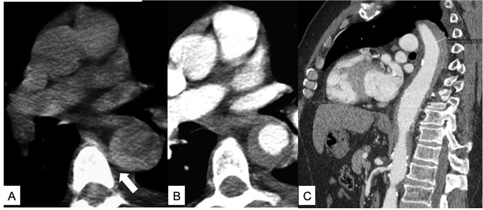
In IRAD, IMH accounted for 5.7% of acute aortic syndromes. Patients were older than those with classic dissection and more often had descending involvement. Mortality was 39.1% with ascending IMH and 8.3% with descending IMH, and 16% of patients progressed to classic dissection on serial imaging.
Type A IMH. Uncomplicated type A IMH is treated with prompt open repair (Class 1), and complicated IMH of either type (malperfusion, periaortic hematoma, tamponade, persistent pain, or rupture) with urgent repair (Class 1). The guideline cites mortality of up to 40% with nonoperative management of type A IMH. Repair within 24 hours carries low mortality, and a delay of 24 to 72 hours is acceptable in stable patients. In selected high-risk surgical patients without high-risk imaging features, medical management with close imaging may be considered (Class 2b). This approach is used more widely in East Asian centers, where outcomes of medical therapy for type A IMH have been more favorable.
Type B IMH. Uncomplicated type B IMH is treated medically (Class 1), with in-hospital mortality of 4% to 6% and 1-year mortality of 9% in the guideline's summary. When repair of zones 2 to 5 is needed, endovascular repair is reasonable with favorable anatomy (Class 2a) and open repair with unfavorable anatomy (Class 2a). Intervention may be reasonable in uncomplicated type B IMH with high-risk imaging features (Class 2b).
| IMH location | High-risk imaging features |
|---|---|
| Type A | Aortic diameter >45–50 mm; hematoma thickness ≥10 mm; ulcer-like projection in the ascending aorta or arch; pericardial effusion on admission |
| Type B | Aortic diameter >47–50 mm; hematoma thickness ≥13 mm; focal intimal disruption in the descending aorta in the acute phase; increasing pleural effusion |
| Either type | Progression on serial imaging, increasing aortic diameter, or increasing hematoma thickness |
High-risk imaging features of intramural hematoma, from the 2022 ACC/AHA guideline (Table 30).
An ulcer-like projection is a contrast-filled outpouching from the lumen into the hematoma that communicates with the lumen through a visible orifice. Ulcer-like projections are found in 20% to 50% of IMH and are associated with progression and late enlargement. An intramural blood pool is a focal contrast collection within the hematoma at the origin of an intercostal, bronchial, or lumbar artery, with a connection to the lumen of less than 2 mm or none visible. Intramural blood pools are generally considered benign, although their prognostic significance is uncertain. The distinction is made on thin-section CT and affects the intensity of follow-up.
Penetrating atherosclerotic ulcer
A PAU appears on CT as a contrast-filled outpouching through calcified plaque beyond the expected aortic contour, often with adjacent wall thickening or hematoma. It is distinguished from an ulcerated plaque that remains within the intima and from an ulcer-like projection in IMH, which arises in a wall without extensive plaque. Patients are usually older than 70 years with hypertension, smoking history, coronary disease, and abdominal aneurysm. Multiple ulcers are common.
| Clinical setting | Recommendation (2022 ACC/AHA) | Class |
|---|---|---|
| PAU with rupture | Urgent repair | 1 |
| Ascending aortic PAU with IMH | Urgent repair | 1 |
| Arch or descending PAU with IMH | Urgent repair is reasonable | 2a |
| Abdominal PAU with IMH | Urgent repair may be considered | 2b |
| Symptomatic isolated PAU (pain that corresponds to the lesion) | Repair | 1 |
| Asymptomatic PAU with high-risk features (diameter ≥13–20 mm, depth ≥10 mm, significant growth, associated saccular aneurysm, increasing pleural effusion) | Elective repair may be considered | 2b |
| PAU of the ascending aorta or proximal arch (zones 0–1) | Open surgical repair | 1 |
| PAU of the distal arch (zones 2–3), descending, or abdominal aorta | Open or endovascular repair, according to anatomy and comorbidity | 2a |
Management of penetrating atherosclerotic ulcer, from the 2022 ACC/AHA guideline (section 7.6). The 2024 ESC guideline recommends endovascular treatment of uncomplicated type B PAU with high-risk imaging features (Class IIa).
The natural history depends on symptoms and associated hematoma. Ascending PAU with IMH ruptures in 33% to 75% of reported cases. In type B PAU with IMH, mortality was 17.6% with conservative management and 7.1% with repair in the series cited by the guideline. Asymptomatic PAUs without high-risk features progressed in 3.6% at 5 years and 6.5% at 10 years. Endovascular repair is attractive because the lesions are focal, but stent grafts in these older patients with diffuse atheroma carry endoleak rates of 4% to 8% and new ulcer formation in about 5%.
Blunt traumatic thoracic aortic injury
Blunt traumatic thoracic aortic injury results from rapid deceleration, most often in motor vehicle collisions and falls. The tear is usually at the isthmus, where the mobile arch meets the descending aorta fixed by the ligamentum arteriosum. It is the second most common cause of death from trauma, and most patients with full-thickness injury die at the scene. CT angiography of the chest, performed as part of the trauma survey, establishes the diagnosis and grade.
| Grade | Injury | Management (2022 ACC/AHA) |
|---|---|---|
| 1 | Intimal tear | Nonoperative management with follow-up imaging (Class 1) |
| 2 | Intramural hematoma | Intervention reasonable with high-risk imaging features (Class 2a); nonoperative management with surveillance may be reasonable without them (Class 2b) |
| 3 | Pseudoaneurysm | Aortic intervention when comorbidities and other injuries are not prohibitive (Class 1) |
| 4 | Rupture | Aortic intervention when comorbidities and other injuries are not prohibitive (Class 1) |
Grading of blunt traumatic thoracic aortic injury (SVS 2011, after Azizzadeh 2009) and management. High-risk imaging features include posterior mediastinal hematoma >10 mm, a lesion-to-normal aortic diameter ratio >1.4, mediastinal mass effect, pseudocoarctation, large left hemothorax, and involvement of the ascending aorta, arch, or great vessels. When repair is indicated and anatomy is suitable, TEVAR is recommended over open repair (Class 1).
Management takes place at a trauma center with aortic expertise (Class 1). Anti-impulse therapy limits extension and rupture (Class 1) unless the patient is hypotensive or in hypovolemic shock from other injuries. In the series cited by the guideline, rupture occurred in about 12% of patients awaiting repair without medical therapy, and in small studies of beta-blocker protocols no ruptures occurred. Repair timing is coordinated with treatment of brain, abdominal, and pelvic injuries. The 2011 SVS guideline favored urgent repair, usually within 24 hours in stable patients. Later registry data showed higher adjusted mortality with repair in the first 24 hours, which supports delayed repair under medical therapy when the aorta is stable. In the SVS meta-analysis of 7,768 patients, mortality was 9% after endovascular repair, 19% after open repair, and 46% with nonoperative management, in largely unmatched cohorts. Unrepaired injuries are followed with CT at 1, 6, and 12 months (Class 2b), and repaired injuries with surveillance suited to the repair (Class 2a).
Acute aortic syndrome in pregnancy
Pregnancy raises cardiac output, blood volume, and heart rate, and hormonal changes alter the media. In IRAD, pregnancy-related dissection accounted for 0.3% of all dissections but 19% of dissections in women younger than 35 years. Of 29 women, 69% had an aortopathy or a family history, most often Marfan syndrome, and the aortopathy was recognized only after the dissection in 47%. Type A dissections occurred in dilated aortas (mean ascending diameter about 55 mm), whereas type B dissections often occurred in aortas of normal size. Of the 27 events with known timing, 11 occurred in the third trimester and 12 after delivery.
The 2022 ACC/AHA guideline gives Class 1 recommendations for pregnant patients with type A dissection: urgent aortic surgery with fetal monitoring in the first or second trimester, and urgent cesarean delivery followed immediately by aortic repair in the third trimester. Type B dissection in pregnancy is treated medically unless complications require endovascular or open repair (Class 1). Beta blockers are continued through pregnancy and postpartum in women with heritable aortopathy, MRI without gadolinium is preferred over CT for imaging the arch and distal aorta, and delivery is planned at a center able to perform emergency aortic surgery. Women with a history of chronic aortic dissection are delivered by cesarean section (Class 1).
Long-term medical therapy, activity, and imaging surveillance
Every survivor of an acute aortic syndrome has residual aortic disease: a dissected distal aorta after type A repair, a dissected or stented aorta after type B dissection, or a diseased wall after IMH or PAU. Late events are aneurysmal degeneration of the false lumen, rupture, new dissection, and malperfusion. Long-term beta blockade is recommended unless contraindicated (Class 1), with angiotensin receptor blockers or angiotensin-converting enzyme inhibitors added as needed. Calcium channel blockers are avoided in Marfan syndrome unless required for blood pressure. The guideline's general recommendation for patients with aortic disease is to treat a systolic pressure of 130 mm Hg or more or a diastolic pressure of 80 mm Hg or more, and many centers aim lower after dissection. Smoking cessation and statin therapy follow general atherosclerosis guidance.
Patients receive education and guidance to avoid intense isometric exercise, burst exertion, and collision sports (Class 1). Heavy lifting with a Valsalva maneuver can raise systolic pressure above 300 mm Hg. Mild to moderate aerobic activity, 30 to 60 minutes 3 to 4 days per week, is reasonable once pressure is controlled (Class 2a), and cardiac rehabilitation after aortic surgery is recommended (Class 1).
| Condition | 2022 ACC/AHA imaging schedule | Class |
|---|---|---|
| Dissection or IMH after repair with residual aortic disease | CT or MRI at 1, 6, and 12 months, then annually if stable | 1 |
| Dissection or IMH managed medically | CT or MRI at 1, 6, and 12 months, then annually if stable | 1 |
| PAU managed medically | CT at 1 month, then every 6 months for 2 years, then at intervals based on age and ulcer features | 2a |
| PAU after repair | Surveillance at intervals appropriate to the repair | 2a |
| Marfan syndrome after root replacement | MRI or CT annually, then every 2 years after 2 stable years | 1 |
| Chronic residual dissection with total diameter ≥5.5 cm | Elective repair | 1 |
Surveillance after acute aortic syndrome, from the 2022 ACC/AHA guideline (section 7.8). The 2024 ESC guideline adds an imaging study at 3 months after medically treated type B dissection or IMH and, after open repair, suggests CT every 2 years once 5 years pass without complications.
Serial studies are interpreted on the same modality, with diameters measured perpendicular to the centerline at the same anatomic levels, because measurement error can exceed true annual growth. MRI reduces cumulative radiation in young patients who need decades of imaging. After TEVAR, CT detects endoleak, stent-graft migration, distal new entry tears, and continued growth of the abdominal false lumen, which is not covered by the graft. Reintervention after TEVAR for dissection occurs in 27% to 39% of patients at midterm follow-up in the series cited by the guideline.
Genetic testing and screening of relatives
A family history is taken in every patient with dissection, covering thoracic aortic disease, peripheral and intracranial aneurysms, and unexplained sudden death in first- and second-degree relatives (Class 1). Genetic testing is recommended in patients with aortic dissection when there is a feature that suggests heritable thoracic aortic disease (Class 1): syndromic features of Marfan, Loeys-Dietz, or vascular Ehlers-Danlos syndrome; presentation before age 60 years; or a family history of thoracic aortic disease, peripheral or intracranial aneurysm, or unexplained early sudden death.
| Finding in the index patient | Screening of relatives (2022 ACC/AHA) |
|---|---|
| Pathogenic or likely pathogenic variant identified | Cascade genetic testing; aortic imaging of relatives who carry the variant, with TTE when the root and ascending aorta are well seen and CT or MRI otherwise (Class 1) |
| Familial disease without an identified variant | Screening aortic imaging of at-risk relatives (Class 1); when normal, imaging is repeated at intervals set by the relative's age and aortic size |
| Dissection with no known family history and no variant | Screening aortic imaging of first-degree relatives (Class 1) |
| Type A dissection | Root and ascending diameters recorded in the operative note for family counseling (Class 1) |
Family evaluation after aortic dissection, from the 2022 ACC/AHA guideline (section 6.1.2.1).
The result changes care for the patient and the family. A pathogenic variant sets gene-specific thresholds for prophylactic surgery in relatives, guides pregnancy counseling, and identifies extra-aortic disease that needs imaging (head-to-pelvis arterial imaging in Loeys-Dietz syndrome). Some genes cause dissection at diameters that would not prompt surgery in sporadic disease: type A dissection can occur below 4.5 cm with ACTA2 variants and below 5.0 cm with MYH11 variants. A negative genetic test does not remove the need for imaging of relatives, because many families with heritable disease have no identified variant.
Prognosis after acute aortic syndrome
Early prognosis is determined by type, malperfusion, and the timeliness of surgery. In the population-based Oxford study, 30-day fatality after hospital admission was 47.4% for type A and 13.3% for type B dissection, and 5-year survival among those who survived the acute phase was 85.7% and 83.3%. Late prognosis depends on false-lumen behavior, blood pressure control, heritable disease, and adherence to surveillance. Patients with a patent or partially thrombosed false lumen, a large residual entry tear, or a total diameter above 40 mm in the acute phase are at the highest risk of late aneurysm formation and reintervention.
Sources of error in acute aortic syndromes
- Treatment of an inferior STEMI with fibrinolysis or anticoagulation in a patient with abrupt chest or back pain, a pulse or pressure differential, or a new diastolic murmur, without bedside imaging of the aortic root.
- Exclusion of dissection by a normal chest radiograph, a normal TTE, or an ADD-RS of 0 alone; none of these rules out an acute aortic syndrome.
- Use of D-dimer to exclude dissection in a patient with an ADD-RS of 2 or 3, or more than a few days after onset, when the false lumen may already be thrombosed.
- A pulmonary embolism or coronary CT protocol for suspected dissection: no noncontrast phase for IMH, no ECG synchronization of the root, and incomplete coverage of the abdomen and iliac arteries.
- Vasodilator therapy before beta blockade, which raises heart rate and dP/dt, and titration of therapy to the lower reading from an arm with subclavian involvement.
- Delay of type A repair for further imaging, for completion of a stroke workup, or because of age alone.
- Unrestricted pericardiocentesis for tamponade from hemopericardium when the operating room is available.
- Classification of a type B dissection as uncomplicated without repeat imaging after recurrent pain, refractory hypertension, a rising lactate, or a falling urine output.
- Discharge of a patient with IMH or PAU without a surveillance schedule, and discharge of any patient with dissection without a family history, genetic evaluation when indicated, and screening of relatives.
Self-assessment
A 61-year-old man is on day 4 of medical therapy for acute type B aortic dissection. Pain resolved on the first day, he has no malperfusion, and his blood pressure is controlled on two drugs. Repeat CT angiography shows a maximal descending aortic diameter of 44 mm, a false lumen of 24 mm, and a primary entry tear of 12 mm on the lesser curvature, unchanged from admission. How is this dissection classified, and what does the 2022 ACC/AHA guideline say about endovascular repair?
Cardiovascular Medicine Board ReviewReferences
- Isselbacher EM, Preventza O, Hamilton Black J III, et al. 2022 ACC/AHA guideline for the diagnosis and management of aortic disease: a report of the American Heart Association/American College of Cardiology Joint Committee on Clinical Practice Guidelines. Circulation. 2022;146:e334–e482. doi:10.1161/CIR.0000000000001106
- Mazzolai L, Teixido-Tura G, Lanzi S, et al. 2024 ESC guidelines for the management of peripheral arterial and aortic diseases. Eur Heart J. 2024;45:3538–3700. doi:10.1093/eurheartj/ehae179
- Hiratzka LF, Bakris GL, Beckman JA, et al. 2010 ACCF/AHA/AATS/ACR/ASA/SCA/SCAI/SIR/STS/SVM guidelines for the diagnosis and management of patients with thoracic aortic disease. Circulation. 2010;121:e266–e369. doi:10.1161/CIR.0b013e3181d4739e
- Lombardi JV, Hughes GC, Appoo JJ, et al. Society for Vascular Surgery (SVS) and Society of Thoracic Surgeons (STS) reporting standards for type B aortic dissections. J Vasc Surg. 2020;71:723–747. doi:10.1016/j.jvs.2019.11.013
- Sievers HH, Rylski B, Czerny M, et al. Aortic dissection reconsidered: type, entry site, malperfusion classification adding clarity and enabling outcome prediction. Interact Cardiovasc Thorac Surg. 2020;30:451–457. doi:10.1093/icvts/ivz281
- Hagan PG, Nienaber CA, Isselbacher EM, et al. The International Registry of Acute Aortic Dissection (IRAD): new insights into an old disease. JAMA. 2000;283:897–903. doi:10.1001/jama.283.7.897
- Pape LA, Awais M, Woznicki EM, et al. Presentation, diagnosis, and outcomes of acute aortic dissection: 17-year trends from the International Registry of Acute Aortic Dissection. J Am Coll Cardiol. 2015;66:350–358. doi:10.1016/j.jacc.2015.05.029
- Evangelista A, Isselbacher EM, Bossone E, et al. Insights from the International Registry of Acute Aortic Dissection: a 20-year experience of collaborative clinical research. Circulation. 2018;137:1846–1860. doi:10.1161/CIRCULATIONAHA.117.031264
- Howard DP, Banerjee A, Fairhead JF, et al. Population-based study of incidence and outcome of acute aortic dissection and premorbid risk factor control: 10-year results from the Oxford Vascular Study. Circulation. 2013;127:2031–2037. doi:10.1161/CIRCULATIONAHA.112.000483
- Mussa FF, Horton JD, Moridzadeh R, et al. Acute aortic dissection and intramural hematoma: a systematic review. JAMA. 2016;316:754–763. doi:10.1001/jama.2016.10026
- Nienaber CA, Fattori R, Mehta RH, et al. Gender-related differences in acute aortic dissection. Circulation. 2004;109:3014–3021. doi:10.1161/01.CIR.0000130644.78677.2C
- Rogers AM, Hermann LK, Booher AM, et al. Sensitivity of the aortic dissection detection risk score, a novel guideline-based tool for identification of acute aortic dissection at initial presentation: results from the International Registry of Acute Aortic Dissection. Circulation. 2011;123:2213–2218. doi:10.1161/CIRCULATIONAHA.110.988568
- Nazerian P, Mueller C, Soeiro AM, et al. Diagnostic accuracy of the aortic dissection detection risk score plus D-dimer for acute aortic syndromes: the ADvISED prospective multicenter study. Circulation. 2018;137:250–258. doi:10.1161/CIRCULATIONAHA.117.029457
- Suzuki T, Distante A, Zizza A, et al. Diagnosis of acute aortic dissection by D-dimer: the International Registry of Acute Aortic Dissection Substudy on Biomarkers (IRAD-Bio) experience. Circulation. 2009;119:2702–2707. doi:10.1161/CIRCULATIONAHA.108.833004
- Evangelista A, Mukherjee D, Mehta RH, et al. Acute intramural hematoma of the aorta: a mystery in evolution. Circulation. 2005;111:1063–1070. doi:10.1161/01.CIR.0000156444.26393.80
- Tsai TT, Evangelista A, Nienaber CA, et al. Partial thrombosis of the false lumen in patients with acute type B aortic dissection. N Engl J Med. 2007;357:349–359. doi:10.1056/NEJMoa063232
- Nienaber CA, Rousseau H, Eggebrecht H, et al. Randomized comparison of strategies for type B aortic dissection: the INvestigation of STEnt Grafts in Aortic Dissection (INSTEAD) trial. Circulation. 2009;120:2519–2528. doi:10.1161/CIRCULATIONAHA.109.886408
- Nienaber CA, Kische S, Rousseau H, et al. Endovascular repair of type B aortic dissection: long-term results of the randomized investigation of stent grafts in aortic dissection trial. Circ Cardiovasc Interv. 2013;6:407–416. doi:10.1161/CIRCINTERVENTIONS.113.000463
- Brunkwall J, Kasprzak P, Verhoeven E, et al. Endovascular repair of acute uncomplicated aortic type B dissection promotes aortic remodelling: 1 year results of the ADSORB trial. Eur J Vasc Endovasc Surg. 2014;48:285–291. doi:10.1016/j.ejvs.2014.05.012
- Ali-Hasan-Al-Saegh S, Halloum N, Scali S, et al. A systematic review and meta-analysis of retrograde type A aortic dissection after thoracic endovascular aortic repair in patients with type B aortic dissection. Medicine (Baltimore). 2023;102:e32944. doi:10.1097/MD.0000000000032944
- Coselli JS, LeMaire SA, Köksoy C, et al. Cerebrospinal fluid drainage reduces paraplegia after thoracoabdominal aortic aneurysm repair: results of a randomized clinical trial. J Vasc Surg. 2002;35:631–639. doi:10.1067/mva.2002.122024
- Hayashi T, Tsukube T, Yamashita T, et al. Impact of controlled pericardial drainage on critical cardiac tamponade with acute type A aortic dissection. Circulation. 2012;126(11 Suppl 1):S97–S101. doi:10.1161/CIRCULATIONAHA.111.082685
- Azizzadeh A, Keyhani K, Miller CC III, et al. Blunt traumatic aortic injury: initial experience with endovascular repair. J Vasc Surg. 2009;49:1403–1408. doi:10.1016/j.jvs.2009.02.234
- Lee WA, Matsumura JS, Mitchell RS, et al. Endovascular repair of traumatic thoracic aortic injury: clinical practice guidelines of the Society for Vascular Surgery. J Vasc Surg. 2011;53:187–192. doi:10.1016/j.jvs.2010.08.027
- Braverman AC, Mittauer E, Harris KM, et al. Clinical features and outcomes of pregnancy-related acute aortic dissection. JAMA Cardiol. 2021;6:58–66. doi:10.1001/jamacardio.2020.4876
- Dunning DW, Kahn JK, Hawkins ET, O'Neill WW. Iatrogenic coronary artery dissections extending into and involving the aortic root. Catheter Cardiovasc Interv. 2000;51:387–393. doi:10.1002/1522-726X(200012)51:4<387::AID-CCD3>3.0.CO;2-B
- Pasternak B, Inghammar M, Svanström H. Fluoroquinolone use and risk of aortic aneurysm and dissection: nationwide cohort study. BMJ. 2018;360:k678. doi:10.1136/bmj.k678
- US Food and Drug Administration. FDA warns about increased risk of ruptures or tears in the aorta blood vessel with fluoroquinolone antibiotics in certain patients. Drug Safety Communication. December 20, 2018. fda.gov
- Greve D, Funke J, Khairi T, et al. Cocaine-related aortic dissection: what do we know? Braz J Cardiovasc Surg. 2020;35:764–769. doi:10.21470/1678-9741-2020-0333
- Sica G, Rea G, Lieto R, et al. CT diagnosis and destiny of acute aortic intramural hematoma. Front Radiol. 2025;5:1552644. doi:10.3389/fradi.2025.1552644
- AlAbsi MN, Sabri GAI, Alqudah KF, et al. Acute Stanford type A aortic dissection mimicking acalculous cholecystitis: a case report. Cureus. 2026;18:e106022. doi:10.7759/cureus.106022
- Nienaber CA, Kische S, Zeller T, et al. Provisional extension to induce complete attachment after stent-graft placement in type B aortic dissection: the PETTICOAT concept. J Endovasc Ther. 2006;13:738–746. doi:10.1583/06-1923.1
- Hofferberth SC, Nixon IK, Boston RC, McLachlan CS, Mossop PJ. Stent-assisted balloon-induced intimal disruption and relamination in aortic dissection repair: the STABILISE concept. J Thorac Cardiovasc Surg. 2014;147:1240–1245. doi:10.1016/j.jtcvs.2013.03.036
- Morello F, Bima P, Pivetta E, et al. Development and validation of a simplified probability assessment score integrated with age-adjusted D-dimer for diagnosis of acute aortic syndromes. J Am Heart Assoc. 2021;10:e018425. doi:10.1161/JAHA.120.018425