Library › Valvular heart disease › Prosthetic heart valves
Prosthetic heart valves
Mechanical, bioprosthetic, and transcatheter heart valves in adults, from the choice of prosthesis and antithrombotic therapy through imaging of prosthetic dysfunction, valve-in-valve treatment, pregnancy, and surveillance.
Every mechanical valve requires lifelong vitamin K antagonist (VKA) therapy. The target international normalized ratio (INR) is 2.5 for a bileaflet or current-generation tilting-disc aortic valve without risk factors and 3.0 for a mechanical mitral valve, an aortic valve with thromboembolic risk factors, or an older-generation prosthesis. Direct oral anticoagulants are contraindicated. A rising prosthetic gradient is judged against the patient's own postimplant baseline, and acquired obstruction, patient–prosthesis mismatch, high flow, and pressure recovery are separated by flow-independent Doppler indices and by direct imaging of leaflet motion.
Mechanical, bioprosthetic, homograft, autograft, and transcatheter valves
Replacement heart valves fall into two families. Mechanical valves are built from pyrolytic carbon and metal and are durable for the life of the patient, but their surfaces and hinge regions are thrombogenic. Biological valves are made from glutaraldehyde-fixed porcine aortic valve tissue or bovine pericardium, need no lifelong anticoagulation for the valve itself, and deteriorate over time. Transcatheter valves are biological valves mounted in a metal frame that is crimped onto a catheter and expanded inside the native valve, a failed surgical bioprosthesis, or an annuloplasty ring.
Mechanical valves. The caged-ball valve, the first prosthesis to achieve durable clinical success in 1960, uses a silicone ball within a metal cage. Blood flows around the ball, so gradients are high for the valve size, and thrombogenicity is the highest of any design. It has not been implanted for many years but is still encountered in long-term survivors. The single tilting-disc valve has one disc that pivots on struts and creates a larger major orifice and a smaller minor orifice, which produces an eccentric jet. The bileaflet valve, the standard contemporary design, has two semicircular pyrolytic carbon leaflets hinged inside a ring. When open, the leaflets lie nearly parallel to flow and form two larger lateral orifices and a narrow central slit; the localized high velocity in that central slit is the basis of pressure recovery. The On-X design adds a flared inlet and leaflet guards and is the only mechanical valve with a guideline-endorsed lower INR target in the aortic position.
Stented and stentless bioprostheses. A stented surgical bioprosthesis mounts three leaflets on a frame with three posts and a sewing ring. In most designs the leaflets are sewn inside the posts; in some pericardial designs they are mounted outside the posts, a feature that raises the risk of coronary obstruction when a transcatheter valve is later implanted inside them. Supra-annular designs seat the sewing ring above the annulus and allow a larger valve for a given annulus. Stentless porcine valves and roots have no frame, so the effective orifice is larger for a given size, at the cost of a more complex implantation. Rapid-deployment (sutureless) valves use a self-anchoring frame that needs few or no annular sutures, which shortens cross-clamp time in combined procedures.
Homografts and the pulmonary autograft. A cryopreserved human aortic homograft is used most often for aortic root replacement in destructive endocarditis with root abscess. In the Ross procedure, the patient's own pulmonary valve (the autograft) replaces the aortic valve and a pulmonary homograft reconstructs the right ventricular outflow tract. The operation avoids anticoagulation and provides near-native hemodynamics, but both the neoaortic valve and the pulmonary homograft can fail, and the operation is performed at Comprehensive Valve Centers.
Transcatheter heart valves. Balloon-expandable valves have a cobalt-chromium frame with intra-annular leaflets. Self-expanding valves have a nitinol frame, and the leaflets of the most widely used self-expanding designs sit above the annulus, which yields larger effective orifice areas in small annuli. Transcatheter valves are also implanted in the pulmonary position, in failed surgical bioprostheses in all four positions, and inside annuloplasty rings.
| Design | Flow and normal Doppler pattern | Normal (physiologic) regurgitation | Antithrombotic requirement | Durability |
|---|---|---|---|---|
| Caged ball | Flow around the ball; high gradients for size | Small closing volume, little or no true leak | Lifelong VKA; INR target 3.0 in the aortic position | Lifelong |
| Single tilting disc | Major and minor orifices; eccentric jet | Small jets where the disc meets the housing; some designs add a central jet through the disc | Lifelong VKA | Lifelong |
| Bileaflet | Two lateral orifices and a central slit; pressure recovery in small valves | Several narrow washing jets at the pivots and the closure line | Lifelong VKA | Lifelong |
| Stented bioprosthesis | Central flow; gradient depends strongly on size | Trivial central jet is common | Aspirin or a short course of VKA early; long-term therapy only for another indication | Structural deterioration rises after 10 years and earlier in the young |
| Stentless valve, homograft, autograft | Near-native orifice and gradients | Minor jets more common than with stented valves | As for stented bioprostheses | Regurgitation is the usual mode of failure |
| Transcatheter valve | Low gradients, lowest with supra-annular leaflets | Small central jets are rare; paravalvular jets are the usual leak | Single antiplatelet therapy unless another indication for anticoagulation exists | Data to 10 years in older patients |
Prosthetic valve families. Physiologic regurgitation from the 2024 American Society of Echocardiography (ASE) guideline; antithrombotic requirements from the 2020 American College of Cardiology/American Heart Association (ACC/AHA) guideline.
Long-term outcomes and complication rates after valve replacement
Valve replacement exchanges native valve disease for a new set of long-term risks: thromboembolism, valve thrombosis, bleeding from anticoagulation, structural deterioration, endocarditis, paravalvular regurgitation, hemolysis, and patient–prosthesis mismatch. Survival after replacement depends more on age, ventricular function, and comorbidity than on the type of valve.
The thrombotic risk of a mechanical valve is substantial without treatment. In a pooled analysis of 13,088 patients followed for 53,647 patient-years, major embolism occurred at 4 per 100 patient-years without antithrombotic therapy, and major bleeding occurred at 1.4 per 100 patient-years with coumarin therapy. With a bileaflet mechanical aortic valve and vitamin K antagonist therapy, the 2020 ACC/AHA guideline cites a thromboembolic rate of about 0.53% per patient-year. Mechanical valve thrombosis occurs at 0.1% to 5.7% per year, with higher rates in the first 3 months after implantation, with some valve types, and in the mitral and tricuspid positions.
Bioprosthetic valves in sinus rhythm carry a thromboembolic risk of about 0.7% per year, higher with mitral than with aortic prostheses (2.4% versus 1.9% per patient-year in one series). About 30% of patients with a surgical aortic bioprosthesis show valve dysfunction within 10 years of implantation, defined as a rise in mean gradient of 10 mm Hg or more or worsening transprosthetic regurgitation. Clinically important structural deterioration increases markedly after 10 years. Reoperation remains safe but carries more risk than the first operation: in Society of Thoracic Surgeons data on elective isolated reoperative aortic valve replacement, operative mortality was 4.6% compared with 2.2% for a first replacement.
Choosing a mechanical or bioprosthetic valve
Valve choice weighs the lifelong anticoagulation and bleeding risk of a mechanical valve against the finite durability of a bioprosthesis and the risk of reintervention. The 2020 ACC/AHA guideline makes the choice a shared decision (Class 1) and anchors it to age because durability depends steeply on age at implantation. The predicted 15-year risk of reoperation for structural deterioration of a surgical aortic bioprosthesis is about 22% at age 50, 30% at age 40, and 50% at age 20. After age 65, the risk of structural deterioration at 15 to 20 years is only about 10%. In one observational series of mitral bioprostheses, expected durability was 11.4 years in patients younger than 60, 16.6 years at 60 to 70, and 19.4 years in those older than 70.
| Patient group | Recommendation | COR (LOE) |
|---|---|---|
| Any patient requiring valve replacement | Choice based on shared decision-making about anticoagulation and reintervention | 1 (C-LD) |
| Any age, anticoagulation contraindicated, unmanageable, or not desired | Bioprosthetic valve recommended | 1 (C-EO) |
| Aortic, age <50 years, no contraindication to anticoagulation | Mechanical valve reasonable | 2a (B-NR) |
| Aortic, age 50 to 65 years, no contraindication | Individualized choice after shared decision-making | 2a (B-NR) |
| Aortic, age >65 years | Bioprosthesis reasonable | 2a (B-NR) |
| Mitral, age <65 years, repair not possible, no contraindication | Mechanical valve reasonable | 2a (B-NR) |
| Mitral, age ≥65 years, repair not possible | Bioprosthesis reasonable | 2a (B-NR) |
| Aortic, age <50 years, prefers a biological valve, suitable anatomy | Ross procedure may be considered at a Comprehensive Valve Center | 2b (B-NR) |
Prosthesis selection in the 2020 ACC/AHA valve guideline. COR, class of recommendation; LOE, level of evidence.
The evidence in the middle age band is observational. In propensity-matched New York data on patients aged 50 to 69 years having isolated aortic valve replacement, 15-year survival was similar with bioprosthetic and mechanical valves (60.6% versus 62.1%), as was stroke. Reoperation was higher with bioprostheses (12.1% versus 6.9%), and major bleeding was higher with mechanical valves (13.0% versus 6.6%). In California data, a mechanical aortic valve was associated with lower 15-year mortality in patients aged 45 to 54 years but not at 55 to 64 years. A mechanical mitral valve was associated with lower mortality up to age 70, at the cost of more bleeding and, in some age groups, more stroke.
| Factors favoring a mechanical valve | Factors favoring a bioprosthesis |
|---|---|
| Age <50 years, with a high risk of bioprosthetic deterioration | Age >65 years, with a low risk of deterioration and a higher bleeding risk |
| Preference to avoid reintervention | Preference to avoid the risk and inconvenience of anticoagulation |
| Low risk of long-term anticoagulation; reliable INR monitoring, including home monitoring | High bleeding risk, limited access to care, or inability to regulate a VKA |
| Another indication for long-term anticoagulation, such as atrial fibrillation | Access to transcatheter valve-in-valve replacement |
| High-risk reintervention, such as porcelain aorta or prior chest radiation | Pregnancy planned (anticoagulation in pregnancy is high risk) |
| Small aortic root that may preclude a later valve-in-valve procedure | A transcatheter valve, with a larger effective orifice for small sizes, when mismatch is a concern |
Factors in the choice of prosthesis, after Table 22 of the 2020 ACC/AHA guideline.
Planning for later valve-in-valve procedures. Because a failed surgical bioprosthesis can be treated with a transcatheter valve-in-valve procedure, the first valve is increasingly chosen with the second and third procedures in mind. A larger first valve, a design with a fracturable frame, and anatomy that leaves room between the future transcatheter frame and the coronary ostia all make a later valve-in-valve procedure safer and its residual gradient lower. A small surgical valve in a small root combines patient–prosthesis mismatch at the first operation with a high residual gradient after valve-in-valve treatment. A transcatheter valve placed first raises different questions about later access to the coronary arteries and about the feasibility of a second transcatheter valve.
The Ross procedure. The pulmonary autograft offers a living valve without anticoagulation, but it converts single-valve disease into potential two-valve disease. Failure is most often due to regurgitation of the neoaortic valve in the second decade, and at least half of pulmonary homografts require reintervention within 10 to 20 years. The 2020 ACC/AHA guideline reserves it for younger patients with suitable anatomy at experienced centers.
Valve choice in women who may become pregnant. A mechanical valve exposes a future pregnancy to the risks of valve thrombosis and of warfarin embryopathy, and a bioprosthesis exposes the woman to reoperation. Pregnancy plans are therefore part of the shared decision before the first valve operation.
INR targets and vitamin K antagonist therapy for mechanical valves
All mechanical valves require lifelong vitamin K antagonist therapy (Class 1). The target INR depends on the valve design, its position, and patient risk factors. The 2020 ACC/AHA guideline specifies a single target for each patient, with an acceptable range of 0.5 INR units on each side. A single target reduces the time spent near the boundaries of a range, and fluctuation of the INR is itself associated with complications.
| Valve and patient | Therapy | COR (LOE) |
|---|---|---|
| Any mechanical valve | Vitamin K antagonist (warfarin) | 1 (A) |
| Bileaflet or current-generation single tilting-disc aortic valve, no thromboembolic risk factors | INR 2.5 (range 2.0–3.0) | 1 (B-NR) |
| Mechanical aortic valve with risk factors (atrial fibrillation, prior thromboembolism, left ventricular (LV) dysfunction, hypercoagulable state) or an older-generation valve such as a caged ball | INR 3.0 (range 2.5–3.5) | 1 (B-NR) |
| Mechanical mitral valve | INR 3.0 (range 2.5–3.5) | 1 (B-NR) |
| On-X aortic valve, no thromboembolic risk factors, ≥3 months after surgery | INR 1.5–2.0 with aspirin 75–100 mg daily | 2b (B-R) |
| Mechanical valve, another indication for antiplatelet therapy, low bleeding risk | Aspirin 75–100 mg daily may be added | 2b (B-R) |
| Mechanical aortic valve, stroke or embolism with a therapeutic INR | INR goal raised to 3.0, or aspirin 75–100 mg added, after assessment of bleeding risk | 2a (C-EO) |
| Mechanical mitral valve, stroke or embolism with a therapeutic INR | INR goal raised to 4.0, or aspirin 75–100 mg added, after assessment of bleeding risk | 2a (C-EO) |
| Dabigatran | Contraindicated | 3: Harm (B-R) |
| Anti-Xa direct oral anticoagulants | Not recommended | 3: Harm (C-EO) |
Antithrombotic therapy for mechanical valves in the 2020 ACC/AHA guideline.
The aortic position is the least thrombogenic because flow through it is fast; the mitral position has lower velocities and more stasis. With a current mechanical aortic valve, a target of 2.0 to 3.0 produced embolic rates similar to a target of 3.0 to 4.5 and less bleeding. In a mechanical mitral valve, a lower target range was associated with lower survival in a German registry. Aspirin is no longer added routinely: the earlier practice derived from trials of older prostheses, and the 2020 guideline limits aspirin to patients who have a separate indication for antiplatelet therapy and a low bleeding risk.
Why direct oral anticoagulants fail. A mechanical valve activates coagulation through contact activation at its artificial surfaces and through high shear at the hinges and sewing ring. Warfarin lowers several vitamin K-dependent factors at once and suppresses this thrombin generation at moderate intensity; a direct inhibitor of a single factor does not achieve the same protection at tolerable doses. In RE-ALIGN, dabigatran was compared with warfarin in patients who had aortic or mitral mechanical valve replacement within the previous 7 days or mitral replacement at least 3 months earlier. The trial was stopped early after 252 patients were randomized, with stroke in 5% versus 0% and major bleeding in 4% versus 2%. In PROACT Xa, apixaban was compared with warfarin (INR 2.0 to 3.0) in 863 patients at least 3 months after On-X aortic valve replacement. The trial was stopped early for excess valve thrombosis or valve-related thromboembolism with apixaban (about 4.2% versus 1.3% per patient-year). The On-X aortic valve was the prosthesis judged most likely to tolerate a less intense regimen.
The On-X aortic valve. In PROACT, 375 patients with an On-X aortic valve and at least one thromboembolic risk factor received standard warfarin for 3 months. They were then randomized to an INR of 1.5 to 2.0 or 2.0 to 3.0, all with aspirin 81 mg. The lower target reduced major bleeding (1.48% versus 3.26% per patient-year) and minor bleeding (1.32% versus 3.41% per patient-year) with similar rates of stroke, transient ischemic attack, neurologic events, and death. A separate PROACT comparison showed harm when dual antiplatelet therapy replaced low-intensity warfarin plus aspirin. The lower target applies only to the On-X valve, only in the aortic position, and only after the first 3 months.
INR monitoring and warfarin management. Time in the therapeutic range and the stability of the INR predict events. Home INR testing and patient self-management are options for trained, reliable patients, and the 2025 European Society of Cardiology/European Association for Cardio-Thoracic Surgery (ESC/EACTS) guideline recommends INR self-monitoring or self-management in selected trained patients. Warfarin dose requirements vary several-fold with common variants in CYP2C9, which metabolizes S-warfarin, and VKORC1, which encodes the drug target, and with age, body size, diet, and liver function. Drugs that inhibit warfarin metabolism (amiodarone, azole antifungals, metronidazole, and trimethoprim-sulfamethoxazole) raise the INR, enzyme inducers such as rifampin and carbamazepine lower it, and changes in dietary vitamin K intake shift it. An intercurrent illness with poor oral intake is a frequent setting for both supratherapeutic and subtherapeutic values. Each new drug, illness, or dietary change in a patient with a mechanical valve is followed by an early INR check.
Excessive anticoagulation. For a supratherapeutic INR above 5.0 without bleeding, the value of oral vitamin K in addition to withholding warfarin is uncertain (Class 2b), because rapid reversal to a subtherapeutic level exposes the valve to thrombosis. Reversal of uncontrollable bleeding is described in the section on procedures.
Antithrombotic therapy after bioprosthetic valves and TAVR
The risk of ischemic stroke is highest in the first 90 to 180 days after a surgical bioprosthesis, before the sewing ring and leaflets are endothelialized, and afterward the valve itself does not require anticoagulation. Antithrombotic therapy after transcatheter aortic valve replacement (TAVR) has been shaped by randomized trials that moved practice from dual antiplatelet therapy to a single antiplatelet agent and showed harm from routine anticoagulation in patients without another indication.
| Setting | Recommendation | COR (LOE) |
|---|---|---|
| TAVR, no other indication for oral anticoagulation | Aspirin 75–100 mg daily is reasonable | 2a (B-R) |
| Surgical aortic or mitral bioprosthesis, no other indication | Aspirin 75–100 mg daily is reasonable | 2a (B-NR) |
| Surgical aortic or mitral bioprosthesis, low bleeding risk | VKA to INR 2.5 for at least 3 and up to 6 months is reasonable | 2a (B-NR) |
| TAVR, low bleeding risk | Aspirin plus clopidogrel 75 mg for 3 to 6 months may be reasonable | 2b (B-R) |
| TAVR, low bleeding risk | VKA to INR 2.5 for at least 3 months may be reasonable | 2b (B-NR) |
| TAVR, no other indication for oral anticoagulation | Rivaroxaban 10 mg plus aspirin is contraindicated | 3: Harm (B-R) |
| Atrial fibrillation with a bioprosthesis implanted >3 months earlier | A non-vitamin K oral anticoagulant is an effective alternative to a VKA | 1 (A) |
| New atrial fibrillation ≤3 months after a surgical or transcatheter bioprosthesis | VKA anticoagulation is reasonable | 2a (B-NR) |
| Stroke or embolism on antiplatelet therapy with a bioprosthesis | VKA in place of antiplatelet therapy may be considered | 2b (C-EO) |
Antithrombotic therapy for bioprosthetic valves in the 2020 ACC/AHA guideline (Sections 2.4.3, 11.2, and 11.5).
| Trial | Population and comparison | Principal result |
|---|---|---|
| POPular TAVI cohort A (2020) | 665 TAVR patients without an anticoagulation indication; aspirin alone versus aspirin plus clopidogrel | Bleeding at 1 year 15.1% versus 26.6%; thromboembolic events 9.7% versus 9.9% |
| POPular TAVI cohort B (2020) | 313 TAVR patients receiving oral anticoagulation; anticoagulation alone versus anticoagulation plus 3 months of clopidogrel | Bleeding at 1 year 21.7% versus 34.6%; no ischemic benefit from clopidogrel |
| GALILEO (2020) | TAVR without an anticoagulation indication; rivaroxaban 10 mg plus aspirin versus aspirin plus clopidogrel | Stopped early; more death or thromboembolism and more bleeding with rivaroxaban |
| ATLANTIS (2022) | TAVR; apixaban versus standard care (VKA with an anticoagulation indication, antiplatelet therapy without one) | Apixaban not superior; less subclinical leaflet thrombosis without clinical benefit |
| ENVISAGE-TAVI AF (2021) | 1,426 TAVR patients with atrial fibrillation; edoxaban versus VKA | Net adverse clinical events noninferior (17.3 versus 16.5 per 100 person-years); major bleeding higher with edoxaban (9.7 versus 7.0 per 100 person-years), mostly gastrointestinal |
| RIVER (2020) | 1,005 patients with atrial fibrillation or flutter and a bioprosthetic mitral valve; rivaroxaban 20 mg versus warfarin | Noninferior for death, major cardiovascular events, or major bleeding at 12 months; major bleeding 1.4% versus 2.6% |
Randomized trials of antithrombotic therapy after TAVR and with bioprosthetic valves. Rates are experimental arm versus comparator.
After TAVR, a single antiplatelet agent is the default in patients without another indication for anticoagulation. Patients who need anticoagulation, most often for atrial fibrillation, receive it without routine added clopidogrel unless a recent coronary event or stent requires it. Anticoagulation given only to prevent leaflet thrombosis is not indicated: GALILEO showed harm, and in ATLANTIS apixaban reduced subclinical leaflet thrombosis in patients without an anticoagulation indication without improving clinical outcomes. Among anticoagulants for atrial fibrillation after TAVR, edoxaban matched a VKA for net clinical events but caused more gastrointestinal bleeding. The bleeding risk of an anticoagulant therefore weighs heavily in this older population.
Two exceptions to the use of direct oral anticoagulants with valve disease remain: mechanical valves and rheumatic mitral stenosis. In INVICTUS, among 4,565 patients with rheumatic heart disease and atrial fibrillation, rivaroxaban caused more events of the composite of stroke, systemic embolism, myocardial infarction, or vascular or unknown death than dose-adjusted VKA therapy, and mortality was higher. A patient with a bioprosthetic valve implanted for rheumatic disease who has residual rheumatic mitral stenosis and atrial fibrillation is therefore treated with a VKA.
Bridging, minor procedures, and reversal of anticoagulation
Management of warfarin around a procedure balances the thrombotic risk of the valve against the bleeding risk of the procedure. The first question is whether warfarin needs to be interrupted at all. Dental extraction, cleaning and restoration, cataract surgery, and superficial skin surgery are performed with a therapeutic INR (Class 1), because bleeding is easily controlled and interruption carries thrombotic risk without benefit.
| Valve and procedure | Strategy | COR (LOE) |
|---|---|---|
| Any mechanical valve, minor procedure with easily controlled bleeding | VKA continued with a therapeutic INR | 1 (C-EO) |
| Bileaflet mechanical aortic valve, no other thromboembolic risk factor, invasive procedure | Temporary VKA interruption without bridging | 1 (C-LD) |
| Mechanical aortic valve with any risk factor, older-generation aortic valve, or mechanical mitral valve | Bridging with UFH or LMWH while the INR is subtherapeutic is reasonable, individualized for bleeding risk | 2a (C-LD) |
| Bioprosthetic valve or annuloplasty ring, anticoagulated for atrial fibrillation | Bridging decided by CHA₂DS₂-VASc score and bleeding risk | 2a (C-LD) |
| Mechanical valve, emergency surgery or invasive procedure | Four-factor prothrombin complex concentrate is reasonable | 2a (C-LD) |
Periprocedural management in the 2020 ACC/AHA guideline. UFH, unfractionated heparin; LMWH, low-molecular-weight heparin.
When interruption is necessary, warfarin is stopped 2 to 4 days before the procedure in a low-risk patient, or 3 to 4 days before in a patient who will be bridged. It is restarted as soon as bleeding risk allows, typically about 24 hours after surgery. Bridging with intravenous UFH or subcutaneous LMWH begins when the INR falls below the therapeutic threshold, usually 36 to 48 hours before surgery. It stops 4 to 6 hours (intravenous UFH) or about 12 hours (subcutaneous LMWH) before the procedure. Postoperative therapeutic heparin is delayed in patients at high bleeding risk.
The evidence for bridging in valve patients is observational. Trials in atrial fibrillation without a mechanical valve showed more bleeding without less thromboembolism, and those results do not transfer directly to mechanical valves. In PERIOP2, 1,471 patients with atrial fibrillation or a mechanical valve whose warfarin was interrupted received preoperative dalteparin and were randomized to postoperative dalteparin or placebo. Major thromboembolism within 90 days did not differ (1.0% with dalteparin versus 1.2% with placebo). Patients with more than one mechanical valve, a caged-ball valve, or prior stroke or transient ischemic attack were excluded, so the highest-risk valve patients remain outside the trial evidence.
Reversal for bleeding or emergency surgery. Four-factor prothrombin complex concentrate replaces factors II, VII, IX, and X, acts within 5 to 15 minutes, and lasts 12 to 24 hours. Fresh frozen plasma acts more slowly (1 to 4 hours) and for a shorter time (under 6 hours). For uncontrollable bleeding in a patient with a mechanical valve, prothrombin complex concentrate is reasonable (Class 2a), and intravenous vitamin K is added when warfarin will not be resumed for 7 days (Class 2a), at a dose of 10 mg for life-threatening bleeding. In patients with a bioprosthetic valve or annuloplasty ring receiving a direct oral anticoagulant, the 2020 ACC/AHA guideline rated idarucizumab (for dabigatran) and andexanet alfa (for the factor Xa inhibitors) as reasonable (Class 2a). Andexanet alfa has since been withdrawn from the US market after the US Food and Drug Administration concluded that its risks outweigh its benefits. In the United States, bleeding on apixaban, rivaroxaban, or edoxaban is treated with four-factor prothrombin complex concentrate. Andexanet remains available in some other countries.
Examination, chest radiograph, fluoroscopy, and MRI safety
Auscultation. A mechanical valve produces crisp, high-pitched closing clicks, and caged-ball and some tilting-disc valves also produce prominent opening clicks. A soft early systolic ejection murmur across a normally functioning aortic prosthesis reflects the small orifice and is expected. Muffled or absent closing clicks indicate impaired occluder motion, and a new or louder stenotic murmur raises suspicion of obstruction. A diastolic murmur at the left sternal border with an aortic prosthesis, or a holosystolic apical murmur with a mitral prosthesis, indicates regurgitation until shown otherwise. A bioprosthesis has native-sounding closure and often a soft ejection murmur. A change in valve sounds, a new murmur, hemolysis, heart failure, or an embolic event in a patient with a prosthetic valve leads to echocardiography and review of recent INR values.
Chest radiograph. The radiograph locates the prosthesis and suggests its design. On the frontal view, aortic prostheses lie above and mitral prostheses below a line drawn from the right cardiophrenic angle toward the left hilum. On the lateral view, aortic prostheses lie above and anterior, and mitral prostheses below and posterior, to a line from the carina to the anterior costophrenic angle. The direction of flow through the ring also differs: toward the ascending aorta for an aortic valve and toward the apex for a mitral valve. A caged-ball valve shows its cage, a tilting-disc valve a ring with a disc seen edge-on, and a bileaflet valve a ring with two leaflets seen edge-on when the projection is tangential. Stented bioprostheses show radiopaque ring and stent-post markers in many models, stentless valves may be invisible, and transcatheter valves show the mesh of their frames.
Cine fluoroscopy and CT of leaflet motion. Fluoroscopy is the fastest method to assess mechanical leaflet excursion. The projection is adjusted until the leaflets are seen in profile, and the opening and closing angles are measured and compared with the manufacturer's values; intermittent obstruction may require prolonged observation. Gated multiphase CT measures the same angles and also shows the tissue that restricts them. The 2024 ASE guideline lists normal opening angles of about 73° to 90° for bileaflet valves and 60° to 80° for single-leaflet valves. Rocking of the sewing ring of more than 15° in an aortic prosthesis implies dehiscence with significant paravalvular regurgitation. In mechanical valve stenosis, fluoroscopy or cine CT to assess leaflet motion is recommended (Class 1).
MRI safety. The 2024 ASE guideline states that prosthetic heart valves can be safely imaged at 1.5 T and 3 T, the field strengths in common clinical use; the device-specific conditions in the manufacturer's labeling apply. Metal produces local artifact that ranges from minor to severe by valve type, and cardiac magnetic resonance does not reliably show mechanical occluder motion. It is valuable for quantifying regurgitant volume and ventricular volumes when echocardiographic windows are limited, especially for pulmonary prostheses.
Normal prosthetic hemodynamics and the baseline echocardiogram
A prosthesis is intrinsically smaller than the native valve it replaces, so it produces a gradient even when it functions normally. The expected gradient differs with design, labeled size, position, cardiac output, and heart rate, and the same mean gradient can be normal for one prosthesis and abnormal for another. Native valve stenosis thresholds do not apply to prostheses. The 2024 ASE guideline therefore relies on three references: published normal values for each model and size, flow-independent indices, and, above all, the patient's own early postimplant study.
Pressure recovery. In a bileaflet valve, flow through the narrow central slit reaches a localized high velocity, and pressure partly recovers downstream. Continuous-wave Doppler records the highest velocity, so the Doppler gradient exceeds the net pressure loss measured by catheter. The effect is largest with small bileaflet valves and a small ascending aorta and is one reason a high gradient in a small mechanical valve does not by itself indicate obstruction.
Physiologic regurgitation. Mechanical valves have two kinds of normal regurgitation: the closing volume displaced as the occluder closes, and leakage through designed gaps in the closed valve. These washing jets clear sites of stasis within the sewing ring. In bileaflet valves, usually two jets arise at each pivot point and one or more along the closure line. Tilting-disc valves produce jets at the disc margin, and some designs a central jet through the disc. Washing jets are narrow at their origin, homogeneous in color with aliasing confined to the base, and arise within the sewing ring. The regurgitant fraction is usually no more than 10% to 15%, although a jet can reach 5 cm in length. A trivial central jet is common in normal biological valves, more often with stentless valves and homografts. Transcatheter valves rarely show central regurgitation; their leaks are usually paravalvular.
The baseline study. An initial postprocedural transthoracic echocardiogram is recommended after every surgical or transcatheter valve implantation and after valve repair (Class 1). The ASE defines the baseline as a study performed under stable hemodynamic conditions, usually 1 to 3 months after implantation, once anemia, tachycardia, and postoperative high-output states have resolved. The report records the prosthesis type and labeled size, the date and type of procedure, heart rate, and blood pressure. It lists peak velocity, mean gradient, Doppler velocity index (DVI), effective orifice area (EOA), acceleration time, and the outflow diameter used. It also records the location and severity of any regurgitation, ventricular size and function, and pulmonary pressure. The same outflow diameter is reused on later studies so that serial change reflects the velocities.
Doppler assessment of prosthetic aortic valves
The aortic prosthetic jet is recorded with continuous-wave Doppler from apical, right parasternal, suprasternal, and subcostal windows, with a small nonimaging transducer where possible, and the highest dense velocity is reported. The outflow tract velocity is recorded with pulsed-wave Doppler just proximal to the zone of flow acceleration. With a transcatheter valve, flow accelerates within the stent below the leaflets, so the outflow diameter and velocity are measured immediately proximal to the stent; a sample placed inside the stent overestimates the EOA.
The 2024 ASE guideline grades prosthetic aortic stenosis with flow-dependent parameters (peak velocity and mean gradient) and flow-independent parameters (DVI and EOA), supported by jet contour and acceleration time. Significant stenosis requires at least one abnormal flow-dependent and one abnormal flow-independent parameter. A surgical valve is graded by absolute values. A transcatheter valve, whose gradients are low and vary less among sizes, is graded by change from its baseline study.
| Parameter | Normal | Possible stenosis | Suggests significant stenosis |
|---|---|---|---|
| Jet contour | Triangular, early peaking | Triangular to intermediate | Rounded, symmetric |
| Acceleration time | <80 ms | 80–100 ms | >100 ms |
| Acceleration time ÷ LV ejection time | <0.32 | 0.32–0.37 | >0.37 |
| Peak velocity* | <3 m/s | 3–4 m/s | ≥4 m/s |
| Mean gradient, surgical valve | <20 mm Hg | 20–34 mm Hg | ≥35 mm Hg |
| DVI, surgical valve | >0.35 | 0.25–0.35 | <0.25 |
| EOA, surgical valve | Reference EOA ± 1 standard deviation (SD) | 1 SD below reference | 2 SD below reference |
| Mean gradient, transcatheter valve | Change <10 mm Hg from baseline | Rise of 10–19 mm Hg | Rise ≥20 mm Hg |
| DVI, transcatheter valve | Change <0.1 or <20% | Fall of 0.1–0.19 or 20%–39% | Fall ≥0.2 or ≥40% |
| EOA, transcatheter valve | Change <0.3 cm² or <25% | Fall of 0.3–0.59 cm² or 25%–49% | Fall ≥0.6 cm² or ≥50% |
Doppler criteria for stenosis of a prosthetic aortic valve, from the 2024 ASE guideline (Table 5). *Peak velocity criteria assume a normal stroke volume (50–90 mL) and flow rate (200–300 mL/s). Acceleration time is affected by LV function and heart rate. DVI = left ventricular outflow tract (LVOT) velocity-time integral (VTI) ÷ prosthetic VTI.
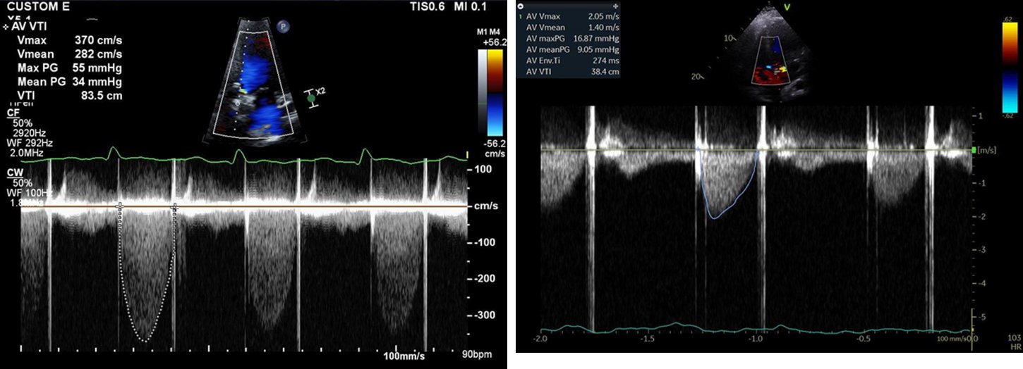
The DVI compares the velocity in the outflow tract with the velocity through the prosthesis. It removes the outflow diameter from the calculation and changes little with flow, because a high-output state raises both velocities in proportion. Its weakness is the pulsed-wave sample: a sample placed too close to the prosthesis, within the flow acceleration, raises the outflow VTI and therefore the DVI and EOA and makes an obstructed valve look normal. A sample placed too far toward the apex has the opposite effect.
A high prosthetic velocity has four common explanations, and the jet contour, acceleration time, DVI, and the course since implantation separate them.
| Cause | Course relative to baseline | Doppler pattern | Leaflets or occluders |
|---|---|---|---|
| Acquired obstruction (thrombus, pannus, structural deterioration) | Rise from a lower baseline | Low DVI, long acceleration time, rounded jet | Restricted or thickened |
| Patient–prosthesis mismatch | High from implantation and stable | Small indexed EOA; normal DVI, acceleration time, and contour | Normal motion |
| High flow (anemia, sepsis, fever, thyrotoxicosis, pregnancy, aortic regurgitation) | Rises with the flow state and falls when it resolves | High outflow velocity; normal DVI and acceleration time | Normal motion |
| Pressure recovery (small bileaflet valve) | Stable | Localized high velocity; Doppler gradient exceeds the catheter gradient | Normal motion |
Differential diagnosis of a high prosthetic aortic gradient, after the 2024 ASE guideline and the 2020 ACC/AHA guideline.
Prosthetic obstruction is usually defined as an increase in mean gradient of more than 50%, or more than 10 mm Hg across an aortic prosthesis, compared with baseline after exclusion of a high-output state. When obstruction is suspected, transthoracic echocardiography is followed by transesophageal echocardiography (TEE), fluoroscopy, or CT to define leaflet motion and the cause, even when the transthoracic study does not show dysfunction (Class 1). Transthoracic imaging shows the posterior aspect of an aortic prosthesis poorly and TEE the anterior aspect, so the two are complementary.
Prosthetic valve Doppler and mismatch calculator
The calculator applies the continuity equation to an aortic or mitral prosthesis. For an aortic prosthesis, EOA = LVOT area × LVOT VTI ÷ prosthetic VTI and DVI = LVOT VTI ÷ prosthetic VTI. For a mitral prosthesis, EOA = LVOT stroke volume ÷ prosthetic mitral VTI and the VTI ratio = prosthetic mitral VTI ÷ LVOT VTI, which rises with obstruction and with mitral regurgitation. The indexed EOA is graded for patient–prosthesis mismatch with the body mass index-adjusted cutoffs of the 2024 ASE guideline.
LVOT area = π × (diameter ÷ 2)². Aortic thresholds for a surgical valve: DVI >0.35 normal, 0.25–0.35 possible stenosis, <0.25 significant; a transcatheter valve is graded by the change from its baseline study; acceleration time ÷ ejection time >0.37 significant. Mitral thresholds: VTI ratio <2.2 normal, >2.5 significant; EOA <1 cm² significant. Mismatch is graded only when valve structure and motion are normal and the measured EOA is within 1 SD of the reference value for the model and size. The continuity method for a mitral prosthesis is invalid with significant aortic or mitral regurgitation.
Doppler assessment of prosthetic mitral, tricuspid, and pulmonary valves
Mitral prosthetic gradients depend strongly on flow and heart rate, because a short diastole requires a higher gradient to fill the ventricle. The mean gradient is therefore always reported with the heart rate, and in atrial fibrillation several cycles are averaged. The native formula of 220 divided by the pressure half-time does not apply to a prosthesis; pressure half-time is a supporting index only.
| Parameter | Normal | Possible stenosis | Suggests significant stenosis |
|---|---|---|---|
| Peak early velocity | <1.9 m/s | 1.9–2.5 m/s | ≥2.5 m/s |
| Mean gradient | ≤5 mm Hg | 6–10 mm Hg | >10 mm Hg |
| VTI ratio (prosthetic mitral ÷ LVOT) | <2.2 | 2.2–2.5 | >2.5 |
| EOA | ≥2.0 cm² | 1–2 cm² | <1 cm² |
| Pressure half-time | <130 ms | 130–200 ms | >200 ms |
Doppler findings suggestive of prosthetic mitral stenosis, from the 2024 ASE guideline (Table 11). Peak velocity, mean gradient, and VTI ratio are also abnormal with significant prosthetic mitral regurgitation; slightly higher values may be seen in some bioprostheses. Values in the possible range prompt consideration of high flow, tachycardia, and mismatch.
Hidden mitral regurgitation. From apical transthoracic windows, a mitral prosthesis shadows the left atrium, so a regurgitant jet can be invisible. Increased forward flow across the valve with a normal pressure half-time is the signature of regurgitation, while a prolonged half-time supports obstruction. In mechanical mitral valves with a normal pressure half-time, a peak mitral velocity of 1.9 m/s or more (sensitivity 90%, specificity 89%) and a VTI ratio of 2.5 or more (sensitivity 89%, specificity 91%) on transthoracic imaging suggest significant prosthetic regurgitation. Flow convergence on the ventricular side of the prosthesis, a dense continuous-wave regurgitant signal, systolic flow reversal in the pulmonary veins, and rising pulmonary pressure support the diagnosis. TEE views the atrial side directly and is required to locate the jet and to separate transvalvular from paravalvular regurgitation.
Tricuspid prostheses. Right-sided gradients vary with respiration, so several cycles across the respiratory cycle are averaged and read with the heart rate. The 2024 ASE guideline uses separate thresholds for biological and mechanical tricuspid prostheses. Findings suggestive of stenosis in a bioprosthesis are a peak early velocity of 2.1 m/s or more, a mean gradient of 9 mm Hg or more, a pressure half-time of 200 ms or more, an EOA below 1.5 cm², and a VTI ratio of 3.3 or more. In a mechanical prosthesis the corresponding values are 1.9 m/s or more, 6 mm Hg or more, 130 ms or more, below 2.0 cm², and 2.1 or more. Mitral criteria are not applied to the tricuspid position. Prosthetic tricuspid regurgitation is graded by integrating vena contracta width, quantitative methods where feasible, hepatic vein flow reversal, continuous-wave density, and right heart size.
Pulmonary prostheses and conduits. These are most common in adults with repaired congenital heart disease. The ASE thresholds for possible obstruction are a peak velocity of 3.2 m/s or more (or a mean gradient of 20 mm Hg or more) for a bioprosthesis and 2.5 m/s or more (or 15 mm Hg or more) for a homograft. Pulmonary regurgitation is quantified most reliably by cardiac magnetic resonance, which also measures right ventricular volumes, and CT defines thrombus, calcification, and conduit anatomy.
Patient–prosthesis mismatch
Patient–prosthesis mismatch is present when a normally functioning prosthesis has an effective orifice that is too small for the patient's body size and cardiac output. The valve itself is not diseased: leaflet or occluder motion is normal and the measured EOA matches the reference value for the model and size. The gradient is high from the day of implantation and remains stable, which distinguishes mismatch from acquired obstruction. It is graded by the EOA indexed to body surface area. Indexing to a large body surface area overstates mismatch in obesity, so the ASE uses lower cutoffs when the body mass index is 30 kg/m² or more.
| Position and body mass index (BMI) | Not significant | Moderate | Severe |
|---|---|---|---|
| Aortic, BMI <30 kg/m² | >0.85 cm²/m² | 0.66–0.85 cm²/m² | ≤0.65 cm²/m² |
| Aortic, BMI ≥30 kg/m² | >0.70 cm²/m² | 0.56–0.70 cm²/m² | ≤0.55 cm²/m² |
| Mitral, BMI <30 kg/m² | >1.2 cm²/m² | 0.91–1.2 cm²/m² | ≤0.90 cm²/m² |
| Mitral, BMI ≥30 kg/m² | >1.0 cm²/m² | 0.76–1.0 cm²/m² | ≤0.75 cm²/m² |
Patient–prosthesis mismatch by indexed effective orifice area, from the 2024 ASE guideline (Table 7). The criteria assume normal valve structure and motion and a measured EOA within 1 SD of the reference EOA.
Consequences of mismatch. Mismatch leaves a residual pressure load, so left ventricular hypertrophy regresses less and symptoms may persist. In a meta-analysis of 34 observational studies with 27,186 patients after aortic valve replacement, all-cause mortality was higher with moderate mismatch (hazard ratio 1.19) and with severe mismatch (hazard ratio 1.84). Mismatch also raises the residual gradient after any future valve-in-valve procedure, because the transcatheter valve is constrained by the small surgical frame.
Prevention of mismatch at the first operation. Mismatch is predicted before implantation by dividing the reference EOA of the planned model and size by the patient's body surface area. When the projected indexed EOA falls in the mismatch range, the surgical options are a more efficient prosthesis (a supra-annular or stentless bioprosthesis or a current mechanical valve) or enlargement of the aortic annulus or root (posterior enlargement procedures and the more recent Y-incision technique) to seat a larger valve. In older patients a transcatheter valve is an alternative. Suture technique affects the seated orifice as well. Non-everting pledgeted mattress sutures add material at the annulus, and some clinical and bench studies show larger effective orifices with simple interrupted sutures, although a multicenter analysis of a supra-annular bioprosthesis found no hemodynamic difference. Mechanical valves and stentless valves produce less mismatch than stented biological valves of the same labeled size. A small annulus is therefore one of the few hemodynamic arguments that can shift a borderline patient toward a mechanical or stentless valve.
Transcatheter valves, especially those with supra-annular leaflets, have larger effective orifices in small annuli. In the SMART trial of patients with a small aortic annulus, a self-expanding supra-annular valve produced a lower mean gradient at 12 months than a balloon-expandable valve (7.7 versus 15.7 mm Hg), a larger EOA (1.99 versus 1.50 cm²), and less bioprosthetic valve dysfunction (9.4% versus 41.6%). Rates of death, disabling stroke, or heart failure hospitalization were noninferior. Whether these hemodynamic differences change long-term clinical outcomes is not yet established.
Symptoms with a normal resting study. When symptoms exceed the resting findings in a patient with a small prosthesis, exercise or dobutamine stress echocardiography shows the gradient response to flow. The response is compared with the expected values for that valve type and size; native stenosis stress criteria do not apply.
Prosthetic valve obstruction: thrombus versus pannus
Acquired obstruction of a mechanical valve results from thrombus, pannus, or both; obstruction of a bioprosthesis results from leaflet thrombosis, structural deterioration, pannus, or endocarditis. Thrombus responds to anticoagulation or fibrinolysis and pannus does not, so the distinction changes treatment. Mixed lesions are common, and pannus can provide a nidus for recurrent thrombosis.
| Feature | Thrombus | Pannus |
|---|---|---|
| Time course | Days to weeks; often early after implantation or after a lapse in anticoagulation | Gradual over years; typically late after implantation |
| Anticoagulation history | Subtherapeutic INR common | Often adequate |
| Position | Mitral and tricuspid prostheses more often than aortic | Aortic prostheses, ventricular side of the sewing ring |
| Appearance on TEE | Larger, soft, often mobile mass on the occluder or leaflets, extending into the atrium with mitral valves | Small, dense, fixed tissue that grows inward from the sewing ring |
| CT attenuation | Lower; complete lysis more likely below 90 HU | Higher; a cutoff of 145 HU separates pannus (above) from thrombus (below) with 87.5% sensitivity and 96% specificity |
| Leaflet motion | Often markedly restricted or immobile | Restricted, sometimes only mildly |
| Response to anticoagulation or fibrinolysis | Yes | No; reoperation or redo valve |
Thrombus and pannus, after the 2024 ASE guideline (Table 9) and the 2020 ACC/AHA guideline. HU, Hounsfield units. Attenuation is one feature among several; values between 90 and 145 HU overlap, and morphology, timing, location, and leaflet motion are weighed together.
Acute mechanical valve thrombosis. Mechanical valve thrombosis presents as a subacute to acute event, from progressive dyspnea with a rising gradient to pulmonary edema, shock, or embolism. Examination may reveal a stenotic murmur and muffled closing clicks. Urgent evaluation with transthoracic echocardiography, TEE, fluoroscopy, or multidetector CT to assess valve function, leaflet motion, and thrombus burden is recommended (Class 1). For a thrombosed left-sided mechanical valve with symptoms of obstruction, urgent treatment with either slow-infusion, low-dose fibrinolytic therapy or emergency surgery is recommended (Class 1), and the choice is made by the heart valve team with the patient.
| Favors surgery | Favors fibrinolysis |
|---|---|
| Surgical expertise readily available | No surgical expertise available |
| Low surgical risk | High surgical risk |
| Contraindication to fibrinolysis | No contraindication to fibrinolysis |
| Recurrent valve thrombosis | First episode |
| NYHA class IV | NYHA class I, II, or III |
| Large clot (>0.8 cm²) | Small clot (≤0.8 cm²) |
| Left atrial thrombus | No left atrial thrombus |
| Coronary disease requiring revascularization, or other valve disease | No or mild coronary disease; no other valve disease |
| Possible pannus | Thrombus visualized |
Surgery versus systemic fibrinolysis for mechanical valve thrombosis, from Table 23 of the 2020 ACC/AHA guideline. NYHA, New York Heart Association.
Overall 30-day mortality with surgery is 10% to 15% and below 5% in patients in NYHA class I or II. Echocardiography-guided, slow-infusion, low-dose fibrinolytic protocols have achieved hemodynamic success above 90% with embolic and major bleeding rates each below 2%. In the single-center PROMETEE study, an ultraslow regimen of 25 mg alteplase infused over 25 hours, repeated when needed, was successful in 90% of 120 episodes in 114 patients, with an overall complication rate of 6.7% and a mortality of 0.8%. Higher NYHA class predicted failure. TEE before and during fibrinolysis measures thrombus size and response. After successful treatment, the anticoagulation target and adherence are reviewed, and recurrent thrombosis favors reoperation.
Bioprosthetic valve thrombosis. Clinical bioprosthetic valve thrombosis is most common in the first 3 months after implantation but has been reported years later. It presents with a rising gradient, leaflet thickening, restricted motion, or new regurgitation. When bioprosthetic thrombosis is suspected, 3D TEE or 4D CT can be useful to rule out leaflet thrombosis (Class 2a). In a hemodynamically stable patient without contraindications, initial treatment with a VKA is reasonable (Class 2a). Anticoagulation often restores leaflet motion and gradients within weeks to months. Recurrence when anticoagulation is stopped and early structural deterioration are risks, so these patients are followed closely.
Subclinical leaflet thrombosis after TAVR and surgical bioprostheses
Contrast-enhanced, ECG-gated CT after TAVR and surgical bioprosthetic replacement detects thin layers of thrombus on the aortic side of the leaflets that cause no symptoms and little change in gradient. Hypoattenuated leaflet thickening (HALT) is a meniscus-shaped, low-attenuation thickening that begins at the base of a leaflet; reduced leaflet motion (RLM) is restricted systolic excursion on four-dimensional CT, and hypoattenuation affecting motion (HAM) combines the two. HALT is first and best seen on CT; on echocardiography a rising mean gradient on routine surveillance may be the only clue.
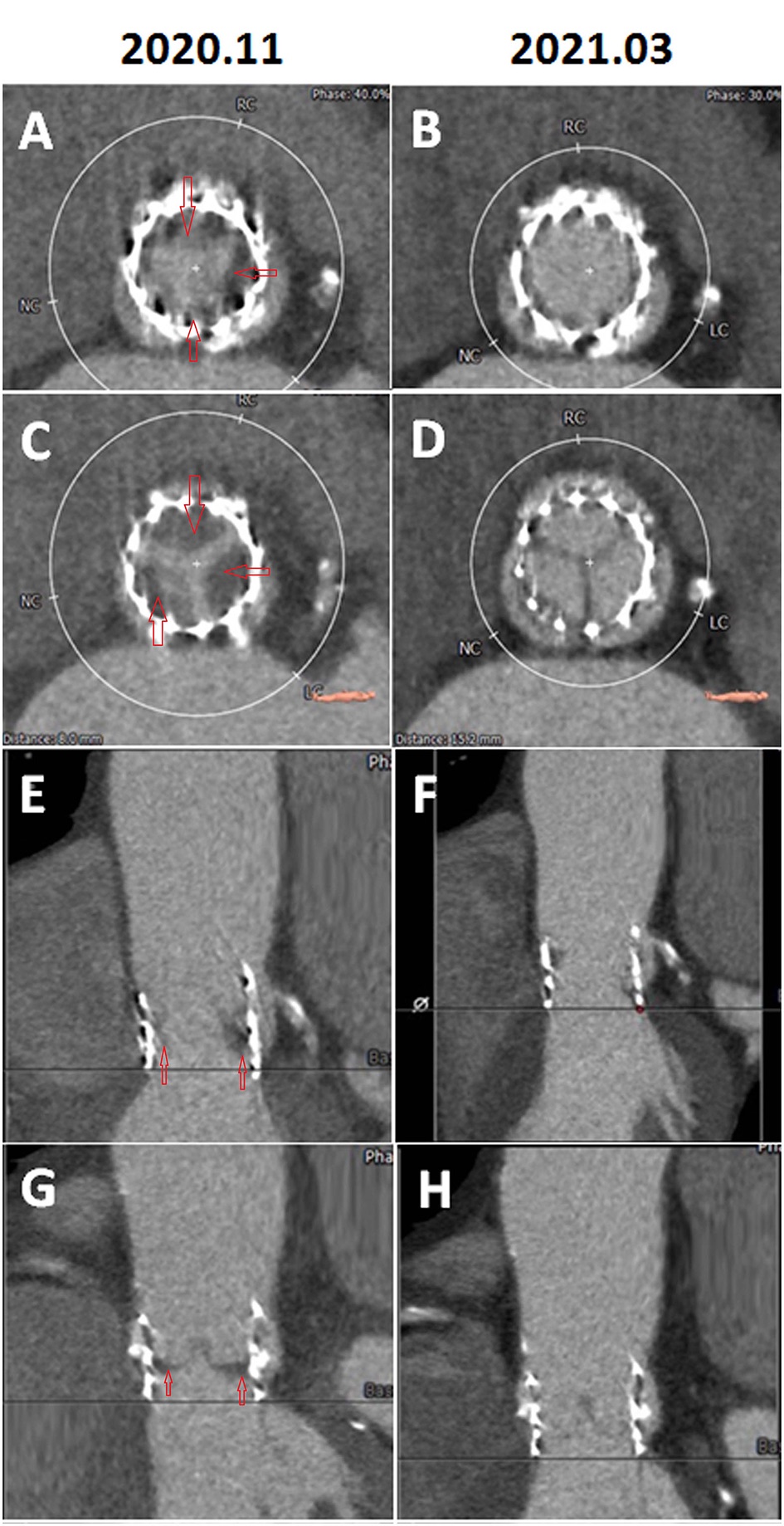
Subclinical leaflet thrombosis was described in 2015 in patients after TAVR and surgical bioprosthetic replacement, with resolution of reduced leaflet motion on anticoagulation. In pooled registry data from 890 patients with interpretable CT, it was present in 12% overall, in 13% of transcatheter and 4% of surgical valves. Anticoagulation with either warfarin or a non-vitamin K oral anticoagulant, but not dual antiplatelet therapy, was associated with less leaflet thrombosis, and leaflet thrombosis was associated with more transient ischemic attacks. HALT can resolve spontaneously and can recur when anticoagulation is stopped.
Randomized trials show that preventing the imaging finding does not prevent clinical events. In the CT substudies of GALILEO and ATLANTIS, anticoagulation reduced HALT and RLM in patients without an anticoagulation indication, but GALILEO showed more death, thromboembolism, and bleeding with rivaroxaban, and ATLANTIS showed no clinical benefit with apixaban. Routine CT screening after TAVR and anticoagulation for incidental, nonobstructive HALT are therefore not established practice. Anticoagulation is reserved for clinically significant bioprosthetic valve thrombosis: a rising gradient, symptoms, heart failure, or embolism with leaflet thickening and restricted motion. Whether subclinical thrombosis shortens bioprosthetic durability is not yet established.
Paravalvular regurgitation and its closure
Paravalvular regurgitation flows between the sewing ring or frame and the native annulus. After surgical replacement, it results from suture dehiscence, a heavily calcified annulus (mitral annular calcification is a classic setting for late leaks), friable tissue, or endocarditis. After TAVR, it results from undersizing, asymmetric calcium that prevents frame apposition, or malposition, and it is usually multiple and eccentric. A new paravalvular leak late after implantation, especially with rocking of the prosthesis, raises the concern of endocarditis, which is excluded first because infection requires antibiotics before surgery and contraindicates transcatheter closure.
Paravalvular jets arise outside the sewing ring, with flow convergence outside the ring confirmed in more than one plane. The jets are graded together, because several small jets can amount to significant regurgitation. In the short-axis view, the circumferential extent of the jets is the sum of their vena contracta lengths divided by the circumference of the outer edge of the valve, and it is integrated with vena contracta width or area and volumetric data.
| Parameter | Mild | Moderate | Severe |
|---|---|---|---|
| Valve structure and motion | Usually normal | Abnormal | Abnormal (dehiscence or rocking with paravalvular leak) |
| LV size (chronic, late postoperative) | Normal | Normal or mildly dilated | Dilated |
| Central jet width, % of LVOT diameter | ≤25% | 26%–64% | ≥65% |
| Vena contracta width | <0.3 cm | 0.3–0.6 cm | >0.6 cm |
| Vena contracta area (2D or 3D) | <0.10 cm² | 0.10–0.29 cm² | ≥0.30 cm² |
| Circumferential extent of paravalvular jets | <10% | 10%–29% | ≥30% |
| Pressure half-time | Slow (>500 ms) | 200–500 ms | Steep (<200 ms) |
| Diastolic flow reversal, descending aorta | Absent or brief early | Intermediate | Prominent, holodiastolic |
| Regurgitant volume | <30 mL/beat | 30–59 mL/beat | ≥60 mL/beat |
| Regurgitant fraction | <30% | 30%–50% | ≥50% |
Severity of prosthetic aortic regurgitation, including paravalvular regurgitation, from the 2024 ASE guideline (Table 8). Circumferential extent is used with vena contracta width or area, not alone.
After TAVR, paravalvular regurgitation imposes a volume load on a hypertrophied, noncompliant ventricle that tolerates it poorly. In the early PARTNER experience, paravalvular regurgitation after TAVR, even when mild, was associated with higher late mortality. Improved frame skirts and CT-based sizing have reduced its incidence.
Treatment of prosthetic regurgitation. In patients with intractable hemolysis or heart failure due to transvalvular or paravalvular prosthetic regurgitation, surgery is recommended unless surgical risk is high or prohibitive (Class 1). In asymptomatic severe prosthetic regurgitation with low operative risk, surgery is reasonable (Class 2a). Percutaneous repair of a paravalvular leak is reasonable at a Comprehensive Valve Center in patients with intractable hemolysis or NYHA class III or IV symptoms who are at high or prohibitive surgical risk and have suitable anatomy (Class 2a). Three-dimensional TEE is recommended to guide the procedure (Class 1). Mitral leaks are approached antegrade through a transseptal puncture, retrograde across the aortic valve, or transapically; aortic leaks are approached retrograde. The defect is crossed with a wire and closed with one or more occluder devices, most often vascular plugs. In a long-term series, percutaneous repair provided durable symptom relief in selected patients, mortality in symptomatic patients remained substantial, and long-term success depended on the amount of residual regurgitation. Surgical repair of a leak carries operative mortality of about 3% in contemporary series, higher for mitral than for aortic leaks.
Hemolysis after valve replacement
Low-grade, subclinical red cell destruction is common with mechanical valves, and clinically important hemolytic anemia is uncommon with normally functioning contemporary prostheses. Significant hemolysis usually indicates a paravalvular leak, where blood is forced at high velocity through a small channel and the red cells are fragmented by shear. Less common causes are transvalvular regurgitation through a degenerated bioprosthesis, severe patient–prosthesis mismatch, and prosthetic obstruction.
The laboratory pattern is a raised lactate dehydrogenase, a low or undetectable haptoglobin, raised indirect bilirubin and reticulocyte count, and schistocytes on the blood film, with a negative direct antiglobulin test. Chronic hemoglobinuria and hemosiderinuria cause iron deficiency, and increased red cell turnover depletes folate. A new or worsening anemia in a patient with a prosthetic valve, particularly with dark urine, leads to these tests and to TEE to identify a paravalvular leak.
Mild anemia is managed with iron and folate replacement and, when needed, periodic transfusion. Intractable hemolysis, meaning anemia that requires repeated transfusion or remains symptomatic despite supplementation, is an indication for surgical repair of the leak, or for percutaneous closure in a patient at high surgical risk with suitable anatomy. Persistent hemolysis after closure usually reflects a residual leak, sometimes a smaller high-velocity jet through or around the occluder.
Structural valve deterioration and bioprosthetic valve failure
Structural valve deterioration is intrinsic, permanent change in the prosthesis itself: leaflet calcification, fibrosis, thickening, tear, or flail, or disruption of the frame, leading to stenosis, regurgitation, or both. It is distinct from nonstructural dysfunction (patient–prosthesis mismatch, paravalvular regurgitation, malposition), from thrombosis, and from endocarditis. Because thrombosis responds to anticoagulation, a rising gradient in a bioprosthesis is investigated for leaflet thrombosis before it is attributed to deterioration.
Risk factors for accelerated deterioration (within 5 years) identified in the 2020 ACC/AHA guideline are age younger than 60 at implantation, smoking, diabetes, chronic kidney disease, an initial mean gradient of 15 mm Hg or more, and valve type. Up to 13% of surgical aortic bioprostheses show hemodynamic dysfunction at a median of 6.7 to 9.9 years after implantation. Most patients remain asymptomatic until deterioration is advanced, and a leaflet tear can cause sudden severe regurgitation.
| Category | Possible (VARC-3 stage 2) structural deterioration | Significant (VARC-3 stage 3) structural deterioration |
|---|---|---|
| Mean gradient | Increase ≥10 mm Hg to a mean gradient ≥20 mm Hg | Increase ≥20 mm Hg to a mean gradient ≥30 mm Hg |
| With a concomitant fall in EOA | ≥0.3 cm² or ≥25% | ≥0.6 cm² or ≥50% |
| And/or a fall in DVI | ≥0.1 or ≥20% | ≥0.2 or ≥40% |
| Intraprosthetic regurgitation | New, or increase of ≥1 grade, to moderate or greater | New, or increase of ≥2 grades, to moderate-to-severe or severe |
Hemodynamic criteria for structural valve deterioration from the 2024 ASE guideline (Table 6), which follows the Valve Academic Research Consortium 3 (VARC-3) definition of hemodynamic valve deterioration. Changes are measured against the baseline study 1 to 3 months after the procedure, assume stable LV function and blood pressure, and require morphologic change in the prosthesis. With combined stenosis and regurgitation, significant deterioration may be present at lower thresholds.
VARC-3 places these hemodynamic stages within a broader definition of bioprosthetic valve dysfunction (structural deterioration, nonstructural dysfunction, thrombosis, and endocarditis) and defines bioprosthetic valve failure in three stages. Stage 1 is dysfunction with clinical consequences (new or worsening symptoms, left ventricular dilation, hypertrophy, or dysfunction, or pulmonary hypertension) or irreversible stage 3 hemodynamic deterioration. Stage 2 is aortic valve reintervention, and stage 3 is valve-related death. The 2017 European Association of Percutaneous Cardiovascular Interventions consensus uses a similar framework. These definitions allow durability to be compared across surgical and transcatheter valves.
Durability of transcatheter compared with surgical valves. Transcatheter durability data are less extensive than the 20-year surgical experience and come mostly from older patients. In the NOTION trial of lower-risk patients, severe structural valve deterioration at 10 years was less frequent after TAVR than after surgical replacement (1.5% versus 10.0%), while bioprosthetic valve failure did not differ significantly (9.7% versus 13.8%). Whether these results apply to patients younger than 70, who will live longer than the follow-up of any trial, is uncertain, and this uncertainty is part of the shared decision about the first valve.
Failed bioprostheses: redo surgery, valve-in-valve, and lifetime management
For symptomatic severe stenosis of a bioprosthetic or mechanical valve, repeat surgery is recommended unless surgical risk is high or prohibitive (Class 1). For severely symptomatic bioprosthetic aortic stenosis at high or prohibitive surgical risk, a transcatheter valve-in-valve procedure at a Comprehensive Valve Center is reasonable (Class 2a). The same applies to severe heart failure due to bioprosthetic regurgitation at high or prohibitive risk (Class 2a). When stenosis is attributed to leaflet thrombosis, a VKA is tried first (Class 2a).
| Consideration | Redo surgical replacement | Transcatheter valve-in-valve |
|---|---|---|
| Procedural risk | Higher than the first operation; reoperative aortic replacement mortality about 4.6% in Society of Thoracic Surgeons data | Lower stroke and bleeding risk in systematic reviews |
| Residual gradient and mismatch | Lower gradients; mismatch can be corrected with a larger valve or root enlargement | High residual gradients inside small surgical valves |
| Paravalvular leak and pacemaker | Low paravalvular leak | Paravalvular leak and pacemaker rates lower than with TAVR in native valves |
| Coronary obstruction | Not a concern | More common than with TAVR in native valves |
| Concomitant disease | Treats endocarditis, root disease, other valves, and coronary disease | Contraindicated with active endocarditis |
| Future options | Larger valve preserves a future valve-in-valve option | Later coronary access and a further transcatheter valve may be limited |
Redo surgery and valve-in-valve treatment for a failed surgical aortic bioprosthesis, after the 2020 ACC/AHA guideline.
In the international VIVID registry of 459 patients treated with aortic valve-in-valve implantation, 30-day mortality was 7.6% and major stroke 1.7%, 93% of survivors were in NYHA class I or II, and 1-year survival was 83.2%. Outcomes were worse with small surgical valves and with stenosis as the mode of failure.
Coronary obstruction after valve-in-valve implantation. The transcatheter valve pushes the surgical leaflets outward into a cylinder that can cover a coronary ostium. In VIVID, clinically evident coronary obstruction occurred in 2.3% of 1,612 valve-in-valve procedures and carried a 30-day mortality of 52.9%. It was more frequent with stented valves whose leaflets are mounted outside the frame (6.1%) and with stentless valves (3.7%) than with stented valves with internally mounted leaflets (0.8%). On CT, the virtual transcatheter valve-to-coronary distance (VTC), measured from a simulated frame to the coronary ostium, predicted obstruction; a VTC below 4 mm identifies high risk. A narrow, low sinotubular junction can also pin the displaced leaflets against the aortic wall and seal the sinus (sinus sequestration). In high-risk anatomy, the surgical or native leaflet can be split before valve implantation with an electrified wire (BASILICA). In the prospective BASILICA trial of 30 patients, the primary success end point was met in 93%, with no coronary obstruction.
Small surgical valves and bioprosthetic valve fracture. The labeled size of a surgical valve is not its true internal diameter, so planning starts from the model's true internal diameter and frame design. A transcatheter valve inside a small surgical valve is constrained by the surgical frame and leaves a high gradient. A high-pressure noncompliant balloon can fracture or stretch the frame of many, but not all, surgical bioprostheses. In bench testing, fracture required about 8 to 24 atmospheres depending on the valve, and only the internal frame broke while the sewing cuff remained intact. Fracture allows fuller expansion of the transcatheter valve and lowers the residual gradient. Supra-annular transcatheter valves also yield lower gradients in small surgical valves.
Mitral and tricuspid valve-in-valve. Transseptal mitral valve-in-valve implantation is an established treatment for failed surgical mitral bioprostheses in patients at high surgical risk. Results are less favorable inside annuloplasty rings and in mitral annular calcification. In a registry of 521 patients, 1-year mortality was 14.0% after valve-in-valve, 30.6% after valve-in-ring, and 62.8% after valve-in-annular-calcification implantation, and left ventricular outflow tract obstruction was most frequent with annular calcification. CT predicts the neo-outflow tract, and laceration of the anterior mitral leaflet (LAMPOON) is used to reduce outflow obstruction when the native leaflet remains. Tricuspid and pulmonary valve-in-valve implantation is performed for failed bioprostheses and conduits.
Prosthetic valve endocarditis and embolic events
Prosthetic valve endocarditis affects mechanical, surgical biological, and transcatheter valves, carries higher mortality than native valve endocarditis, and more often requires surgery. Infection in the months after implantation is usually acquired perioperatively and is commonly staphylococcal; later infection follows bacteremia from another source. Annular abscess, pseudoaneurysm, fistula, and dehiscence with a new paravalvular leak or rocking prosthesis are characteristic. TEE is more sensitive for vegetations and leaflet perforation; CT is more accurate for root abscess and pseudoaneurysm and less affected by prosthetic shadowing; and fluorodeoxyglucose positron emission tomography (FDG PET)/CT shows perivalvular infection around prostheses and transcatheter frames. The diagnosis, antibiotic regimens, and surgical indications are described on the infective endocarditis page.
Patients with a prosthetic valve (including a transcatheter valve) or prosthetic material used for valve repair are in the highest-risk group. For this group, the 2021 American Heart Association (AHA) scientific statement recommends antibiotic prophylaxis before dental procedures that manipulate the gingival tissue or the periapical region of the teeth or perforate the oral mucosa. Regular dental care is part of prosthetic valve follow-up.
An embolic event in a patient with a prosthetic valve leads to review of time in the therapeutic range and adherence, evaluation for endocarditis and new atrial fibrillation, and imaging for valve or leaflet thrombosis by TEE or CT. Intensification of anticoagulation after an embolic event with a therapeutic INR follows the recommendations in the INR section. In patients with a bioprosthesis who have an embolic event on antiplatelet therapy, a VKA may be considered (Class 2b).
Pregnancy with a prosthetic valve
Pregnancy with a mechanical valve is classified as World Health Organization risk category III. Pregnancy is hypercoagulable, warfarin crosses the placenta, and heparin protects the valve less reliably, so no anticoagulation strategy is consistently safe for both mother and fetus. The 2020 ACC/AHA guideline cites a maternal mortality of about 1% and a valve thrombosis risk of about 5%. In the European ROPAC registry, valve thrombosis complicated 10 of 212 pregnancies (4.7%) in women with mechanical valves. Maternal mortality was 1.4%, hemorrhagic events occurred in 23.1%, and only 58% had an uncomplicated pregnancy with a live birth, compared with 79% of women with tissue valves and 78% of women without a prosthetic valve. Miscarriage was more frequent with first-trimester VKA than with heparin (28.6% versus 9.2%), as was late fetal death (7.1% versus 0.7%). Five of the 10 thromboses occurred in the first trimester, all in women switched to heparin.
Fetal risk of warfarin depends on dose. In a series of 58 pregnancies, fetal complications occurred in 22 of 25 pregnancies with a warfarin requirement above 5 mg daily and in 5 of 33 with 5 mg or less. Warfarin embryopathy (nasal hypoplasia and stippled epiphyses) follows exposure during roughly weeks 6 to 12, and fetal intracranial hemorrhage can occur at any stage while the fetus is anticoagulated.
| Recommendation | COR (LOE) |
|---|---|
| Prepregnancy assessment, including echocardiography, by a cardiologist with expertise in valve disease in pregnancy | 1 (C-EO) |
| Care in a tertiary center with a pregnancy heart team (cardiology, cardiac surgery, anesthesiology, maternal-fetal medicine) | 1 (C-EO) |
| Counseling that pregnancy is high risk and that no anticoagulation strategy is consistently safe for mother and baby | 1 (B-NR) |
| Therapeutic anticoagulation with frequent monitoring throughout pregnancy; women who cannot maintain it are counseled against pregnancy | 1 (B-NR) |
| Shared decision on strategy, with counseling that VKA carries the lowest maternal risk and the highest risk of miscarriage, fetal death, and anomalies, especially in the first trimester and above 5 mg/day | 1 (B-NR) |
| Warfarin ≤5 mg/day: continuation for all three trimesters is reasonable | 2a (B-NR) |
| Warfarin >5 mg/day: dose-adjusted LMWH at least twice daily (anti-Xa 0.8–1.2 U/mL 4 to 6 hours after the dose) in the first trimester, then warfarin, is reasonable; continuous IV UFH (aPTT 2 times control) in the first trimester if LMWH is unavailable | 2a (B-NR) |
| Warfarin >5 mg/day: dose-adjusted LMWH for all three trimesters may be considered | 2b (B-NR) |
| Warfarin ≤5 mg/day: LMWH in the first trimester followed by warfarin may be considered | 2b (B-NR) |
| Switch from warfarin to twice-daily LMWH or IV UFH at least 1 week before planned delivery; from LMWH to UFH at least 36 hours before; stop UFH at least 6 hours before planned vaginal delivery | 1 (C-LD) |
| Labor or urgent delivery while therapeutically anticoagulated with a VKA: cesarean delivery after reversal | 1 (C-LD) |
| Valve obstruction or embolic event in pregnancy: TEE | 1 (B-NR) |
| Hemodynamically stable obstructive left-sided mechanical valve thrombosis: slow-infusion, low-dose fibrinolysis is reasonable | 2a (B-NR) |
| LMWH without anti-Xa monitoring 4 to 6 hours after the dose | 3: Harm (B-NR) |
| Dabigatran; anti-Xa direct oral anticoagulants | 3: Harm (B-R; C-EO) |
Prosthetic valves in pregnancy in the 2020 ACC/AHA guideline (Sections 13.2.1 and 13.2.2). aPTT, activated partial thromboplastin time.
Much of the maternal morbidity occurs with underdosed or unmonitored heparin. LMWH requirements rise as pregnancy advances, so anti-Xa levels are checked frequently and doses adjusted. Transvalvular gradients rise during pregnancy with increased heart rate, plasma volume, and stroke volume, so a higher gradient is expected and is judged with these changes in mind. A bioprosthesis avoids anticoagulation during pregnancy but deteriorates faster in young women, and reoperation is expected.
Surveillance after valve replacement
Lifelong follow-up of a prosthetic valve has two purposes: maintaining the antithrombotic therapy the valve requires and detecting dysfunction before it becomes an emergency. Clinical review includes symptoms, valve sounds and murmurs, INR control or antiplatelet adherence, blood pressure, rhythm, dental care, and signs of hemolysis.
| Situation | Imaging | COR (LOE) |
|---|---|---|
| Any surgical or transcatheter prosthesis or valve repair | Initial postprocedural transthoracic echocardiogram (TTE; baseline, usually 1 to 3 months after implantation) | 1 (B-NR) |
| Change in symptoms or signs suggesting valve dysfunction | Repeat TTE | 1 (C-EO) |
| Symptoms or signs of prosthetic dysfunction | TEE, gated cardiac CT, or fluoroscopy, even if TTE shows no dysfunction | 1 (C-LD) |
| Surgical bioprosthesis | TTE at 5 and 10 years after implantation, then annually | 2a (C-LD) |
| Transcatheter bioprosthesis | TTE annually | 2a (C-LD) |
| Mechanical valve with a normal baseline study and no clinical change | Routine annual TTE not needed | — |
Imaging after valve replacement in the 2020 ACC/AHA guideline (Section 11.1.1). Earlier annual imaging may be considered for surgical bioprostheses at risk of accelerated deterioration.
Each surveillance study is compared side by side with the baseline, with the same outflow diameter, windows, and transducer. Any new symptom, new murmur, change in valve sounds, embolic event, hemolysis, fever with bacteremia, or a sustained subtherapeutic INR in a patient with a mechanical valve leads to imaging outside the routine schedule.
Sources of error in prosthetic valve Doppler
- Native valve thresholds applied to a prosthesis: a mean gradient of 25 mm Hg can be normal for a small stented valve and abnormal for a large one.
- A pulsed-wave sample in the zone of flow acceleration at the sewing ring or inside a transcatheter stent: the outflow VTI, DVI, and EOA are overestimated and obstruction is missed.
- A study read without the baseline: a high but stable gradient from mismatch is mistaken for obstruction, or a rise from a low baseline is accepted as normal.
- Gradients recorded during anemia, fever, tachycardia, or pregnancy: flow-dependent values rise while DVI and acceleration time stay normal.
- Pressure recovery in a small bileaflet valve: the Doppler gradient exceeds the net pressure loss, and catheter and Doppler values disagree without obstruction.
- Mitral gradients read without the heart rate, or mitral area estimated with 220 ÷ pressure half-time: both misgrade prosthetic mitral function.
- Mitral regurgitation hidden by acoustic shadowing: a high E velocity and VTI ratio with a normal pressure half-time are attributed to stenosis.
- Washing jets mistaken for pathologic regurgitation, or a paravalvular jet mistaken for a washing jet: the origin relative to the sewing ring decides.
- Indexed EOA cutoffs for normal body mass index applied in obesity: mismatch is overclassified.
- Several small paravalvular jets graded individually: total regurgitation after TAVR is underestimated.
Self-assessment
A 63-year-old man with a 23-mm bileaflet mechanical aortic valve implanted 9 years earlier has gradually worsening exertional dyspnea. His INR values over the past year have all been between 2.3 and 3.0. The mean gradient has risen from 12 mm Hg on the baseline study to 36 mm Hg, the DVI is 0.22, and the acceleration time is 112 ms. Cine fluoroscopy shows a reduced opening angle of one leaflet, and contrast CT shows a fixed crescent of tissue on the ventricular side of the sewing ring measuring 230 Hounsfield units. What is the most likely mechanism, and what is the treatment?
Echocardiography Board ReviewReferences
- Otto CM, Nishimura RA, Bonow RO, et al. 2020 ACC/AHA guideline for the management of patients with valvular heart disease: a report of the American College of Cardiology/American Heart Association Joint Committee on Clinical Practice Guidelines. Circulation. 2021;143:e72–e227. doi:10.1161/CIR.0000000000000923
- Praz F, Borger MA, Lanz J, et al. 2025 ESC/EACTS guidelines for the management of valvular heart disease. Eur Heart J. 2025;46:4635–4736. doi:10.1093/eurheartj/ehaf194
- Zoghbi WA, Jone PN, Chamsi-Pasha MA, et al. Guidelines for the evaluation of prosthetic valve function with cardiovascular imaging: a report from the American Society of Echocardiography developed in collaboration with the Society for Cardiovascular Magnetic Resonance and the Society of Cardiovascular Computed Tomography. J Am Soc Echocardiogr. 2024;37:2–63. doi:10.1016/j.echo.2023.10.004
- Zoghbi WA, Asch FM, Bruce C, et al. Guidelines for the evaluation of valvular regurgitation after percutaneous valve repair or replacement: a report from the American Society of Echocardiography. J Am Soc Echocardiogr. 2019;32:431–475. doi:10.1016/j.echo.2019.01.003
- Généreux P, Piazza N, Alu MC, et al. Valve Academic Research Consortium 3: updated endpoint definitions for aortic valve clinical research. J Am Coll Cardiol. 2021;77:2717–2746. doi:10.1016/j.jacc.2021.02.038
- Capodanno D, Petronio AS, Prendergast B, et al. Standardized definitions of structural deterioration and valve failure in assessing long-term durability of transcatheter and surgical aortic bioprosthetic valves: a consensus statement from the European Association of Percutaneous Cardiovascular Interventions (EAPCI) endorsed by the European Society of Cardiology (ESC) and the European Association for Cardio-Thoracic Surgery (EACTS). Eur Heart J. 2017;38:3382–3390. doi:10.1093/eurheartj/ehx303
- Wilson WR, Gewitz M, Lockhart PB, et al. Prevention of viridans group streptococcal infective endocarditis: a scientific statement from the American Heart Association. Circulation. 2021;143:e963–e978. doi:10.1161/CIR.0000000000000969
- Vongpatanasin W, Hillis LD, Lange RA. Prosthetic heart valves. N Engl J Med. 1996;335:407–416. doi:10.1056/NEJM199608083350607
- Cannegieter SC, Rosendaal FR, Briët E. Thromboembolic and bleeding complications in patients with mechanical heart valve prostheses. Circulation. 1994;89:635–641. doi:10.1161/01.cir.89.2.635
- Chiang YP, Chikwe J, Moskowitz AJ, et al. Survival and long-term outcomes following bioprosthetic vs mechanical aortic valve replacement in patients aged 50 to 69 years. JAMA. 2014;312:1323–1329. doi:10.1001/jama.2014.12679
- Goldstone AB, Chiu P, Baiocchi M, et al. Mechanical or biologic prostheses for aortic-valve and mitral-valve replacement. N Engl J Med. 2017;377:1847–1857. doi:10.1056/NEJMoa1613792
- Eikelboom JW, Connolly SJ, Brueckmann M, et al. Dabigatran versus warfarin in patients with mechanical heart valves. N Engl J Med. 2013;369:1206–1214. doi:10.1056/NEJMoa1300615
- Wang TY, Svensson LG, Wen J, et al. Apixaban or warfarin in patients with an On-X mechanical aortic valve. NEJM Evid. 2023;2(7):EVIDoa2300067. doi:10.1056/EVIDoa2300067
- Puskas J, Gerdisch M, Nichols D, et al. Reduced anticoagulation after mechanical aortic valve replacement: interim results from the Prospective Randomized On-X Valve Anticoagulation Clinical Trial randomized Food and Drug Administration investigational device exemption trial. J Thorac Cardiovasc Surg. 2014;147:1202–1211. doi:10.1016/j.jtcvs.2014.01.004
- Kovacs MJ, Wells PS, Anderson DR, et al. Postoperative low molecular weight heparin bridging treatment for patients at high risk of arterial thromboembolism (PERIOP2): double blind randomised controlled trial. BMJ. 2021;373:n1205. doi:10.1136/bmj.n1205
- Brouwer J, Nijenhuis VJ, Delewi R, et al. Aspirin with or without clopidogrel after transcatheter aortic-valve implantation. N Engl J Med. 2020;383:1447–1457. doi:10.1056/NEJMoa2017815
- Nijenhuis VJ, Brouwer J, Delewi R, et al. Anticoagulation with or without clopidogrel after transcatheter aortic-valve implantation. N Engl J Med. 2020;382:1696–1707. doi:10.1056/NEJMoa1915152
- Dangas GD, Tijssen JGP, Wöhrle J, et al. A controlled trial of rivaroxaban after transcatheter aortic-valve replacement. N Engl J Med. 2020;382:120–129. doi:10.1056/NEJMoa1911425
- Collet JP, Van Belle E, Thiele H, et al. Apixaban vs. standard of care after transcatheter aortic valve implantation: the ATLANTIS trial. Eur Heart J. 2022;43:2783–2797. doi:10.1093/eurheartj/ehac242
- Van Mieghem NM, Unverdorben M, Hengstenberg C, et al. Edoxaban versus vitamin K antagonist for atrial fibrillation after TAVR. N Engl J Med. 2021;385:2150–2160. doi:10.1056/NEJMoa2111016
- Guimarães HP, Lopes RD, de Barros e Silva PGM, et al. Rivaroxaban in patients with atrial fibrillation and a bioprosthetic mitral valve. N Engl J Med. 2020;383:2117–2126. doi:10.1056/NEJMoa2029603
- Connolly SJ, Karthikeyan G, Ntsekhe M, et al. Rivaroxaban in rheumatic heart disease-associated atrial fibrillation. N Engl J Med. 2022;387:978–988. doi:10.1056/NEJMoa2209051
- Makkar RR, Fontana G, Jilaihawi H, et al. Possible subclinical leaflet thrombosis in bioprosthetic aortic valves. N Engl J Med. 2015;373:2015–2024. doi:10.1056/NEJMoa1509233
- Chakravarty T, Søndergaard L, Friedman J, et al. Subclinical leaflet thrombosis in surgical and transcatheter bioprosthetic aortic valves: an observational study. Lancet. 2017;389:2383–2392. doi:10.1016/S0140-6736(17)30757-2
- Özkan M, Gündüz S, Gürsoy OM, et al. Ultraslow thrombolytic therapy: a novel strategy in the management of PROsthetic MEchanical valve Thrombosis and the prEdictors of outcomE: the Ultra-slow PROMETEE trial. Am Heart J. 2015;170:409–418. doi:10.1016/j.ahj.2015.04.025
- Head SJ, Mokhles MM, Osnabrugge RLJ, et al. The impact of prosthesis–patient mismatch on long-term survival after aortic valve replacement: a systematic review and meta-analysis of 34 observational studies comprising 27 186 patients with 133 141 patient-years. Eur Heart J. 2012;33:1518–1529. doi:10.1093/eurheartj/ehs003
- Herrmann HC, Mehran R, Blackman DJ, et al. Self-expanding or balloon-expandable TAVR in patients with a small aortic annulus. N Engl J Med. 2024;390:1959–1971. doi:10.1056/NEJMoa2312573
- Kodali SK, Williams MR, Smith CR, et al. Two-year outcomes after transcatheter or surgical aortic-valve replacement. N Engl J Med. 2012;366:1686–1695. doi:10.1056/NEJMoa1200384
- Sorajja P, Cabalka AK, Hagler DJ, Rihal CS. Long-term follow-up of percutaneous repair of paravalvular prosthetic regurgitation. J Am Coll Cardiol. 2011;58:2218–2224. doi:10.1016/j.jacc.2011.07.041
- Thyregod HGH, Jørgensen TH, Ihlemann N, et al. Transcatheter or surgical aortic valve implantation: 10-year outcomes of the NOTION trial. Eur Heart J. 2024;45:1116–1124. doi:10.1093/eurheartj/ehae043
- Dvir D, Webb JG, Bleiziffer S, et al. Transcatheter aortic valve implantation in failed bioprosthetic surgical valves. JAMA. 2014;312:162–170. doi:10.1001/jama.2014.7246
- Ribeiro HB, Rodés-Cabau J, Blanke P, et al. Incidence, predictors, and clinical outcomes of coronary obstruction following transcatheter aortic valve replacement for degenerative bioprosthetic surgical valves: insights from the VIVID registry. Eur Heart J. 2018;39:687–695. doi:10.1093/eurheartj/ehx455
- Khan JM, Greenbaum AB, Babaliaros VC, et al. The BASILICA trial: prospective multicenter investigation of intentional leaflet laceration to prevent TAVR coronary obstruction. JACC Cardiovasc Interv. 2019;12:1240–1252. doi:10.1016/j.jcin.2019.03.035
- Allen KB, Chhatriwalla AK, Cohen DJ, et al. Bioprosthetic valve fracture to facilitate transcatheter valve-in-valve implantation. Ann Thorac Surg. 2017;104:1501–1508. doi:10.1016/j.athoracsur.2017.04.007
- Yoon SH, Whisenant BK, Bleiziffer S, et al. Outcomes of transcatheter mitral valve replacement for degenerated bioprostheses, failed annuloplasty rings, and mitral annular calcification. Eur Heart J. 2019;40:441–451. doi:10.1093/eurheartj/ehy590
- van Hagen IM, Roos-Hesselink JW, Ruys TPE, et al. Pregnancy in women with a mechanical heart valve: data of the European Society of Cardiology Registry of Pregnancy and Cardiac Disease (ROPAC). Circulation. 2015;132:132–142. doi:10.1161/CIRCULATIONAHA.115.015242
- Vitale N, De Feo M, De Santo LS, et al. Dose-dependent fetal complications of warfarin in pregnant women with mechanical heart valves. J Am Coll Cardiol. 1999;33:1637–1641. doi:10.1016/S0735-1097(99)00044-3
- Liang KW, Yu CL, Lin WW, Lee WL. Case report: leaflet thrombosis after transcatheter aortic valve replacement with worsening heart failure: a successful resolution using non-vitamin K antagonist oral anticoagulant. Front Cardiovasc Med. 2021;8:731427. doi:10.3389/fcvm.2021.731427
- Munawar G, Gupta V, Laiq Z, Sleik K. Mechanical aortic valve thrombosis: treatment with tissue plasminogen activator. JACC Case Rep. 2025;30(19):104006. doi:10.1016/j.jaccas.2025.104006
- US Food and Drug Administration. Update on the safety of Andexxa by AstraZeneca: FDA safety communication. 2025. fda.gov