Library › Valvular heart disease › Infective endocarditis
Infective endocarditis
Native valve, prosthetic valve, and device endocarditis in adults from risk and microbiology through diagnosis, multimodality imaging, antibiotic therapy, surgery, special populations, follow-up, and prophylaxis.
Infective endocarditis is diagnosed by the 2023 Duke-ISCVID criteria: two major criteria, one major and three minor, or five minor criteria establish definite disease. Transesophageal echocardiography is indicated when the transthoracic study is nondiagnostic, when a prosthetic valve or device is present, and when complications are suspected. Heart failure from valve destruction, uncontrolled infection, and prevention of embolism are the three indications for surgery, which is performed during the initial hospitalization once an indication is present.
Definition and classification of infective endocarditis
Infective endocarditis is microbial infection of the endocardial surface of the heart. The usual site is a heart valve, but the infection also involves the mural endocardium, the chordae, a septal defect, a prosthetic valve or annuloplasty ring, an intracardiac lead, or the endothelium of a great vessel next to a valve. The characteristic lesion is the vegetation, an infected mass of platelets, fibrin, microorganisms, and inflammatory cells. Infection that extends beyond the leaflet into the annulus and surrounding tissue produces abscess, pseudoaneurysm, and fistula, and these lesions, with leaflet perforation and prosthetic dehiscence, are the destructive complications that most often require surgery.
The older division into acute and subacute endocarditis described tempo and organism. Acute disease, typically from Staphylococcus aureus, destroys a valve within days and seeds distant sites. Subacute disease, typically from viridans group streptococci, progresses over weeks with fever, weight loss, and immunologic phenomena. Current classification is anatomic and epidemiologic, because the substrate predicts the organism, the imaging strategy, and the surgical stakes.
| Category | Definition | Clinical significance |
|---|---|---|
| Native valve endocarditis | Infection of a native valve, left- or right-sided | Organism spectrum dominated by S. aureus, streptococci, and enterococci |
| Prosthetic valve endocarditis, early | Within the first year after valve implantation | Perioperative and healthcare acquisition; coagulase-negative staphylococci, S. aureus, gram-negative bacilli, and fungi; high rate of perivalvular extension |
| Prosthetic valve endocarditis, late | More than 1 year after implantation | Organisms resemble native valve disease; prosthetic material still lowers echocardiographic sensitivity and raises surgical risk |
| Transcatheter valve endocarditis | Infection of a transcatheter aortic, mitral, tricuspid, or pulmonary valve | Older, frail patients; enterococci prominent; frame artifact limits echocardiography |
| Device-related endocarditis | Infection of a pacemaker or defibrillator lead, with or without valve involvement | Complete system removal is required |
| Right-sided endocarditis | Tricuspid or pulmonary valve, or right-sided lead | Injection drug use, catheters, and devices; septic pulmonary emboli; better prognosis than left-sided disease |
| Healthcare-associated endocarditis | Onset more than 48 hours after admission, or community onset with recent intravenous therapy, hemodialysis, or hospitalization | S. aureus and enterococci; vascular catheters and dialysis access as sources |
| Blood culture-negative endocarditis | Endocarditis without growth in standard blood cultures | Prior antibiotics most often; otherwise fastidious or intracellular organisms that require serology and molecular testing |
Classification of infective endocarditis by substrate, timing, and microbiology. The cutoff for early prosthetic valve endocarditis is 1 year in most series; the 2023 European Society of Cardiology (ESC) guideline defines early prosthetic valve endocarditis as infection within 6 months of valve surgery.
Epidemiology and the changing risk profile
Infective endocarditis is uncommon, with an incidence in high-income countries of roughly 3 to 10 cases per 100,000 people each year. The 2023 ESC guideline cites a global estimate of 13.8 cases per 100,000 people and 66,300 deaths in 2019. In-hospital mortality has stayed in the range of 15% to 30% for two decades. The International Collaboration on Endocarditis-Prospective Cohort Study (ICE-PCS) enrolled 2,781 adults with definite endocarditis in 25 countries between 2000 and 2005. S. aureus caused 31.2% of episodes, nearly half of patients underwent surgery, and in-hospital mortality was 17.7%.
The disease has moved from the young patient with rheumatic heart disease to an older patient with degenerative valve disease, prosthetic material, and frequent healthcare contact. The drivers of that change are an aging population, more valve replacement and repair, more transcatheter valve implantation, more pacemakers and defibrillators, long-term vascular catheters, hemodialysis, and, in the United States, the opioid epidemic. Rheumatic heart disease remains the main substrate where rheumatic fever is endemic.
Cardiac predisposition. Prior infective endocarditis carries the highest recurrence risk of any substrate. Prosthetic valves of every type, surgical and transcatheter, and prosthetic material used for valve repair are the next most important. Other predisposing lesions include degenerative mitral and aortic valve disease with more than mild regurgitation or stenosis, mitral valve prolapse with regurgitation, a bicuspid aortic valve, rheumatic disease, hypertrophic obstructive cardiomyopathy, and congenital heart disease. The congenital lesions of highest risk are unrepaired cyanotic lesions and repairs with residual shunts or regurgitation next to prosthetic patches. An implanted electronic device is a substrate for lead infection and for valve infection.
Transcatheter valves. In an international registry of 20,006 patients after transcatheter aortic valve replacement (TAVR), the incidence of endocarditis was 1.1% per person-year, with a median onset 5.3 months after implantation. Enterococci (24.6%) and S. aureus (23.3%) were the most frequent organisms, and in-hospital mortality was 36%. Younger age, male sex, diabetes, and moderate or severe residual aortic regurgitation were associated with higher risk.
People who inject drugs. Hospitalizations for endocarditis among young people who inject drugs rose in the United States during the opioid epidemic, as documented in national discharge data. Injection introduces skin flora directly into the bloodstream and particulate matter damages the tricuspid endothelium. These patients have a high rate of right-sided disease, of S. aureus infection, and of reinfection after cure.
Hemodialysis, immunosuppression, and diabetes. Hemodialysis combines repeated vascular access, mitral annular and valvular calcification, and impaired host defense, and it carries one of the highest incidence rates and worst outcomes of any risk group. Diabetes, malignancy, solid organ transplantation, and immunosuppressive therapy raise the risk of S. aureus bacteremia, fungal infection, and healthcare-associated endocarditis.
Oral sources of bacteremia. Viridans group streptococci from the mouth remain a leading cause of native valve endocarditis. Tooth brushing, flossing, and chewing produce frequent low-grade bacteremia, which makes daily oral hygiene the main preventive measure.
Pathogenesis: endothelial injury, vegetation, and biofilm
Normal valve endothelium resists bacterial adherence. Endocarditis begins where the endothelium is damaged. High-velocity jets, turbulent flow, and the contact of a jet with the opposite wall injure the endothelial surface, and the exposed subendothelial matrix attracts platelets and fibrin. The result is a sterile platelet-fibrin deposit, called nonbacterial thrombotic endocarditis, which forms a nidus for organisms that enter the blood during transient bacteremia.
Lesions form on the low-pressure side of a jet: the atrial surface of the mitral and tricuspid valves, the ventricular surface of the aortic and pulmonary valves, and the site where a regurgitant aortic jet strikes the anterior mitral leaflet or the chordae. A ventricular septal defect seeds the right ventricular surface opposite the jet.
Organisms that cause endocarditis express adhesins that bind fibronectin, fibrinogen, and platelets. Once attached, they multiply inside a growing mesh of fibrin and platelets, where host phagocytes have poor access. Bacterial density inside a vegetation is very high, and many organisms are in a slow-growing, metabolically inactive state. Biofilm on prosthetic material and leads adds a protective matrix. These features explain why therapy requires prolonged, high-dose, bactericidal antibiotics, why combination therapy is used for some organisms, and why infected prosthetic material is often removed.
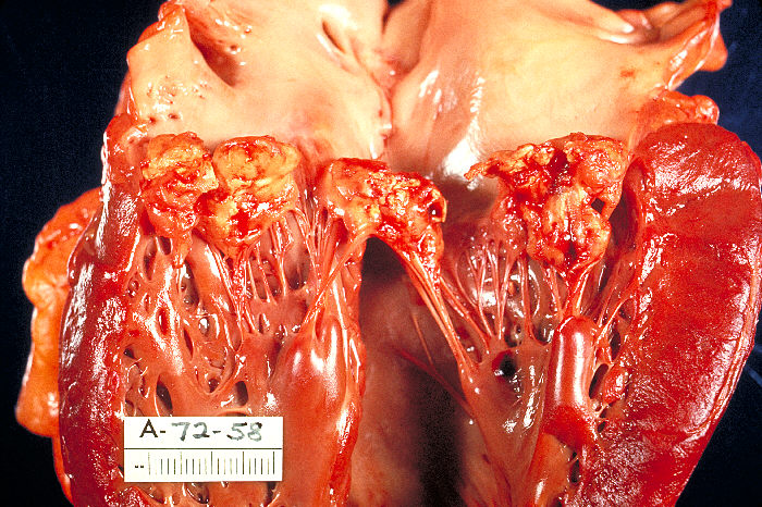

Destruction follows. Proteolytic enzymes and the inflammatory response perforate leaflets, rupture chordae, and erode into the annulus. Aortic valve infection spreads into the aortic root and the mitral-aortic intervalvular fibrosa, and erosion near the membranous septum interrupts the atrioventricular node and His bundle. Fragments of vegetation embolize to the brain, spleen, kidneys, coronary arteries, and limbs from left-sided lesions, and to the lungs from right-sided lesions. Sustained antigenemia generates circulating immune complexes, which deposit in the glomeruli, skin, and retina and account for glomerulonephritis, Osler nodes, Roth spots, and a positive rheumatoid factor.
Microbiology of infective endocarditis by organism and substrate
Staphylococci, streptococci, and enterococci cause most episodes. The organism determines the tempo of disease, the risk of embolism and abscess, the antibiotic regimen, the need for surgery, and the search for a source.
| Organism | Typical setting | Clinical features and implications |
|---|---|---|
| Staphylococcus aureus | Most common cause overall; healthcare contact, intravascular catheters, hemodialysis, injection drug use, devices | Acute course, high embolic and abscess rates, metastatic infection (spine, joints, psoas, epidural space); independent predictor of death; early surgery threshold is low for left-sided disease |
| Coagulase-negative staphylococci | Prosthetic valves, especially in the first year; leads | Biofilm formation; methicillin resistance common; Staphylococcus lugdunensis behaves like S. aureus and destroys native valves |
| Viridans group streptococci | Native valves with prior disease; oral source | Subacute course; usually highly penicillin-susceptible; immunologic phenomena; good medical cure rate |
| Streptococcus gallolyticus (formerly S. bovis biotype I) | Older adults | Association with colorectal adenoma and carcinoma; colonoscopy after stabilization |
| Enterococci (Enterococcus faecalis most often) | Older adults, men, genitourinary and gastrointestinal sources, healthcare exposure, transcatheter valves | Subacute course; intrinsic resistance to cephalosporins; requires synergistic combination therapy; colonoscopy reasonable |
| HACEK group | Haemophilus, Aggregatibacter, Cardiobacterium, Eikenella, Kingella; oral flora | Fastidious gram-negative bacilli; large vegetations and emboli; ceftriaxone is standard therapy |
| Pseudomonas aeruginosa and other gram-negative bacilli | Healthcare exposure, injection drug use, prosthetic valves | Uncommon; high mortality; combination therapy and early surgery |
| Streptococcus pneumoniae | Alcohol use disorder, asplenia | Aggressive aortic valve destruction; endocarditis with pneumonia and meningitis is Austrian syndrome |
| Fungi (Candida, Aspergillus) | Prosthetic valves, long-term catheters, parenteral nutrition, immunosuppression, injection drug use | Bulky vegetations and major emboli; medical therapy alone rarely cures; surgery plus prolonged antifungal therapy |
| Coxiella burnetii, Bartonella species, Brucella, Tropheryma whipplei | Animal exposure, homelessness and body lice (B. quintana), cats (B. henselae), unpasteurized dairy | Blood culture-negative endocarditis; diagnosis by serology, polymerase chain reaction (PCR), and valve tissue |
Principal organisms in adult infective endocarditis and their implications.
The 2023 Duke-ISCVID criteria define typical organisms as S. aureus, S. lugdunensis, E. faecalis, all streptococci except S. pneumoniae and S. pyogenes, Granulicatella and Abiotrophia, Gemella, and the HACEK group. With intracardiac prosthetic material, coagulase-negative staphylococci, Corynebacterium striatum and C. jeikeium, Serratia marcescens, P. aeruginosa, Cutibacterium acnes, nontuberculous mycobacteria (especially Mycobacterium chimaera), and Candida species are also typical. E. faecalis counts as typical whether the infection was acquired in the community or the hospital and whether or not a primary focus is known.
Clinical presentation and physical findings
Fever, the most common symptom, is present in most patients, although it can be blunted in older adults, in patients with kidney failure or immunosuppression, and after antibiotics. Subacute disease presents with weeks of malaise, night sweats, anorexia, weight loss, arthralgia, and back pain. Acute disease presents with high fever, rigors, sepsis, and early complications. A new or changed regurgitant murmur is common, but many patients have preexisting murmurs and a murmur may be absent early, in right-sided disease, and in device infection. Heart failure, a stroke, a cold limb, flank pain from renal infarction, left upper quadrant pain from splenic infarction, or new heart block can be the first presentation.
Endocarditis is suspected in any patient with fever and a predisposing substrate, with bacteremia from a typical organism, with an unexplained embolic event, or with a new regurgitant lesion. S. aureus bacteremia carries a substantial probability of endocarditis, and echocardiography is part of its evaluation. Endocarditis is also on the differential of every stroke in a febrile patient, because thrombolysis is hazardous in septic embolism.
| Finding | Description | Mechanism and significance |
|---|---|---|
| Petechiae | Conjunctival, palatal, or skin petechiae | Vascular phenomenon; conjunctival hemorrhage is a Duke-ISCVID minor criterion |
| Splinter hemorrhages | Linear dark-red streaks under the nails | Nonspecific; common with manual labor and trauma |
| Janeway lesions | Painless hemorrhagic macules on the palms and soles | Septic microemboli with microabscesses; more common in acute, often staphylococcal, disease; vascular minor criterion |
| Osler nodes | Tender subcutaneous nodules on the finger and toe pulps | Immune-complex vasculitis; subacute disease; immunologic minor criterion |
| Roth spots | Retinal hemorrhages with pale centers | Immunologic minor criterion; also seen in anemia, leukemia, and diabetes |
| Purulent purpura | Purpuric skin lesions with pus | Vascular minor criterion added in 2023 |
| Splenomegaly, clubbing | Long-standing subacute infection | Uncommon in contemporary practice |
Peripheral stigmata of infective endocarditis. All have low sensitivity, and their absence does not lower the probability of endocarditis enough to defer blood cultures and echocardiography.
Embolic, neurologic, cardiac, and immunologic complications
Systemic embolism. Embolic events occur in 20% to 50% of patients, and in the European EURO-ENDO registry 25.3% had embolic complications. Many emboli are clinically silent. Risk is highest before and during the first days of antibiotic therapy and falls steadily over the first 2 weeks of effective treatment, which is the rationale for operating early when embolic risk is the indication. Vegetation length and mobility predict embolism: in a prospective multicenter study, a length above 10 mm and severe mobility independently predicted new embolic events after antibiotics were started, and a length above 15 mm predicted 1-year mortality. S. aureus, S. gallolyticus, fungi, mitral (especially anterior leaflet) location, and a prior embolus add risk.
Neurologic complications. The brain is the most important embolic target. Symptomatic stroke, transient ischemic attack, encephalopathy, meningitis, brain abscess, and intracranial hemorrhage all occur. On systematic magnetic resonance imaging, central nervous system lesions are found in up to 60% to 80% of patients, most of them small, often asymptomatic, ischemic lesions; hemorrhage, abscess, or infectious (mycotic) aneurysm is found in fewer than 10%. Cerebral microbleeds are present in 50% to 60% of patients, are not a Duke-ISCVID criterion, and do not by themselves delay surgery.
Infectious intracranial aneurysms arise from septic embolism to the vasa vasorum or the arterial lumen, usually at distal branch points of the middle cerebral artery. They can rupture before, during, or after therapy and are a cause of intracranial hemorrhage. Computed tomographic or magnetic resonance angiography is performed in patients with neurologic symptoms or hemorrhage and before cardiac surgery when an aneurysm is suspected, and catheter angiography is used when noninvasive imaging is inconclusive. Treatment is antibiotic therapy with endovascular or surgical exclusion of aneurysms that are large, enlarging, or ruptured.
Splenic, renal, and other emboli. Splenic infarcts are common and usually silent. Splenic abscess presents with persistent fever and left upper quadrant pain and may require drainage or splenectomy, ideally before valve surgery. Renal infarction causes flank pain and hematuria. Coronary embolism causes myocardial infarction, and peripheral emboli cause limb ischemia and mycotic aneurysms of the visceral and limb arteries.
Septic pulmonary emboli are the hallmark of tricuspid and right-sided lead infection: multiple peripheral nodules, often cavitating, with pleuritic pain, cough, hemoptysis, empyema, or pneumothorax.
Heart failure. This is the most frequent complication and the most frequent indication for surgery. Acute severe regurgitation follows cusp perforation or flail in the aortic valve and leaflet perforation or chordal rupture in the mitral valve. A fistula or obstruction of a prosthesis by a bulky vegetation has a similar effect. Each produces sudden volume overload in a ventricle that has not dilated. Pulmonary edema and cardiogenic shock follow. Antibiotics do not restore a destroyed valve.
Perivalvular extension and conduction block. Abscess and pseudoaneurysm are most frequent in aortic and prosthetic valve endocarditis and in S. aureus infection. Extension into the membranous septum interrupts conduction. New PR prolongation, atrioventricular block, or bundle branch block in aortic endocarditis indicates perivalvular extension until proven otherwise, and transesophageal echocardiography or cardiac CT follows promptly. A daily electrocardiogram during aortic valve infection detects this change.
Immune complex disease and the kidney. Kidney injury in endocarditis has several causes: immune complex glomerulonephritis, renal infarction, renal abscess, sepsis, and nephrotoxic drugs (aminoglycosides, vancomycin, contrast). Immune complex glomerulonephritis is a Duke-ISCVID minor criterion when unexplained acute or acute-on-chronic kidney injury is accompanied by two of hematuria, proteinuria, cellular casts, or serologic abnormalities, or when a biopsy shows immune complex disease. Hypocomplementemia and a positive rheumatoid factor are common in subacute disease.
Vertebral osteomyelitis and discitis. Back pain in endocarditis can reflect spondylodiscitis or an epidural abscess, most often with S. aureus or streptococci. Magnetic resonance imaging of the spine is obtained for localized pain or neurologic signs. Conversely, pyogenic spondylodiscitis with a typical organism leads to echocardiography. Musculoskeletal foci may extend the duration of therapy.
Blood culture strategy and laboratory evaluation
Blood cultures establish the diagnosis and direct therapy. In a patient who is clinically stable, at least three sets of blood cultures (each an aerobic and an anaerobic bottle, with adequate blood volume) are drawn from separate peripheral venipunctures before antibiotics are given. The 2023 ESC guideline recommends three sets at 30-minute intervals; in a patient with sepsis, the sets are drawn over a short interval and antibiotics are started without delay. Cultures from an indwelling catheter are paired with peripheral cultures because catheter colonization produces false positives.
Bacteremia in endocarditis is continuous, so most or all sets are positive when the organism is a common one and no antibiotics were given. A single positive set of a skin commensal, such as a coagulase-negative staphylococcus or Cutibacterium, usually represents contamination in a patient without prosthetic material. The 2023 Duke-ISCVID criteria count isolation of a typical organism from two or more separate sets as a major criterion and no longer require the older timing rules or separate venipunctures for classification. Collection practice still follows the microbiologic standard, because it determines yield and interpretation.
After therapy begins, cultures are repeated every 24 to 48 hours until they are negative. The duration of therapy is counted from the first day of effective therapy, and with initially positive cultures, from the first negative culture. Persistent bacteremia beyond 5 to 7 days of appropriate therapy indicates uncontrolled infection: an abscess, an infected device or catheter, a metastatic focus, or inadequate therapy.
Supportive laboratory findings include normocytic anemia, leukocytosis in acute disease, thrombocytopenia, elevated C-reactive protein and erythrocyte sedimentation rate, hypocomplementemia, a positive rheumatoid factor, microscopic hematuria, and proteinuria. None is specific. C-reactive protein is followed during therapy, and its fall is one of the criteria for oral step-down. Procalcitonin does not establish or exclude the diagnosis.
S. aureus bacteremia. Every patient with S. aureus bacteremia undergoes transthoracic echocardiography. The 2020 American College of Cardiology/American Heart Association (ACC/AHA) valve guideline rates transesophageal echocardiography as reasonable in S. aureus bacteremia without a known source (Class 2a). Prosthetic valves, a cardiac device, persistent bacteremia, community acquisition, injection drug use, embolic or metastatic foci, and a vertebral focus all increase the probability of endocarditis and lower the threshold for transesophageal imaging. The 2023 ESC guideline allows validated risk scores to identify patients in whom transesophageal imaging can be omitted.
Blood culture-negative endocarditis: serology and molecular diagnosis
Blood culture-negative endocarditis accounts for a minority of cases in North America and a larger share where antibiotics are given before cultures are drawn. The most common cause is prior antibiotic therapy, which suppresses growth of streptococci and other susceptible organisms for days to weeks. When antibiotics have been given and the patient is stable, additional cultures are drawn, and antibiotics are sometimes withheld briefly to improve yield.
The remaining cases are caused by organisms that grow poorly or not at all in standard media. In a prospective French study of 819 patients with blood culture-negative endocarditis, systematic serology, PCR of blood and valve tissue, and histopathology identified a cause in most cases. Coxiella burnetii and Bartonella species were the most frequent organisms identified. Noninfectious causes, including nonbacterial thrombotic endocarditis and Libman-Sacks endocarditis, were also found.
| Organism | Exposure and setting | Diagnostic test |
|---|---|---|
| Coxiella burnetii (Q fever) | Farm animals, parturient animals, unpasteurized milk; prosthetic valves and aortic grafts | Phase I IgG titer above 1:800 or a single positive blood culture (major criterion); PCR of blood or valve |
| Bartonella quintana, B. henselae | Homelessness, body lice, alcohol use disorder; cats | IgG titer of 1:800 or more (major criterion); PCR of blood or valve |
| Tropheryma whipplei | Older men; arthralgia; often afebrile | PCR of blood or valve tissue; periodic acid-Schiff staining of valve |
| Brucella species | Unpasteurized dairy, livestock; travel to endemic regions | Serology; prolonged blood culture incubation |
| Legionella, Mycoplasma, Chlamydia | Rare; prosthetic valves | Serology, PCR |
| Fungi | Prosthetic valves, catheters, immunosuppression | Fungal blood cultures, beta-D-glucan, galactomannan, histology and culture of emboli or valve |
Causes of blood culture-negative endocarditis and the tests that identify them. Serologic thresholds are those of the 2023 Duke-ISCVID criteria.
Molecular diagnosis. Positive PCR or other nucleic acid testing (including amplicon or metagenomic sequencing) from blood for C. burnetii, Bartonella, or T. whipplei is a Duke-ISCVID major criterion, and positive blood sequencing for other organisms is a minor criterion. The 2026 AHA scientific statement advises saving a plasma sample drawn before antibiotics, so that metagenomic sequencing can be performed when cultures remain negative at 72 hours. Excised valve tissue, vegetations, and arterial emboli are sent for culture, histopathology, and broad-range PCR. Bacterial DNA persists in valve tissue after successful treatment, so a positive molecular result from a resected valve does not prove viable infection and does not by itself extend therapy.
The 2023 Duke-ISCVID diagnostic criteria
The Duke criteria, introduced in 1994 and modified in 2000, combined microbiologic and echocardiographic evidence into a standard case definition. The 2023 revision by the International Society for Cardiovascular Infectious Diseases (ISCVID) updated them for contemporary practice. The 2026 AHA scientific statement adopts the Duke-ISCVID criteria. The principal changes concern microbiology, imaging, and predisposition. Microbiologic changes are an expanded list of typical organisms, especially with prosthetic material; removal of the blood culture timing and separate-venipuncture requirements; and molecular and serologic criteria for Coxiella, Bartonella, and T. whipplei. Imaging and surgical changes are cardiac CT and fluorodeoxyglucose (FDG) positron emission tomography/CT (PET/CT) as major imaging criteria and a new major surgical criterion. The list of predisposing conditions is broader and now includes transcatheter valves, endovascular devices, and prior valve repair.
| Criterion | Requirements |
|---|---|
| Pathologic (definite) | Microorganisms identified by culture, staining, PCR, sequencing, or in situ hybridization, or active endocarditis on histology, in a vegetation, cardiac tissue, explanted prosthetic valve or sewing ring, ascending aortic graft with valve involvement, endovascular device, or arterial embolus |
| Major microbiologic | Typical organism from 2 or more separate blood culture sets; nontypical organism from 3 or more sets; positive blood PCR or sequencing for C. burnetii, Bartonella, or T. whipplei; C. burnetii phase I IgG above 1:800 or isolation from a single blood culture; B. henselae or B. quintana IgG 1:800 or higher |
| Major imaging | Echocardiography or cardiac CT showing vegetation (including on a lead), leaflet perforation, leaflet aneurysm, abscess, pseudoaneurysm, or intracardiac fistula; significant new valvular regurgitation compared with prior imaging (worsening of known regurgitation does not count); new partial dehiscence of a prosthetic valve compared with prior imaging. FDG PET/CT showing abnormal uptake at a native or prosthetic valve, ascending aortic graft, intracardiac lead, or other prosthetic material; for a surgical prosthesis, uptake counts only 3 months or more after implantation. Isolated generator pocket uptake does not qualify |
| Major surgical | Endocarditis seen on direct inspection at heart surgery, when neither a major imaging criterion nor subsequent histologic or microbiologic confirmation is available |
| Minor: predisposition | Prior endocarditis; prosthetic valve, surgical or transcatheter; prior valve repair; congenital heart disease; more than mild regurgitation or stenosis of any cause; endovascular electronic device; hypertrophic obstructive cardiomyopathy; injection drug use |
| Minor: fever | Temperature above 38.0 °C (100.4 °F) |
| Minor: vascular phenomena | Arterial emboli, septic pulmonary infarcts, cerebral or splenic abscess, mycotic aneurysm, intracranial hemorrhage, conjunctival hemorrhage, Janeway lesions, purulent purpura |
| Minor: immunologic phenomena | Positive rheumatoid factor, Osler nodes, Roth spots, immune complex glomerulonephritis |
| Minor: microbiologic | Positive blood cultures that do not meet the major criterion; positive culture, PCR, or sequencing from a sterile site other than the heart or an embolus; positive blood sequencing for organisms other than Coxiella, Bartonella, or T. whipplei |
| Minor: imaging | Abnormal FDG PET/CT uptake within 3 months of implantation of a prosthetic valve, ascending aortic graft, or other prosthetic material |
| Minor: physical examination | New regurgitant murmur on auscultation, used only when echocardiography is unavailable |
The 2023 Duke-ISCVID criteria. Definite: pathologic criteria, or 2 major, 1 major and 3 minor, or 5 minor criteria. Possible: 1 major and 1 minor, or 3 minor criteria. Rejected: a firm alternative diagnosis, resolution with less than 4 days of antibiotics, no evidence at surgery or autopsy after less than 4 days of antibiotics, or criteria for possible endocarditis not met.
Comparison with earlier criteria and the 2023 ESC criteria. The 2000 modified Duke criteria, still embedded in many references, required one of three patterns: typical organisms in two separate cultures; persistently positive cultures drawn 12 hours apart; or all of three, or most of four or more, cultures with the first and last drawn at least 1 hour apart. They recognized only echocardiography for the major imaging criterion and used a narrower list of predisposing conditions. The 2023 ESC guideline uses its own modification of the Duke criteria. It adds cardiac CT, FDG PET/CT, and leukocyte scintigraphy to the major imaging criterion and, unlike Duke-ISCVID, does not apply a fixed 3-month interval for prosthetic valve uptake; uptake pattern and technical quality are judged instead. In validation cohorts, both revisions are more sensitive than the 2000 criteria, and specificity varies with the population. Criteria are classification tools for research and teaching: a patient with possible endocarditis and a high clinical probability is treated as having endocarditis.
Duke-ISCVID classification checklist
The checklist applies the 2023 classification rules. The major imaging and major surgical criteria count as one major criterion when both are present, because the surgical criterion is used only in the absence of a major imaging criterion.
2023 Duke-ISCVID classification
Definite, possible, or rejectedClassification rules from the 2023 Duke-ISCVID criteria. Each minor category counts once, however many findings it contains. A patient with possible endocarditis and high clinical probability is managed as having endocarditis.
Echocardiography: transthoracic, transesophageal, and repeat studies
Transthoracic echocardiography (TTE) is performed promptly in every patient with suspected endocarditis (2020 ACC/AHA, Class 1). It defines valve function, ventricular size and function, pulmonary pressure, and pericardial effusion, and it often shows large vegetations. Its specificity exceeds 90%, but its sensitivity for vegetations is about 40% to 66%, and it is lower still for perivalvular complications, small vegetations, prostheses, and leads. A good-quality negative transthoracic study in a patient with low clinical probability makes endocarditis unlikely, and an alternative diagnosis is sought.
Transesophageal echocardiography (TEE) has sensitivity and specificity above 90% for native valve vegetations. The 2020 ACC/AHA guideline recommends TEE when TTE is nondiagnostic, when complications are suspected, and when intracardiac leads are present (Class 1). It rates TEE as reasonable in S. aureus bacteremia without a known source and in patients with a prosthetic valve and persistent fever without bacteremia (Class 2a). The 2026 AHA statement adds TEE within 2 days before an oral step-down. In native valve disease, the threshold for TEE is low and guided by pretest probability and image quality. The 2023 ESC guideline recommends TEE in all patients with suspected endocarditis, even with a positive TTE, except isolated right-sided native valve infection with a good-quality, unequivocal TTE.
Repeat imaging. Early in the illness, a vegetation can be below the threshold of detection and an abscess can be an area of thickening that cavitates days later. When the initial TTE and TEE are negative and suspicion remains high, the study is repeated within 1 week (5 to 7 days in the ESC guideline). Any new murmur, heart failure, conduction disturbance, embolism, or persistent fever or bacteremia prompts immediate repeat imaging. A TTE at completion of therapy establishes a new baseline, and a TTE after valve surgery documents the repair or prosthesis.
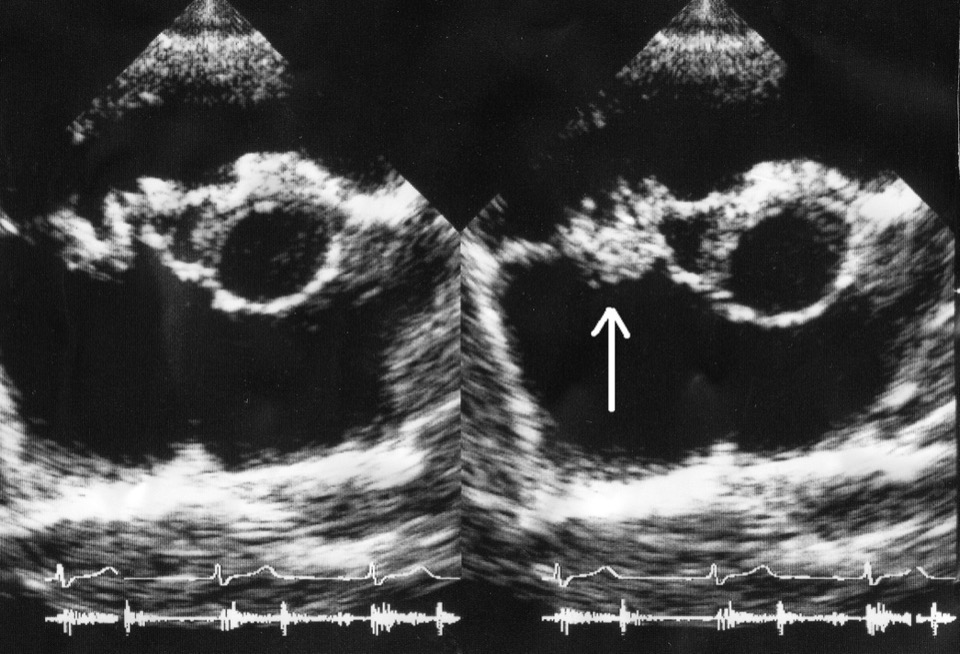

Vegetation features. A vegetation is an irregular, soft-tissue mass attached on the upstream (low-pressure) side of the valve near the coaptation line, with motion independent of the leaflet. The report records the valve and leaflet, the attachment site, maximal length in the plane of greatest extent, mobility, and the view used. Serial measurements use the same modality and view, because oblique or cross-modality measurements differ by more than true change. Vegetation length of 10 mm is the threshold used in surgical decisions in both US and European guidance, and very large vegetations (above 30 mm) carry a particularly high neurologic risk.
| Lesion | Echocardiographic appearance | Doppler and significance |
|---|---|---|
| Abscess | Perivalvular thickening or an echolucent or echodense cavity in the annulus, root, or adjacent myocardium | No flow into the cavity; uncontrolled infection and a surgical indication |
| Pseudoaneurysm | Pulsatile echo-free space communicating with a chamber or the aorta; the mitral-aortic intervalvular fibrosa is a classic site | Flow in and out through a neck; expands in the phase of higher pressure in the connected chamber |
| Fistula | Tract between two chambers or between the aorta and a chamber | Jet timed by the pressure difference; an aorta-to-right atrium fistula produces a continuous high-velocity jet |
| Leaflet perforation | Defect in the leaflet body | Regurgitant jet through the leaflet, away from the coaptation line |
| Leaflet aneurysm | Saccular bulge of the leaflet toward the low-pressure side, often on the anterior mitral leaflet from an aortic jet | Can perforate at its tip |
| Prosthetic dehiscence | Paravalvular defect; rocking of the prosthesis when extensive | New paravalvular regurgitation compared with prior imaging |
| Acute severe regurgitation | Flail cusp or leaflet, ruptured chordae | Aortic: short pressure half-time and premature mitral closure; mitral: early-peaking continuous-wave envelope with a normal-sized left atrium |
Destructive and perivalvular lesions of infective endocarditis on echocardiography. Each is a Duke-ISCVID major imaging criterion.
Cardiac CT, FDG PET/CT, leukocyte scintigraphy, and brain imaging
Cardiac CT. Electrocardiogram-gated contrast-enhanced CT is less sensitive than TEE for vegetations and leaflet perforation but defines abscess, pseudoaneurysm, fistula, prosthetic dehiscence, and root anatomy, especially when prosthetic material shadows the echocardiographic image. It is a Duke-ISCVID major imaging modality. CT also assesses the coronary arteries before surgery in patients at low or intermediate risk of coronary disease and in those with aortic vegetations, in whom catheter manipulation across the aortic root carries a risk of dislodging a vegetation. Combined CT angiography of the head, chest, abdomen, and pelvis detects emboli, abscesses, and mycotic aneurysms when clinically indicated.
FDG PET/CT. Activated leukocytes take up 18F-FDG, so PET/CT localizes metabolically active inflammation around valves, grafts, leads, and generator pockets, and across the whole body. Its value is greatest in prosthetic valve and device infection with negative or equivocal echocardiography, where pooled sensitivity in prosthetic valve endocarditis is 86% and specificity 84%. The 2020 ACC/AHA guideline rates FDG PET/CT as reasonable in possible endocarditis (Class 2a), and the 2026 AHA statement places it as a second-line test in suspected prosthetic valve endocarditis. Its sensitivity for native valve endocarditis is low. Uptake identifies inflammation and not infection: surgical adhesives, recent implantation, foreign-body reaction, vasculitis, and atherosclerosis produce uptake. Interpretation therefore integrates the pattern (intense, focal or multifocal, or heterogeneous uptake favors infection; diffuse homogeneous ring uptake soon after surgery favors healing), the interval since implantation, CT morphology, and microbiology. Physiologic myocardial glucose uptake obscures perivalvular and intracardiac lead activity, so the study requires a high-fat, very-low-carbohydrate diet and prolonged fasting, and a study with failed suppression is reported as nondiagnostic. Prior antibiotic therapy lowers sensitivity. PET/CT also identifies extracardiac foci, such as spondylodiscitis, septic emboli, and vascular graft infection, that change the duration of therapy and source control.
Leukocyte scintigraphy. Single-photon emission CT (SPECT) with radiolabeled autologous leukocytes is more specific for infection than FDG and is less affected by recent surgery. It is less sensitive, especially for native valves and vegetations smaller than 1 cm, and it requires cell labeling and delayed imaging. It is used when PET/CT is unavailable or when early postoperative inflammation limits FDG interpretation. It is part of the ESC imaging criteria but not of Duke-ISCVID.
Brain imaging. Cerebral CT or magnetic resonance imaging is obtained in every patient with neurologic symptoms. In the 2023 ESC guideline, brain and whole-body imaging is recommended in symptomatic patients (Class 1) and may be considered for screening in asymptomatic patients (Class 2b). The 2026 AHA statement regards brain magnetic resonance imaging before surgery as reasonable in asymptomatic patients and states that silent emboli do not delay surgery. Detection of a silent embolus adds a vascular minor criterion and can convert possible to definite endocarditis. Vascular imaging is added when an infectious aneurysm is suspected.
| Modality | Strengths | Limitations | Principal role |
|---|---|---|---|
| TTE | Universal availability; hemodynamics; ventricular function; tricuspid valve near the transducer | Sensitivity about 40% to 66% for vegetations; poor for prostheses, leads, and abscess | First study in all patients; baseline and end-of-therapy studies |
| TEE | Highest sensitivity for vegetations, perforation, and abscess; posterior prosthetic structures | Anterior aortic root shadowed by prosthetic rings; frame artifact in transcatheter valves | Prosthetic valves, devices, nondiagnostic TTE, suspected complications, before oral step-down |
| Cardiac CT | Abscess, pseudoaneurysm, fistula, dehiscence; coronary anatomy; surgical planning | Lower sensitivity for small vegetations and perforations; contrast; radiation | Perivalvular extension, prosthetic valve infection, preoperative coronary assessment |
| FDG PET/CT | Prosthetic valve, graft, and device infection; whole-body search for emboli and foci | Low sensitivity for native valves; false positives after surgery; requires myocardial suppression | Second-line in suspected prosthetic valve or device infection with nondiagnostic echocardiography |
| Leukocyte SPECT/CT | Specific for infection; less affected by recent surgery | Lower sensitivity and resolution; labor-intensive | Alternative when PET/CT is unavailable or inconclusive |
| Brain MRI and angiography | Ischemic lesions, hemorrhage, abscess, infectious aneurysms | Microbleeds of uncertain significance | Neurologic symptoms; timing of surgery; detection of silent emboli |
Imaging modalities in infective endocarditis. The TTE sensitivity figure is that cited in the 2026 AHA scientific statement; the FDG PET/CT figure is the meta-analysis estimate cited in the 2023 ESC guideline.
Differential diagnosis of valvular masses and sterile endocarditis
A mass on a valve is not synonymous with endocarditis. The diagnosis requires the clinical and microbiologic context, the attachment and motion of the mass, and comparison with prior studies.
| Entity | Typical appearance | Distinguishing context |
|---|---|---|
| Nonbacterial thrombotic (marantic) endocarditis | Small sessile masses along the closure line, often bilateral leaflet surfaces, usually mitral or aortic | Advanced adenocarcinoma, other malignancy, hypercoagulable states; sterile cultures; systemic and cerebral emboli; treated with anticoagulation and control of the underlying disease |
| Libman-Sacks endocarditis | Verrucous sessile lesions on either surface of the mitral or aortic leaflets, often near the base | Systemic lupus erythematosus and antiphospholipid syndrome; sterile; can cause regurgitation and emboli |
| Papillary fibroelastoma | Small, mobile, frond-like mass on a stalk, often mid-leaflet on the aortic valve | Afebrile; stable on serial imaging; embolic risk leads to resection |
| Myxoma | Atrial mass attached to the interatrial septum by a stalk | Constitutional symptoms and emboli can mimic endocarditis; attachment site is diagnostic |
| Lambl excrescences | Thin filamentous strands on the aortic closure line | Stable over years; no infection |
| Thrombus on a prosthesis or lead | Soft mass on a prosthetic valve or lead; fibrin strands on leads are common | Subtherapeutic anticoagulation (prosthetic valves); no bacteremia; lead strands alone do not establish infection |
| Degenerative tissue | Calcified nodules, myxomatous redundancy, ruptured chordae | Unchanged from prior imaging; no infection |
Mimics of infective endocarditis on echocardiography. Nonbacterial thrombotic and Libman-Sacks lesions embolize, and morphology alone does not separate them from infection.
Nonbacterial thrombotic endocarditis is the counterpart of the early infective lesion before organisms arrive. It is found in patients with mucin-producing adenocarcinomas (pancreas, lung, gastrointestinal tract, ovary), disseminated intravascular coagulation, and other hypercoagulable states, and it often presents as multiterritory cerebral infarction. Sterile cultures with embolic events lead to evaluation for malignancy and antiphospholipid antibodies. Other mimics of the clinical syndrome include acute rheumatic fever, adult-onset Still disease, vasculitis, atrial myxoma with constitutional symptoms, and lymphoma.
Antibiotic therapy by organism
Antibiotic therapy for endocarditis is bactericidal, intravenous in its initial phase, high-dose, and prolonged. The regimen depends on the organism, its minimum inhibitory concentration (MIC), the valve (native or prosthetic), renal function, allergy, and the surgical plan. Empiric therapy, started after cultures in an unstable patient, covers staphylococci (including methicillin-resistant strains), streptococci, and enterococci. For native valves and late prosthetic infection, vancomycin is commonly combined with an agent active against streptococci and HACEK organisms. Early prosthetic infection adds gram-negative coverage. Therapy is narrowed once the organism and susceptibilities are known, with infectious diseases consultation.
The 2026 AHA scientific statement replaced the 2015 statement. It presents consensus recommendations without formal classes of recommendation and changes several long-standing regimens. Gentamicin is no longer added in staphylococcal prosthetic valve endocarditis, because nephrotoxicity outweighs an unproven benefit. Native and prosthetic valve S. aureus endocarditis are both treated for 6 weeks. E. faecalis endocarditis is treated for 6 weeks with a two-beta-lactam combination for native and prosthetic valves. Duration is counted from the first day of negative blood cultures on effective therapy.
| Organism and valve | Preferred regimen (adult doses, normal renal function) | Duration |
|---|---|---|
| Methicillin-susceptible S. aureus (MSSA), native valve | Nafcillin or oxacillin 12 g/24 h IV in 6 doses, or cefazolin 6 g/24 h IV in 3 doses | 6 weeks |
| Methicillin-resistant S. aureus (MRSA), native valve | Vancomycin dosed to an area under the concentration-time curve (AUC) of 400 to 600 mg·h/L, or daptomycin 8 to 12 mg/kg/day; ceftaroline or linezolid are alternatives or additions in selected cases | 6 weeks |
| Staphylococci, prosthetic valve | Antistaphylococcal beta-lactam (MSSA) or vancomycin (MRSA, coagulase-negative staphylococci); rifampin 900 mg/24 h in 3 doses individualized, started after blood cultures clear; no gentamicin | 6 weeks |
| Viridans streptococci and S. gallolyticus, penicillin-susceptible, native valve | Aqueous penicillin G 24 million units/24 h IV, or ceftriaxone 2 g/24 h IV, without gentamicin | 4 weeks |
| Streptococci, prosthetic valve | Penicillin G 24 million units/24 h IV or ceftriaxone 2 g/24 h IV | 6 weeks |
| Streptococci, penicillin MIC 0.25 to 2 mg/L | Ceftriaxone monotherapy when the ceftriaxone MIC is 0.5 mg/L or less; penicillin-resistant isolates: ceftriaxone with gentamicin for the first 2 weeks | 4 to 6 weeks |
| E. faecalis, native or prosthetic | Ampicillin 2 g IV every 4 hours plus ceftriaxone 2 g IV every 12 hours | 6 weeks |
| HACEK organisms | Ceftriaxone 2 g/24 h IV; ciprofloxacin is the alternative when beta-lactams cannot be used | 4 weeks native; 6 weeks prosthetic |
| P. aeruginosa | Antipseudomonal beta-lactam (cefepime, piperacillin-tazobactam, or meropenem) plus tobramycin or a fluoroquinolone; early surgery | 6 weeks |
| Candida or Aspergillus | Valve surgery plus a lipid formulation of amphotericin B with or without flucytosine, or a high-dose echinocandin, followed by long-term oral azole suppression when indicated | At least 6 weeks |
| C. burnetii | Doxycycline plus hydroxychloroquine | At least 18 months |
Principal antibiotic regimens in adult infective endocarditis, after the 2026 AHA scientific statement, with doses from the 2015 AHA statement where unchanged. Durations are counted from the first negative blood culture on effective therapy. Doses are adjusted for renal function and body weight with pharmacist and infectious diseases input.
Staphylococci. For MSSA, an antistaphylococcal penicillin or cefazolin is superior to vancomycin, and cefazolin is an equivalent first-line option in the 2026 statement. In a randomized trial of S. aureus bacteremia and right-sided endocarditis, daptomycin 6 mg/kg/day was noninferior to standard therapy; higher doses are used in endocarditis because of the bacterial burden and the risk of resistance. Low-dose gentamicin added to staphylococcal therapy increased nephrotoxicity without clinical benefit, and aminoglycosides are not used in staphylococcal native valve endocarditis. When a native valve infected with S. aureus is replaced during therapy, the native valve regimen is continued and the prosthetic valve regimen is not substituted.
Streptococci. The penicillin MIC divides viridans group streptococci into susceptible, intermediate (0.25 to 2 mg/L), and resistant (4 mg/L or more) strains. Nutritionally variant streptococci (Granulicatella, Abiotrophia) and Gemella are treated with enterococcal-type regimens. Colonoscopy follows S. gallolyticus endocarditis.
Enterococci. E. faecalis requires two synergistic agents for cure. Ampicillin plus ceftriaxone saturates complementary penicillin-binding proteins. In a multicenter observational study, it was as effective as ampicillin plus gentamicin, with much less renal failure. Under the 2026 statement, it is the initial regimen for every patient with E. faecalis endocarditis, regardless of aminoglycoside susceptibility or kidney function, and ampicillin plus gentamicin is no longer listed. The 2026 statement regards colonoscopy as reasonable in every patient with E. faecalis endocarditis. Vancomycin-resistant Enterococcus faecium is treated with daptomycin- or linezolid-based regimens chosen with infectious diseases input, and surgery is often required.
Antibiotics after valve surgery. When surgery takes place during therapy, the planned total course is completed, counting preoperative days, provided at least 14 days follow surgery; the course is not restarted. A positive valve culture or molecular test does not by itself extend duration. When therapy was complete before surgery, valve cultures are negative, and no active infection is present, no further antibiotic course is needed. Metastatic foci and incomplete source control justify longer therapy.
Monitoring and adverse effects. Vancomycin is dosed by area under the curve, with trough-only monitoring no longer recommended for serious MRSA infection, and it carries nephrotoxicity that rises with exposure and with concurrent piperacillin-tazobactam or aminoglycosides. Daptomycin causes myopathy, so creatine kinase is measured weekly, and it causes eosinophilic pneumonia. Beta-lactams cause neutropenia, rash, and interstitial nephritis, and oxacillin causes hepatotoxicity. Rifampin induces hepatic enzymes and interacts with warfarin, direct oral anticoagulants, antiretrovirals, and many other drugs. Weekly blood counts, renal and hepatic function, and drug levels are monitored throughout a prolonged course.
Partial oral step-down, outpatient therapy, and long-acting lipoglycopeptides
The Partial Oral Treatment of Endocarditis (POET) trial randomized 400 stable patients with left-sided endocarditis to continued intravenous therapy or to two-drug oral regimens. Eligible patients had streptococcal, E. faecalis, S. aureus, or coagulase-negative staphylococcal infection and had completed at least 10 days of intravenous therapy (at least 7 days after valve surgery). The composite of all-cause mortality, unplanned cardiac surgery, embolic events, and relapse at 6 months occurred in 12.1% of the intravenous group and 9.0% of the oral group, meeting noninferiority. The median hospital stay after randomization fell from 19 to 3 days. About 20% of screened patients qualified, 27% had prosthetic valves, and only 5 injected drugs. The 2020 ACC/AHA guideline states that a change to oral therapy in stable patients with left-sided endocarditis may be considered (Class 2b). The 2026 AHA statement accepts partial oral therapy in carefully selected patients who meet POET-type criteria.
| Requirement | Criterion |
|---|---|
| Intravenous phase | At least 10 days of appropriate intravenous therapy, and at least 7 days after valve surgery |
| Organism | Streptococci, E. faecalis, MSSA, or coagulase-negative staphylococci with known susceptibilities |
| Clinical response | Afebrile for at least 3 days; blood cultures negative; white cell count below 15 × 10⁹/L; C-reactive protein below 25% of peak or below 20 mg/L |
| Imaging | TEE within 2 days before transition showing no abscess, no progression, and no new surgical indication |
| Exclusions | Body mass index above 40 kg/m², suspected malabsorption, another infection requiring intravenous therapy, doubtful adherence |
Criteria for partial oral step-down in left-sided endocarditis, from the POET trial as adopted in the 2026 AHA scientific statement. Oral regimens in POET combined two active drugs, for example amoxicillin with rifampin or moxifloxacin for streptococci and E. faecalis, and dicloxacillin or linezolid with rifampin or fusidic acid for MSSA; the statement reached no consensus on oral monotherapy.
Response-tailored treatment duration (POET II). The trial, published in 2026, tested whether total duration can be shortened in stable patients with left-sided endocarditis caused by streptococci, E. faecalis, or S. aureus. In 508 patients, a response-tailored strategy shortened median treatment from 41 to 26 days and met noninferiority for the composite of death, unplanned cardiac surgery, and symptomatic embolism. Relapse of bacteremia or endocarditis was more frequent (5.1% versus 1.6%). The 2026 AHA statement, published online 11 days after the trial, retains the standard durations, and these remain the reference.
Outpatient parenteral therapy. After the critical phase (the first 2 weeks, when complications concentrate), stable patients can complete parenteral therapy at home or in an infusion center with daily nursing contact and physician review at least weekly. Heart failure, uncontrolled infection, and neurologic complications exclude outpatient therapy.
Long-acting lipoglycopeptides. Dalbavancin and oritavancin have half-lives above 200 hours and allow weekly or less frequent dosing. They are used off-label to complete therapy of gram-positive endocarditis in patients who responded to initial intravenous therapy and cannot receive conventional outpatient therapy or oral step-down, including some people who inject drugs. The ESC guideline describes a dalbavancin regimen of a 1.5 g loading dose followed by 0.5 to 1 g weekly to complete 6 weeks. Randomized data in endocarditis are lacking, resistance has emerged during therapy, and oritavancin falsely prolongs the activated partial thromboplastin time for up to 5 days, which complicates heparin monitoring.
Indications for surgery in infective endocarditis
In international registries, about half of patients with definite endocarditis undergo surgery during the index hospitalization. Surgery removes infected tissue, drains abscesses, restores valve competence, and eliminates embolic sources. The three indications are heart failure, uncontrolled infection, and prevention of embolism. Once an indication is established, surgery is performed during the initial hospitalization, before the antibiotic course is complete.
| Indication | 2020 ACC/AHA class | 2023 ESC timing |
|---|---|---|
| Valve dysfunction causing symptoms of heart failure | 1 | Emergency (within 24 hours) for refractory pulmonary edema or cardiogenic shock; urgent (3 to 5 days) for other heart failure |
| Left-sided endocarditis caused by S. aureus, fungi, or other highly resistant organisms | 1 | Urgent for fungi and multiresistant organisms; individualized for S. aureus |
| Heart block, annular or aortic abscess, or destructive penetrating lesions | 1 | Urgent |
| Persistent bacteremia or fever for more than 5 to 7 days despite appropriate therapy | 1 | Urgent, after metastatic foci are excluded or controlled |
| Prosthetic valve endocarditis with relapsing infection after a complete course | 1 | Urgent surgery for early prosthetic valve endocarditis within 6 months of valve surgery (Class 1); individualized for late infection, with S. aureus favoring urgent surgery |
| Recurrent emboli with persistent vegetation despite appropriate antibiotics | 2a | Urgent; vegetation of 10 mm or more after an embolic episode is Class 1 in the ESC guideline |
| Native valve endocarditis with a mobile vegetation longer than 10 mm, with or without embolism | 2b | Urgent surgery may be considered with low operative risk |
| Device-related infection | 1 (complete system removal) | Complete extraction without delay |
Indications for surgery in active infective endocarditis. Surgery is performed during the initial hospitalization, before completion of the antibiotic course. The ESC guideline defines emergency surgery as within 24 hours, urgent as within 3 to 5 days, and nonurgent as within the same admission.
Heart failure. Valve destruction is the most common indication and the strongest predictor of death without surgery. Refractory pulmonary edema or cardiogenic shock from acute aortic or mitral regurgitation, a fistula, or prosthetic obstruction requires emergency surgery. Vasodilators, diuretics, and inotropes are bridges to the operating room, and an intra-aortic balloon pump is contraindicated in acute aortic regurgitation.
Uncontrolled infection. Perivalvular extension, persistent bacteremia without another source, enlarging vegetations, and organisms that antibiotics rarely eradicate (fungi, multidrug-resistant organisms, P. aeruginosa) indicate surgery. S. aureus prosthetic valve endocarditis carries high mortality with medical therapy alone, and early surgery is the usual course.
Prevention of embolism. Embolic risk is highest early and falls with therapy, so the benefit of surgery to prevent embolism is greatest in the first days. Vegetation size, mobility, location, organism, prior emboli, and operative risk are weighed together. The 2026 AHA statement lists vegetations larger than 10 mm with recurrent embolism or high embolic risk among the indications and favors surgery within 48 to 72 hours once an indication is established in the active phase.
Timing of surgery: the EASE trial and early operation
The Early Surgery versus Conventional Treatment in Infective Endocarditis (EASE) trial randomized 76 patients with left-sided native valve endocarditis, severe valve disease, and vegetations longer than 10 mm. Patients received either surgery within 48 hours of randomization or conventional care, in which surgery was performed only for complications. The composite of in-hospital death or embolic events at 6 weeks occurred in 1 patient (3%) in the early-surgery group and 9 patients (23%) in the conventional group, a difference driven by fewer embolic events. At 6 months, death, embolism, or recurrence occurred in 3% and 28%. The trial excluded patients with heart failure requiring emergency surgery, prosthetic valves, large strokes, and abscesses, and it was small, but it supports early operation when embolic risk is high and operative risk is acceptable.
Observational data favor early surgery when an indication is present, with the caveat of selection bias: patients too sick to operate on and patients with less severe disease both fall into the medical-therapy group. In-hospital mortality after surgery for endocarditis is about 10% to 20% and higher in patients older than 75 years. Operative risk rises with heart failure, shock, kidney failure, abscess, prosthetic valve infection, and S. aureus. Delay does not sterilize a destroyed valve or drain an abscess, and the risk of embolism, heart failure, and perivalvular extension accrues while the operation waits.
When surgery is indicated but the operative risk is prohibitive, the decision is made by the endocarditis team with the patient and family. Options include prolonged antibiotic therapy followed by suppressive therapy, percutaneous aspiration of right-sided vegetations, and palliation.
Surgery after stroke and intracranial hemorrhage
Cardiopulmonary bypass requires full heparinization and causes hypotension, so a fresh cerebral infarct can undergo hemorrhagic transformation or extension. Delay, however, exposes the patient to recurrent embolism and to the progression of heart failure and infection. Current guidance resolves this conflict by the type and size of the neurologic lesion.
Ischemic stroke without hemorrhage. A transient ischemic attack, silent embolism, or a small infarct without hemorrhage does not delay indicated surgery. The 2020 ACC/AHA guideline states that surgery without delay may be considered after a stroke without intracranial hemorrhage or extensive neurologic damage (Class 2b). The 2023 ESC guideline recommends surgery without delay after a transient ischemic attack or ischemic stroke when heart failure, uncontrolled infection, abscess, or persistent high embolic risk is present, provided coma is absent and hemorrhage has been excluded (Class 1).
Intracranial hemorrhage and extensive infarction. The 2020 ACC/AHA guideline states that delaying surgery for at least 4 weeks may be considered after major ischemic stroke or intracranial hemorrhage in a hemodynamically stable patient (Class 2b). The 2026 AHA statement defers surgery for at least 2 weeks, and preferably 4, after hemorrhagic or extensive ischemic stroke, and it regards cardiac surgery as inappropriate in a comatose patient. Retrospective series report acceptable outcomes from earlier surgery in selected patients with small hemorrhages when heart failure or uncontrolled infection makes delay dangerous. Brain imaging is repeated during the delay.
Acute stroke therapy. Intravenous thrombolysis is avoided in endocarditis-related ischemic stroke because of the risk of hemorrhage from septic arteritis and infectious aneurysms (ESC Class 3). Mechanical thrombectomy for large-vessel occlusion may be considered in selected patients (ESC Class 2b). An infectious aneurysm is excluded by computed tomographic or magnetic resonance angiography before cardiac surgery when one is suspected.
Surgical technique: repair, replacement, and root reconstruction
The principles are radical debridement of all infected and necrotic tissue, closure of cavities and fistulas, reconstruction of the annulus and root, and restoration of valve function. Infected prosthetic material and leads are removed.
Mitral valve repair is preferred when infection is limited to the leaflets, because it avoids a prosthesis in an infected field and preserves ventricular function. Perforations are closed with autologous or bovine pericardial patches, and chordal rupture is repaired with artificial chords. Extensive destruction requires replacement. Aortic valve infection usually requires replacement. Root abscess requires debridement and reconstruction with pericardial patches, aortic root replacement with a composite graft or homograft, or, for extensive destruction of the intervalvular fibrosa, a complex reconstruction of the aorto-mitral curtain.
The choice between mechanical and biological prostheses follows the usual criteria of age, life expectancy, bleeding risk, and patient preference; infection does not mandate a particular type. Homografts and stentless grafts are useful in extensive root destruction, and they have not been shown to lower the rate of reinfection compared with conventional prostheses. Tricuspid valve infection is managed by vegetectomy, leaflet repair, or, when necessary, valvectomy or replacement, and repair is preferred over replacement in people who inject drugs.
Complete heart block after surgery for aortic endocarditis with perivalvular extension is common; in one series of 444 patients operated on for endocarditis, 12.8% required a permanent pacemaker for atrioventricular block. Epicardial leads are often placed at operation, and a transvenous system is avoided until infection is cured.
Anticoagulation and antiplatelet therapy during endocarditis
Endocarditis is not an indication for anticoagulation, and anticoagulation does not prevent embolism from a vegetation while it raises the risk of intracranial hemorrhage from septic infarcts and infectious aneurysms. In a randomized trial, aspirin started at diagnosis did not reduce embolic events and was associated with a trend toward more bleeding, and antiplatelet therapy is not started to prevent embolism.
Patients with a separate indication for anticoagulation, such as a mechanical valve or atrial fibrillation, present a balance of thromboembolic and hemorrhagic risk. The 2023 ESC guideline recommends continuing anticoagulation in left-sided endocarditis with an established indication when there is no contraindication; in observational data this did not increase stroke, cerebral hemorrhage, or mortality at 10 weeks. Vitamin K antagonists are often replaced by unfractionated heparin in patients likely to need surgery, because heparin can be stopped and reversed quickly. Anticoagulation is interrupted after intracranial hemorrhage, with the timing of resumption decided with neurology; in a mechanical valve recipient, unfractionated heparin is restarted cautiously once repeat imaging shows stability. Direct oral anticoagulants are not used with mechanical valves.
Prosthetic valve and transcatheter valve endocarditis
Prosthetic valve endocarditis accounts for a growing share of all endocarditis and carries higher mortality than native valve disease. Early infection, within the first year, is acquired at operation or in hospital and is dominated by coagulase-negative staphylococci, S. aureus, enterococci, gram-negative bacilli, and fungi; late infection resembles native valve disease in organism spectrum. Infection of a mechanical valve begins at the sewing ring and spreads into the annulus, causing dehiscence, abscess, and fistula. Infection of a bioprosthesis also involves the leaflets and can cause vegetation, leaflet perforation, and obstruction.
Diagnosis is harder than in native valve disease. Fever with bacteremia from a typical or prosthesis-associated organism in a patient with a prosthesis is prosthetic valve endocarditis until proven otherwise. TTE assesses gradients and ventricular function, TEE is required in all patients, and cardiac CT and FDG PET/CT are added when echocardiography is nondiagnostic. A visible vegetation is not required. New paravalvular regurgitation or dehiscence compared with prior imaging satisfies a major criterion, and a rising gradient in a bioprosthesis during bacteremia is attributed to infection or thrombosis before it is attributed to degeneration.
Staphylococcal prosthetic valve endocarditis is treated for 6 weeks with rifampin added after blood cultures clear and without gentamicin. Early surgery is indicated for heart failure, perivalvular extension, dehiscence, S. aureus or fungal infection, persistent bacteremia, and relapse. Selected patients with late streptococcal or HACEK infection, no perivalvular extension, and prompt response can be treated medically. Prosthesis interpretation on imaging is detailed on the prosthetic heart valves page.
Transcatheter valve endocarditis. Patients are older and frailer, enterococci are prominent, and in-hospital mortality was 36% in the largest registry. The metal frame produces shadowing and reverberation on both TTE and TEE, and multimodality imaging with CT and FDG PET/CT is often required. Endocarditis on a transcatheter valve is managed as prosthetic valve endocarditis at experienced centers. Surgical explantation is technically demanding, and a benefit of explantation over medical therapy has not been convincingly shown, so the decision is individualized by the heart team. Infection after transcatheter edge-to-edge mitral repair and after percutaneous closure devices is managed by the same principles.
Cardiac implantable electronic device infection and lead extraction
Infection of a pacemaker or defibrillator presents as a pocket infection (erythema, swelling, warmth, drainage, erosion of the generator or leads through the skin), as lead infection with bacteremia, or as lead-related endocarditis with vegetations on the leads or the tricuspid valve. Staphylococci cause most infections. Erosion of hardware through the skin indicates infection of the whole system even when the pocket appears clean.
Diagnosis combines blood cultures, TEE of the full intracardiac course of the leads and the tricuspid valve, and, when the diagnosis remains uncertain, FDG PET/CT, which localizes pocket and extracardiac lead uptake. Intracardiac echocardiography is an option when transthoracic and transesophageal studies are negative and bacteremia persists. Fibrin strands and small thrombi on leads are common in patients without infection, so a lead mass by itself does not establish infection; bacteremia, pocket findings, septic pulmonary emboli, and comparison with prior studies do.
Complete extraction. Definite device infection requires complete removal of the generator and all leads, combined with antibiotic therapy (2020 ACC/AHA, Class 1; 2017 Heart Rhythm Society [HRS] consensus; 2024 AHA scientific statement). Antibiotics alone are followed by high relapse rates. In valvular endocarditis without evidence of device or lead infection, the 2020 ACC/AHA guideline rates complete removal as reasonable when the organism is S. aureus or a fungus and when the patient undergoes valve surgery (Class 2a), and the 2026 AHA statement favors complete extraction in valvular endocarditis even without lead vegetations or pocket findings. Lead extraction is performed at centers with experienced operators, cardiac surgical backup, and imaging, because vascular tear, tamponade, and tricuspid injury can be immediately fatal. Most lead vegetations are removed with the leads by transvenous extraction. Very large vegetations raise the risk of pulmonary embolization during extraction, and percutaneous aspiration before extraction or surgical removal is used in selected patients.
Reimplantation. The continuing need for a device is reassessed after extraction, and many patients no longer require one. When a new device is needed, it is implanted at a different site (the contralateral side, the iliac vein, or an epicardial position). Implantation follows resolution of local and systemic infection and at least 72 hours of negative blood cultures; in patients with valvular endocarditis, it also waits until 14 days after extraction. Leadless pacemakers and subcutaneous or extravascular defibrillators avoid endovascular leads. Pacemaker-dependent patients are bridged with a temporary active-fixation lead connected to an externalized generator, or with a leadless system.
Prevention of device infection. Preprocedural antistaphylococcal prophylaxis, meticulous pocket technique, and avoidance of unnecessary generator changes reduce infection. In the WRAP-IT trial of 6,983 patients undergoing generator replacement, upgrade, revision, or initial defibrillator implantation with cardiac resynchronization, an absorbable antibacterial envelope reduced major device infection at 12 months from 1.2% to 0.7%.
Ventricular assist device infection. Infections in patients with left ventricular assist devices are classified as device-specific (driveline, pump pocket, pump or cannula), device-related (bloodstream infection or endocarditis in which the device may be involved), and non-device infections. Driveline infections are the most frequent. Device components cannot be sterilized by antibiotics, so pump-related infection is managed with prolonged or lifelong suppressive therapy, surgical debridement, device exchange, or urgent transplantation. FDG PET/CT localizes driveline and pump infection.
Right-sided endocarditis and endocarditis in people who inject drugs
Right-sided endocarditis involves the tricuspid valve most often, and less commonly the pulmonary valve, the Eustachian valve, or a lead. It arises from injection drug use, central venous catheters, hemodialysis access, devices, and congenital heart disease. S. aureus is the usual organism. The presentation is pulmonary: fever, bacteremia, cough, pleuritic pain, and multiple cavitating septic pulmonary emboli. The tricuspid valve lies close to the chest wall, and TTE is often diagnostic, with TEE added for poor windows, devices, prostheses, and suspected left-sided involvement.
Right-sided disease has a better prognosis than left-sided disease, and most uncomplicated cases are cured with antibiotics. The 2026 AHA statement lists persistent bacteremia, recurrent septic pulmonary emboli, lung abscess after 7 days of antibiotics, and right-sided vegetations larger than 2 cm as indications for intervention. The 2023 ESC guideline lists right heart failure from severe tricuspid regurgitation unresponsive to diuretics, a vegetation larger than 20 mm with recurrent pulmonary emboli, and concomitant left-sided involvement. Tricuspid repair is preferred over replacement.
Percutaneous aspiration. Percutaneous mechanical aspiration of large tricuspid or lead vegetations with a vacuum system is used in selected patients with vegetations larger than 2 cm, persistent bacteremia or septic emboli despite antibiotics, and high surgical risk. No randomized trials establish its efficacy or safety, and it is performed at experienced centers. Aspiration debulks the infected mass; it does not replace extraction of an infected device or repair of a destroyed valve. The 2023 ESC guideline states it may be considered for right-sided septic masses in patients at high surgical risk (Class 2b).
Endocarditis in people who inject drugs. The 2022 AHA scientific statement frames endocarditis in people who inject drugs as a complication of a treatable substance use disorder. Addiction medicine consultation is a core component of endocarditis team care. Medications for opioid use disorder (buprenorphine, methadone, extended-release naltrexone) are started during hospitalization, and harm-reduction services are linked at discharge. The decision to operate rests on the same cardiac indications as in other patients and is not withheld because of drug use, although reoperation for reinfection of a prosthesis is a recognized challenge. When a full intravenous course in hospital is not completed, partial oral therapy or a long-acting lipoglycopeptide is preferable to an incomplete course. Valve repair, when feasible, avoids a prosthesis at risk of later reinfection.
Endocarditis in pregnancy and adult congenital heart disease
Pregnancy. Endocarditis in pregnancy is rare but threatens mother and fetus. In a systematic review of pregnancy-associated endocarditis, maternal mortality was 11%, and 14% of pregnancies ended in fetal death. Injection drug use, congenital heart disease, and prosthetic valves are the main substrates in contemporary series; rheumatic disease has declined. Diagnosis follows the same criteria, and TEE is safe with appropriate sedation and monitoring. Antibiotic choice favors beta-lactams, vancomycin, and daptomycin; aminoglycosides, tetracyclines, and fluoroquinolones are avoided when an alternative exists because of fetal toxicity. Indications for surgery do not change, but cardiopulmonary bypass carries a substantial fetal risk. When the fetus is viable, delivery before cardiopulmonary bypass is often planned, and cardiology, cardiac surgery, obstetrics, and anesthesia decide the timing together.
Adult congenital heart disease. Congenital heart disease carries a lifelong risk of endocarditis that varies with the lesion. The highest risks are with unrepaired cyanotic disease, palliative shunts and conduits, prosthetic valves and conduits (including transcatheter pulmonary valves), and repairs with residual defects next to prosthetic material. A small ventricular septal defect, left-sided obstructive lesions, and a bicuspid aortic valve are common substrates. Endocarditis of a right ventricle-to-pulmonary artery conduit or a transcatheter pulmonary valve presents with fever and a rising conduit gradient, and that combination is treated as endocarditis until excluded. Imaging is limited by complex anatomy and prosthetic material, and cardiac CT and FDG PET/CT are used frequently.
Composition and role of the multidisciplinary endocarditis team
The 2020 ACC/AHA guideline specifies that patients with endocarditis are managed by a multispecialty heart valve team of cardiology, cardiac surgery, and infectious diseases specialists (Class 1). The 2026 AHA and 2023 ESC documents describe an endocarditis team of cardiology, infectious diseases, cardiac surgery, cardiac imaging, microbiology, and, as needed, neurology, neurosurgery, nephrology, electrophysiology, addiction medicine, pharmacy, and nursing. Patients with complicated endocarditis are managed at, or in close contact with, a reference center with on-site cardiac surgery. Team management has been associated with earlier surgery, more consistent antibiotic therapy, and lower mortality in observational studies.
The team meets at diagnosis and at each decision point: the regimen and its duration, the need and timing of surgery, management of neurologic complications and anticoagulation, the step-down to oral or outpatient therapy, device extraction and reimplantation, and the follow-up plan.
Prognosis, relapse, recurrence, and follow-up
In-hospital mortality is 15% to 30%. Predictors of death include older age, comorbidity (diabetes, kidney failure, malignancy), prosthetic valve infection, S. aureus and fungal infection, heart failure, perivalvular extension, stroke, septic shock, and an indication for surgery that is not followed by surgery. In the ICE-PCS cohort, S. aureus infection was independently associated with in-hospital death. Long-term survival is reduced by the underlying comorbidity, valve dysfunction, and recurrence.
Relapse and reinfection. Recurrence after a first episode is reported in 2% to 9% of survivors in contemporary series. A relapse is a repeat episode with the same organism, usually within 6 months, and indicates insufficient therapy or a persistent focus. A reinfection is an episode with a different organism, or a late episode, and reflects ongoing risk, most often in people who inject drugs, patients on hemodialysis, and those with prosthetic valves. Relapse with a prosthesis is an indication for surgery.
Suppressive therapy. When infected prosthetic material cannot be removed (a prosthetic valve or device with prohibitive operative risk, a retained lead, a ventricular assist device) or in fungal endocarditis without source control, long-term oral suppressive therapy directed at the organism is reasonable after the curative course. It does not replace surgery when surgery is feasible.
| Time | Action |
|---|---|
| Completion of therapy | TTE as a new baseline of valve and ventricular function; removal of intravenous catheters; dental evaluation if not done during admission |
| After valve surgery | TTE to document the repair or prosthesis, with a follow-up study 6 to 12 weeks after surgery |
| First weeks after discharge | Clinical review for fever, heart failure, and drug toxicity; blood cultures before any antibiotic for fever |
| Months 1 to 12 | Clinical review at intervals; surveillance for late heart failure, progressive regurgitation, and need for delayed valve surgery |
| Lifelong | Dental review at least twice a year for high-risk patients; skin hygiene; addiction care; prophylaxis before qualifying dental procedures |
Follow-up after infective endocarditis. A new murmur, recurrent fever, heart failure, embolism, or new conduction disease after treatment is evaluated as possible relapse.
Patient education. Education covers the symptoms of relapse, the need for blood cultures before any antibiotic is given for fever, oral hygiene and dental care, avoidance of tattooing and body piercing, and care of skin and intravenous access. For people who inject drugs, it also covers safe injection practice and addiction treatment. Patients with a highest-risk condition carry information on their prophylaxis needs.
Antibiotic prophylaxis: patients and procedures
Prophylaxis has been progressively restricted since 2007 because bacteremia from daily activities far exceeds that from dental procedures, because the absolute risk reduction from a single prophylactic dose is small, and because antibiotics carry their own adverse effects. The 2021 AHA scientific statement retained the 2007 selection of patients, and the 2020 ACC/AHA guideline rates prophylaxis before qualifying dental procedures as reasonable in these patients (Class 2a). An interrupted time-series analysis in England found an increase in endocarditis above the projected trend after prophylaxis prescribing fell sharply in 2008. The design cannot establish causation, and observational data remain the basis of current policy.
| Element | US guidance (2020 ACC/AHA; 2021 AHA) | 2023 ESC guideline |
|---|---|---|
| Highest-risk patients | Prosthetic valves, including transcatheter valves and homografts; prosthetic material for valve repair (annuloplasty rings, chords, clips); prior endocarditis; unrepaired cyanotic congenital heart disease, including palliative shunts and conduits; completely repaired congenital defects with prosthetic material during the first 6 months; repaired congenital defects with residual shunts or regurgitation at or next to a prosthetic patch or device; cardiac transplant recipients with valve regurgitation from a structurally abnormal valve | Prior endocarditis; surgical or transcatheter prosthetic valves and valve repair material; untreated cyanotic congenital disease and congenital disease treated with prosthetic material (up to 6 months, or lifelong with residual shunt or regurgitation); ventricular assist devices as destination therapy |
| Intermediate-risk patients | Not a category; prophylaxis not recommended | Rheumatic disease, degenerative valve disease, congenital valve anomalies including bicuspid aortic valve, cardiac electronic devices, hypertrophic cardiomyopathy: not routinely recommended; may be considered individually |
| Dental procedures | Procedures involving manipulation of gingival tissue or the periapical region of teeth, or perforation of the oral mucosa | Extraction, oral surgery, and procedures requiring manipulation of the gingival or periapical region |
| Nondental procedures | Not recommended for gastrointestinal, genitourinary, or transesophageal procedures without active infection (Class 3: No Benefit) | May be considered for selected invasive respiratory, gastrointestinal, genitourinary, skin, or musculoskeletal procedures in high-risk patients (Class 2b) |
| Regimen | Amoxicillin 2 g orally 30 to 60 minutes before; ampicillin 2 g or cefazolin or ceftriaxone 1 g IM or IV; cephalexin 2 g, azithromycin or clarithromycin 500 mg, or doxycycline 100 mg with penicillin allergy; clindamycin no longer recommended | Amoxicillin or ampicillin 2 g; cefazolin or ceftriaxone 1 g; cephalexin 2 g; azithromycin or clarithromycin 500 mg; doxycycline 100 mg |
Antibiotic prophylaxis against infective endocarditis in adults. Cephalosporins are avoided after anaphylaxis, angioedema, or urticaria with penicillin or ampicillin. A dose not given before the procedure can be given up to 2 hours afterward.
Routine anesthetic injections through noninfected tissue, dental radiographs, placement or adjustment of removable prosthodontic or orthodontic appliances, and bleeding from trauma to the lips or oral mucosa do not warrant prophylaxis. When a patient already takes an antibiotic, prophylaxis uses a different class. Prophylaxis is not a substitute for oral hygiene: regular dental care, treatment of periodontal disease, and avoidance of skin breaches are the main preventive measures in every patient at risk.
Prevention in hospitalized and surgical patients. Healthcare-associated endocarditis is prevented by limiting the use and duration of central venous catheters, by aseptic insertion and care of vascular access, by prompt removal of infected catheters, and by treating S. aureus bacteremia with source control and adequate therapy. Patients scheduled for valve surgery or device implantation undergo dental evaluation and treatment of dental infection beforehand when time allows, and screening and decolonization for S. aureus are used before cardiac surgery.
Prophylaxis eligibility and regimen
2020 ACC/AHA and 2021 AHAProphylaxis eligibility and adult regimens from the 2020 ACC/AHA valve guideline and the 2021 AHA scientific statement, with the 2023 ESC position where it differs.
Self-assessment
A 78-year-old man had transcatheter aortic valve replacement 8 months ago. He has a temperature of 38.4 °C, and Enterococcus faecalis grows in three of three blood culture sets. Transthoracic and transesophageal echocardiography are nondiagnostic because of frame artifact. FDG PET/CT with adequate myocardial suppression shows intense focal uptake at the valve frame. How is this episode classified by the 2023 Duke-ISCVID criteria, and what antibiotic combination and duration does current AHA guidance favor?
Cardiovascular Medicine Board ReviewReferences
- DeSimone DC, Marks L, Dayer MJ, et al. Infective endocarditis: diagnosis, antibiotic therapy, and management: a scientific statement from the American Heart Association. Circulation. 2026;154(15):e593–e619. doi:10.1161/CIR.0000000000001466
- Baddour LM, Wilson WR, Bayer AS, et al. Infective endocarditis in adults: diagnosis, antimicrobial therapy, and management of complications: a scientific statement for healthcare professionals from the American Heart Association. Circulation. 2015;132:1435–1486. doi:10.1161/CIR.0000000000000296
- Fowler VG Jr, Durack DT, Selton-Suty C, et al. The 2023 Duke-International Society for Cardiovascular Infectious Diseases criteria for infective endocarditis: updating the modified Duke criteria. Clin Infect Dis. 2023;77:518–526. doi:10.1093/cid/ciad271
- Otto CM, Nishimura RA, Bonow RO, et al. 2020 ACC/AHA guideline for the management of patients with valvular heart disease: a report of the American College of Cardiology/American Heart Association Joint Committee on Clinical Practice Guidelines. Circulation. 2021;143:e72–e227. doi:10.1161/CIR.0000000000000923
- Delgado V, Ajmone Marsan N, de Waha S, et al. 2023 ESC guidelines for the management of endocarditis. Eur Heart J. 2023;44:3948–4042. doi:10.1093/eurheartj/ehad193
- Wilson WR, Gewitz M, Lockhart PB, et al. Prevention of viridans group streptococcal infective endocarditis: a scientific statement from the American Heart Association. Circulation. 2021;143:e963–e978. doi:10.1161/CIR.0000000000000969
- Kusumoto FM, Schoenfeld MH, Wilkoff BL, et al. 2017 HRS expert consensus statement on cardiovascular implantable electronic device lead management and extraction. Heart Rhythm. 2017;14:e503–e551. doi:10.1016/j.hrthm.2017.09.001
- Baddour LM, Esquer Garrigos Z, Rizwan Sohail M, et al. Update on cardiovascular implantable electronic device infections and their prevention, diagnosis, and management: a scientific statement from the American Heart Association. Circulation. 2024;149:e201–e216. doi:10.1161/CIR.0000000000001187
- Baddour LM, Weimer MB, Wurcel AG, et al. Management of infective endocarditis in people who inject drugs: a scientific statement from the American Heart Association. Circulation. 2022;146:e187–e201. doi:10.1161/CIR.0000000000001090
- Pettersson GB, Coselli JS, Hussain ST, et al. 2016 The American Association for Thoracic Surgery (AATS) consensus guidelines: surgical treatment of infective endocarditis: executive summary. J Thorac Cardiovasc Surg. 2017;153:1241–1258.e29. doi:10.1016/j.jtcvs.2016.09.093
- Zoghbi WA, Jone PN, Chamsi-Pasha MA, et al. Guidelines for the evaluation of prosthetic valve function with cardiovascular imaging: a report from the American Society of Echocardiography developed in collaboration with the Society for Cardiovascular Magnetic Resonance and the Society of Cardiovascular Computed Tomography. J Am Soc Echocardiogr. 2024;37:2–63. doi:10.1016/j.echo.2023.10.004
- Li JS, Sexton DJ, Mick N, et al. Proposed modifications to the Duke criteria for the diagnosis of infective endocarditis. Clin Infect Dis. 2000;30:633–638. doi:10.1086/313753
- Durack DT, Lukes AS, Bright DK. New criteria for diagnosis of infective endocarditis: utilization of specific echocardiographic findings. Duke Endocarditis Service. Am J Med. 1994;96:200–209. doi:10.1016/0002-9343(94)90143-0
- Kang DH, Kim YJ, Kim SH, et al. Early surgery versus conventional treatment for infective endocarditis. N Engl J Med. 2012;366:2466–2473. doi:10.1056/NEJMoa1112843
- Chan KL, Dumesnil JG, Cujec B, et al. A randomized trial of aspirin on the risk of embolic events in patients with infective endocarditis. J Am Coll Cardiol. 2003;42:775–780. doi:10.1016/S0735-1097(03)00829-5
- Iversen K, Ihlemann N, Gill SU, et al. Partial oral versus intravenous antibiotic treatment of endocarditis. N Engl J Med. 2019;380:415–424. doi:10.1056/NEJMoa1808312
- Bundgaard H, Pries-Heje M, Hjulmand J, et al. Response-tailored or standard-duration antibiotic treatment for infective endocarditis. N Engl J Med. 2026;395:1600–1609. doi:10.1056/NEJMoa2607887
- Murdoch DR, Corey GR, Hoen B, et al. Clinical presentation, etiology, and outcome of infective endocarditis in the 21st century: the International Collaboration on Endocarditis-Prospective Cohort Study. Arch Intern Med. 2009;169:463–473. doi:10.1001/archinternmed.2008.603
- Habib G, Erba PA, Iung B, et al. Clinical presentation, aetiology and outcome of infective endocarditis. Results of the ESC-EORP EURO-ENDO (European infective endocarditis) registry: a prospective cohort study. Eur Heart J. 2019;40:3222–3232. doi:10.1093/eurheartj/ehz620
- Thuny F, Di Salvo G, Belliard O, et al. Risk of embolism and death in infective endocarditis: prognostic value of echocardiography: a prospective multicenter study. Circulation. 2005;112:69–75. doi:10.1161/CIRCULATIONAHA.104.493155
- Regueiro A, Linke A, Latib A, et al. Association between transcatheter aortic valve replacement and subsequent infective endocarditis and in-hospital death. JAMA. 2016;316:1083–1092. doi:10.1001/jama.2016.12347
- Fournier PE, Thuny F, Richet H, et al. Comprehensive diagnostic strategy for blood culture-negative endocarditis: a prospective study of 819 new cases. Clin Infect Dis. 2010;51:131–140. doi:10.1086/653675
- Fowler VG Jr, Boucher HW, Corey GR, et al. Daptomycin versus standard therapy for bacteremia and endocarditis caused by Staphylococcus aureus. N Engl J Med. 2006;355:653–665. doi:10.1056/NEJMoa053783
- Cosgrove SE, Vigliani GA, Campion M, et al. Initial low-dose gentamicin for Staphylococcus aureus bacteremia and endocarditis is nephrotoxic. Clin Infect Dis. 2009;48:713–721. doi:10.1086/597031
- Fernández-Hidalgo N, Almirante B, Gavaldà J, et al. Ampicillin plus ceftriaxone is as effective as ampicillin plus gentamicin for treating Enterococcus faecalis infective endocarditis. Clin Infect Dis. 2013;56:1261–1268. doi:10.1093/cid/cit052
- Rybak MJ, Le J, Lodise TP, et al. Therapeutic monitoring of vancomycin for serious methicillin-resistant Staphylococcus aureus infections: a revised consensus guideline and review by the American Society of Health-System Pharmacists, the Infectious Diseases Society of America, the Pediatric Infectious Diseases Society, and the Society of Infectious Diseases Pharmacists. Am J Health Syst Pharm. 2020;77:835–864. doi:10.1093/ajhp/zxaa036
- Tarakji KG, Mittal S, Kennergren C, et al. Antibacterial envelope to prevent cardiac implantable device infection. N Engl J Med. 2019;380:1895–1905. doi:10.1056/NEJMoa1901111
- Hannan MM, Husain S, Mattner F, et al. Working formulation for the standardization of definitions of infections in patients using ventricular assist devices. J Heart Lung Transplant. 2011;30:375–384. doi:10.1016/j.healun.2011.01.717
- Wurcel AG, Anderson JE, Chui KKH, et al. Increasing infectious endocarditis admissions among young people who inject drugs. Open Forum Infect Dis. 2016;3:ofw157. doi:10.1093/ofid/ofw157
- Kebed KY, Bishu K, Al Adham RI, et al. Pregnancy and postpartum infective endocarditis: a systematic review. Mayo Clin Proc. 2014;89:1143–1152. doi:10.1016/j.mayocp.2014.04.024
- Dayer MJ, Jones S, Prendergast B, et al. Incidence of infective endocarditis in England, 2000–13: a secular trend, interrupted time-series analysis. Lancet. 2015;385:1219–1228. doi:10.1016/S0140-6736(14)62007-9