Library › Imaging › Coronary CTA and calcium scoring
Coronary CT angiography and calcium scoring
Coronary CT angiography and coronary calcium scoring in adults, from indications, preparation, and acquisition through Agatston scoring, CAD-RADS 2.0 reporting, plaque, CT physiology, stents, grafts, anomalies, artifacts, and the outcome trials.
Coronary CT angiography (CCTA) is a Class 1 test for intermediate-risk acute chest pain without known coronary disease and for intermediate- to high-risk stable chest pain. Its negative predictive value for obstructive disease is high, and in SCOT-HEART a CCTA-guided strategy reduced coronary death or myocardial infarction at 5 and 10 years, largely through earlier preventive therapy. The Coronary Artery Disease Reporting and Data System (CAD-RADS) 2.0 report combines the maximal stenosis (0 to 5), the plaque burden (P1 to P4), and modifiers for nondiagnostic segments, high-risk plaque, ischemia, stents, grafts, and exceptions. A coronary calcium score measures calcified plaque on a noncontrast scan: a lesion counts at 130 Hounsfield units (HU) or more over an area of at least 1 mm².
Indications for coronary CT angiography and calcium scoring
CCTA answers anatomic questions: whether coronary plaque is present, how much, of what composition, and whether it narrows the lumen. Calcium scoring answers a narrower question with a noncontrast scan: how much calcified plaque is present. Stress testing answers whether ischemia is present. The 2021 American Heart Association/American College of Cardiology (AHA/ACC) multisociety chest pain guideline matches the test to the question and to the patient's risk, and the 2023 multimodality appropriate use criteria rate CCTA in symptomatic patients across the same scenarios.
| Clinical setting | Role of cardiac CT | Class |
|---|---|---|
| Acute chest pain, low risk (30-day death or MACE below 1%) | Discharge home without admission or urgent cardiac testing | 2a |
| Acute chest pain, intermediate risk, no known CAD, ACS excluded or evaluation inconclusive | CCTA to exclude atherosclerotic plaque and obstructive CAD | 1 (A) |
| Stable chest pain, low risk | Testing can be deferred; coronary calcium scoring as a first-line test to exclude calcified plaque and identify a low likelihood of obstructive CAD | 2a |
| Stable chest pain, intermediate to high risk, no known CAD | CCTA for diagnosis of CAD, risk stratification, and guidance of treatment | 1 |
| CCTA stenosis of 40% to 90% in a proximal or middle segment | FFR-CT for vessel-specific ischemia and revascularization decisions | 2a |
| Inconclusive or abnormal stress test | CCTA | 2a |
| Known nonobstructive CAD with new or persistent stable chest pain | CCTA to assess plaque progression and new obstruction | 2a |
| Prior CABG or coronary stents of 3 mm or larger, stable chest pain | CCTA to evaluate graft patency or stent patency | 2a |
| Suspected anomalous coronary origin or course | CCTA defines origin, course, ostium, and intramural segment | — |
Recommendations from the 2021 AHA/ACC/ASE/CHEST/SAEM/SCCT/SCMR chest pain guideline, with Class of Recommendation (Level of Evidence where cited). ACS, acute coronary syndrome; CABG, coronary artery bypass grafting; CAD, coronary artery disease; FFR-CT, fractional flow reserve derived from CT; MACE, major adverse cardiac events.
Age and expected calcium shift the balance between anatomic and functional testing. The 2021 guideline synopsis favors CCTA in patients younger than 65 years who are not taking preventive therapy, because plaque is less calcified and the identification of nonobstructive disease changes prevention. Stress testing may be favored at 65 years and older, when obstructive disease and ischemia are more likely and dense calcium lowers the specificity of CCTA. The choice between CCTA and stress imaging in established coronary disease is covered on the chronic coronary disease page under the 2023 AHA/ACC guideline. The 2024 European Society of Cardiology (ESC) guideline recommends CCTA as an initial test in symptomatic patients with a low or moderate (above 5% to 50%) clinical likelihood of obstructive disease (Class I, Level A).
A calcium score is a risk test for asymptomatic adults. It does not image the lumen or noncalcified plaque, and it does not answer the diagnostic question in a patient with suspected obstructive disease. Its use to set statin intensity and low-density lipoprotein (LDL) cholesterol goals under the 2026 ACC/AHA dyslipidemia guideline, including the Multi-Ethnic Study of Atherosclerosis (MESA) risk score and age-, sex-, and race-specific percentiles, is covered on the cardiovascular risk assessment page.
Factors that predict a nondiagnostic study. Most failed studies can be predicted before the scan. A fast or irregular rhythm that cannot be controlled, inability to hold the breath or follow instructions, severe obesity, dense calcium in the segment of interest, a small or heavily calcified stent, a barrier to iodinated contrast, and a scanner without the needed temporal resolution each lower the chance of a diagnostic result. A stress test or invasive angiography answers the question better in a patient who combines several of these factors, for example atrial fibrillation with a variable ventricular rate, an Agatston score above 1,000, and severe obesity.
Patient preparation, contrast safety, and breath-holding
Screening before CCTA reviews the indication, kidney function, prior reactions to iodinated contrast, current medications (beta-blockers, nondihydropyridine calcium channel blockers, ivabradine, nitrates, phosphodiesterase-5 inhibitors, and metformin), pregnancy status in women of childbearing potential, heart rhythm, and the ability to hold the breath for the length of the acquisition. Patients usually avoid caffeine on the day of the scan because it raises the heart rate.
Kidney function. The American College of Radiology and National Kidney Foundation consensus (2020) concluded that the risk of acute kidney injury from intravenous contrast has been overstated. Prophylaxis with intravenous normal saline is indicated for patients with acute kidney injury or an estimated glomerular filtration rate (eGFR) below 30 mL/min/1.73 m² who are not receiving maintenance dialysis, and it may be considered at an eGFR of 30 to 44 mL/min/1.73 m² in selected high-risk patients. A patient on maintenance dialysis can receive contrast without urgent dialysis afterward unless the volume load is a concern. Metformin is withheld after contrast in patients with acute kidney injury or an eGFR below 30 mL/min/1.73 m².
Prior contrast reaction. A prior allergic-like reaction to iodinated contrast is the strongest predictor of a repeat reaction. The usual response is premedication with corticosteroids and an antihistamine, a change to a different contrast agent, or an alternative test. Premedication lowers the rate of reactions but does not prevent all of them, so the scan is performed where a reaction can be treated. A shellfish or iodine allergy is not a contraindication. Physiologic reactions, such as warmth and nausea, are not allergic and do not require premedication.
Venous access, electrocardiographic leads, and breath-hold. A large-bore antecubital cannula, usually 18 or 20 gauge and preferably in the right arm, supports injection rates of 5 to 7 mL/s and avoids dense contrast crossing the left brachiocephalic vein. The electrocardiographic leads are placed off the scan field to give a clear R wave for triggering. The breath-hold is rehearsed before contrast is given. A Valsalva maneuver or a large inspiration changes the heart rate, dilutes the contrast bolus with unopacified blood from the inferior vena cava, and shifts the heart between the planning scan and the acquisition. A moderate inspiration, held without straining, is the standard instruction.
Heart-rate control and nitroglycerin before coronary CT angiography
The coronary arteries move with every beat, and the right coronary artery moves most. Motion is least during diastasis, the slow-filling period of mid-diastole, and a slower heart rate lengthens that window. The Society of Cardiovascular Computed Tomography (SCCT) acquisition guideline uses a heart rate of 60 beats/min or less as the standard target when rate control is safe and useful. The target is a guide. A regular rate of 62 to 65 beats/min may produce excellent images on a dual-source scanner, while the same rate may blur the right coronary artery on a single-source scanner with slower rotation. Beat-to-beat variability degrades images more than a modest, stable elevation of the rate, because the reconstruction assumes that each beat repeats the one before it.
A stable, low heart rate also lowers radiation dose. It allows a prospectively triggered acquisition with a narrow window, or a high-pitch helical acquisition that covers the heart in one beat, in place of a retrospectively gated scan.
| Agent | Regimens in published protocols | Action | Main cautions |
|---|---|---|---|
| Oral metoprolol tartrate | 50 to 100 mg, given 1 hour before the scan, with the dose scaled to the resting rate; some protocols add 50 mg 12 hours before the scan (immediate-release forms only) | First-line rate control | Hypotension, decompensated heart failure, second- or third-degree atrioventricular block without a pacemaker, active bronchospasm, a home beta-blocker dose already taken |
| Intravenous metoprolol | 5 mg increments at the scanner, each followed by about 5 minutes of monitoring, typically to a maximum of 20 to 25 mg | Titration while rate and blood pressure are monitored | As for oral metoprolol; each dose follows a new measurement of rate, rhythm, and blood pressure |
| Ivabradine (oral) | 15 mg orally 1 hour before the scan, or 7.5 mg in patients on chronic beta-blockade; an investigational use, and an add-on dose in beta-blocked patients did not lower the scan heart rate further in one ambulatory series | Inhibits the sinus node funny current (If); slows the sinus rate without lowering blood pressure or contractility | Effective only in sinus rhythm; slower onset; no effect on the ventricular rate in atrial fibrillation |
| Diltiazem (oral, immediate release) | Small oral doses (30 mg, repeated once in trial protocols) when a beta-blocker is contraindicated | Slows the sinus rate and the ventricular response in atrial fibrillation | Hypotension, reduced ejection fraction, conduction disease; additive effect with beta-blockers |
| Sublingual nitroglycerin | 0.4 to 0.8 mg (one or two tablets or sprays) about 5 minutes before the contrast acquisition; effect lasts about 20 to 30 minutes | Dilates the epicardial arteries; improves visualization of distal segments and branches | Systolic hypotension; sildenafil or vardenafil within 24 hours or tadalafil within 48 hours; severe aortic stenosis; obstructive hypertrophic cardiomyopathy |
Heart-rate control and vasodilation before coronary CTA. Beta-blocker, ivabradine, and nitroglycerin regimens follow the SCCT acquisition guideline (2016); the diltiazem regimen comes from published trial protocols, and each laboratory sets its own. Beta-blockers and calcium channel blockers taken at home count toward the dose.
Each dose is a decision made on fresh measurements. A patient who took a beta-blocker at home may already be at target, and a template that adds a dose automatically can cause symptomatic bradycardia or hypotension. Repeated intravenous doses given to reach exactly 60 beats/min trade blood pressure for a gain of one or two beats per minute. When the target cannot be reached safely, the acquisition is adapted: an end-systolic reconstruction window, a scanner with better temporal resolution, or acceptance of the higher rate when the question concerns a large proximal structure such as an anomalous origin.
The quietest phase moves with heart rate. At low rates it lies in mid-diastole, around 70% to 80% of the R-R interval; at higher rates diastasis shortens and end-systole, around 35% to 45%, becomes the more stable phase. The right and left coronary arteries may be sharpest at different phases. An acquisition planned for a higher rate must include end-systole, because a phase that was not acquired cannot be reconstructed later.
Nitroglycerin. Nitroglycerin is given to improve the images, not to treat findings. When it is withheld because of hypotension or a phosphodiesterase-5 inhibitor, the report says so, because distal vessel caliber and visualization may be reduced. Differences in distal caliber between two studies of the same patient are compared only after checking whether nitroglycerin was given both times.
Rhythm problems. Atrial fibrillation combines a variable rate with irregular timing, and a controlled average rate does not make the rhythm regular. Wide-detector scanners that cover the heart in one rotation, dual-source scanners, and retrospective gating with electrocardiographic editing tolerate irregular rhythms best. Premature beats in a prospectively triggered sequential scan misregister one slab and produce a step that can mimic a stenosis or occlusion. A pacemaker in stable continuous pacing provides a regular trigger; a beta-blocker does not slow a paced rhythm, and the lead produces streak artifact near the right coronary artery.
Scanner technology and ECG-synchronized acquisition
Temporal resolution, the time needed to acquire the data for one image, determines how well coronary motion is frozen. A single-source scanner reconstructs an image from about half a gantry rotation, so its temporal resolution is roughly half the rotation time; with rotation times of 0.25 to 0.35 seconds this is about 125 to 175 ms. A dual-source scanner uses two tube-detector pairs mounted about 90° apart and needs only a quarter rotation, which gives a temporal resolution of about 66 to 75 ms. Multisegment reconstruction combines data from several beats to shorten the effective window but depends on a perfectly regular rhythm.
Coverage along the patient axis determines how many beats the acquisition spans. A 64-row scanner covers about 4 cm per rotation and builds the heart from several beats. A wide-detector scanner with 256 or 320 rows covers about 16 cm and can image the whole heart in a single beat, which removes slab misregistration. Spatial resolution of about 0.5 mm in-plane on conventional detectors limits the evaluation of small branches, stents, and dense calcium.
| Acquisition mode | How data are acquired | Requirements | Radiation dose | Main use |
|---|---|---|---|---|
| Prospective ECG triggering (step-and-shoot or single-beat volume) | Exposure only during a preselected window of each beat; table moves between beats, or a wide detector covers the heart in one beat | Regular rhythm; low or controlled rate; a wider window (padding) for higher or less stable rates | Low; scales with window width | Routine CCTA |
| Retrospective ECG gating | Continuous low-pitch helical acquisition through the whole cycle; any phase can be reconstructed | Tolerates higher and irregular rates; allows electrocardiographic editing | Highest; reduced by ECG-based tube current modulation | Ventricular function, valve motion, irregular rhythm |
| High-pitch helical (dual source) | Very fast table movement covers the heart within one diastole | Low, regular heart rate; dual-source scanner | Lowest, often below 2 mSv and below 1 mSv in selected patients | Low-dose CCTA; combined coronary and aortic studies |
| Noncontrast calcium scan | Prospectively triggered axial acquisition, 120 kVp, 3-mm slices | Standardized technique for comparison with reference data | About 1 mSv on contemporary scanners | Agatston score |
ECG, electrocardiographic. Doses vary widely with scanner, body size, tube voltage, and protocol.
Contrast injection and timing. Coronary studies use agents containing 270 to 400 mg of iodine per milliliter, injected at about 5 to 7 mL/s and followed by a saline chaser of 40 to 50 mL. The volume follows from the injection rate and the length of the acquisition: fast and wide-detector scanners often need 50 to 70 mL, and older protocols used up to about 120 mL. The iodine delivery rate, concentration multiplied by injection rate, predicts arterial enhancement better than either alone; 350 mg/mL at 5 mL/s delivers 1.75 g of iodine per second. Arrival of the bolus is detected by bolus tracking, which monitors attenuation in the aorta and starts the scan when a threshold is crossed, or by a test bolus of 10 to 20 mL that records the time to peak enhancement. The SCCT acquisition guideline uses an intraluminal attenuation above 250 HU as a typical target. The saline chaser also clears dense contrast from the superior vena cava and right heart, which otherwise throws streaks across the right coronary artery.
Tube voltage. Lower tube voltage brings the photon spectrum closer to the k-edge of iodine (33.2 keV), which increases vessel attenuation and allows lower contrast and radiation doses at the cost of more noise. In the randomized PROTECTION II trial of nonobese patients, a 100-kVp protocol gave image quality noninferior to 120 kVp with a 31% lower dose-length product (599 vs. 868 mGy·cm). In the PROTECTION VI survey (2017), 56% of coronary CTA scans used 100 kVp or less, compared with 5% in 2007. Body size sets the lowest voltage that keeps noise acceptable.
Radiation dose in cardiac CT: CTDIvol, DLP, and effective dose
The scanner reports two dose quantities. The volume CT dose index (CTDIvol, mGy) describes the average dose within the scanned slab for a standard phantom. The dose-length product (DLP, mGy·cm) is CTDIvol multiplied by the scan length and describes the total scanner output. Effective dose (mSv) is estimated by multiplying the DLP by a region-specific conversion factor. The common adult chest factor is 0.014 mSv·mGy⁻¹·cm⁻¹. A cardiac-specific analysis found an average factor of 0.026 (range 0.020 to 0.035) and showed that the chest factor underestimates effective dose by an average of 46%. The same DLP therefore yields estimates that differ almost twofold, and effective dose is a tool for comparing protocols and populations, not a measure of an individual patient's risk.
Doses have fallen steeply. In the PROTECTION I registry (2007), the median DLP of CCTA across 50 sites was 885 mGy·cm, about 12 mSv. In PROTECTION VI (2017), with 4,502 patients at 61 hospitals in 32 countries, the median DLP was 195 mGy·cm (about 2.7 mSv with the chest factor, or 5.1 mSv with the cardiac factor), 78% lower than a decade earlier, with no increase in nondiagnostic studies. The median DLP still varied 37-fold between hospitals. Body weight, heart rate, sinus rhythm, tube voltage, iterative reconstruction, and the choice of acquisition mode predicted dose.
The 2018 ACC/HRS/NASCI/SCAI/SCCT expert consensus on ionizing radiation frames dose management as justification and optimization. Justification means the test is indicated and its result will change management. Optimization means the lowest dose that still answers the question. Measures include heart-rate control, prospective triggering with the narrowest adequate window, high-pitch acquisition when the rhythm allows, tube voltage and current adapted to body size, ECG-based tube current modulation when retrospective gating is needed, a scan range limited to the heart, and iterative or deep-learning reconstruction. A diagnostic study that answers the question at a few millisieverts is safer than a low-dose study that must be repeated or followed by another test.
Image reconstruction and post-processing
Raw data are reconstructed at one or more cardiac phases, with a slice thickness of about 0.5 to 0.75 mm, a kernel, and a reconstruction algorithm. Smooth kernels lower noise and suit soft plaque and myocardium. Sharp kernels improve edge definition and reduce blooming from calcium and stent struts at the cost of more noise. Iterative reconstruction models the acquisition physics and noise and allows lower doses at similar image quality. Deep-learning reconstruction reduces noise and preserves edges; it cannot restore anatomy outside the scan range or motion that was never frozen.
Axial source images show origins, course, extracardiac structures, and artifacts, and every finding made on a reformat is confirmed on them. Multiplanar reformats show any plane through the volume. Curved planar reformats follow a centerline along a whole vessel and display its length, plaque distribution, and branch relationships in one image, but an off-center centerline creates false stenoses and can omit an ostial lesion. Cross-sections perpendicular to the centerline are the plane for measuring stenosis and characterizing plaque. Maximum intensity projections give an angiogram-like overview but superimpose dense calcium on the lumen. Volume-rendered images display anatomy for communication, especially anomalies and grafts, and are not used to grade stenosis.
Spectral CT. Dual-energy and photon-counting scanners separate photon energies and produce virtual monoenergetic images, iodine maps, and virtual noncontrast images. Low-energy images (around 40 to 70 keV) raise iodine attenuation and rescue a poorly enhanced study, with more noise. High-energy images reduce blooming and beam hardening from calcium and metal. Virtual noncontrast images allow calcium scoring from a contrast-enhanced acquisition on some platforms, with scores that are not interchangeable with a standard Agatston scan.
Coronary anatomy and the SCCT segment model
CCTA reports use the SCCT segment model, which extends the American Heart Association model to 18 segments and allows each lesion to be located the same way on every study. Coronary segments larger than 1.5 mm in diameter are graded for stenosis.
| Vessel | Segments (SCCT number) | Boundaries |
|---|---|---|
| Right coronary artery | Proximal (1), mid (2), distal (3), right posterior descending (4), right posterolateral (16) | Proximal: ostium to half the distance to the acute margin; mid: to the acute margin; distal: acute margin to the origin of the posterior descending artery |
| Left main | Left main (5) | Ostium to the bifurcation or trifurcation; a distal bifurcation plaque is assigned to the left main |
| Left anterior descending | Proximal (6), mid (7), distal (8), first diagonal (9), second diagonal (10) | Proximal: left main to the first large septal or diagonal branch; mid: to half the distance to the apex; distal: to the end of the vessel |
| Circumflex | Proximal (11), first obtuse marginal (12), mid-distal (13), second obtuse marginal (14), left posterior descending (15), left posterolateral (18) | Proximal: left main to the first obtuse marginal; segments 15 and 18 arise from the circumflex in left dominance |
| Ramus intermedius | Ramus (17) | The middle branch of a true left main trifurcation |
SCCT 18-segment coronary model (SCCT interpretation and reporting guideline). Disease at the junction of two segments is assigned to the more proximal segment.
Dominance is defined by the artery that gives rise to the posterior descending artery, not by vessel size. The circulation is right dominant in most people, left dominant when the posterior descending artery arises from the circumflex, and codominant when both arteries supply the posterior descending territory. In a left-dominant circulation the right coronary artery is small throughout and smooth tapering is not disease. Separate ostia of the left anterior descending and circumflex arteries from the left sinus, without a left main segment, are a normal variant and not an anomalous origin.
The Agatston calcium score: acquisition and method
The Agatston score was described in 1990 for electron-beam CT, and its rules have been kept so that scores remain comparable with the reference populations on which risk data rest. The scan is a noncontrast, ECG-triggered axial acquisition at 120 kVp with 3-mm slices, reconstructed with a standard kernel. Contrast in the lumen raises its attenuation above the scoring threshold, so a contrast-enhanced CCTA cannot provide a standard Agatston score.
A calcified lesion is any focus of at least 130 HU covering an area of at least 1 mm²; both conditions must be met. Each lesion's area is multiplied by a density weighting factor set by its peak attenuation, not its mean: 1 for 130 to 199 HU, 2 for 200 to 299 HU, 3 for 300 to 399 HU, and 4 for 400 HU or more. Lesion scores are summed across all slices and all coronary arteries to give the total score. The report lists the score of each artery (left main, left anterior descending, circumflex, and right coronary) alongside the total.
Lesion score is the area (mm²) multiplied by the density factor. Lesions below 130 HU or smaller than 1 mm² are not scored. The CAD-RADS plaque burden category is derived from the same total.
The volume score (mm³ of calcium above the threshold) and the mass score (calibrated to hydroxyapatite) are less sensitive to slice position and density weighting and are more reproducible on serial scans, but outcome data and treatment thresholds rest on the Agatston score. Serial scans are compared only when acquired and reconstructed with the same technique, because slice thickness, kernel, tube voltage, and iterative reconstruction change the measured area and peak.
- Noncoronary calcium scored as coronary: aortic wall calcium near an ostium, aortic valve or mitral annular calcium, and pericardial calcium are excluded; tracing the focus on adjacent slices shows whether it lies in the artery.
- Metal scored as calcium: stents, surgical clips, and sternal wires are removed from the score, and the report states that a stented segment was excluded.
- Single bright pixels or noise counted as lesions: the 1 mm² area criterion excludes them, and high noise in obese patients inflates small scores.
- Motion misrepresenting lesion size: blurring or duplication of a calcified focus changes its area and peak, most often in the right coronary artery.
- Density factor taken from mean attenuation: the factor is set by the peak attenuation of the lesion.
- Score assigned to unmeasured calcium: calcium seen on a nongated or contrast-enhanced scan is not given a number without a measurement.
CAC-DRS reporting, the zero score, and calcium on chest CT
The Coronary Artery Calcium Data and Reporting System (CAC-DRS, SCCT 2018) standardizes the calcium report. The category is set by the absolute Agatston score, and a modifier records the number of coronary arteries containing calcium.
| Element | Definition | Meaning |
|---|---|---|
| A0 | Agatston score 0 | No coronary calcium |
| A1 | 1–99 | Mild calcified plaque burden |
| A2 | 100–299 | Moderate calcified plaque burden |
| A3 | 300 or more | Severe calcified plaque burden |
| N1 to N4 | Number of arteries with calcium (left main, left anterior descending, circumflex, right coronary) | Appended to the category after a slash, for example A2/N3 |
| V0 to V3 | Visual grade: absent, mild, moderate, severe | Nongated chest CT; the same N modifier applies |
Coronary Artery Calcium Data and Reporting System (SCCT, 2018). The document prints A3 as a score above 300; because A2 ends at 299, a score of exactly 300 is assigned A3. The age-, sex-, and race-specific percentile from MESA may be reported alongside the category; the category itself is always based on the absolute score.
The absolute score measures plaque burden and tracks absolute event risk, while the percentile compares the score with people of the same age, sex, and race or ethnicity. A score of 30 is A1 by absolute burden and can still lie above the 90th percentile in a woman in her forties. Treatment decisions under the 2026 ACC/AHA dyslipidemia guideline use both: in primary prevention at intermediate or selected borderline risk, any score above 0 leads to recommended LDL-lowering therapy, particularly at a score of 100 or more or at or above the 75th percentile, and the details are on the cardiovascular risk assessment page.
A score of zero in a symptomatic patient. A zero score means no calcium at the scoring threshold. It does not exclude noncalcified plaque, which is more common in younger patients, smokers, and patients with acute coronary syndromes. In the CONFIRM registry of 10,037 symptomatic patients who underwent CCTA, 51% had a calcium score of zero. Among them, 84% had no coronary disease, 13% had nonobstructive plaque, 3.5% had a stenosis of 50% or more, and 1.4% had a stenosis of 70% or more. Patients with a zero score and obstructive disease had a higher event rate than those with no coronary disease (3.9% vs. 0.8%; hazard ratio [HR] 5.7). A zero score lowers the probability of obstructive disease in a symptomatic patient and does not rule out an acute coronary syndrome.
Calcium on nongated chest CT. Coronary calcium is visible on many chest CT scans performed for other reasons, including lung cancer screening. The 2016 SCCT/Society of Thoracic Radiology guideline recommends that coronary calcium be evaluated and reported on all noncontrast chest CT scans, by visual grade (none, mild, moderate, or severe) or by an estimated Agatston score. Nongated scans underestimate small amounts of calcium because of motion and thicker slices, so a visual grade of none on a nongated scan does not equal a gated score of zero.
Systematic reading of a coronary CT angiogram
- Image quality: coverage, enhancement, noise, motion, and artifacts are judged globally and then segment by segment. Alternate phases and reconstructions are reviewed for any degraded segment before it is graded or called nondiagnostic.
- Anatomy: the ostia, left main, major branches, and dominance are identified, and any anomalous origin or course is traced in relation to the aorta and pulmonary artery.
- Plaque: every segment of the 18-segment model is reviewed for plaque, which is described as calcified, noncalcified, or partially calcified, with its extent.
- Stenosis: the maximal diameter stenosis of each lesion is graded on cross-sections perpendicular to the centerline against a normal reference, and confirmed on a second plane.
- Plaque burden and high-risk features: the segment involvement score or calcium score is recorded, and positive remodeling, low attenuation, spotty calcium, and the napkin-ring sign are sought.
- Stents, grafts, and nonatherosclerotic findings: each is assessed for interpretability and patency, and bridges, fistulas, aneurysms, and dissections are described in their own terms.
- Myocardium, valves, pericardium, and extracardiac structures: wall thinning, fat, or calcification in a coronary territory, valve calcium, and findings in the lungs, mediastinum, and upper abdomen are reviewed on a full field of view.
- Report: the impression identifies the artery, segment, and severity of the lesion that sets the category, followed by the full CAD-RADS 2.0 code, limitations, and any actionable finding.
Grading coronary stenosis on CT and accuracy against invasive angiography
Stenosis is graded as percent diameter reduction relative to a normal reference segment, usually the average of the normal vessel proximal and distal to the lesion. CCTA grades stenosis in ranges, not single values, because spatial resolution limits precision: 0% (no plaque or stenosis), 1% to 24% (minimal), 25% to 49% (mild), 50% to 69% (moderate), 70% to 99% (severe), and 100% (occlusion). Measurement is made on cross-sections; an oblique or off-center long-axis view can overestimate a stenosis substantially. Diffuse disease leaves no normal reference, and the report then states that the estimate is limited and describes the diffuse burden.
The accuracy of CCTA depends on the prevalence of disease and on calcium. In ACCURACY (230 patients without known coronary disease and a prevalence of obstructive disease near 25%), 64-row CCTA had a per-patient sensitivity of 95%, specificity of 83%, positive predictive value of 64%, and negative predictive value of 99% for a stenosis of 50% or more on invasive angiography. CORE-64 enrolled a higher-risk population, with a 56% prevalence of obstructive disease, and excluded calcium scores above 600. The area under the curve was 0.93, sensitivity 85%, specificity 90%, positive predictive value 91%, and negative predictive value 83%. The negative predictive value is therefore highest where CCTA is recommended, in patients at low to intermediate pretest probability, and falls as prevalence and calcium rise.
CCTA tends to overestimate stenosis compared with invasive angiography, most of all in heavily calcified segments, where blooming narrows the displayed lumen. Many stenoses of 50% to 69% on CCTA are not ischemic, and a stenosis of 70% or more is not always ischemic. This anatomic and physiologic mismatch is the reason for CT-derived fractional flow reserve and CT perfusion. In NXT, CCTA alone had a sensitivity of 94% but a specificity of 34% for lesion-specific ischemia defined by invasive fractional flow reserve.
CAD-RADS 2.0 reporting categories and management
The Coronary Artery Disease Reporting and Data System (CAD-RADS 2.0, 2022) is the expert consensus document of the SCCT, ACC, American College of Radiology, and North American Society for Cardiovascular Imaging. It assigns one patient-level code made of the maximal stenosis category, the plaque burden category, and modifiers, in that order, separated by slashes. The category reflects the single most severe stenosis in any segment larger than 1.5 mm; stenoses in different vessels are never averaged. Management suggestions differ between stable and acute chest pain, and they inform clinical decisions without replacing them.
| Category | Maximal stenosis | Stable chest pain: suggested management | Acute chest pain: interpretation and suggested management |
|---|---|---|---|
| CAD-RADS 0 | 0%: no plaque or stenosis | No further cardiac investigation; reassurance; nonatherosclerotic causes of chest pain evaluated | ACS highly unlikely; no further evaluation for ACS |
| CAD-RADS 1 | 1–24%, or plaque with positive remodeling and no stenosis | No further testing for obstructive disease; preventive therapy matched to plaque burden | ACS unlikely; no further evaluation for ACS; outpatient follow-up for preventive therapy matched to plaque burden |
| CAD-RADS 2 | 25–49% | No further testing for obstructive disease; risk factor modification and preventive pharmacotherapy | ACS less likely; no further evaluation for ACS; hospital admission with cardiology consultation considered when clinical suspicion is high, troponin is positive, or high-risk plaque is present |
| CAD-RADS 3 | 50–69% | Functional assessment may be performed (FFR-CT, CT perfusion, or stress testing), particularly with persistent symptoms on medical therapy; preventive and antianginal therapy | ACS possible; hospital admission with cardiology consultation considered; functional assessment, with invasive angiography considered when ischemia is present (I+) |
| CAD-RADS 4A | 70–99% in one or two vessels | Invasive angiography or functional assessment, according to location, extent, and symptoms | ACS likely; hospital admission with cardiology consultation; invasive angiography or functional assessment |
| CAD-RADS 4B | Left main ≥50%, or ≥70% in all three vessels | Invasive angiography recommended, with revascularization as appropriate | ACS likely; hospital admission with cardiology consultation; invasive angiography recommended |
| CAD-RADS 5 | 100%: total occlusion | Invasive angiography, functional assessment, or viability assessment | ACS very likely; expedited invasive angiography and revascularization if an acute occlusion is suspected |
| CAD-RADS N | Nondiagnostic segment, with no stenosis of 50% or more in the diagnostic segments | Obstructive disease cannot be excluded; additional or alternative evaluation | ACS cannot be excluded; additional or alternative evaluation for ACS |
CAD-RADS 2.0 (SCCT/ACC/ACR/NASCI, 2022). ACS, acute coronary syndrome. In the acute setting, troponin, the electrocardiogram, and the clinical picture take precedence; a patient with a CAD-RADS 2 result and ongoing high-risk features of ACS follows the ACS pathway.
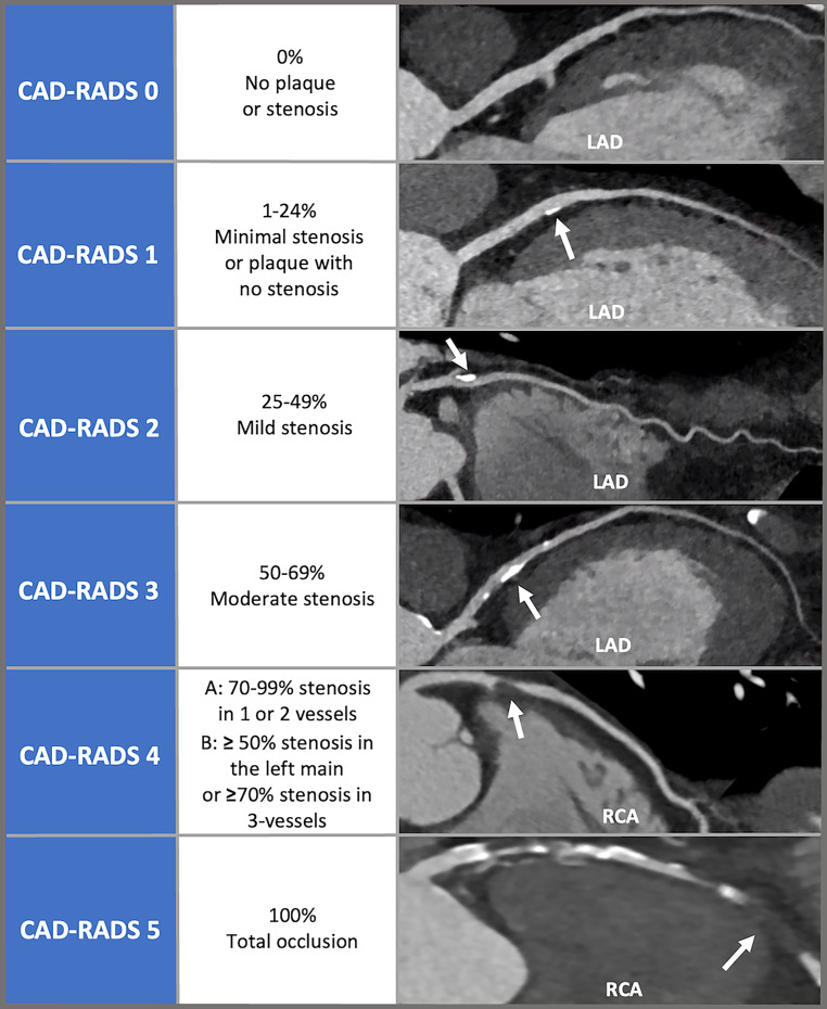
Plaque burden (P). CAD-RADS 2.0 added a plaque burden category to every study with plaque, because diffuse nonobstructive disease and a single mild plaque carried the same code under the first version. Burden is graded by the calcium score, the segment involvement score (the number of the 16 coronary segments containing any plaque), or a visual estimate, by whichever method the laboratory uses. When methods disagree, the highest category is reported, and a calcium score alone underestimates burden when noncalcified plaque is extensive. CAD-RADS 0 has no P category.
| Category | Plaque amount | Agatston score | Segment involvement score | Visual estimate |
|---|---|---|---|---|
| P1 | Mild | 1–100 | 1–2 | Mild plaque in 1–2 vessels |
| P2 | Moderate | 101–300 | 3–4 | Moderate plaque in 1–2 vessels, or mild plaque in 3 vessels |
| P3 | Severe | 301–999 | 5–7 | Moderate plaque in 3 vessels, or severe plaque in 1 vessel |
| P4 | Extensive | 1,000 or more | 8 or more | Severe plaque in more than one vessel |
CAD-RADS 2.0 plaque burden. The document lists P4 as an Agatston score above 1,000; because P3 ends at 999, a score of exactly 1,000 is assigned P4. Higher burden warrants more intensive preventive therapy, including in patients with nonobstructive stenosis.
Modifiers. Modifiers follow the plaque category in a fixed order: N, HRP, I, S, G, and E.
| Modifier | Definition | Rule |
|---|---|---|
| N (nondiagnostic) | At least one segment larger than 1.5 mm cannot be evaluated after review of alternate phases and reconstructions | If no diagnostic segment has a stenosis of 50% or more, the study is coded CAD-RADS N and N precedes the plaque category (CAD-RADS N/P1). If a diagnostic segment has a stenosis of 50% or more, the category is kept and N follows the plaque category (CAD-RADS 3/P2/N) |
| HRP (high-risk plaque) | Two or more of positive remodeling, low-attenuation plaque, spotty calcification, and the napkin-ring sign | One feature alone does not qualify |
| I (ischemia) | CT-FFR or CT perfusion performed: I+ (FFR-CT 0.75 or less, or reversible perfusion defect), I± (FFR-CT 0.76–0.80, or indeterminate perfusion), I− (FFR-CT above 0.80, or no ischemia or a fixed defect) | Used only when a CT physiology test was performed; it adds to the category and does not change it |
| S (stent) | Coronary stent present | The category reflects the worst stenosis, in a stent or a native vessel (CAD-RADS 4A/P2/S) |
| G (graft) | Coronary bypass grafts present | The category reflects the most severe stenosis in a graft or a native vessel that is not bypassed; bypassed native segments are not graded; plaque burden includes native vessels and grafts |
| E (exception) | Nonatherosclerotic finding: anomalous origin, dissection, aneurysm or pseudoaneurysm, vasculitis, fistula, extrinsic compression, arteriovenous malformation | The finding is described in its own terms and is not forced into a stenosis category |
CAD-RADS 2.0 modifiers. A complete code reads, for example, CAD-RADS 3/P3/N/HRP/I+.
In a PROMISE substudy of 3,840 patients followed for a median of 25 months, the unadjusted hazard ratio for death, hospitalization for unstable angina, or myocardial infarction rose from 2.66 for CAD-RADS 1 to 30.80 for CAD-RADS 4B and 5, relative to CAD-RADS 0 (2.43 and 21.84 after adjustment for the ASCVD risk score). Stenosis and burden can diverge: CAD-RADS 2/P4/HRP describes a mild maximal stenosis with extensive plaque and high-risk features, a report that is not reassuring, because most myocardial infarctions arise from segments that were not obstructive at the time of imaging.
Assigning the CAD-RADS 2.0 stenosis category, plaque burden, and modifiers
A complete CAD-RADS 2.0 code combines the maximal stenosis category, the plaque burden category, and the modifiers. The entries below produce the code and the consensus management suggestion for the stable or acute setting.
Plaque burden is the highest category from the calcium score, segment involvement score, or visual estimate; P1 is assigned when plaque is present and no burden measure is entered. A lesion-specific FFR-CT entry takes precedence over the perfusion result for the I modifier. Management text follows CAD-RADS 2.0 and is integrated with symptoms, troponin, the electrocardiogram, and image quality.
Plaque composition, burden, and high-risk plaque features
Plaque is described as calcified, noncalcified, or partially calcified (mixed); "soft plaque" is avoided because attenuation reflects composition only approximately. Calcified plaque is easy to see and blooms, so its size and the stenosis it causes are overestimated. Noncalcified plaque has lower attenuation than the contrast-filled lumen and higher attenuation than surrounding fat; it often grows outward, so a large plaque may narrow the lumen very little. A plaque reported only by its narrowing therefore understates the disease. Plaque without stenosis is still coronary atherosclerosis, and "normal coronary CTA" is reserved for studies with no plaque at all.
High-risk features. The SCCT/NASCI plaque consensus (2021) and CAD-RADS 2.0 define four features associated with future acute coronary syndromes. Each is confirmed on thin, high-quality images and in more than one plane before it is counted.
| Feature | Definition on CT | Common error |
|---|---|---|
| Positive remodeling | Outer vessel diameter at the plaque divided by the mean outer diameter of the proximal and distal reference segments greater than 1.1 | Lumen dimensions used in place of outer vessel dimensions |
| Low-attenuation plaque | Focal noncalcified plaque component below 30 HU, consistent with a lipid-rich necrotic core | Low values from partial-volume averaging next to dense calcium, or a region of interest that includes fat or lumen |
| Spotty calcification | Small calcium foci (under 3 mm) embedded in noncalcified plaque | Bulky or confluent calcium counted as spotty |
| Napkin-ring sign | Central low-attenuation core abutting the lumen, surrounded by a rim of higher attenuation, on cross-section | Concentric plaque without the low-attenuation core and brighter rim |
High-risk plaque features (SCCT/NASCI 2021; CAD-RADS 2.0). The HRP modifier requires two or more features. Interobserver agreement for high-risk features is moderate: in one prospective study of 100 patients, κ was 0.4 for the napkin-ring sign and 0.4 for combined positive remodeling and low attenuation.
In 1,059 patients followed for about 27 months, acute coronary syndrome developed in 22.2% of patients whose plaque showed both positive remodeling and low attenuation, in 3.7% with one feature, and in 0.5% with neither. In a PROMISE substudy of 4,415 outpatients, high-risk plaque was present in 15.3%. Major adverse cardiovascular events occurred in 6.4% of those with it and 2.4% of those without (adjusted HR 1.72 [1.13–2.62] after accounting for risk score and significant stenosis), and the association was strongest in patients with nonobstructive disease. In SCOT-HEART, quantitative low-attenuation plaque burden above 4% carried a hazard ratio of 4.65 (2.06–10.5) for myocardial infarction and was the strongest predictor of it, ahead of the calcium score and stenosis severity. High-risk features raise the probability of future events; they do not identify a lesion as unstable, flow-limiting, or responsible for current symptoms, and they are not an indication for revascularization of a nonobstructive plaque.
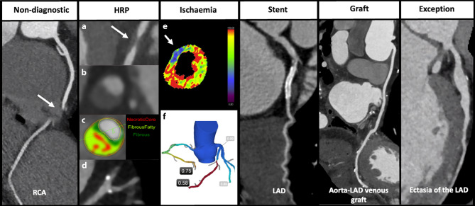
Quantitative plaque analysis. Semiautomated and artificial intelligence–based software measures vessel and lumen volumes and reports total plaque volume, percent atheroma volume (plaque volume divided by vessel volume), and the volumes of calcified, noncalcified, and low-attenuation components. These measures predict events beyond stenosis and track change on serial scans. Values depend on tube voltage, kernel, reconstruction, and software version, so serial comparisons require matched technique and matched segment endpoints, and thresholds validated on one platform are not applied to another.
Pericoronary adipose tissue. Inflammation in the coronary wall shifts the adjacent fat toward a more aqueous, less lipid-rich state, which raises its CT attenuation. The perivascular fat attenuation index (FAI) quantifies this signal around the proximal right coronary artery. In the CRISP-CT study, an FAI of −70.1 HU or higher predicted cardiac mortality, with hazard ratios of 9.04 in the derivation cohort and 5.62 in the validation cohort, and improved risk reclassification. FAI is a research and emerging clinical marker; it is not part of CAD-RADS 2.0.
Plaque findings and preventive therapy. Coronary plaque on CCTA, obstructive or not, establishes atherosclerosis, and the plaque category, extent, and high-risk features guide the intensity of preventive therapy. In SCOT-HEART, patients in the CCTA arm were more often started on preventive therapy (odds ratio 1.40) and antianginal therapy, and this change in treatment is the most likely explanation for fewer myocardial infarctions without more revascularization. LDL-lowering intensity and goals for patients with coronary plaque are set out on the dyslipidemia page.
CT-derived fractional flow reserve
CT-derived fractional flow reserve (FFR-CT) applies computational fluid dynamics or machine-learning models to the coronary tree segmented from a resting CCTA. The model assigns boundary conditions for hyperemic flow from myocardial mass and vessel size and estimates the pressure ratio at every point along each vessel, without additional imaging or drugs. The value depends on the lumen geometry supplied to the model. Motion, misregistration, poor opacification, and heavy calcium all propagate into it, and the study must be acquired with nitroglycerin and adequate heart-rate control.
In NXT (254 patients), FFR-CT had a per-patient area under the curve of 0.90 against invasive fractional flow reserve, compared with 0.81 for CCTA stenosis alone, with a sensitivity of 86% and a specificity of 79%. In PLATFORM, patients scheduled for invasive angiography had similar radiation exposure with the FFR-CT strategy and 32% lower costs at 90 days ($7,343 vs. $10,734). In the international ADVANCE registry (5,083 patients), FFR-CT changed management in 66.9% compared with CCTA alone. Nonobstructive disease at invasive angiography was found in 14.4% of patients with an FFR-CT of 0.80 or less and 43.8% of those above 0.80. No patient with a value above 0.80 died or had a myocardial infarction within 90 days.
| Element | Standard |
|---|---|
| Lesions tested | Stenoses of 40% to 90% in proximal or middle segments, when the result will change management (2021 chest pain guideline, Class 2a); the 2026 SCCT/SCAI consensus recommends it for diameter stenoses of 50% to 90%; not used for CAD-RADS 0, minimal plaque, or total occlusion |
| Where the value is read | 1 to 2 cm distal to the stenosis (CAD-RADS 2.0); the 2026 SCCT/SCAI consensus specifies 2 cm; not the lowest value at the distal tip |
| Thresholds | Above 0.80 normal (I−); 0.76 to 0.80 borderline (I±); 0.75 or less abnormal (I+) |
| Borderline values | Interpreted with symptoms, lesion location, vessel size, and the drop across the lesion; a drop of more than 0.12 supports a hemodynamically significant lesion |
| Diffuse disease | A gradual decline without a focal step reflects diffuse pressure loss and normal tapering; it is not attributed to a single mild plaque |
| Unreliable settings | Severe motion or misregistration at the lesion, poor enhancement, stented segments, bypass grafts, small distal vessels, total occlusions; heavy calcium lowers specificity but does not exclude FFR-CT when image quality is high |
FFR-CT interpretation (CAD-RADS 2.0; SCCT/SCAI 2026 consensus; 2021 AHA/ACC chest pain guideline).
FFR-CT ranks lesions by physiology and can reverse the order suggested by anatomy: a 70% right coronary stenosis with a post-lesion value of 0.84 is less likely to cause ischemia than a 55% left anterior descending stenosis with a value of 0.69. Tandem lesions each produce a step, and the distal value reflects both. An abnormal value in a 1.5-mm distal branch does not by itself warrant angiography. An abnormal proximal value leads to invasive evaluation only when symptoms, territory, and the expected benefit of revascularization support it.
CT myocardial perfusion
Stress CT perfusion images the first pass of iodinated contrast through the myocardium during vasodilator stress, usually adenosine or regadenoson, and compares it with the rest CCTA. Static perfusion captures a single snapshot near peak myocardial enhancement and is read visually or semiquantitatively; a defect involving more than 50% of the wall thickness is transmural. Dynamic perfusion samples the myocardium repeatedly over the first pass and estimates myocardial blood flow, which helps detect balanced multivessel ischemia, at a higher radiation dose; no universal flow threshold applies across platforms. The SCCT perfusion consensus (2020) recommends an interval of about 10 to 20 minutes between nitroglycerin and vasodilator stress, because the combination can cause marked hypotension, and between rest and stress acquisitions.
In CORE320 (381 patients), combined CCTA and CT perfusion had a per-patient area under the curve of 0.87 for a stenosis of 50% or more with a corresponding perfusion defect on single-photon emission CT; sensitivity was 80% and specificity 74%. Against invasive angiography alone, CT perfusion was more sensitive than single-photon emission CT (88% vs. 62%) and less specific (55% vs. 67%). CT perfusion remains interpretable in heavily calcified vessels, where FFR-CT is least reliable, and its results enter the CAD-RADS I modifier in the same way.
Randomized trials of coronary CT angiography
| Trial | Population and comparison | Main result |
|---|---|---|
| SCOT-HEART (2018; 10-year report 2025) | 4,146 patients referred to chest pain clinics in Scotland; CCTA plus standard care vs. standard care | Coronary death or nonfatal MI at 5 years 2.3% vs. 3.9% (HR 0.59, 0.41–0.84), without more angiography or revascularization; at 10 years 6.6% vs. 8.2% (HR 0.79, 0.63–0.99), nonfatal MI HR 0.72; revascularization 15.2% vs. 15.3% |
| PROMISE (2015) | 10,003 symptomatic outpatients in North America; initial CCTA vs. initial functional testing | Death, MI, hospitalization for unstable angina, or major procedural complication 3.3% vs. 3.0% at a median of about 2 years (HR 1.04, 0.83–1.29); fewer catheterizations without obstructive disease (3.4% vs. 4.3%) but more catheterizations within 90 days (12.2% vs. 8.1%); higher cumulative radiation (12.0 vs. 10.1 mSv) |
| DISCHARGE (2022) | 3,561 patients with stable chest pain and intermediate pretest probability referred for invasive angiography at European sites; CT first vs. invasive angiography first | Cardiovascular death, nonfatal MI, or nonfatal stroke at 3.5 years 2.1% vs. 3.0% (HR 0.70, 0.46–1.07); major procedure-related complications 0.5% vs. 1.9% (HR 0.26, 0.13–0.55); angina in the final 4 weeks 8.8% vs. 7.5% |
| PRECISE (2023) | 2,103 stable symptomatic patients; risk-based strategy (deferred testing at minimal risk, CCTA with selective FFR-CT otherwise) vs. usual testing | Death, nonfatal MI, or catheterization without obstructive disease at about 1 year 4.2% vs. 11.3% (HR 0.35, 0.25–0.50), driven by fewer catheterizations without obstructive disease (2.6% vs. 10.2%); death or MI HR 1.52 (0.73–3.15) |
| PLATFORM (2015) | 584 patients; CCTA with FFR-CT vs. usual care | In the planned invasive stratum, angiography without obstructive disease 12% vs. 73%; angiography cancelled in 61%; costs 32% lower |
| ROMICAT-II (2012) | 1,000 emergency department patients with possible ACS; early CCTA vs. standard evaluation | Length of stay 7.6 hours shorter; direct discharge 47% vs. 12%; no missed ACS; more downstream testing and radiation |
| ACT (2012) | 1,370 low- to intermediate-risk emergency department patients; CCTA vs. traditional care | No death or MI at 30 days in 640 patients with negative CCTA; discharge from the emergency department 49.6% vs. 22.7%; median stay 18.0 vs. 24.8 hours |
| CT-STAT (2011) | 699 low-risk emergency department patients; CCTA vs. rest-stress myocardial perfusion imaging | Time to diagnosis 54% shorter (2.9 vs. 6.3 hours); costs 38% lower; MACE after a normal index test 0.8% vs. 0.4% (not significant) |
ACS, acute coronary syndrome; HR, hazard ratio with 95% confidence interval; MACE, major adverse cardiac events; MI, myocardial infarction.
CCTA as the first test in stable chest pain is at least as safe as functional testing, identifies nonobstructive atherosclerosis that functional testing misses, and lowers myocardial infarction when its findings change preventive therapy. It reduces angiography that finds no obstructive disease, especially when paired with FFR-CT, although it can increase the total number of angiograms in the short term. In ISCHEMIA, blinded CCTA was performed before randomization in patients with adequate kidney function to exclude left main stenosis of 50% or more and to exclude patients without obstructive disease; that use of CCTA as a gatekeeper is now part of the management of chronic coronary disease. In patients already referred for invasive angiography at intermediate probability, DISCHARGE showed that CT first produced a similar rate of major events with fewer procedural complications.
Coronary CT angiography in the emergency department
CCTA in acute chest pain is used inside a structured pathway after the electrocardiogram, high-sensitivity troponin testing, and a validated risk assessment. Low-risk patients usually need no urgent testing, and patients with an acute coronary syndrome established by the electrocardiogram or troponin follow the invasive pathway; CCTA has no role as a delaying step for them. ROMICAT-II, ACT, and CT-STAT showed that CCTA shortens time to diagnosis and length of stay and allows safe discharge after a negative study.
A high-quality CAD-RADS 0 study in an intermediate-risk patient with negative serial troponins supports discharge without a stress test. It does not exclude myocarditis, pulmonary embolism, aortic dissection, coronary vasospasm, or microvascular dysfunction. Persistent pain with a rising troponin and normal coronary arteries leads to a search beyond the epicardial vessels, for example cardiac magnetic resonance for myocarditis or myocardial infarction with nonobstructive coronary arteries. High-risk plaque in the emergency setting supports admission or observation even without severe stenosis when the clinical picture is concerning.
Triple rule-out. A single ECG-gated acquisition covering the coronary arteries, the thoracic aorta, and the pulmonary arteries can evaluate acute coronary syndrome, aortic dissection, and pulmonary embolism together. It requires a longer scan range, more contrast, a contrast injection timed to opacify both the pulmonary and systemic circulations, and more radiation than a coronary study. It is reserved for patients in whom all three diagnoses remain plausible after clinical assessment; when one diagnosis is clearly suspected, the dedicated study for that diagnosis is preferred. The dedicated imaging of acute aortic syndromes and pulmonary embolism is covered on those pages.
Coronary stents on CT
Stent struts bloom and cause beam hardening, so the visible in-stent lumen is smaller than the true lumen, and small stents may appear occluded when they are patent. The 2021 chest pain guideline supports CCTA to assess stent patency in stable chest pain when the stent diameter is 3 mm or more (Class 2a). Diameter is one factor among several: strut material and thickness, overlapping stents, adjacent calcium, heart rate, and the scanner's spatial resolution also determine whether the lumen can be seen. A 3.5-mm proximal stent imaged at 55 beats/min on a high-resolution scanner is favorable; a 2.5-mm distal stent in a calcified vessel with tachycardia is not.
Stents are read on thin slices with a sharp kernel, on cross-sections and long-axis views, with wide window settings. The first judgment for each stent is whether its lumen is evaluable. An evaluable stent is described as patent, with in-stent restenosis (low-attenuation neointimal tissue inside the struts narrowing the lumen), or occluded (no contrast in the stent while the vessel before it enhances well, with or without distal collateral filling). Restenosis and new plaque often lie at the stent edges, which are easier to assess than the lumen inside the metal. A stent that cannot be evaluated is reported as nondiagnostic. The nondiagnostic rules apply when the stented segment is larger than 1.5 mm, and functional testing or invasive angiography answers the question. Photon-counting and ultra-high-resolution reconstructions improve in-stent visualization.
Coronary artery bypass grafts on CT
Bypass grafts are larger and move less than native coronary arteries, and CCTA assesses graft patency accurately (Class 2a in stable chest pain after CABG). The protocol differs from a native coronary study: the scan begins above the aortic arch to include the origins of the internal mammary arteries from the subclavian arteries and the proximal anastomoses on the ascending aorta, and the contrast timing must opacify long graft paths. A routine coronary scan range omits the mammary origins, and the omission cannot be corrected after the scan.
Each graft is reported by origin and target, with its patency, stenosis or occlusion, proximal and distal anastomoses, and complications such as aneurysm or thrombus. Saphenous vein grafts develop diffuse wall thickening, bulky irregular plaque, aneurysmal degeneration, and occlusion; an occluded vein graft often appears as a blind stump at the aorta or is not seen at all. Native vessels are reviewed as well, particularly ungrafted territories and segments distal to the anastomoses, although heavily calcified native vessels in this population are often nondiagnostic. Before redo sternotomy, CT shows the distance between the sternum and a patent mammary graft, the right ventricle, or the aorta, which determines the risk of injury on reentry. Surgical clips and sternal wires cause local artifact.
Coronary anomalies, myocardial bridging, fistulas, and aneurysms
CCTA is the reference noninvasive test for coronary anomalies, because it shows the origin, ostium, proximal course, and the relationship of the vessel to the aorta, pulmonary artery, and myocardium in three dimensions. Anomalies are reported with the E modifier and described in anatomic terms.
| Finding | CT features reported | Clinical significance |
|---|---|---|
| Anomalous aortic origin from the opposite sinus | Sinus of origin; ostial shape (slit-like or round); takeoff angle; course (interarterial, subpulmonic or intraseptal, prepulmonic, retroaortic, retrocardiac); intramural segment and its length; proximal narrowing | An interarterial course, slit-like ostium, acute takeoff angle, intramural course, and proximal narrowing are high-risk features (2025 ACC/AHA adult congenital heart disease guideline); a left coronary artery from the right sinus with an interarterial course carries higher risk than the right coronary equivalent |
| Anomalous origin from the pulmonary artery | Left (or right) coronary artery arising from the pulmonary artery; large, tortuous right coronary artery with extensive collaterals and retrograde flow into the pulmonary artery | Myocardial ischemia, left ventricular dysfunction, mitral regurgitation from papillary muscle infarction; surgical repair in adults |
| Myocardial bridge | Segment, usually the mid left anterior descending artery, running within the myocardium; length and depth; systolic compression on multiphase data | Usually benign; the tunneled segment is not atherosclerotic stenosis; plaque often forms just proximal to it; deep, long bridges can cause ischemia |
| Coronary artery fistula | Origin, course, drainage site (right atrium, right ventricle, pulmonary artery, coronary sinus, left heart), aneurysmal dilation along the path | Left-to-right shunt, coronary steal, endocarditis, rupture of an associated aneurysm |
| Aneurysm and ectasia | Focal aneurysm or diffuse ectasia; segments involved; thrombus; calcification; stenosis at inflow or outflow | Atherosclerosis, Kawasaki disease (calcified giant proximal aneurysms in young adults), vasculitis, connective tissue disease, or iatrogenic injury |
Nonatherosclerotic coronary findings on CCTA (CAD-RADS E modifier). Risk features for anomalous aortic origin follow the 2025 ACC/AHA/HRS/ISACHD/SCAI adult congenital heart disease guideline.
Origin and course are separate findings. An anomalous right coronary artery from the left sinus can pass between the great arteries, in front of the pulmonary artery, or behind the aorta, and only the full trace through the volume classifies it. An interarterial segment usually runs at the level of the aortic wall and may be intramural; a long intramural segment with a slit-like, laterally compressed lumen is the anatomy that surgery unroofs. Origin and course can usually be defined at heart rates above the usual target, which matters in a young patient with exertional syncope whose blood pressure does not tolerate rate control. Further evaluation of ischemia (stress imaging or invasive physiology) and of arrhythmia follows the adult congenital guideline.
Artifacts that mimic or hide coronary stenosis
Artifacts can mimic, hide, or exaggerate stenosis. Each is identified by its mechanism and tested with another phase, plane, or kernel, or with a spectral reconstruction.
| Artifact | Mechanism | Appearance | Remedy |
|---|---|---|---|
| Motion | Coronary displacement during the reconstruction window, most often in the right coronary artery and circumflex | Blurred edges, doubled contours, pseudostenosis | Heart-rate control; alternate phases; end-systolic reconstruction; motion-correction algorithms; dual-source or wide-detector scanning |
| Stair-step and slab misregistration | Data from different beats joined at slab boundaries after a premature beat or irregular rhythm, or breathing during the scan | Displacement of every cardiac structure at one level, mimicking stenosis or occlusion | Electrocardiographic editing; arrhythmia rejection; single-beat acquisition |
| Calcium blooming | Partial-volume averaging and limited spatial resolution enlarge dense calcium | Calcified plaque appears larger and encroaches on the lumen; stenosis overestimated | Thin slices; sharp kernel; wide window; high-energy monoenergetic or photon-counting reconstruction |
| Beam hardening | Preferential absorption of low-energy photons by dense material | Dark bands next to calcium, metal, or dense contrast, mimicking low-attenuation plaque or perfusion defects | Saline chaser; beam-hardening correction; high-energy monoenergetic images |
| Streak from dense contrast or metal | Undiluted contrast in the superior vena cava or right heart; pacemaker leads, clips, wires, valves | Bright and dark streaks across the right coronary artery or stented segment | Saline chaser; arm position; metal-artifact reduction |
| Poor contrast timing or low enhancement | Early or late trigger, low iodine delivery, low cardiac output | Faint lumen; plaque-lumen contrast lost; distal vessels and grafts appear occluded | Test bolus or bolus tracking adjusted for output; low-energy monoenergetic images |
| Partial volume in small vessels | Vessel diameter close to spatial resolution | Apparent narrowing of normal small branches | Segments of 1.5 mm or less are not graded |
Artifacts at CCTA and their correction (Kalisz et al., Radiographics 2016; SCCT acquisition guideline).
Ventricular function, valves, atrial anatomy, and incidental findings on cardiac CT
When the acquisition covers the whole cardiac cycle, ventricular volumes, ejection fraction, and regional wall motion are measured from multiphase reconstructions. A single-phase prospectively triggered study cannot provide an ejection fraction, so a request for function is planned into the acquisition. Even single-phase images show myocardial thinning, fatty replacement, or calcification in a coronary territory, which indicate prior infarction, and delayed-phase imaging can show late iodine enhancement and estimate extracellular volume. CT also defines aortic root and annular anatomy and valve calcium before transcatheter aortic valve replacement, as covered on the aortic stenosis page. It also maps the pulmonary veins and left atrial appendage before catheter ablation or appendage occlusion. A filling defect in the appendage on an early phase may be slow flow or thrombus, and a delayed acquisition distinguishes them.
Incidental noncardiac findings. A cardiac CT scan includes part of the lungs, mediastinum, chest wall, spine, and upper abdomen. Data outside a cropped cardiac field of view were still acquired, and a full field-of-view reconstruction is reviewed on every study; anatomy outside the scanned range was never acquired and cannot be recovered. Incidental findings are common in this population and are mostly benign. The clinically important ones include lung nodules and masses, which are managed by Fleischner Society criteria (or the Lung CT Screening Reporting and Data System [Lung-RADS] in a screening program), aortic aneurysm and dissection, pulmonary embolism, mediastinal masses and lymphadenopathy, hiatal hernia, and liver, adrenal, and breast lesions. The report states each finding, its significance, and the recommended follow-up, so that a nodule seen on a scan ordered for chest pain does not go unmanaged. Limiting the calcium scan field of view to the heart reduces incidental findings and the follow-up imaging they generate.
Spectral, photon-counting, and artificial intelligence–assisted coronary CT
Photon-counting detector CT converts each x-ray photon directly into an electrical signal and sorts it by energy. Compared with energy-integrating detectors it gives smaller voxels, less electronic noise, and inherent spectral data with every scan. Ultra-high-resolution coronary reconstructions reduce blooming from calcium and stents; in one study they reclassified 62 of 114 patients (54.4%) to a lower CAD-RADS category than standard-resolution images. These gains are most relevant to patients with heavy calcium and stents, the groups in whom conventional CCTA is least accurate.
Artificial intelligence in cardiac CT serves two purposes. Deep-learning reconstruction reduces noise and dose. Diagnostic algorithms segment the coronary tree, grade stenosis, quantify plaque volume and composition, compute FFR-CT, score calcium automatically on gated and nongated scans, and measure pericoronary fat attenuation. Their outputs depend on image quality and on the population in which they were trained, and every automated contour and value is reviewed by the reader before it enters the report.
Self-assessment
A 58-year-old man with exertional chest pressure undergoes coronary CT angiography. The proximal left anterior descending artery has a 60% stenosis caused by a plaque with a remodeling index of 1.3 and a core of 22 HU. The Agatston score is 420. The proximal right coronary artery cannot be evaluated because of motion on every reconstructed phase. CT-FFR measured 2 cm distal to the left anterior descending stenosis is 0.72. What is the complete CAD-RADS 2.0 code, and what is the next step?
Cardiac CT Board ReviewReferences
- Cury RC, Leipsic J, Abbara S, et al. CAD-RADS 2.0 – 2022 Coronary Artery Disease–Reporting and Data System: an expert consensus document of the Society of Cardiovascular Computed Tomography (SCCT), the American College of Cardiology (ACC), the American College of Radiology (ACR), and the North America Society of Cardiovascular Imaging (NASCI). J Cardiovasc Comput Tomogr. 2022;16:536–557. doi:10.1016/j.jcct.2022.07.002
- Gulati M, Levy PD, Mukherjee D, et al. 2021 AHA/ACC/ASE/CHEST/SAEM/SCCT/SCMR guideline for the evaluation and diagnosis of chest pain: a report of the American College of Cardiology/American Heart Association Joint Committee on Clinical Practice Guidelines. Circulation. 2021;144:e368–e454. doi:10.1161/CIR.0000000000001029
- Virani SS, Newby LK, Arnold SV, et al. 2023 AHA/ACC/ACCP/ASPC/NLA/PCNA guideline for the management of patients with chronic coronary disease: a report of the American Heart Association/American College of Cardiology Joint Committee on Clinical Practice Guidelines. Circulation. 2023;148:e9–e119. doi:10.1161/CIR.0000000000001168
- Blumenthal RS, Morris PB, Gaudino M, et al. 2026 ACC/AHA/AACVPR/ABC/ACPM/ADA/AGS/APhA/ASPC/NLA/PCNA guideline on the management of dyslipidemia: a report of the American College of Cardiology/American Heart Association Joint Committee on Clinical Practice Guidelines. J Am Coll Cardiol. 2026;87:2624–2757. doi:10.1016/j.jacc.2025.11.016
- Multimodality Writing Group for Chronic Coronary Disease; Winchester DE, Maron DJ, Blankstein R, et al. ACC/AHA/ASE/ASNC/ASPC/HFSA/HRS/SCAI/SCCT/SCMR/STS 2023 multimodality appropriate use criteria for the detection and risk assessment of chronic coronary disease. J Am Coll Cardiol. 2023;81:2445–2467. doi:10.1016/j.jacc.2023.03.410
- Vrints C, Andreotti F, Koskinas KC, et al. 2024 ESC guidelines for the management of chronic coronary syndromes. Eur Heart J. 2024;45:3415–3537. doi:10.1093/eurheartj/ehae177
- Abbara S, Blanke P, Maroules CD, et al. SCCT guidelines for the performance and acquisition of coronary computed tomographic angiography: a report of the Society of Cardiovascular Computed Tomography Guidelines Committee. J Cardiovasc Comput Tomogr. 2016;10:435–449. doi:10.1016/j.jcct.2016.10.002
- Leipsic J, Abbara S, Achenbach S, et al. SCCT guidelines for the interpretation and reporting of coronary CT angiography: a report of the Society of Cardiovascular Computed Tomography Guidelines Committee. J Cardiovasc Comput Tomogr. 2014;8:342–358. doi:10.1016/j.jcct.2014.07.003
- Hecht HS, Blaha MJ, Kazerooni EA, et al. CAC-DRS: Coronary Artery Calcium Data and Reporting System. An expert consensus document of the Society of Cardiovascular Computed Tomography (SCCT). J Cardiovasc Comput Tomogr. 2018;12:185–191. doi:10.1016/j.jcct.2018.03.008
- Hecht HS, Cronin P, Blaha MJ, et al. 2016 SCCT/STR guidelines for coronary artery calcium scoring of noncontrast noncardiac chest CT scans: a report of the Society of Cardiovascular Computed Tomography and Society of Thoracic Radiology. J Cardiovasc Comput Tomogr. 2017;11:74–84. doi:10.1016/j.jcct.2016.11.003
- Agatston AS, Janowitz WR, Hildner FJ, et al. Quantification of coronary artery calcium using ultrafast computed tomography. J Am Coll Cardiol. 1990;15:827–832. doi:10.1016/0735-1097(90)90282-T
- Shaw LJ, Blankstein R, Bax JJ, et al. Society of Cardiovascular Computed Tomography / North American Society of Cardiovascular Imaging – expert consensus document on coronary CT imaging of atherosclerotic plaque. J Cardiovasc Comput Tomogr. 2021;15:93–109. doi:10.1016/j.jcct.2020.11.002
- Weir-McCall JR, Koweek L, Suchá D, et al. Fractional flow reserve in coronary computed tomography angiography: an expert consensus document of the Society of Cardiovascular Computed Tomography (SCCT) and the Society for Cardiovascular Angiography and Interventions (SCAI). Endorsed by the American College of Cardiology (ACC). J Cardiovasc Comput Tomogr. 2026;20:316–332. doi:10.1016/j.jcct.2026.05.006
- Patel AR, Bamberg F, Branch K, et al. Society of Cardiovascular Computed Tomography expert consensus document on myocardial computed tomography perfusion imaging. J Cardiovasc Comput Tomogr. 2020;14:87–100. doi:10.1016/j.jcct.2019.10.003
- Hirshfeld JW Jr, Ferrari VA, Bengel FM, et al. 2018 ACC/HRS/NASCI/SCAI/SCCT expert consensus document on optimal use of ionizing radiation in cardiovascular imaging: best practices for safety and effectiveness. J Am Coll Cardiol. 2018;71:e283–e351. doi:10.1016/j.jacc.2018.02.016
- Trattner S, Halliburton S, Thompson CM, et al. Cardiac-specific conversion factors to estimate radiation effective dose from dose-length product in computed tomography. JACC Cardiovasc Imaging. 2018;11:64–74. doi:10.1016/j.jcmg.2017.06.006
- Hausleiter J, Meyer T, Hermann F, et al. Estimated radiation dose associated with cardiac CT angiography. JAMA. 2009;301:500–507. doi:10.1001/jama.2009.54
- Stocker TJ, Deseive S, Leipsic J, et al. Reduction in radiation exposure in cardiovascular computed tomography imaging: results from the PROspective multicenter registry on radiaTion dose Estimates of cardiac CT angIOgraphy iN daily practice in 2017 (PROTECTION VI). Eur Heart J. 2018;39:3715–3723. doi:10.1093/eurheartj/ehy546
- Davenport MS, Perazella MA, Yee J, et al. Use of intravenous iodinated contrast media in patients with kidney disease: consensus statements from the American College of Radiology and the National Kidney Foundation. Radiology. 2020;294:660–668. doi:10.1148/radiol.2019192094
- Muster V, Wallner M, Schmidt A, et al. Heart rate-reducing therapy with add-on ivabradine and bisoprolol before coronary computed tomographic angiography in a fast-track ambulatory setting. J Int Med Res. 2018;46:2249–2257. doi:10.1177/0300060518761302
- SCOT-HEART Investigators; Newby DE, Adamson PD, Berry C, et al. Coronary CT angiography and 5-year risk of myocardial infarction. N Engl J Med. 2018;379:924–933. doi:10.1056/NEJMoa1805971
- Williams MC, Wereski R, Tuck C, et al. Coronary CT angiography-guided management of patients with stable chest pain: 10-year outcomes from the SCOT-HEART randomised controlled trial in Scotland. Lancet. 2025;405:329–337. doi:10.1016/S0140-6736(24)02679-5
- Douglas PS, Hoffmann U, Patel MR, et al. Outcomes of anatomical versus functional testing for coronary artery disease. N Engl J Med. 2015;372:1291–1300. doi:10.1056/NEJMoa1415516
- DISCHARGE Trial Group; Maurovich-Horvat P, Bosserdt M, Kofoed KF, et al. CT or invasive coronary angiography in stable chest pain. N Engl J Med. 2022;386:1591–1602. doi:10.1056/NEJMoa2200963
- Douglas PS, Nanna MG, Kelsey MD, et al. Comparison of an initial risk-based testing strategy vs usual testing in stable symptomatic patients with suspected coronary artery disease: the PRECISE randomized clinical trial. JAMA Cardiol. 2023;8:904–914. doi:10.1001/jamacardio.2023.2595
- Maron DJ, Hochman JS, Reynolds HR, et al. Initial invasive or conservative strategy for stable coronary disease. N Engl J Med. 2020;382:1395–1407. doi:10.1056/NEJMoa1915922
- Hoffmann U, Truong QA, Schoenfeld DA, et al. Coronary CT angiography versus standard evaluation in acute chest pain. N Engl J Med. 2012;367:299–308. doi:10.1056/NEJMoa1201161
- Litt HI, Gatsonis C, Snyder B, et al. CT angiography for safe discharge of patients with possible acute coronary syndromes. N Engl J Med. 2012;366:1393–1403. doi:10.1056/NEJMoa1201163
- Goldstein JA, Chinnaiyan KM, Abidov A, et al. The CT-STAT (Coronary Computed Tomographic Angiography for Systematic Triage of Acute Chest Pain Patients to Treatment) trial. J Am Coll Cardiol. 2011;58:1414–1422. doi:10.1016/j.jacc.2011.03.068
- Budoff MJ, Dowe D, Jollis JG, et al. Diagnostic performance of 64-multidetector row coronary computed tomographic angiography for evaluation of coronary artery stenosis in individuals without known coronary artery disease: results from the prospective multicenter ACCURACY trial. J Am Coll Cardiol. 2008;52:1724–1732. doi:10.1016/j.jacc.2008.07.031
- Miller JM, Rochitte CE, Dewey M, et al. Diagnostic performance of coronary angiography by 64-row CT. N Engl J Med. 2008;359:2324–2336. doi:10.1056/NEJMoa0806576
- Villines TC, Hulten EA, Shaw LJ, et al. Prevalence and severity of coronary artery disease and adverse events among symptomatic patients with coronary artery calcification scores of zero undergoing coronary computed tomography angiography: results from the CONFIRM (Coronary CT Angiography Evaluation for Clinical Outcomes: An International Multicenter) registry. J Am Coll Cardiol. 2011;58:2533–2540. doi:10.1016/j.jacc.2011.10.851
- Nørgaard BL, Leipsic J, Gaur S, et al. Diagnostic performance of noninvasive fractional flow reserve derived from coronary computed tomography angiography in suspected coronary artery disease: the NXT trial (Analysis of Coronary Blood Flow Using CT Angiography: Next Steps). J Am Coll Cardiol. 2014;63:1145–1155. doi:10.1016/j.jacc.2013.11.043
- Douglas PS, Pontone G, Hlatky MA, et al. Clinical outcomes of fractional flow reserve by computed tomographic angiography-guided diagnostic strategies vs. usual care in patients with suspected coronary artery disease: the prospective longitudinal trial of FFRCT: outcome and resource impacts study (PLATFORM). Eur Heart J. 2015;36:3359–3367. doi:10.1093/eurheartj/ehv444
- Hlatky MA, De Bruyne B, Pontone G, et al. Quality-of-life and economic outcomes of assessing fractional flow reserve with computed tomography angiography: PLATFORM. J Am Coll Cardiol. 2015;66:2315–2323. doi:10.1016/j.jacc.2015.09.051
- Fairbairn TA, Nieman K, Akasaka T, et al. Real-world clinical utility and impact on clinical decision-making of coronary computed tomography angiography-derived fractional flow reserve: lessons from the ADVANCE Registry. Eur Heart J. 2018;39:3701–3711. doi:10.1093/eurheartj/ehy530
- Rochitte CE, George RT, Chen MY, et al. Computed tomography angiography and perfusion to assess coronary artery stenosis causing perfusion defects by single photon emission computed tomography: the CORE320 study. Eur Heart J. 2014;35:1120–1130. doi:10.1093/eurheartj/eht488
- Motoyama S, Sarai M, Harigaya H, et al. Computed tomographic angiography characteristics of atherosclerotic plaques subsequently resulting in acute coronary syndrome. J Am Coll Cardiol. 2009;54:49–57. doi:10.1016/j.jacc.2009.02.068
- Ferencik M, Mayrhofer T, Bittner DO, et al. Use of high-risk coronary atherosclerotic plaque detection for risk stratification of patients with stable chest pain: a secondary analysis of the PROMISE randomized clinical trial. JAMA Cardiol. 2018;3:144–152. doi:10.1001/jamacardio.2017.4973
- Williams MC, Kwiecinski J, Doris M, et al. Low-attenuation noncalcified plaque on coronary computed tomography angiography predicts myocardial infarction: results from the multicenter SCOT-HEART trial (Scottish Computed Tomography of the HEART). Circulation. 2020;141:1452–1462. doi:10.1161/CIRCULATIONAHA.119.044720
- Oikonomou EK, Marwan M, Desai MY, et al. Non-invasive detection of coronary inflammation using computed tomography and prediction of residual cardiovascular risk (the CRISP CT study): a post-hoc analysis of prospective outcome data. Lancet. 2018;392:929–939. doi:10.1016/S0140-6736(18)31114-0
- Gurvitz M, Krieger EV, Fuller S, et al. 2025 ACC/AHA/HRS/ISACHD/SCAI guideline for the management of adults with congenital heart disease. J Am Coll Cardiol. 2026;87:822–976. doi:10.1016/j.jacc.2025.09.006
- Kalisz K, Buethe J, Saboo SS, et al. Artifacts at cardiac CT: physics and solutions. Radiographics. 2016;36:2064–2083. doi:10.1148/rg.2016160079
- MacMahon H, Naidich DP, Goo JM, et al. Guidelines for management of incidental pulmonary nodules detected on CT images: from the Fleischner Society 2017. Radiology. 2017;284:228–243. doi:10.1148/radiol.2017161659
- Celeng C, Takx RAP. Moving towards a uniform diagnosis of coronary artery disease on coronary CTA: Coronary Artery Disease–Reporting and Data System 2.0. Neth Heart J. 2024;32:378–385. doi:10.1007/s12471-024-01903-6
- Bittner DO, Mayrhofer T, Budoff M, et al. Prognostic value of coronary CTA in stable chest pain: CAD-RADS, CAC, and cardiovascular events in PROMISE. JACC Cardiovasc Imaging. 2020;13:1534–1545. doi:10.1016/j.jcmg.2019.09.012
- Halfmann MC, Bockius S, Emrich T, et al. Ultrahigh-spatial-resolution photon-counting detector CT angiography of coronary artery disease for stenosis assessment. Radiology. 2024;310:e231956. doi:10.1148/radiol.231956
- Salem AM, Davis J, Gopalan D, et al. Characteristics of conventional high-risk coronary plaques and a novel CT defined thin-cap fibroatheroma in patients undergoing CCTA with stable chest pain. Clin Imaging. 2023;101:69–76. doi:10.1016/j.clinimag.2023.06.009
- George RT, Mehra VC, Chen MY, et al. Myocardial CT perfusion imaging and SPECT for the diagnosis of coronary artery disease: a head-to-head comparison from the CORE320 multicenter diagnostic performance study. Radiology. 2014;272:407–416. doi:10.1148/radiol.14140806
- Hausleiter J, Martinoff S, Hadamitzky M, et al. Image quality and radiation exposure with a low tube voltage protocol for coronary CT angiography: results of the PROTECTION II Trial. JACC Cardiovasc Imaging. 2010;3:1113–1123. doi:10.1016/j.jcmg.2010.08.016