Library › Coronary artery disease › Chronic coronary disease
Chronic coronary disease and stable angina
Chronic coronary disease and stable angina in adults, from pretest probability and choice of the first test through antianginal and preventive therapy, revascularization, INOCA, vasospastic angina, and follow-up.
Chronic coronary disease is managed on three separate axes: event prevention for the atherosclerotic disease, relief of angina, and revascularization where anatomy or ventricular function predicts a survival or symptom benefit. In stable patients, an initial invasive strategy does not reduce death or myocardial infarction compared with guideline-directed medical therapy (ISCHEMIA), and it does relieve angina in proportion to the angina present. Left main stenosis of 50% or more and multivessel disease with an ejection fraction of 35% or less are the settings in which coronary artery bypass grafting (CABG) carries a Class 1 recommendation to improve survival.
Definition of chronic coronary disease in the 2023 AHA/ACC guideline
The 2023 American Heart Association/American College of Cardiology (AHA/ACC) multisociety guideline replaced the term stable ischemic heart disease with chronic coronary disease (CCD). It describes CCD as a heterogeneous group of conditions. These include obstructive and nonobstructive coronary artery disease, with or without previous myocardial infarction or revascularization, ischemic heart disease diagnosed only by noninvasive testing, and chronic angina syndromes with varying underlying causes. The guideline applies to outpatients after an acute coronary syndrome or revascularization once acute issues have stabilized. It also applies to patients with ischemic cardiomyopathy, stable angina or an ischemic equivalent, angina with vasospasm or microvascular dysfunction, or coronary disease found on a screening study such as a stress test or coronary CT angiography (CCTA).
The European Society of Cardiology (ESC) uses the term chronic coronary syndromes (2019 and 2024 guidelines) for the same population and emphasizes that plaque biology and symptoms remain dynamic during clinically quiet periods.
Prevalence, sex differences, and changing patient mix in chronic coronary disease
The 2023 guideline reports National Health and Nutrition Examination Survey estimates for 2015 to 2018. About 20.1 million US adults (7.2%) live with coronary heart disease, 11 million (4.1%) have angina pectoris, and 8.8 million (3.1%) have had a myocardial infarction. About 200,000 of the myocardial infarctions that occur each year in the United States, roughly one-quarter, occur in people with a previous infarction. Coronary heart disease mortality fell by about 25% over the preceding decade, and coronary disease remains the leading cause of death in the United States and worldwide.
Prevalence rises with age and differs by sex: 8.3% of men and 6.2% of women carry the diagnosis, whereas self-reported angina is nearly equal (4.2% of men and 4.0% of women). Women more often present with angina and nonobstructive coronary arteries, and the relative contribution of microvascular dysfunction and vasospasm is larger in women.
Patients are older than in the 2012 stable ischemic heart disease guideline era, with more chronic kidney disease, atrial fibrillation, diabetes, cancer, and polypharmacy. Among patients referred for noninvasive testing for suspected coronary disease, obstructive disease is now present in about 10%, and fewer than 10% report typical exertional angina. In US practice, 50% to 60% of elective invasive angiograms show no obstructive disease.
Pathophysiology: oxygen supply, demand, and plaque
Determinants of demand and supply. Myocardial oxygen demand rises with heart rate, systolic wall stress (pressure and cavity radius), and contractility. The heart extracts about 70% of the oxygen delivered at rest, so increased demand is met almost entirely by increased flow. Angina occurs when demand exceeds the flow that the epicardial artery and the microcirculation together can deliver.
The subendocardium. The inner wall has the highest wall stress and resting oxygen extraction and is perfused almost entirely in diastole, because systolic contraction compresses its intramural vessels. Tachycardia shortens diastole, and ischemia raises left ventricular diastolic pressure, so the two narrow the perfusion gradient in a self-amplifying loop. This explains why exercise-induced ischemia is subendocardial and why ST depression, not elevation, is the usual stress finding.
Stenosis hemodynamics. A stenosis that is trivial at resting flow can become flow-limiting during exertion, which is why physiologic significance is measured at maximal hyperemia (fractional flow reserve) or in the wave-free period of diastole. Resting flow is preserved until a stenosis reaches about 85% to 90% diameter, whereas maximal flow begins to fall at about 50% to 70%.
The ischemic cascade. Ischemia proceeds from perfusion heterogeneity through metabolic change, diastolic dysfunction, regional systolic dysfunction, and electrocardiographic change to angina. Relaxation fails before contraction, and the stiff ventricle raises filling pressure, so exertional dyspnea often precedes chest discomfort. Tests directed at earlier steps in the cascade (perfusion imaging, wall motion) detect ischemia that the exercise electrocardiogram can miss.
Plaque biology. Stable plaques usually have a thick fibrous cap and a smaller lipid core. Thin-cap fibroatheromas, with a large necrotic core, macrophage inflammation, and a cap less than 65 µm thick on pathology, are the substrate for rupture. Positive (outward) remodeling preserves the lumen despite substantial plaque burden, so a nonobstructive angiogram can conceal extensive disease, and most myocardial infarctions arise from lesions that were not the tightest stenosis on prior angiography. Plaque erosion produces thrombosis over a denuded endothelium without cap rupture. These observations explain why preventive therapy, which acts on the whole coronary tree, reduces infarction while focal revascularization of a stable stenosis does not.
Collaterals, stunning, and hibernation. Collateral vessels develop with chronic severe stenosis. Collateral-dependent myocardium is vulnerable to coronary steal during vasodilator stress. Stunning is reversible contractile dysfunction that persists for hours to days after an ischemic episode despite restored flow. Hibernation is chronic dysfunction in viable myocardium subtended by severe stenosis, and the window for recovery after revascularization narrows as fibrosis develops.
Stable angina, anginal equivalents, and the Canadian Cardiovascular Society class
The history establishes whether symptoms are likely ischemic and how limiting they are. Stable angina is a pressure, tightness, heaviness, or squeezing discomfort, usually retrosternal, that is provoked by exertion or emotional stress, lasts minutes, and is relieved within about 5 minutes by rest or sublingual nitroglycerin. Pain that is sharp, fleeting (seconds), positional, pleuritic, reproduced by palpation, or localized to a fingertip-sized area is unlikely to be ischemic.
The traditional classification counts three features: substernal discomfort of characteristic quality and duration, provocation by exertion or emotional stress, and relief by rest or nitroglycerin. Three features constitute typical angina, two atypical angina, and one or none nonanginal chest pain. These categories drive the pretest probability tables. The 2021 AHA/ACC chest pain guideline discourages the term atypical, because it is widely misread as noncardiac. It recommends describing chest pain as cardiac, possible cardiac, or noncardiac, with attention to the specific features that point toward ischemia.
Anginal equivalents. Exertional dyspnea, fatigue, and discomfort in the jaw, arm, shoulder, interscapular area, or epigastrium without chest pain can represent ischemia, particularly when reproducible with exertion and otherwise unexplained. Dyspnea as the dominant symptom has its own column in the contemporary pretest probability model and, in some studies, carries a worse prognosis than chest pain. Nitroglycerin relief supports but does not establish an ischemic cause, because esophageal spasm and other smooth-muscle disorders also respond.
Silent ischemia. Objective ischemia without the patient's usual symptoms is more frequent in diabetes with autonomic neuropathy, in older adults, and after infarction. It carries the same prognostic weight as symptomatic ischemia of similar extent.
Patterns that are not stable angina. Angina at rest, severe new-onset angina, and angina that increases in frequency, duration, or intensity or occurs at a lower threshold define unstable angina, which is managed as an acute coronary syndrome. Angina mainly at rest, at night or in the early morning, with preserved exercise tolerance, suggests vasospasm.
| Class | Canadian Cardiovascular Society definition |
|---|---|
| I | Ordinary physical activity, such as walking or climbing stairs, does not cause angina. Angina occurs with strenuous, rapid, or prolonged exertion at work or recreation. |
| II | Slight limitation of ordinary activity. Angina with walking or climbing stairs rapidly, walking uphill, walking or stair climbing after meals, in cold or wind, under emotional stress, or in the first hours after waking; walking more than two blocks on the level or climbing more than one flight of stairs at a normal pace provokes angina. |
| III | Marked limitation of ordinary activity. Angina with walking one to two blocks on the level or climbing one flight of stairs at a normal pace. |
| IV | Inability to carry out any physical activity without discomfort; angina may be present at rest. |
Canadian Cardiovascular Society grading of angina. A validated patient-reported measure such as the Seattle Angina Questionnaire may be reasonable to assess symptoms, function, and quality of life (Class 2b, 2023 guideline); it was the health-status instrument in ISCHEMIA and CorMicA.
Physical examination, resting ECG, echocardiography, and laboratory testing
The examination in stable angina is often normal. It identifies atherosclerosis elsewhere (carotid or femoral bruits, reduced pedal pulses), risk factors (hypertension, xanthelasma, tendon xanthomas in familial hypercholesterolemia, obesity), and alternative causes (anemia, thyroid disease). During or immediately after an episode of ischemia, a fourth heart sound, a new apical murmur of ischemic mitral regurgitation, a paradoxically split second sound, or pulmonary crackles may be present. A systolic ejection murmur raises the possibility of aortic stenosis or obstructive hypertrophic cardiomyopathy, both of which cause angina with normal coronary arteries.
Resting electrocardiogram. A resting 12-lead electrocardiogram is obtained in every patient. It is frequently normal in stable angina, and a normal tracing does not exclude severe coronary disease. Pathological Q waves indicate prior infarction; left bundle branch block, left ventricular hypertrophy, resting ST-T abnormalities, atrial fibrillation, and conduction disease each carry adverse prognostic weight and influence the choice of stress test. A tracing recorded during pain can be diagnostic: transient ST depression supports demand ischemia, and transient ST elevation suggests vasospasm or acute occlusion.
Transthoracic echocardiography. Transthoracic echocardiography is recommended in intermediate-high risk patients with stable chest pain who have pathological Q waves, symptoms or signs of heart failure, complex ventricular arrhythmias, or a murmur of unclear cause (Class 1, 2021 chest pain guideline). It defines ventricular function and detects valvular, myocardial, or pericardial disease. Regional wall motion abnormalities in a coronary distribution at rest suggest prior infarction or hibernation. The ESC guidelines recommend resting echocardiography in all patients with suspected chronic coronary syndrome.
Chest radiograph. The chest radiograph contributes little in stable angina and is obtained for signs of heart failure or suspected pulmonary disease.
Laboratory testing. The initial evaluation includes a fasting or nonfasting lipid panel, glycated hemoglobin or fasting glucose, serum creatinine with estimated glomerular filtration rate, hemoglobin (anemia lowers the ischemic threshold), and thyroid function when clinically indicated. High-sensitivity C-reactive protein reflects residual inflammatory risk but is not a requirement for colchicine therapy in the 2023 guideline.
Differential diagnosis of stable chest pain
| Category | Conditions | Features that separate them from stable angina |
|---|---|---|
| Cardiac, nonatherosclerotic | Aortic stenosis, hypertrophic cardiomyopathy, coronary vasospasm, microvascular dysfunction, myocardial bridging, anomalous coronary origin, coronary aneurysm or ectasia, pericarditis | Murmur or abnormal echocardiogram; rest or nocturnal pain (spasm); pleuritic and positional pain with friction rub (pericarditis); young patient with exertional syncope (anomalous origin) |
| Vascular and pulmonary | Pulmonary hypertension, pulmonary embolism, aortic dissection, pleuritis, pneumothorax | Exertional dyspnea with right heart findings; acute onset with tearing or pleuritic pain; hypoxemia |
| Gastrointestinal | Gastroesophageal reflux, esophageal spasm, peptic ulcer disease, biliary disease | Relation to meals or lying flat; burning quality; response to acid suppression; esophageal spasm can respond to nitroglycerin |
| Musculoskeletal | Costochondritis, cervical or thoracic radiculopathy, muscle strain | Pain reproduced by palpation or movement; brief, sharp, or positional pain |
| Psychological | Panic disorder, anxiety, depression | Episodes with hyperventilation and paresthesias; no relation to exertion; the diagnosis of exclusion when ischemia has been reasonably excluded |
| Increased demand or reduced oxygen delivery | Anemia, thyrotoxicosis, uncontrolled hypertension, tachyarrhythmia, hypoxemia, stimulant use | Angina appears with the secondary condition and resolves with its treatment, although coronary disease may coexist |
Differential diagnosis of stable chest pain in adults. Acute coronary syndrome, aortic dissection, pulmonary embolism, and tension pneumothorax are excluded first when chest pain is new, at rest, or accelerating.
Pretest probability of obstructive coronary disease
The decision to test, and which test, starts from the probability of obstructive disease. The Diamond-Forrester model (1979), based on age, sex, and symptom type in referral and autopsy series, overestimates obstructive disease in contemporary patients, especially women. The CAD Consortium models (basic: age, sex, symptoms, setting; clinical: adds diabetes, hypertension, dyslipidemia, and smoking; extended: adds coronary calcium) were validated across hospitals and countries, and each step adds discrimination.
A pooled analysis of 15,815 symptomatic patients in three contemporary cohorts found that obstructive disease was about one-third as prevalent as the earlier model predicted. These values form the pretest probability table of the 2019 ESC guideline. Figure 11 of the 2021 AHA/ACC chest pain guideline condenses them into a single chest pain column, using the anginal values as upper limits (for example, 52% or less for men aged 70 years or older), keeps the dyspnea column, and adds a panel that adjusts the estimate by coronary calcium score (1 to 99, 100 to 999, and 1,000 or more). The 2021 guideline defines low risk as a pretest probability of 15% or less, a group whose annual risk of cardiovascular death or myocardial infarction was below 1% in contemporary cohorts. Patients younger than 40 years and those with symptoms of low ischemic likelihood are also classified as low risk.
| Age, years | Typical angina | Atypical angina | Nonanginal pain | Dyspnea | ||||
|---|---|---|---|---|---|---|---|---|
| Men | Women | Men | Women | Men | Women | Men | Women | |
| 30–39 | 3% | 5% | 4% | 3% | 1% | 1% | 0% | 3% |
| 40–49 | 22% | 10% | 10% | 6% | 3% | 2% | 12% | 3% |
| 50–59 | 32% | 13% | 17% | 6% | 11% | 3% | 20% | 9% |
| 60–69 | 44% | 16% | 26% | 11% | 22% | 6% | 27% | 14% |
| ≥70 | 52% | 27% | 34% | 19% | 24% | 10% | 32% | 12% |
Pretest probability of obstructive coronary disease by age, sex, and symptom type in 15,815 symptomatic patients (Juarez-Orozco et al., 2019), the basis of the 2019 ESC table and of Figure 11 in the 2021 AHA/ACC chest pain guideline. Highlighted cells exceed 15%, the threshold above which the 2021 guideline classifies a patient as intermediate-high risk.
Clinical modifiers. Diabetes, smoking, dyslipidemia, hypertension, family history of premature coronary disease, peripheral artery disease, an abnormal resting electrocardiogram (Q waves, ST-T changes), left ventricular dysfunction, and visible coronary calcium on prior chest CT raise the probability; a coronary artery calcium score of zero lowers it. The 2024 ESC guideline replaced the table with a risk factor–weighted clinical likelihood model that adds the number of risk factors to age, sex, and symptoms. It recommends deferral of testing at a likelihood of 5% or less, coronary calcium scoring to reclassify patients at more than 5% to 15%, and CCTA as the preferred test for patients at more than 5% to 50%.
Coronary calcium in symptomatic patients. In patients with stable chest pain at low risk, coronary artery calcium testing is reasonable as a first-line test to exclude calcified plaque and identify a low likelihood of obstructive disease (Class 2a).
Pretest probability and exercise risk calculators
Probabilities from the pooled contemporary cohorts (Juarez-Orozco 2019, the 2019 ESC table). The 2021 AHA/ACC chest pain guideline uses the typical angina values as the upper limit for any chest pain and applies its 15% threshold to them. The table estimates obstructive disease; it does not estimate event risk, microvascular dysfunction, or vasospasm.
Duke treadmill score = exercise time (minutes, Bruce protocol) − 5 × maximal ST deviation (mm, any lead except aVR) − 4 × angina index. Risk groups and survival from the outpatient derivation cohort (Mark et al., 1991). The score does not apply with left bundle branch block, ventricular pacing, pre-excitation, digoxin, or more than 1 mm of resting ST depression.
Choosing the first test: CCTA, stress imaging, exercise ECG, or no test
For patients with stable chest pain and no known coronary disease, the 2021 AHA/ACC chest pain guideline matches the test to the clinical question, anatomy or ischemia. Pretest risk, ability to exercise, the resting electrocardiogram, and local expertise also steer the choice.
| Setting | Recommendation | Class |
|---|---|---|
| Any outpatient with stable chest pain and no known CAD | A model to estimate pretest probability is used to identify low-risk patients in whom testing can be deferred | 1 |
| Low risk | Coronary artery calcium testing as a first-line test to exclude calcified plaque | 2a |
| Low risk with an interpretable ECG | Exercise testing without imaging to exclude ischemia and determine functional capacity | 2a |
| Intermediate-high risk | CCTA for diagnosis, risk stratification, and treatment decisions | 1 |
| Intermediate-high risk | Stress imaging (echocardiography, PET or SPECT perfusion, or CMR) for diagnosis of ischemia and estimation of risk | 1 |
| Intermediate-high risk, nuclear imaging selected | PET in preference to SPECT, if available | 2a |
| Intermediate-high risk, interpretable ECG, able to reach at least 5 METs | Exercise electrocardiography | 2a |
| Stenosis of 40% to 90% in a proximal or middle segment on CCTA | FFR-CT for vessel-specific ischemia and revascularization decisions | 2a |
| Inconclusive or abnormal exercise ECG or stress imaging | CCTA | 2a |
| Inconclusive CCTA | Stress imaging | 2a |
Recommendations for stable chest pain without known coronary disease, 2021 AHA/ACC/ASE/CHEST/SAEM/SCCT/SCMR chest pain guideline. CAD, coronary artery disease; CMR, cardiovascular magnetic resonance; FFR-CT, fractional flow reserve derived from CT; MET, metabolic equivalent; PET, positron emission tomography; SPECT, single-photon emission CT.
Anatomic or functional testing. CCTA answers whether plaque and obstructive stenosis are present and how extensive the disease is. Stress imaging answers whether ischemia is present, where, and how extensive, and it carries prognostic information through exercise capacity and hemodynamic response. The 2021 guideline synopsis notes that CCTA is preferable in patients younger than 65 years who are not on preventive therapy. Stress testing may be advantageous at 65 years or older, when obstructive disease and ischemia are more likely and coronary calcium is heavier.
| Patient feature | Test favored | Reason |
|---|---|---|
| Younger patient, no known CAD, regular rhythm, low to moderate calcium expected | CCTA | High negative predictive value; identifies nonobstructive plaque for prevention |
| Able to exercise, interpretable resting ECG | Exercise ECG or exercise stress imaging | Exercise capacity and hemodynamic response add prognostic information |
| Left bundle branch block or ventricular pacing | Vasodilator perfusion imaging (PET or SPECT) or CCTA | Exercise and dobutamine produce septal perfusion and wall motion artifacts |
| Resting ST depression ≥1 mm, LVH with repolarization changes, digoxin, pre-excitation | Stress imaging | Exercise ST changes are uninterpretable |
| Unable to exercise | Pharmacologic stress imaging or CCTA | Exercise ECG is nondiagnostic without adequate workload |
| Heavy coronary calcium, atrial fibrillation with rapid or irregular rate, extensive stents | Functional imaging | Blooming and motion artifact make CT stenosis grading unreliable |
| Severe obesity | PET, CMR, or contrast-enhanced stress echocardiography; CT according to scanner capability | Attenuation and image noise degrade SPECT and noncontrast echocardiography |
| Severe kidney dysfunction not on dialysis | Stress echocardiography or nuclear perfusion | Avoids iodinated contrast and the risk of contrast-associated kidney injury |
| Persistent symptoms with nonobstructive arteries | PET or CMR with myocardial blood flow reserve, or invasive coronary function testing | Detects microvascular dysfunction and vasospasm that anatomy cannot show |
Patient features that steer the choice of the first test, adapted from Tables 5 and 6 and Figure 12 of the 2021 chest pain guideline and the 2023 multimodality appropriate use criteria. eGFR, estimated glomerular filtration rate; LVH, left ventricular hypertrophy.
Repeating the same nondiagnostic stress test without a new reason is rated low value, whereas CCTA after an inconclusive or discordant functional test is appropriate when better anatomic information will change management.
Coronary CT angiography, CAD-RADS, and FFR-CT
CCTA images the coronary lumen and wall with an average radiation dose that, on current scanners with prospective electrocardiographic triggering, is commonly a few millisieverts or less. Its negative predictive value for obstructive disease is high.
Outcome trials. In PROMISE (10,003 symptomatic outpatients), initial CCTA and initial functional testing produced similar rates of death, myocardial infarction, hospitalization for unstable angina, or major procedural complication over a median of about 2 years (3.3% vs. 3.0%). In SCOT-HEART (4,146 patients referred to a chest pain clinic), adding CCTA to standard care reduced death from coronary heart disease or nonfatal myocardial infarction at 5 years (2.3% vs. 3.9%; hazard ratio 0.59), without an increase in invasive angiography or revascularization over 5 years. The benefit was attributed to more preventive and antianginal therapy started on the basis of plaque detection. In DISCHARGE (3,561 patients with stable chest pain and intermediate pretest probability referred for invasive angiography), a CT-first strategy produced a similar rate of cardiovascular death, myocardial infarction, or stroke at 3.5 years (2.1% vs. 3.0%; hazard ratio 0.70, not significant). Major procedure-related complications were less frequent (0.5% vs. 1.9%).
CAD-RADS 2.0. The 2022 Coronary Artery Disease Reporting and Data System standardizes the CCTA report by the maximal stenosis.
| Category | Maximal stenosis | Interpretation and usual next step in stable symptoms |
|---|---|---|
| CAD-RADS 0 | 0% | No plaque or stenosis; noncoronary and nonatherosclerotic causes are evaluated if symptoms persist |
| CAD-RADS 1 | 1–24% | Minimal nonobstructive disease; preventive therapy according to plaque burden and risk |
| CAD-RADS 2 | 25–49% | Mild nonobstructive disease; preventive therapy; reported as coronary artery disease, never as a negative study |
| CAD-RADS 3 | 50–69% | Moderate stenosis; functional assessment (FFR-CT, CT perfusion, stress imaging) when the result will change management |
| CAD-RADS 4A | 70–99% | Severe stenosis; invasive angiography or functional assessment according to symptoms and territory |
| CAD-RADS 4B | Left main ≥50% or three-vessel ≥70% | High-risk anatomy; invasive angiography |
| CAD-RADS 5 | 100% | Total occlusion; invasive angiography or viability assessment according to symptoms and ventricular function |
| CAD-RADS N | Nondiagnostic | Obstructive disease cannot be excluded; further testing |
CAD-RADS 2.0 categories (SCCT/ACC/ACR/NASCI, 2022). Modifiers: P1–P4 plaque burden, HRP high-risk plaque, I ischemia (I+ for FFR-CT ≤0.75, I± for 0.76–0.80, I− for >0.80), S stent, G graft, E exceptions.
High-risk plaque and plaque burden. Positive remodeling (remodeling index above 1.1), low-attenuation plaque (mean attenuation below 30 Hounsfield units), the napkin-ring sign, and spotty calcification identify plaques associated with future events, as listed in the 2023 guideline's higher-risk features. Total plaque burden predicts events even without obstructive stenosis. These findings lead to more intensive preventive therapy; they are not indications for invasive angiography or revascularization by themselves.
FFR-CT and CT perfusion. Computational fluid dynamics applied to the CCTA dataset estimates fractional flow reserve along each vessel without additional imaging or drugs. The lowest value at the distal vessel tip overestimates lesion-specific ischemia in long, diffusely diseased vessels; the value 1 to 2 cm distal to the stenosis and the drop across the lesion are reported. Heavy calcification, motion, and poor contrast opacification reduce accuracy. Stress CT perfusion is an alternative where available.
CCTA after revascularization and in known disease. CCTA is reasonable to evaluate bypass graft patency and stents of 3 mm or larger (Class 2a). Bypass grafts are large, less calcified, and less mobile than native vessels, and patency is assessed accurately; native vessels distal to grafts are often heavily calcified and nondiagnostic. In symptomatic patients with known nonobstructive disease, CCTA is reasonable to assess plaque progression and new obstruction (Class 2a).
Functional testing: exercise ECG, stress echocardiography, nuclear perfusion, and stress CMR
Exercise electrocardiography. A standard positive response is 1 mm or more of horizontal or downsloping ST depression 80 ms after the J point; upsloping depression is less specific. Exercise capacity is among the most powerful predictors of outcome in cardiology, and failure to complete the first stage of the Bruce protocol (fewer than 5 METs) or to reach 85% of age-predicted maximal heart rate increases risk. Patients who exercise into Bruce stage III or beyond 10 METs with a negative electrocardiogram have a low risk of coronary events.
Stress echocardiography. Exercise or dobutamine stress echocardiography detects ischemia as new or worsening regional wall motion abnormalities, and it carries no radiation. Ultrasound-enhancing agents improve endocardial definition in patients with poor windows. A normal exercise echocardiogram at a good workload predicts a low event rate. Extensive inducible abnormalities, ischemia at a low dobutamine dose or heart rate, and left ventricular cavity dilation with stress identify high risk.
Nuclear perfusion: SPECT and PET. Perfusion imaging shows the extent and severity of ischemia and scar, and gated images add ejection fraction and transient ischemic dilation. Ischemia involving 10% or more of the left ventricle is a high-risk finding. PET has higher spatial resolution, routine attenuation correction, and fewer nondiagnostic studies, and when nuclear perfusion is selected, PET is reasonable in preference to SPECT if available (Class 2a). PET (and quantitative CMR) measures absolute myocardial blood flow and myocardial blood flow reserve; adding flow reserve is useful to improve diagnostic accuracy and risk stratification (Class 2a). A global flow reserve below 2 indicates more extensive or diffuse disease, microvascular dysfunction, or both.
Relative perfusion imaging compares each region with the best-perfused region. When all three territories are similarly ischemic, as in left main or balanced three-vessel disease, the relative images can appear normal. Transient ischemic dilation, a fall in ejection fraction or new wall motion abnormalities after stress, ST depression during the infusion, and reduced global flow reserve on PET are the clues.
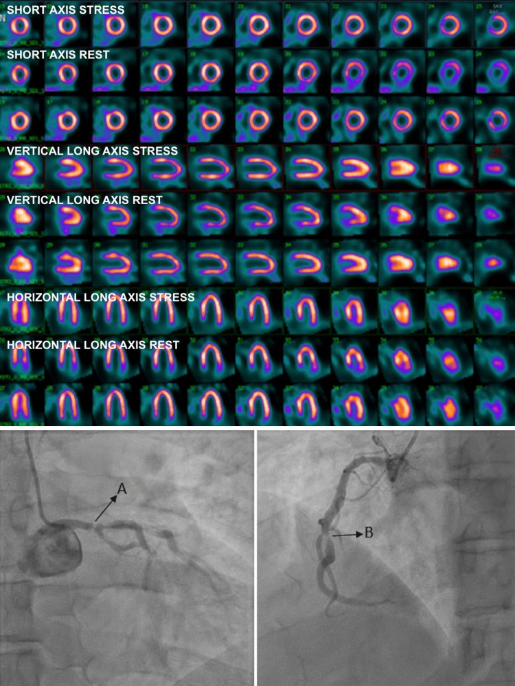
Stress cardiovascular magnetic resonance. Vasodilator perfusion CMR, or dobutamine wall motion CMR, has high diagnostic accuracy without ionizing radiation and adds late gadolinium enhancement to define scar and viability. In patients with known coronary disease, an abnormal stress CMR study was associated with about twice the mortality hazard in a 2,496-patient registry cited by the 2023 guideline.
Stress testing in known coronary disease. For patients with obstructive coronary disease and persistent stable chest pain despite guideline-directed therapy, stress PET or SPECT, CMR, or echocardiography is recommended to detect and quantify ischemia, estimate risk, and guide therapy (Class 1, 2023 guideline). Exercise treadmill testing can be useful to judge whether symptoms are angina, their severity, and functional capacity (Class 2a).
Risk stratification of patients with established chronic coronary disease
The 2023 guideline recommends that risk stratification integrate all clinical and test information, or use a validated risk score, to classify the annual risk of cardiovascular death or nonfatal myocardial infarction as low (below 1%), intermediate (1% to 3%), or high (above 3%) (Class 1). Test results alone are insufficient: in one externally validated cohort, the Duke treadmill score alone had a c-index of 0.62 for death. Adding clinical variables raised it to 0.83 and reclassified 64% of low-risk treadmill scores as intermediate or high risk. In a meta-analysis of 165 studies, a normal functional test did not uniformly predict an annual risk below 1%; a normal CCTA did.
Clinical features associated with higher risk include older age, prior myocardial infarction or revascularization, heart failure, reduced ejection fraction, diabetes, chronic kidney disease, peripheral artery disease, atrial fibrillation, smoking, depression, poor adherence, poverty, and poor social support.
| Annual risk of death or MI | Noninvasive findings |
|---|---|
| High (>3%) | Resting LVEF <35% not explained by noncoronary causes; resting perfusion abnormality ≥10% of myocardium without prior MI; ST depression ≥2 mm at low workload or persisting into recovery, exercise-induced ST elevation, or exercise-induced VT or VF; severe stress-induced LV dysfunction (peak exercise LVEF <45% or a fall of ≥10% with stress); stress perfusion abnormality ≥10% of myocardium or in multiple territories; stress-induced LV dilation; inducible wall motion abnormality in more than 2 segments or 2 coronary territories; wall motion abnormality at dobutamine ≤10 µg/kg/min or heart rate <120 beats/min; coronary calcium score >400; multivessel stenosis ≥70% or left main ≥50% on CCTA |
| Intermediate (1% to 3%) | Resting LVEF 35% to 49%; resting perfusion abnormality in 5% to 9.9% of myocardium without prior MI; ST depression ≥1 mm with exertional symptoms; stress perfusion abnormality in 5% to 9.9% of myocardium or one territory without LV dilation; small inducible wall motion abnormality in 1 to 2 segments of one territory; calcium score 100 to 399; one-vessel stenosis ≥70% or 50% to 69% stenosis in two or more arteries on CCTA |
| Low (<1%) | Duke treadmill score ≥5, or no new ST changes or angina at maximal exercise; normal perfusion or a defect involving <5% of myocardium; normal stress wall motion; calcium score <100; no stenosis >50% on CCTA |
Noninvasive risk stratification in stable ischemic heart disease, adapted from Table 4 of the 2012 ACCF/AHA stable ischemic heart disease guideline, the framework that the 2023 guideline retains for annual risk categories. LV, left ventricular; LVEF, left ventricular ejection fraction; MI, myocardial infarction; VF, ventricular fibrillation; VT, ventricular tachycardia.
Left ventricular dysfunction. Ejection fraction is the strongest single predictor of survival in chronic coronary disease. In patients with newly reduced left ventricular systolic function, clinical heart failure, or both, invasive coronary angiography is recommended to define anatomy and guide potential revascularization (Class 1); CCTA may be an initial strategy in selected patients in whom a nonischemic cardiomyopathy is suspected.
Invasive coronary angiography: indications and limits
Invasive angiography is a decision tool for revascularization, not a risk-stratification test for every patient. The 2023 guideline recommends it for patients whose symptoms or functional capacity change and persist despite guideline-directed therapy, to guide treatment aimed at improving angina (Class 1). It is also recommended for new left ventricular dysfunction or heart failure (Class 1). It is not routinely recommended for risk stratification in patients without left ventricular dysfunction, heart failure, angina refractory to therapy, or noninvasive findings suggesting left main disease above 50% (Class 3: No Benefit). The 2021 chest pain guideline recommends it for patients with obstructive disease and persistent angina with moderate to severe ischemia (Class 1). It is also recommended when CCTA shows left main stenosis of 50% or more, three-vessel stenosis of 70% or more, or obstructive disease with FFR-CT of 0.80 or less (Class 1).
Angiography shows the lumen, not the wall. It underestimates plaque burden because of positive remodeling, cannot judge the physiology of intermediate stenoses, and visually overestimates or underestimates stenosis severity with interobserver variability that matters most between 50% and 80%. For percutaneous coronary intervention (PCI), radial access is recommended over femoral access to reduce bleeding and vascular complications (Class 1, 2021 revascularization guideline).
Coronary physiology: FFR, iFR, and other pressure indices before revascularization
Fractional flow reserve (FFR) is the ratio of mean distal coronary pressure to mean aortic pressure during maximal hyperemia, usually induced with intravenous or intracoronary adenosine. It estimates the fraction of normal maximal flow that the stenosed vessel can deliver, and a value of 0.80 or less identifies a hemodynamically significant epicardial lesion. The instantaneous wave-free ratio (iFR) and other nonhyperemic pressure ratios (diastolic pressure ratio, resting full-cycle ratio) are measured at rest during the part of the cardiac cycle when microvascular resistance is lowest and most stable; 0.89 or less is abnormal. The thresholds were calibrated separately, so they are not mathematically interchangeable, and the two indices disagree in about one-fifth of lesions.
In patients with angina or an anginal equivalent, no prior ischemia evaluation, and angiographically intermediate stenoses, FFR or a proven nonhyperemic ratio is recommended before PCI (Class 1, Level A), and FFR in this setting is a high-value intervention. The evidence base:
- In FAME (1,005 patients with multivessel disease), FFR-guided PCI reduced death, myocardial infarction, or repeat revascularization at 1 year compared with angiography-guided PCI (13.2% vs. 18.3%) while using fewer stents.
- DEFINE-FLAIR (2,492 patients) and iFR-SWEDEHEART (2,037 patients) found iFR-guided revascularization noninferior to FFR-guided revascularization for major adverse cardiac events at 1 year, with fewer procedural symptoms and shorter procedures.
FFR, CFR, and IMR measure different compartments. Fractional flow reserve and nonhyperemic ratios assess the epicardial conductance vessel. Coronary flow reserve (CFR, hyperemic flow divided by resting flow) integrates the epicardial artery and the microcirculation. The index of microcirculatory resistance (IMR; distal pressure multiplied by the hyperemic mean transit time from thermodilution) assesses the microcirculation. A normal FFR with a reduced coronary flow reserve indicates diffuse disease or microvascular dysfunction, and a lesion with a normal FFR is not stented to treat angina.
Intravascular imaging with IVUS and OCT
Intravascular ultrasound (IVUS) and optical coherence tomography (OCT) image the vessel wall and lumen in cross-section. IVUS penetrates deeper, measures the external elastic membrane and plaque burden, and is preferred for ostial left main lesions. OCT has about ten times the axial resolution, defines calcium thickness, thin caps, thrombus, and stent strut apposition, and requires blood clearance with contrast flush, which limits its use at the aorto-ostial junction.
In the 2021 ACC/AHA/SCAI revascularization guideline, IVUS can be useful for procedural guidance, particularly in left main or complex coronary stenting (Class 2a). OCT is a reasonable alternative to IVUS except in ostial left main disease (Class 2a), and IVUS or OCT is reasonable to determine the mechanism of stent failure (Class 2a).
Myocardial viability and revascularization in ischemic cardiomyopathy
Viability testing identifies dysfunctional myocardium that may recover contractile function after revascularization. Dobutamine echocardiography shows contractile reserve; SPECT and PET show preserved perfusion or metabolism; and late gadolinium enhancement on CMR shows the transmural extent of scar, with segments with less than 50% transmural enhancement more likely to recover.
In the STICH trial (1,212 patients with ejection fraction 35% or less and anatomy suitable for CABG), CABG plus medical therapy did not significantly reduce all-cause death at a median of 56 months (36% vs. 41%), but it reduced cardiovascular death and death or cardiovascular hospitalization. In the 10-year extension (STICHES), all-cause death was lower with CABG (58.9% vs. 66.1%; hazard ratio 0.84). In the STICH viability substudy, viable myocardium was associated with better survival but did not identify patients who benefited from CABG compared with medical therapy. In REVIVED-BCIS2 (700 patients with ejection fraction 35% or less and extensive coronary disease with viability in at least four segments), PCI did not reduce death from any cause or hospitalization for heart failure compared with optimal medical therapy (37.2% vs. 38.0%) over a median of 3.4 years. It did not improve ejection fraction.
Viability imaging informs the likelihood of functional recovery and the extent of scar, and decisions integrate anatomy, symptoms, operative risk, comorbidity, and patient preference. Viability alone neither mandates nor excludes revascularization.
Antianginal drugs: selection, dosing, and combination
Antianginal drugs act by lowering myocardial oxygen demand (beta-blockers, non-dihydropyridine calcium channel blockers, ivabradine), by increasing supply through vasodilation (nitrates, dihydropyridine calcium channel blockers), or by a metabolic mechanism (ranolazine, which inhibits the late inward sodium current). Their justification rests on symptom relief; none of them, beta-blockers outside the settings described below included, has been shown to reduce death in chronic coronary disease.
First-line choice. Either a beta-blocker or a calcium channel blocker is first-line therapy. The choice follows heart rate, blood pressure, ventricular function, conduction disease, suspected vasospasm, and comorbidities. A beta-blocker is preferred after a recent myocardial infarction, with an ejection fraction of 50% or less, with tachyarrhythmias, or with a high resting heart rate. A calcium channel blocker is preferred with vasospasm, asthma or severe peripheral artery disease that limits beta-blockade, or bradycardia (a dihydropyridine in that case). Abrupt withdrawal of a beta-blocker can provoke rebound ischemia.
Combinations. A dihydropyridine calcium channel blocker combines well with a beta-blocker, which blunts its reflex tachycardia. A non-dihydropyridine agent (verapamil or diltiazem) with a beta-blocker risks bradycardia, atrioventricular block, and reduced contractility, and is used with caution. Ranolazine adds antianginal effect without lowering heart rate or blood pressure, which makes it useful when bradycardia or hypotension limits other drugs.
| Drug | Usual adult dose | Selection points and cautions |
|---|---|---|
| Metoprolol succinate | 100–400 mg once daily for angina (12.5–200 mg in heart failure) | Evidence-based in reduced ejection fraction (with carvedilol and bisoprolol); target resting heart rate about 55–60 beats/min |
| Carvedilol | 3.125–25 mg twice daily | Alpha- and beta-blockade; more blood pressure lowering |
| Atenolol | 50–100 mg once daily (up to 200 mg) | Renally cleared; inferior to other antihypertensives for blood pressure outcomes |
| Amlodipine | 5–10 mg once daily (lower in older adults and hepatic impairment) | Peripheral edema; safe in reduced ejection fraction; combines with beta-blockers |
| Nifedipine extended release | 30–90 mg once daily | Short-acting nifedipine is avoided because of reflex tachycardia and hypotension |
| Diltiazem extended release | 120–480 mg daily | Lowers heart rate; avoided with reduced ejection fraction and with conduction disease; CYP3A4 inhibitor |
| Verapamil | 80–120 mg three times daily (immediate release); extended release 120–480 mg daily is labeled for hypertension | Constipation; avoided with reduced ejection fraction; raises digoxin and some statin levels |
| Isosorbide mononitrate extended release | 30–120 mg once daily (240 mg rarely) | Once-daily dosing provides a nitrate-free interval |
| Isosorbide dinitrate | 5–40 mg two or three times daily | Immediate-release form; eccentric dosing with a nitrate-free interval of at least 14 hours |
| Sublingual nitroglycerin | 0.3–0.4 mg tablet or 0.4 mg spray | Every 5 minutes up to 3 doses; also taken before activity that provokes angina |
| Ranolazine extended release | 500 mg twice daily, up to 1,000 mg twice daily | Contraindicated with strong CYP3A inhibitors, CYP3A inducers, and hepatic cirrhosis; maximum 500 mg twice daily with moderate CYP3A inhibitors such as diltiazem and verapamil; dose-related QTc prolongation; simvastatin limited to 20 mg; metformin dose limited |
Antianginal drugs available in the United States. Doses are typical ranges from US prescribing information and are adjusted for heart rate, blood pressure, hepatic and renal function, and interacting drugs. Bisoprolol and carvedilol are labeled in the United States for hypertension or heart failure, not angina, and their doses come from those labels.
Nitrates. Continuous exposure produces tolerance within about 24 hours, which a daily nitrate-free interval prevents; rebound angina can occur during that interval. Nitrates are contraindicated within 24 hours of sildenafil or vardenafil and within 48 hours of tadalafil, because the combination causes severe hypotension; they are also avoided in hypertrophic obstructive cardiomyopathy and severe aortic stenosis with hypotension.
Ranolazine. In CARISA (823 patients) and ERICA (565 patients on maximal amlodipine), ranolazine reduced angina frequency and nitroglycerin use on top of background antianginal therapy. Dizziness, nausea, and constipation are common.
Ivabradine. In SIGNIFY (19,102 patients with stable coronary disease, no clinical heart failure, and heart rate of 70 beats/min or more), ivabradine did not reduce cardiovascular death or myocardial infarction. In the subgroup with activity-limiting angina it increased the primary outcome (7.6% vs. 6.5%), the basis of the Class 3: Harm recommendation. In the United States it is approved for heart failure with reduced ejection fraction, not for angina.
Beta-blockers after myocardial infarction and in chronic coronary disease
Contemporary patients receive early reperfusion, statins, and antiplatelet therapy, and most have preserved ejection fraction. The 2023 guideline therefore separates the indications:
- In CCD with an ejection fraction of 40% or less, with or without previous infarction, beta-blocker therapy is recommended to reduce major adverse cardiovascular events, including cardiovascular death (Class 1).
- With an ejection fraction below 50%, sustained-release metoprolol succinate, carvedilol, or bisoprolol titrated to target doses is recommended in preference to other beta-blockers (Class 1).
- In patients who started a beta-blocker after infarction and have no current or previous ejection fraction of 50% or less, angina, arrhythmias, or uncontrolled hypertension, reassessing the indication beyond 1 year may be reasonable (Class 2b).
- In CCD without previous infarction or an ejection fraction of 50% or less, and without another primary indication, beta-blocker therapy does not reduce major adverse cardiovascular events (Class 3: No Benefit).
Trials since the guideline support this approach. In REDUCE-AMI (5,020 patients with myocardial infarction and ejection fraction of 50% or more), beta-blockade did not reduce death or myocardial infarction over a median of 3.5 years (7.9% vs. 8.3%; hazard ratio 0.96). REBOOT, conducted in patients with ejection fraction above 40%, found no effect on death, reinfarction, or heart failure hospitalization. The combined BETAMI-DANBLOCK analysis, which included patients with ejection fraction of 40% or more, reported a modest reduction in its composite of death or major adverse cardiovascular events (14.2% vs. 16.3%; hazard ratio 0.85) without a difference in mortality; a separate individual-patient meta-analysis found the benefit of beta-blockade concentrated in patients with an ejection fraction of 40% to 49%. An individual-patient meta-analysis of five trials (17,801 patients with ejection fraction of 50% or more) found no reduction in death, myocardial infarction, or heart failure (8.1% vs. 8.3%; hazard ratio 0.97). Reassessment is not abrupt withdrawal: when a beta-blocker that was started after infarction is stopped, it is tapered, and the original indication is reviewed.
Antithrombotic therapy in chronic coronary disease
Antiplatelet therapy reduces atherothrombotic events at the cost of bleeding, and every choice after the first months following an event or PCI balances ischemic and bleeding risk.
| Setting | Recommendation | Class |
|---|---|---|
| CCD without an indication for oral anticoagulation | Aspirin 81 mg daily (75–100 mg) | 1 |
| After PCI for CCD | Aspirin plus clopidogrel for 6 months, then single antiplatelet therapy | 1 |
| Selected patients after PCI with a drug-eluting stent and 1 to 3 months of DAPT | P2Y12 inhibitor monotherapy for at least 12 months to reduce bleeding | 2a |
| High ischemic risk, low to moderate bleeding risk, no indication for anticoagulation or DAPT | Rivaroxaban 2.5 mg twice daily plus aspirin 81 mg daily | 2a |
| On DAPT | Proton pump inhibitor to reduce gastrointestinal bleeding | 2a |
| Elective PCI in a patient requiring oral anticoagulation | DAPT for 1 to 4 weeks, then clopidogrel alone for 6 months, in addition to a DOAC | 1 |
| Oral anticoagulation, low atherothrombotic risk, 1 year after PCI | Aspirin stopped and DOAC continued alone | 2b |
| Oral anticoagulation and no acute indication for antiplatelet therapy | DOAC monotherapy | 2b |
| No recent ACS or PCI-related indication | Clopidogrel added to aspirin | 3: No Benefit |
| Previous stroke, TIA, or intracranial hemorrhage | Vorapaxar added to DAPT; prasugrel | 3: Harm |
| Any CCD | Chronic nonsteroidal anti-inflammatory drugs | 3: Harm |
Antiplatelet and anticoagulant recommendations, 2023 AHA/ACC chronic coronary disease guideline. ACS, acute coronary syndrome; DAPT, dual antiplatelet therapy; DOAC, direct oral anticoagulant; MI, myocardial infarction; TIA, transient ischemic attack.
Aspirin dose and alternatives. In ADAPTABLE (15,076 patients with atherosclerotic disease), aspirin 81 mg and 325 mg daily produced similar rates of death, myocardial infarction, or stroke and similar major bleeding. In HOST-EXAM (5,438 patients who had completed DAPT without events after PCI), clopidogrel monotherapy reduced the composite of death, myocardial infarction, stroke, readmission for acute coronary syndrome, or major bleeding compared with aspirin over 24 months (5.7% vs. 7.7%; hazard ratio 0.73). The 2024 ESC guideline recommends clopidogrel 75 mg daily as an alternative to aspirin monotherapy (Class I).
Dual-pathway inhibition. In COMPASS (27,395 patients with stable coronary or peripheral artery disease), rivaroxaban 2.5 mg twice daily plus aspirin reduced cardiovascular death, stroke, or myocardial infarction compared with aspirin alone (4.1% vs. 5.4%; hazard ratio 0.76). Major bleeding was more frequent (3.1% vs. 1.9%), without a significant increase in fatal or intracranial bleeding.
Atrial fibrillation with chronic coronary disease. For patients beyond the first year after PCI or an acute coronary syndrome, adding antiplatelet therapy to an anticoagulant mainly adds bleeding. In AFIRE (2,236 patients in Japan with atrial fibrillation and coronary disease more than 1 year after revascularization or with angiographically confirmed disease), rivaroxaban monotherapy was noninferior to rivaroxaban plus an antiplatelet agent for cardiovascular events and death and superior for major bleeding. All-cause mortality was lower with monotherapy. In EPIC-CAD (1,040 patients with high-risk atrial fibrillation and stable coronary disease), edoxaban alone reduced net adverse clinical events at 12 months compared with edoxaban plus a single antiplatelet agent (6.8% vs. 16.2%), mainly through less bleeding.
Bleeding prevention. Pantoprazole and other agents with less CYP2C19 inhibition than omeprazole or esomeprazole avoid the pharmacokinetic interaction with clopidogrel, although no clinical outcome harm has been proven in trials.
Risk-factor and preventive therapy: lipids, blood pressure, diabetes, weight, and colchicine
Lipids. High-intensity statin therapy to lower low-density lipoprotein cholesterol (LDL-C) by 50% or more is recommended (Class 1). The 2026 ACC/AHA dyslipidemia guideline sets goals of LDL-C below 55 mg/dL for very high-risk disease and below 70 mg/dL for other atherosclerotic disease; when LDL-C remains above goal on maximally tolerated statin therapy, ezetimibe, bempedoic acid, or a proprotein convertase subtilisin/kexin type 9 (PCSK9) monoclonal antibody is added, and lipoprotein(a) is measured at least once. Icosapent ethyl may be considered for persistent triglycerides of 150 to 499 mg/dL (Class 2b), and fish-oil supplements, niacin, and fenofibrate added to statins do not reduce cardiovascular events (Class 3: No Benefit).
Blood pressure. A target below 130/80 mm Hg is recommended in adults with CCD and hypertension (Class 1), unchanged in the 2025 AHA/ACC guideline. A beta-blocker, angiotensin-converting enzyme (ACE) inhibitor, or angiotensin receptor blocker (ARB) is first-line for compelling indications such as recent infarction or angina (Class 1).
Renin-angiotensin-aldosterone inhibition. ACE inhibitors, or ARBs in patients intolerant of ACE inhibitors, are recommended in CCD with hypertension, diabetes, an ejection fraction of 40% or less, or chronic kidney disease (Class 1). Without these conditions and with an ejection fraction above 40%, they may be considered (Class 2b).
SGLT2 inhibitors and GLP-1 receptor agonists. In CCD with type 2 diabetes, a sodium-glucose cotransporter 2 (SGLT2) inhibitor or a glucagon-like peptide-1 (GLP-1) receptor agonist with proven cardiovascular benefit is recommended to reduce major adverse cardiovascular events (Class 1), independent of glycemic control. With heart failure and an ejection fraction of 40% or less, an SGLT2 inhibitor is recommended irrespective of diabetes (Class 1), and with ejection fraction above 40% it can be beneficial (Class 2a). In SELECT (17,604 patients with established cardiovascular disease, body mass index of 27 or more, and no diabetes), semaglutide 2.4 mg weekly reduced cardiovascular death, myocardial infarction, or stroke (6.5% vs. 8.0%; hazard ratio 0.80). This extended the GLP-1 receptor agonist indication to obesity without diabetes.
Weight. Weight loss is recommended for patients with overweight or obesity, with GLP-1 receptor agonists when pharmacotherapy is warranted.
Colchicine. Low-dose colchicine may be considered for secondary prevention (Class 2b). In LoDoCo2 (5,522 patients with chronic coronary disease), colchicine 0.5 mg daily reduced cardiovascular death, spontaneous myocardial infarction, ischemic stroke, or ischemia-driven revascularization (6.8% vs. 9.6%), with a numerical excess of noncardiovascular death. CLEAR SYNERGY (7,062 patients), which started colchicine within days of infarction, showed no benefit (9.1% vs. 9.3%). Colchicine 0.5 mg daily is approved in the United States to reduce cardiovascular events in adults with established atherosclerotic disease. The 2023 AHA/ACC guideline advises avoiding it at an estimated glomerular filtration rate below 30 mL/min/1.73 m². The LODOCO label contraindicates it at a creatinine clearance below 15 mL/min, in severe hepatic impairment, and with strong CYP3A4 or P-glycoprotein inhibitors. The label also advises against moderate CYP3A4 inhibitors in patients with renal or hepatic impairment. The 2024 ESC guideline gives colchicine a Class 2a recommendation.
Immunization. Annual influenza vaccination is recommended to reduce cardiovascular morbidity, cardiovascular death, and all-cause death (Class 1), COVID-19 vaccination is recommended according to public health guidance (Class 1), and pneumococcal vaccination is reasonable (Class 2a).
Lifestyle, cardiac rehabilitation, mental health, and social determinants
Diet and physical activity. A diet emphasizing vegetables, fruits, legumes, nuts, whole grains, and lean protein is recommended (Class 1). So are at least 150 minutes per week of moderate-intensity or 75 minutes per week of vigorous aerobic activity and resistance training on 2 or more days per week (Class 1). Saturated fat below 6% of calories and sodium below 2,300 mg/day can be beneficial (Class 2a).
Cardiac rehabilitation. Referral to a cardiac rehabilitation program is recommended for all eligible patients with CCD (Class 1): after recent myocardial infarction, PCI, or CABG (Level A), with stable angina or after heart transplant (Level B-R), and after spontaneous coronary artery dissection (Level C-LD). Participation reduces cardiovascular mortality and hospitalization and improves exercise capacity and quality of life, yet it remains underused, particularly by women, older adults, and minority and rural patients.
Tobacco. Tobacco use is assessed and cessation advised at every visit (Class 1). Behavioral support combined with varenicline, bupropion, or combination nicotine replacement is recommended (Class 1), and varenicline may be preferred (Class 2b). Exposure to secondhand smoke is harmful (Class 3: Harm).
Alcohol and substances. Patients who drink are advised to limit alcohol to 1 drink per day for women and 2 for men (Class 2a), and alcohol is not recommended for cardiovascular protection (Class 3: No Benefit).
Mental health. Depression, anxiety, and psychosocial stress are common in CCD and are associated with worse outcomes. Screening and referral are reasonable (Class 2a), and treatment with pharmacologic or nonpharmacologic therapy is reasonable to improve cardiovascular outcomes (Class 2a).
Sleep apnea. Obstructive sleep apnea is common in CCD and contributes to hypertension and atrial fibrillation. In the SAVE trial of patients with cardiovascular disease and moderate to severe sleep apnea, continuous positive airway pressure improved symptoms and quality of life without reducing cardiovascular events.
Team-based care, shared decision-making, and social determinants. A multidisciplinary team-based approach is recommended to improve outcomes and risk-factor control (Class 1, Level A), and clinical follow-up at least annually is recommended (Class 1). Shared decision-making is recommended when the evidence is uncertain or a significant risk-benefit tradeoff exists (Class 1), and a validated decision aid may be considered for patients with angina weighing revascularization (Class 2b). Routine assessment of social determinants of health (health care access, economic stability, food and housing security, transportation, and social context) is recommended to inform treatment decisions and lifestyle recommendations (Class 1), and out-of-pocket cost is discussed because it reduces adherence.
Revascularization versus medical therapy: COURAGE, ISCHEMIA, and ORBITA
Opening a stenosis that causes ischemia was long assumed to prevent infarction and death. Randomized trials in stable patients have separated two effects: revascularization reliably relieves angina in symptomatic patients. In most stable patients without high-risk anatomy or severe left ventricular dysfunction, it does not reduce death or myocardial infarction beyond what intensive medical therapy achieves.
| Trial | Population | Comparison | Main result |
|---|---|---|---|
| COURAGE (2007) | 2,287 patients with stable disease, ≥70% stenosis, and objective ischemia | PCI plus optimal medical therapy vs. medical therapy | Death or MI 19.0% vs. 18.5% at a median of 4.6 years; earlier angina relief with PCI that narrowed over time |
| ISCHEMIA (2020) | 5,179 patients with moderate or severe ischemia; left main ≥50%, LVEF <35%, unacceptable angina, and recent ACS excluded | Initial invasive strategy vs. initial conservative strategy | No significant reduction in the primary composite at a median of 3.2 years; more events with invasive care at 6 months (5.3% vs. 3.4%) and fewer at 5 years (16.4% vs. 18.2%); greater angina relief with invasive care in patients with frequent angina |
| ISCHEMIA-CKD (2020) | 777 patients with eGFR <30 mL/min/1.73 m² or on dialysis and moderate or severe ischemia | Initial invasive vs. conservative strategy | No reduction in death or MI over a median of 2.2 years; more strokes with the invasive strategy; no angina benefit |
| ISCHEMIA-EXTEND (2023) | ISCHEMIA cohort, median follow-up 5.7 years | Same | All-cause death at 7 years 12.7% vs. 13.4% (no difference); cardiovascular death lower (6.4% vs. 8.6%) and noncardiovascular death higher (5.6% vs. 4.4%) with invasive care |
| ORBITA (2018) | 200 randomized patients with single-vessel disease after 6 weeks of antianginal optimization | PCI vs. placebo procedure | No significant difference in exercise time increment (16.6 seconds) |
| ORBITA-2 (2023) | 301 patients with angina and ischemia, antianginal drugs stopped | PCI vs. placebo procedure | Lower angina symptom score with PCI at 12 weeks (odds ratio 2.21); exercise time longer by about 60 seconds |
| FAME 2 (2012, 2018) | 888 patients with at least one lesion with FFR ≤0.80 | FFR-guided PCI plus medical therapy vs. medical therapy | Death, MI, or urgent revascularization 13.9% vs. 27.0% at 5 years, driven by urgent revascularization |
Randomized trials of revascularization versus medical therapy in stable coronary disease. ACS, acute coronary syndrome; eGFR, estimated glomerular filtration rate; FFR, fractional flow reserve; LVEF, left ventricular ejection fraction; MI, myocardial infarction.
What ISCHEMIA showed. The primary outcome was cardiovascular death, myocardial infarction, or hospitalization for unstable angina, heart failure, or resuscitated cardiac arrest. Severity of ischemia was associated with death, but there was no interaction between ischemia severity (mild, moderate, or severe) and the effect of the invasive strategy. About one-third of participants had no angina in the month before enrollment; invasive care improved Seattle Angina Questionnaire scores in patients with daily, weekly, or monthly angina and produced little change in those without angina.
What ISCHEMIA did not test. The trial excluded significant left main disease (by blinded CCTA in most patients), ejection fraction below 35%, recent acute coronary syndrome, unacceptable angina despite medical therapy, and advanced heart failure. Its results do not apply to those groups. A secondary analysis of 398 participants with heart failure or an ejection fraction below 45% found a lower 4-year primary event rate with invasive care (17.2% vs. 29.3%).
Indications for revascularization in chronic coronary disease
| Setting | Recommendation | Class |
|---|---|---|
| Lifestyle-limiting angina despite GDMT with significant stenoses amenable to revascularization | Revascularization to improve symptoms | 1 |
| Significant left main disease | CABG in addition to medical therapy to improve survival | 1 |
| Multivessel disease with LVEF ≤35% | CABG in addition to medical therapy to improve survival | 1 |
| Multivessel disease with LVEF 35% to 50% | CABG to improve survival | 2a |
| Selected significant left main stenosis in which PCI can provide revascularization equivalent to CABG | PCI to improve survival | 2a |
| Multivessel disease appropriate for CABG or PCI | Revascularization in addition to GDMT to lower spontaneous MI, urgent revascularization, or cardiac death | 2a |
| Three-vessel disease with normal LVEF | CABG may be reasonable to improve survival; the survival benefit of PCI is uncertain | 2b |
| Isolated proximal LAD disease | Survival benefit of revascularization uncertain | 2b |
| Angina, no prior ischemia testing, angiographically intermediate stenosis | FFR or a proven nonhyperemic ratio before PCI | 1 |
| Complex three-vessel disease or unclear optimal strategy | Heart Team with interventional cardiology and cardiac surgery | 1 |
Revascularization in chronic coronary disease: 2023 AHA/ACC chronic coronary disease guideline and 2021 ACC/AHA/SCAI revascularization guideline (the LVEF 35% to 50%, normal-LVEF, and proximal LAD rows). The 2021 guideline downgraded CABG for survival in three-vessel disease with normal LVEF from Class 1 to Class 2b, largely because of ISCHEMIA; the American Association for Thoracic Surgery and the Society of Thoracic Surgeons did not endorse the guideline. GDMT, guideline-directed management and therapy; LAD, left anterior descending artery.
Survival evidence in left main disease and ischemic cardiomyopathy comes predominantly from surgical trials. A lesion that is not physiologically significant is not revascularized to treat angina; persistent symptoms in that setting lead to evaluation for diffuse disease, microvascular dysfunction, vasospasm, or a noncardiac cause.
PCI or CABG for left main and multivessel disease
When revascularization is indicated, the choice between PCI and CABG depends on anatomic complexity, diabetes, left ventricular function, completeness of revascularization achievable with each method, surgical risk, frailty, and patient preference. A Heart Team that includes an interventional cardiologist and a cardiac surgeon is recommended for complex three-vessel disease or when the optimal strategy is unclear (Class 1). The SYNTAX score grades anatomic complexity (low 22 or less, intermediate 23 to 32, high 33 or more), although the 2021 guideline notes its interobserver variability and absence of clinical variables.
| Patient group | Recommendation | Class |
|---|---|---|
| Left main disease with high-complexity CAD | CABG in preference to PCI to improve survival | 1 |
| Multivessel disease with complex, diffuse CAD (for example, SYNTAX >33) | CABG over PCI to improve survival | 2a |
| Diabetes and multivessel disease involving the LAD, suitable for CABG | CABG with a left internal mammary artery to the LAD in preference to PCI to reduce mortality and repeat revascularization | 1 |
| Diabetes with left main stenosis and low- or intermediate-complexity CAD (SYNTAX ≤33) | PCI as an alternative to CABG to reduce MACE | 2b |
| Appropriate for revascularization but a poor surgical candidate | PCI over CABG to improve symptoms and reduce MACE | 2a |
PCI versus CABG in chronic coronary disease, 2023 AHA/ACC guideline (modified from the 2021 ACC/AHA/SCAI revascularization guideline). CAD, coronary artery disease; LAD, left anterior descending artery; MACE, major adverse cardiovascular events.
| Trial | Population | Main result |
|---|---|---|
| SYNTAX (5 years) | 1,800 patients with left main or three-vessel disease | Death, stroke, MI, or repeat revascularization 37.3% with PCI vs. 26.9% with CABG; the difference was concentrated in intermediate and high SYNTAX scores |
| FREEDOM | 1,900 patients with diabetes and multivessel disease | Death, MI, or stroke at 5 years 26.6% with PCI vs. 18.7% with CABG; all-cause death 16.3% vs. 10.9%; more strokes with CABG |
| EXCEL (5 years) | 1,905 patients with left main disease and SYNTAX ≤32 | Death, stroke, or MI 22.0% with PCI vs. 19.2% with CABG (no significant difference); all-cause death 13.0% vs. 9.9% |
| NOBLE (5 years) | 1,201 patients with left main disease | Major adverse cardiac or cerebrovascular events 28% with PCI vs. 19% with CABG (hazard ratio 1.58), driven by nonprocedural MI and repeat revascularization; no difference in death |
| FAME 3 (1 year) | 1,500 patients with three-vessel disease, left main excluded | FFR-guided PCI did not meet noninferiority to CABG (10.6% vs. 6.9%) |
Randomized trials of PCI versus CABG. MI, myocardial infarction.
Conduits and completeness. The left internal mammary artery to the left anterior descending artery has the best long-term patency and outcome of any graft. In the 2021 guideline, a radial artery is recommended in preference to saphenous vein to bypass the second most important target with significant stenosis (Class 1), because it improves patency and long-term cardiac outcomes. Saphenous vein grafts develop accelerated atherosclerosis and have substantially lower long-term patency. For PCI, complete revascularization means treating lesions that subtend substantial myocardium and cause ischemia, not every visible stenosis.
Chronic total occlusion. In the 2021 guideline, after treatment of non-occlusive lesions, the benefit of PCI of a chronic total occlusion to improve symptoms in patients with refractory angina and suitable anatomy is uncertain (Class 2b). Randomized trials have shown symptom improvement in some patients and no reduction in death or myocardial infarction.
Recurrent angina after PCI or CABG
Recurrent angina after revascularization requires the mechanism to be established before repeat intervention.
| Cause | Typical timing and features | Evaluation and treatment |
|---|---|---|
| Stent thrombosis | Abrupt; presents as acute coronary syndrome, often ST elevation | Emergency angiography; intravascular imaging for underexpansion, malapposition, or edge dissection; adherence to antiplatelet therapy reviewed |
| In-stent restenosis from neointimal hyperplasia | Gradually returning exertional angina, usually within the first year | Intravascular imaging; drug-coated balloon or repeat drug-eluting stent |
| Neoatherosclerosis | Late after drug-eluting stents; behaves like de novo plaque and can rupture | Intravascular imaging; treatment as for restenosis |
| Stent underexpansion or fracture | Mechanical; often in calcified vessels | Intravascular imaging; high-pressure or lithotripsy-assisted dilation in place of another stent |
| Disease progression or incomplete revascularization | New lesion elsewhere or residual ischemia in an untreated territory | Stress imaging or CCTA; physiology at angiography |
| Saphenous vein graft disease | Accelerated, diffuse, friable atherosclerosis; distal embolization during intervention | Native-vessel PCI preferred when feasible; embolic protection for vein graft PCI |
| Microvascular dysfunction or vasospasm | Persistent angina with widely patent stents and grafts | Coronary function testing; vasodilator therapy |
Mechanisms of recurrent angina after revascularization.
Ischemia with nonobstructive coronary arteries and microvascular dysfunction
Nonobstructive coronary disease is found in more than half of patients undergoing elective angiography and is associated with an increased risk of death and myocardial infarction. Angina with no obstructive coronary arteries (ANOCA) describes the symptom; ischemia with no obstructive coronary arteries (INOCA) requires objective ischemia. Obstructive disease is excluded by a stenosis below 50% on CCTA or angiography, or an FFR above 0.80. INOCA is a group of mechanisms, not a diagnosis: coronary microvascular dysfunction, epicardial vasospasm, microvascular spasm, or a combination, and these endotypes frequently coexist.
Coronary microvascular dysfunction includes impaired vasodilator capacity (reduced coronary flow reserve), increased microvascular resistance, and microvascular spasm. It is more frequent in women and in patients with hypertension, diabetes, obesity, and insulin resistance, and it also occurs with hypertrophic, infiltrative, and other cardiomyopathies.
The Coronary Vasomotion Disorders International Study Group (COVADIS) criteria define definitive microvascular angina by four findings: symptoms of myocardial ischemia, absence of obstructive coronary disease, objective evidence of ischemia, and impaired microvascular function (coronary flow reserve below 2.0 to 2.5 depending on method, IMR above 25, or microvascular spasm). Suspected microvascular angina requires the first two findings and either of the last two.
Testing. For persistent stable chest pain with nonobstructive disease, the 2021 chest pain guideline rates stress PET with myocardial blood flow reserve (Class 2a), stress CMR with blood flow reserve (Class 2a), and invasive coronary function testing in patients with at least mild ischemia on imaging (Class 2a) as reasonable. Stress echocardiography with coronary flow velocity reserve is possibly reasonable (Class 2b). Invasive coronary function testing, performed at the time of angiography with a pressure-temperature wire, measures FFR, coronary flow reserve by thermodilution, and IMR during adenosine hyperemia. Intracoronary acetylcholine then tests endothelial function and provokes epicardial or microvascular spasm. The 2024 ESC guideline gives greater weight to invasive coronary function testing in patients with persistent symptoms despite medical therapy.
| Endotype | Invasive criteria | Linked therapy in CorMicA |
|---|---|---|
| Microvascular angina | IMR ≥25, coronary flow reserve <2.0, resistive reserve ratio <2.0, or microvascular spasm (angina and ischemic ST changes with <90% epicardial constriction on acetylcholine) | Baseline aspirin, statin, and ACE inhibitor considered; beta-blocker first line; non-dihydropyridine calcium channel blocker if beta-blockers fail or are not tolerated; add-on dihydropyridine calcium channel blocker or ranolazine |
| Vasospastic angina | Epicardial spasm: >90% constriction with symptoms and ST deviation on acetylcholine | Calcium channel blocker first line; long-acting nitrate second line; smoking cessation |
| Noncardiac chest pain | Normal FFR, coronary flow reserve, IMR, and acetylcholine response | Antianginal drugs stopped; antiplatelet and statin stopped unless otherwise indicated; noncardiac evaluation |
INOCA endotypes and stratified therapy, adapted from Table 17 of the 2023 AHA/ACC guideline (CorMicA protocol). Nicorandil, listed as an add-on in the trial, is not available in the United States.
Stratified therapy. In CorMicA (151 patients with angina and no obstructive disease, 74% women), invasive testing at the index angiogram with therapy linked to the endotype improved the Seattle Angina Questionnaire summary score by 11.7 units at 6 months compared with usual care. The improvement was sustained at 1 year, with no difference in adverse events. In symptomatic patients with nonobstructive disease, stratified medical therapy guided by invasive coronary physiology can be useful to improve angina and quality of life (Class 2a, 2023 guideline). All patients receive risk-factor control and statin therapy when atherosclerosis is present.
Vasospastic angina and myocardial bridging
Vasospastic (variant, Prinzmetal) angina results from transient, reversible constriction of an epicardial artery causing subtotal or total occlusion. Spasm occurs in angiographically normal arteries or at sites of mild plaque. Smoking is the dominant modifiable risk factor. Vasospasm is more prevalent in East Asian populations, and in Western series it accounts for a substantial fraction of patients with angina and nonobstructive arteries. Triggers include cold exposure, hyperventilation, emotional stress, cocaine and amphetamines, fluoropyrimidine chemotherapy (5-fluorouracil, capecitabine), triptans, ergot alkaloids, and nonselective beta-blockers.
Clinical features. Attacks occur predominantly at rest, between midnight and early morning, last 5 to 15 minutes, and respond promptly to nitroglycerin. Exercise tolerance is usually preserved, although it can be reduced in the morning. Transient ST elevation during an attack indicates transmural ischemia from occlusive spasm; ST depression or new negative U waves indicate subtotal spasm. Prolonged spasm can cause myocardial infarction with nonobstructive arteries, atrioventricular block (more often with right coronary spasm), ventricular tachycardia or fibrillation, and sudden death.
COVADIS criteria define definitive vasospastic angina as nitrate-responsive angina plus either of two findings. One is transient ischemic ECG changes in at least two contiguous leads during spontaneous episodes (ST elevation or depression of 0.1 mV or more, or new negative U waves). The other is documented spasm: transient occlusion with more than 90% constriction, angina, and ischemic ECG changes.
Provocation testing. When the diagnosis is uncertain, intracoronary acetylcholine is given in incremental doses (commonly up to 100 µg into the left coronary artery and 50 µg into the right) with continuous electrocardiographic and symptom monitoring. Intracoronary nitroglycerin follows. A positive test reproduces the usual symptoms with ischemic electrocardiographic changes and more than 90% epicardial constriction. Calcium channel blockers and nitrates are withheld before elective testing when clinically safe, because they suppress spasm.
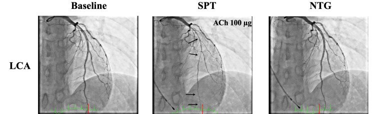
Treatment. Calcium channel blockers are first-line therapy, often at high doses and taken in the evening to cover nocturnal attacks; in refractory cases a dihydropyridine and a non-dihydropyridine agent are combined. Long-acting nitrates are added as second-line therapy, and sublingual nitroglycerin treats attacks. Smoking cessation and avoidance of triggers (stimulants, triptans, ergots) are essential. Nonselective beta-blockers can worsen spasm by leaving alpha-adrenergic vasoconstriction unopposed and are avoided. PCI has no role for isolated spasm without a fixed stenosis. Survivors of cardiac arrest attributed to vasospasm are evaluated for an implantable cardioverter-defibrillator in addition to vasodilator therapy, because recurrent arrest can occur despite medication. With treatment and smoking cessation, long-term survival is generally favorable; multivessel spasm, left main spasm, continued smoking, and ventricular arrhythmia during attacks identify higher risk.
Myocardial bridging. A myocardial bridge is a segment of epicardial artery, usually the mid left anterior descending artery, that tunnels through myocardium and is compressed in systole. Most bridges are incidental, but deep or long bridges can cause angina through diastolic flow impairment, accelerated proximal atherosclerosis, and associated vasospasm. CCTA and intravascular ultrasound (the echolucent half-moon sign) identify them. Beta-blockers and non-dihydropyridine calcium channel blockers are first-line; nitrates can worsen symptoms by increasing systolic compression, and surgical unroofing is reserved for refractory cases. Anomalous aortic origin of a coronary artery with an interarterial course (particularly the left coronary artery from the right sinus) causes exertional ischemia and sudden death in young adults, and coronary aneurysms after Kawasaki disease cause thrombosis and stenosis in adulthood; both are defined by CCTA.
Refractory angina: definition and treatment options when revascularization is not possible
Refractory angina is angina (or an equivalent) that persists despite intensive medical therapy, with objective evidence that the symptoms are ischemic and no remaining options for revascularization; many definitions add Canadian Cardiovascular Society class III or IV symptoms. The label applies only after adherence, doses, and combinations have been reviewed, all three classic antianginal classes and ranolazine have been tried as tolerated, and anemia, uncontrolled hypertension, tachyarrhythmia, and thyroid disease have been corrected. Microvascular dysfunction and vasospasm are evaluated first because they are treatable causes of angina after revascularization.
| Therapy | Evidence and status |
|---|---|
| Enhanced external counterpulsation | Sequential lower-limb cuff inflation in diastole; one sham-controlled trial (MUST-EECP, 1999) showed less angina and longer time to ST depression; cleared by the US Food and Drug Administration; may be considered (Class 2b, 2023 guideline); infrequently used |
| Coronary sinus reducer | Hourglass stent that narrows the coronary sinus and redistributes flow to the subendocardium; in COSIRA (104 patients), 35% vs. 15% improved by at least two angina classes; in ORBITA-COSMIC (51 patients), fewer daily angina episodes with no change in myocardial blood flow; CE marked in Europe since 2011 (Class 2b in the 2024 ESC guideline) and investigational in the United States at the time of the 2023 guideline |
Therapies for refractory angina. Cardiac rehabilitation, psychological support, and palliative care address the functional and emotional burden of refractory symptoms.
Chronic coronary disease in women, older adults, kidney disease, and other populations
Women and pregnancy. Women with suspected coronary disease are older at presentation, more often have angina with nonobstructive arteries, microvascular dysfunction, vasospasm, and spontaneous coronary artery dissection. They are less often referred for cardiac rehabilitation and prescribed guideline-directed therapy. Women with CCD who are contemplating pregnancy or are pregnant are risk-stratified and counseled (Class 1) and cared for by a multidisciplinary cardio-obstetric team before conception through the postpartum period (Class 1). ACE inhibitors, ARBs, angiotensin receptor-neprilysin inhibitors, direct renin inhibitors, and aldosterone antagonists are not used in pregnancy (Class 3: Harm); continuing a statin during pregnancy may be considered (Class 2b). Systemic postmenopausal hormone therapy is not given (Class 3: Harm).
Spontaneous coronary artery dissection. After spontaneous coronary artery dissection, counseling on triggers and recurrence is recommended (Class 1), evaluation for arteriopathy in other vascular beds such as fibromuscular dysplasia is reasonable (Class 2a), and beta-blocker therapy may be reasonable to reduce recurrence (Class 2b).
Older adults. Adults aged 75 years and older have a high prevalence of CCD and more often have left main or multivessel disease, yet are less likely to receive evidence-based therapy. Few randomized trials enrolled them. Frailty, cognitive function, polypharmacy, orthostatic hypotension, bleeding risk, kidney function, and life expectancy shape decisions about antithrombotic intensity, blood pressure targets, invasive testing, and revascularization.
Chronic kidney disease. Mortality on hemodialysis is about 20% per year, about half of it cardiovascular. Noninvasive tests are less accurate in advanced disease, and heavy coronary calcium limits CCTA. In ISCHEMIA-CKD, an invasive strategy did not reduce death or myocardial infarction. Measures to minimize treatment-related acute kidney injury (contrast volume limits, hydration, avoidance of nephrotoxins) are recommended (Class 1).
Diabetes. Diabetes increases the extent and progression of coronary disease and the frequency of silent ischemia. It favors CABG over PCI for multivessel disease (FREEDOM).
Young adults. In young adults with CCD, comprehensive evaluation and treatment of nontraditional risk factors after traditional factors are optimized can be beneficial (Class 2a). The evaluation includes familial hypercholesterolemia, lipoprotein(a), autoimmune and inflammatory disease, HIV, substance use (cocaine, amphetamines, cannabis, anabolic steroids), thrombophilia in selected cases, prior chest radiation, Kawasaki disease, anomalous coronary origin, and spontaneous coronary artery dissection.
HIV and autoimmune disease. In adults with CCD and HIV, antiretroviral therapy reduces cardiovascular events (Class 1), regimens with favorable lipid profiles are reasonable (Class 2a), and lovastatin and simvastatin are not combined with protease inhibitors (Class 3: Harm). In rheumatoid arthritis, disease-modifying therapy reduces cardiovascular events (Class 2a), and long-term high-dose glucocorticoids are avoided when alternatives exist (Class 3: Harm).
Cancer. In patients with CCD and cancer, a multidisciplinary team with cardiology and oncology expertise is recommended (Class 1). Mediastinal radiation causes ostial and proximal coronary disease years later; fluoropyrimidines cause vasospasm; vascular endothelial growth factor inhibitors and some tyrosine kinase inhibitors increase arterial events; and thrombocytopenia complicates antiplatelet therapy.
Valvular disease and heart transplant. Coronary disease commonly coexists with valvular disease, particularly aortic stenosis, and is assessed before valve intervention by invasive angiography or, in selected patients, CCTA.
Noncardiac surgery. Stable CCD without unstable symptoms, decompensated heart failure, or severe valvular disease rarely requires testing before noncardiac surgery beyond what is indicated independently of surgery. In CARP (510 patients with stable coronary disease before major vascular surgery), prophylactic revascularization did not improve long-term survival, and revascularization solely to reduce perioperative events is not recommended. Aspirin is continued when bleeding risk allows, beta-blockers and statins already prescribed are continued, and elective surgery is timed around the minimum duration of DAPT after recent stenting.
Driving, work, and sexual activity. Most patients with stable symptoms continue to drive and work. Commercial driving and safety-sensitive occupations are governed by regulatory standards that address ischemia, ventricular function, and arrhythmias. Sexual activity is comparable in energy cost to moderate exertion (about 3 to 5 METs), and patients who can perform that level of activity without ischemia can generally resume it.
Event rates and determinants of outcome in chronic coronary disease
Rates of cardiovascular death and myocardial infarction in CCD have fallen over the past two decades with statins, antiplatelet therapy, blood pressure control, smoking reduction, and newer cardiometabolic drugs. Exercise capacity and patient-reported angina frequency independently predict outcome.
Persistent angina is associated with worse quality of life and more hospitalization, which is why symptom control is a goal of its own and not only a marker of disease severity.
Follow-up and testing in patients without new symptoms
Patients with CCD are reviewed at least annually (Class 1) for symptoms, functional status, adherence, adequacy of lifestyle and drug therapy, and complications of disease and treatment. Blood pressure, glycemic control, lipid levels, weight, kidney function, tobacco use, physical activity, mood, and social needs are reassessed, and doses of beta-blockers, ACE inhibitors or ARBs, and SGLT2 inhibitors are titrated to target.
| Recommendation (2023 AHA/ACC guideline) | Class |
|---|---|
| Clinical follow-up at least annually | 1 |
| Routine periodic CCTA or stress testing, with or without imaging, without a change in clinical or functional status on optimized GDMT | 3: No Benefit |
| Routine periodic reassessment of left ventricular function without a change in status | 3: No Benefit |
| Routine periodic invasive coronary angiography without a change in status | 3: Harm |
Follow-up and testing in stable patients, 2023 AHA/ACC chronic coronary disease guideline. ACS, acute coronary syndrome; CCTA, coronary CT angiography; GDMT, guideline-directed management and therapy.
The interval since a prior PCI, CABG, or stress test is not by itself an indication for testing. Repeat testing is indicated by a new clinical question whose answer could change management:
- new or worsening exertional symptoms, or angina that persists despite optimized therapy;
- an unexplained decline in functional capacity;
- new heart failure, a new murmur, or new left ventricular dysfunction;
- clinically important ventricular arrhythmia or syncope;
- a new electrocardiographic finding such as Q waves or left bundle branch block;
- planned major change in therapy, such as evaluation for revascularization or enrollment in a program that requires exercise assessment.
Self-assessment
A 61-year-old woman with type 2 diabetes has Canadian Cardiovascular Society class II angina despite metoprolol succinate and amlodipine. LVEF is 55%. Angiography shows 80% proximal left anterior descending, 75% mid circumflex, and 90% mid right coronary artery stenoses, with a SYNTAX score of 28, and she is a suitable surgical candidate. Which revascularization strategy carries a Class 1 recommendation, and which outcomes does it reduce compared with PCI?
Cardiovascular Medicine Board ReviewReferences
- Virani SS, Newby LK, Arnold SV, et al. 2023 AHA/ACC/ACCP/ASPC/NLA/PCNA guideline for the management of patients with chronic coronary disease: a report of the American Heart Association/American College of Cardiology Joint Committee on Clinical Practice Guidelines. Circulation. 2023;148:e9–e119. doi:10.1161/CIR.0000000000001168
- Gulati M, Levy PD, Mukherjee D, et al. 2021 AHA/ACC/ASE/CHEST/SAEM/SCCT/SCMR guideline for the evaluation and diagnosis of chest pain: a report of the American College of Cardiology/American Heart Association Joint Committee on Clinical Practice Guidelines. Circulation. 2021;144:e368–e454. doi:10.1161/CIR.0000000000001029
- Lawton JS, Tamis-Holland JE, Bangalore S, et al. 2021 ACC/AHA/SCAI guideline for coronary artery revascularization: a report of the American College of Cardiology/American Heart Association Joint Committee on Clinical Practice Guidelines. Circulation. 2022;145:e18–e114. doi:10.1161/CIR.0000000000001038
- Vrints C, Andreotti F, Koskinas KC, et al. 2024 ESC guidelines for the management of chronic coronary syndromes. Eur Heart J. 2024;45:3415–3537. doi:10.1093/eurheartj/ehae177
- Blumenthal RS, Morris PB, Gaudino M, et al. 2026 ACC/AHA/AACVPR/ABC/ACPM/ADA/AGS/APhA/ASPC/NLA/PCNA guideline on the management of dyslipidemia. J Am Coll Cardiol. 2026;87:2624–2757. doi:10.1016/j.jacc.2025.11.016
- Jones DW, Ferdinand KC, Taler SJ, et al. 2025 AHA/ACC/AANP/AAPA/ABC/ACCP/ACPM/AGS/AMA/ASPC/NMA/PCNA/SGIM guideline for the prevention, detection, evaluation and management of high blood pressure in adults: a report of the American College of Cardiology/American Heart Association Joint Committee on Clinical Practice Guidelines. Circulation. 2025;152:e114–e218. doi:10.1161/CIR.0000000000001356
- Fihn SD, Gardin JM, Abrams J, et al. 2012 ACCF/AHA/ACP/AATS/PCNA/SCAI/STS guideline for the diagnosis and management of patients with stable ischemic heart disease. Circulation. 2012;126:e354–e471. doi:10.1161/CIR.0b013e318277d6a0
- Winchester DE, Maron DJ, Blankstein R, et al. ACC/AHA/ASE/ASNC/ASPC/HFSA/HRS/SCAI/SCCT/SCMR/STS 2023 multimodality appropriate use criteria for the detection and risk assessment of chronic coronary disease. J Am Coll Cardiol. 2023;81:2445–2467. doi:10.1016/j.jacc.2023.03.410
- Cury RC, Leipsic J, Abbara S, et al. CAD-RADS 2.0 – 2022 Coronary Artery Disease–Reporting and Data System: an expert consensus document of the Society of Cardiovascular Computed Tomography (SCCT), the American College of Cardiology (ACC), the American College of Radiology (ACR), and the North America Society of Cardiovascular Imaging (NASCI). J Cardiovasc Comput Tomogr. 2022;16:536–557. doi:10.1016/j.jcct.2022.07.002
- Juarez-Orozco LE, Saraste A, Capodanno D, et al. Impact of a decreasing pre-test probability on the performance of diagnostic tests for coronary artery disease. Eur Heart J Cardiovasc Imaging. 2019;20:1198–1207. doi:10.1093/ehjci/jez054
- Mark DB, Shaw L, Harrell FE Jr, et al. Prognostic value of a treadmill exercise score in outpatients with suspected coronary artery disease. N Engl J Med. 1991;325:849–853. doi:10.1056/NEJM199109193251204
- Douglas PS, Hoffmann U, Patel MR, et al. Outcomes of anatomical versus functional testing for coronary artery disease (PROMISE). N Engl J Med. 2015;372:1291–1300. doi:10.1056/NEJMoa1415516
- SCOT-HEART Investigators; Newby DE, Adamson PD, Berry C, et al. Coronary CT angiography and 5-year risk of myocardial infarction. N Engl J Med. 2018;379:924–933. doi:10.1056/NEJMoa1805971
- DISCHARGE Trial Group; Maurovich-Horvat P, Bosserdt M, et al. CT or invasive coronary angiography in stable chest pain. N Engl J Med. 2022;386:1591–1602. doi:10.1056/NEJMoa2200963
- Boden WE, O'Rourke RA, Teo KK, et al. Optimal medical therapy with or without PCI for stable coronary disease (COURAGE). N Engl J Med. 2007;356:1503–1516. doi:10.1056/NEJMoa070829
- Maron DJ, Hochman JS, Reynolds HR, et al. Initial invasive or conservative strategy for stable coronary disease (ISCHEMIA). N Engl J Med. 2020;382:1395–1407. doi:10.1056/NEJMoa1915922
- Spertus JA, Jones PG, Maron DJ, et al. Health-status outcomes with invasive or conservative care in coronary disease. N Engl J Med. 2020;382:1408–1419. doi:10.1056/NEJMoa1916370
- Bangalore S, Maron DJ, O'Brien SM, et al. Management of coronary disease in patients with advanced kidney disease (ISCHEMIA-CKD). N Engl J Med. 2020;382:1608–1618. doi:10.1056/NEJMoa1915925
- Hochman JS, Anthopolos R, Reynolds HR, et al. Survival after invasive or conservative management of stable coronary disease (ISCHEMIA-EXTEND). Circulation. 2023;147:8–19. doi:10.1161/CIRCULATIONAHA.122.062714
- Al-Lamee R, Thompson D, Dehbi HM, et al. Percutaneous coronary intervention in stable angina (ORBITA): a double-blind, randomised controlled trial. Lancet. 2018;391:31–40. doi:10.1016/S0140-6736(17)32714-9
- Rajkumar CA, Foley MJ, Ahmed-Jushuf F, et al. A placebo-controlled trial of percutaneous coronary intervention for stable angina (ORBITA-2). N Engl J Med. 2023;389:2319–2330. doi:10.1056/NEJMoa2310610
- Tonino PAL, De Bruyne B, Pijls NHJ, et al. Fractional flow reserve versus angiography for guiding percutaneous coronary intervention (FAME). N Engl J Med. 2009;360:213–224. doi:10.1056/NEJMoa0807611
- Xaplanteris P, Fournier S, Pijls NHJ, et al. Five-year outcomes with PCI guided by fractional flow reserve (FAME 2). N Engl J Med. 2018;379:250–259. doi:10.1056/NEJMoa1803538
- Davies JE, Sen S, Dehbi HM, et al. Use of the instantaneous wave-free ratio or fractional flow reserve in PCI (DEFINE-FLAIR). N Engl J Med. 2017;376:1824–1834. doi:10.1056/NEJMoa1700445
- Götberg M, Christiansen EH, Gudmundsdottir IJ, et al. Instantaneous wave-free ratio versus fractional flow reserve to guide PCI (iFR-SWEDEHEART). N Engl J Med. 2017;376:1813–1823. doi:10.1056/NEJMoa1616540
- Fearon WF, Zimmermann FM, De Bruyne B, et al. Fractional flow reserve–guided PCI as compared with coronary bypass surgery (FAME 3). N Engl J Med. 2022;386:128–137. doi:10.1056/NEJMoa2112299
- Velazquez EJ, Lee KL, Jones RH, et al. Coronary-artery bypass surgery in patients with ischemic cardiomyopathy (STICHES). N Engl J Med. 2016;374:1511–1520. doi:10.1056/NEJMoa1602001
- Perera D, Clayton T, O'Kane PD, et al. Percutaneous revascularization for ischemic left ventricular dysfunction (REVIVED-BCIS2). N Engl J Med. 2022;387:1351–1360. doi:10.1056/NEJMoa2206606
- Mohr FW, Morice MC, Kappetein AP, et al. Coronary artery bypass graft surgery versus percutaneous coronary intervention in patients with three-vessel disease and left main coronary disease: 5-year follow-up of the randomised, clinical SYNTAX trial. Lancet. 2013;381:629–638. doi:10.1016/S0140-6736(13)60141-5
- Farkouh ME, Domanski M, Sleeper LA, et al. Strategies for multivessel revascularization in patients with diabetes (FREEDOM). N Engl J Med. 2012;367:2375–2384. doi:10.1056/NEJMoa1211585
- Stone GW, Kappetein AP, Sabik JF, et al. Five-year outcomes after PCI or CABG for left main coronary disease (EXCEL). N Engl J Med. 2019;381:1820–1830. doi:10.1056/NEJMoa1909406
- Holm NR, Mäkikallio T, Lindsay MM, et al. Percutaneous coronary angioplasty versus coronary artery bypass grafting in the treatment of unprotected left main stenosis: updated 5-year outcomes from the randomised, non-inferiority NOBLE trial. Lancet. 2020;395:191–199. doi:10.1016/S0140-6736(19)32972-1
- Eikelboom JW, Connolly SJ, Bosch J, et al. Rivaroxaban with or without aspirin in stable cardiovascular disease (COMPASS). N Engl J Med. 2017;377:1319–1330. doi:10.1056/NEJMoa1709118
- Koo BK, Kang J, Park KW, et al. Aspirin versus clopidogrel for chronic maintenance monotherapy after percutaneous coronary intervention (HOST-EXAM): an investigator-initiated, prospective, randomised, open-label, multicentre trial. Lancet. 2021;397:2487–2496. doi:10.1016/S0140-6736(21)01063-1
- Yasuda S, Kaikita K, Akao M, et al. Antithrombotic therapy for atrial fibrillation with stable coronary disease (AFIRE). N Engl J Med. 2019;381:1103–1113. doi:10.1056/NEJMoa1904143
- Cho MS, Kang DY, Ahn JM, et al. Edoxaban antithrombotic therapy for atrial fibrillation and stable coronary artery disease (EPIC-CAD). N Engl J Med. 2024;391:2075–2086. doi:10.1056/NEJMoa2407362
- Nidorf SM, Fiolet ATL, Mosterd A, et al. Colchicine in patients with chronic coronary disease (LoDoCo2). N Engl J Med. 2020;383:1838–1847. doi:10.1056/NEJMoa2021372
- Lincoff AM, Brown-Frandsen K, Colhoun HM, et al. Semaglutide and cardiovascular outcomes in obesity without diabetes (SELECT). N Engl J Med. 2023;389:2221–2232. doi:10.1056/NEJMoa2307563
- Nissen SE, Lincoff AM, Brennan D, et al. Bempedoic acid and cardiovascular outcomes in statin-intolerant patients (CLEAR Outcomes). N Engl J Med. 2023;388:1353–1364. doi:10.1056/NEJMoa2215024
- Yndigegn T, Lindahl B, Mars K, et al. Beta-blockers after myocardial infarction and preserved ejection fraction (REDUCE-AMI). N Engl J Med. 2024;390:1372–1381. doi:10.1056/NEJMoa2401479
- Kristensen AMD, Rossello X, Atar D, et al. Beta-blockers after myocardial infarction with normal ejection fraction. N Engl J Med. 2026;394:540–550. doi:10.1056/NEJMoa2512686
- Fox K, Ford I, Steg PG, et al. Ivabradine in stable coronary artery disease without clinical heart failure (SIGNIFY). N Engl J Med. 2014;371:1091–1099. doi:10.1056/NEJMoa1406430
- Ford TJ, Stanley B, Good R, et al. Stratified medical therapy using invasive coronary function testing in angina: the CorMicA trial. J Am Coll Cardiol. 2018;72:2841–2855. doi:10.1016/j.jacc.2018.09.006
- Ong P, Camici PG, Beltrame JF, et al. International standardization of diagnostic criteria for microvascular angina. Int J Cardiol. 2018;250:16–20. doi:10.1016/j.ijcard.2017.08.068
- Beltrame JF, Crea F, Kaski JC, et al. International standardization of diagnostic criteria for vasospastic angina. Eur Heart J. 2017;38:2565–2568. doi:10.1093/eurheartj/ehv351