Library › Congenital heart disease › ASD and PFO
Atrial septal defect and patent foramen ovale
Interatrial shunts in adults, from anatomy and shunt physiology through imaging, closure of atrial septal defects, patent foramen ovale after stroke and in other syndromes, complications, pregnancy, and follow-up.
A secundum atrial septal defect with a pulmonary-to-systemic flow ratio (Qp/Qs) of 1.5 or more and right ventricular dilation is closed when pulmonary vascular resistance is 2 Wood units or less (Class 1). Closure is reasonable up to 5 Wood units (Class 2a). A patent foramen ovale is closed after a nonlacunar stroke of undetermined cause in patients aged 18 to 60 years. The foramen must be the probable route of embolism, as judged by the Risk of Paradoxical Embolism (RoPE) score and high-risk anatomy.
Types of interatrial communication in adults
The atrial septum forms in stages. The septum primum grows from the roof of the common atrium toward the endocardial cushions, and the gap below its leading edge, the ostium primum, closes when it fuses with the cushions. Before that fusion, fenestrations in the upper septum primum coalesce into the ostium secundum. The septum secundum then infolds from the atrial roof to the right of the septum primum and covers most of the ostium secundum, leaving an oval opening, the foramen ovale. The lower septum primum forms its flap valve.
In the fetus, the Eustachian valve directs oxygenated inferior vena caval blood across the foramen ovale into the left atrium. At birth, pulmonary vascular resistance falls, left atrial pressure exceeds right atrial pressure, and the flap is pressed against the septum secundum. The foramen generally closes functionally within the first 2 months of life, and anatomic fusion follows. When fusion fails, the result is a patent foramen ovale (PFO). This potential channel between septum primum and septum secundum has no missing tissue and is open whenever right atrial pressure exceeds left atrial pressure, as during the release phase of a Valsalva maneuver. A true atrial septal defect (ASD) is a deficiency of septal tissue with continuous shunting.
| Communication | Location and anatomy | Share of ASDs | Associated lesions | Usual treatment |
|---|---|---|---|---|
| Patent foramen ovale | Flap-valve channel at the anterosuperior fossa ovalis; no tissue deficiency | Not an ASD; present in about 25% of adults | Atrial septal aneurysm, prominent Eustachian valve, Chiari network | Transcatheter closure for selected embolic or hypoxemic syndromes |
| Secundum ASD | Deficiency of septum primum within the fossa ovalis; single, multiple, or fenestrated | About 80% | Mitral valve prolapse; Holt-Oram syndrome; NKX2-5 and GATA4 variants | Transcatheter device when rims and size allow; otherwise surgery |
| Primum ASD (partial atrioventricular septal defect) | Absent atrioventricular canal septum; common annulus with two orifices | About 15% | Cleft left atrioventricular valve with regurgitation; Down syndrome; left ventricular outflow tract obstruction | Surgery; no device option |
| Superior sinus venosus defect | Absent wall between the superior vena cava and right upper pulmonary vein; SVC overrides the septum | About 5% | Anomalous drainage of the right upper pulmonary vein in most cases | Surgical baffle or Warden procedure; covered-stent closure in selected centers |
| Inferior sinus venosus defect | At the inferior vena cava–right atrial junction | Less than 1% | Anomalous right lower or middle pulmonary veins | Surgery |
| Unroofed coronary sinus | Deficient wall between the coronary sinus and left atrium; shunt through an enlarged coronary sinus ostium | About 1% | Persistent left superior vena cava (Raghib complex) | Surgery; partial unroofing occasionally closed with a device |
Interatrial communications in adults, after the 2015 American Society of Echocardiography/Society for Cardiovascular Angiography and Interventions (ASE/SCAI) echocardiography guideline, the 2020 European Society of Cardiology (ESC) adult congenital guideline, and the 2025 American College of Cardiology/American Heart Association (ACC/AHA) adult congenital guideline (shares of isolated ASD). Common atrium, absence of all septal components, is rare and usually accompanies heterotaxy.
Sinus venosus and coronary sinus defects lie outside the true atrial septum. Their shunt behaves like partial anomalous pulmonary venous return, and they are the communications most often missed by transthoracic imaging. A "high" secundum defect near the superior vena cava, from absence of the superior limbic band, is a different lesion and is not associated with anomalous veins.
Atrial septal aneurysm. An atrial septal aneurysm is a redundant, mobile septum, defined by excursion of more than 10 mm from the septal plane into either atrium or a combined right and left excursion of 15 mm. Its prevalence is 2% to 3%. It is associated with a PFO, a larger PFO, multiple fenestrations, and embolic events, and it is one of the two high-risk PFO features used in stroke trials.
Eustachian valve and Chiari network. The Eustachian valve, the remnant of the inferior vena caval valve, extends from the inferior vena cava–right atrial junction. A prominent valve keeps directing caval blood toward the fossa ovalis and is associated with persistence of the foramen. A Chiari network is a fenestrated, filamentous remnant of the right sinus venosus valve, present in 2% to 3% of people and associated with PFO and atrial septal aneurysm. Both structures can block contrast from the superior vena cava and produce a false-negative bubble study.
Adult congenital classification. The 2025 ACC/AHA anatomic and physiological classification places ostium secundum ASD among simple lesions (anatomic class I). Sinus venosus defect, partial or complete atrioventricular septal defect, and anomalous pulmonary venous connection are lesions of moderate complexity (class II). The physiological stage reflects hemodynamic burden. A repaired defect without residual shunt or other sequelae is stage A. A trivial or small shunt is stage B, and a hemodynamically significant shunt or low-risk pulmonary arterial hypertension is stage C. Eisenmenger syndrome and intermediate- or high-risk pulmonary arterial hypertension are stage D. The 2018 classification had separated small from moderate and large unrepaired secundum defects. A PFO is an anatomic variant outside the scope of the adult congenital guideline.
Prevalence and genetics of interatrial shunts
A PFO is the most common interatrial communication. In an autopsy study of 965 normal hearts, the prevalence was 27.3%, falling from 34.3% in the first three decades to 25.4% in the fourth through eighth decades and 20.2% in the ninth and tenth decades. The mean diameter was 4.9 mm, and it increased with age. In a case-control study of adults younger than 55 years, a PFO was found in 40% of patients with ischemic stroke and 10% of controls, an association strongest in strokes without another identified cause.
ASD is among the most common congenital lesions first diagnosed in adulthood, because the murmur is soft and symptoms are often absent until the fourth or fifth decade. Most cases are sporadic. Familial secundum ASD occurs with NKX2-5 variants, which also cause progressive atrioventricular conduction disease, and with GATA4 variants. Holt-Oram syndrome (TBX5) combines secundum ASD or ventricular septal defect with upper-limb malformations and conduction disease. Partial atrioventricular septal defect is common in Down syndrome. First-degree relatives of a patient with a familial or syndromic defect are offered echocardiography and genetic counseling.
Shunt physiology and right heart volume overload
Flow across an ASD occurs mainly in late systole and diastole. Its direction and size depend on the area of the defect and on the relative filling resistance of the two ventricles, not on the systolic pressure difference. The right ventricle is thinner and more compliant than the left, and pulmonary resistance is lower than systemic resistance, so blood moves left to right. Pulmonary flow rises, and the right atrium, right ventricle, and pulmonary arteries dilate. The left ventricle carries only systemic flow and is normal sized or small.
Left ventricular compliance falls with hypertension, coronary disease, and aging, which raises left atrial pressure and increases left-to-right flow. This is why many patients first develop symptoms after age 40. Mitral stenosis (Lutembacher syndrome) or mitral regurgitation increases the shunt for the same reason. Conversely, a failing or hypertrophied right ventricle, tricuspid regurgitation, or rising pulmonary resistance reduces left-to-right flow. A Qp/Qs near 1 in a large defect therefore reflects balanced resistances or ventricular disease, not a small shunt.
Right-to-left shunting occurs in four settings. A small amount is normal in early systole and with coughing or Valsalva release in any ASD or PFO. It is sustained when right atrial pressure exceeds left atrial pressure, as in pulmonary embolism, right ventricular infarction, severe tricuspid regurgitation, or positive-pressure ventilation. It becomes predominant in Eisenmenger physiology. It can occur at normal pressures when anatomy directs caval flow across the septum, the mechanism of platypnea-orthodeoxia syndrome.
Natural history of unrepaired atrial septal defect
Small defects, generally less than 10 mm without right ventricular enlargement, rarely cause symptoms, and some secundum defects enlarge over time. A hemodynamically significant ASD produces exercise intolerance, dyspnea, and fatigue that worsen with age, atrial flutter and fibrillation from atrial stretch beginning in the fourth decade, right heart failure, and, in a minority, pulmonary arterial hypertension. Paradoxical embolism can occur at any age.
Outcome after repair depends on age at repair. In a 27- to 32-year follow-up of surgically closed isolated ASD, patients repaired before age 25 had survival similar to that of the general population. Those repaired later had lower survival, with late atrial arrhythmias, stroke, and heart failure. In adults older than 40 years, surgical closure improved long-term survival and functional status compared with medical therapy in a nonrandomized series. A randomized trial of 473 patients older than 40 years found that surgery reduced a composite of major cardiovascular events. After adjustment for age, pulmonary artery pressure, and cardiac index, surgery also reduced overall mortality. It did not reduce new atrial flutter or fibrillation. Closure therefore relieves volume overload and symptoms at any age, while the arrhythmia substrate that develops before closure persists.
Symptoms, examination, and the electrocardiogram in atrial septal defect
Many adults with a large shunt describe themselves as asymptomatic until exercise capacity is tested or improves after closure. Presenting problems are exertional dyspnea, fatigue, palpitations from atrial flutter or fibrillation, right heart failure, an incidental finding of right heart enlargement on imaging, and stroke from paradoxical embolism. Recurrent respiratory infection and cyanosis are late features.
The second heart sound is widely split, and the split does not vary with respiration (fixed splitting). The large right-sided stroke volume prolongs right ventricular ejection, and the shunt evens out the respiratory change in venous return between the two sides. The excess pulmonary flow produces a soft midsystolic ejection murmur at the left upper sternal border; flow across the defect itself makes no audible murmur. A large shunt adds a mid-diastolic rumble at the left lower sternal border from increased tricuspid inflow and a parasternal right ventricular lift. In a primum defect, an apical holosystolic murmur reflects left atrioventricular valve regurgitation. With pulmonary arterial hypertension, the split narrows, P2 becomes loud, and the flow murmurs fade. Cyanosis and clubbing indicate shunt reversal.
The electrocardiogram distinguishes defect types. A secundum ASD typically shows right axis deviation and an rsR′ pattern or incomplete right bundle branch block in lead V1, from right ventricular volume overload. A notch near the apex of the R wave in the inferior leads (crochetage) is associated with secundum ASD. A primum defect shows left axis deviation with an rsR′ pattern and often first-degree atrioventricular block, because the conduction axis is displaced inferiorly. A superior sinus venosus defect lies next to the sinus node and can be associated with an ectopic atrial rhythm with negative P waves in the inferior leads. Atrial flutter or fibrillation and first-degree block become more frequent with age; progressive atrioventricular block in a family with ASD suggests an NKX2-5 variant.
The chest radiograph shows an enlarged right atrium and right ventricle, a prominent main pulmonary artery, and increased pulmonary vascular markings (shunt vascularity) with a normal or small left atrium. In Eisenmenger physiology, the central pulmonary arteries are large and the peripheral vessels are attenuated.
Transthoracic echocardiography and shunt quantification
Transthoracic echocardiography is the first test. The subcostal four-chamber and short-axis (sagittal) views place the septum perpendicular to the beam and show secundum and primum defects with the least dropout. The apical four-chamber view produces apparent dropout in the thin fossa ovalis. Color Doppler at a low Nyquist limit shows left-to-right flow, and pulsed-wave Doppler shows its late systolic and diastolic timing. A sinus venosus defect is sought from high right parasternal and subcostal views of the superior vena cava. A dilated coronary sinus suggests an unroofed coronary sinus or a persistent left superior vena cava.
The hemodynamic consequences determine significance. Right ventricular dilation is present when the basal diameter exceeds 41 mm or the mid-cavity diameter exceeds 35 mm in a right ventricle–focused apical view, and three-dimensional volumes are more accurate. Volume overload flattens the septum in diastole with normal systolic shape; pressure overload flattens it in systole. Tricuspid regurgitant velocity and pulmonary acceleration time estimate pulmonary pressure.
Qp/Qs is calculated from the stroke volumes of the right ventricular outflow tract (RVOT) and left ventricular outflow tract (LVOT): Qp/Qs = (RVOT diameter² × RVOT VTI) ÷ (LVOT diameter² × LVOT VTI), where VTI is the velocity-time integral and the constant π/4 cancels. Both diameters are squared, so a 1-mm error in a 25-mm right ventricular outflow tract changes Qp/Qs by about 8%. Significant semilunar valve regurgitation adds regurgitant volume to the stroke volume of that outflow tract and distorts the ratio. A calculated Qp/Qs that conflicts with chamber size is rechecked, and right ventricular enlargement is the more reliable sign of a significant shunt.
Unexplained right ventricular dilation with an intact fossa ovalis is a sinus venosus defect, anomalous pulmonary venous return, or an unroofed coronary sinus until proved otherwise. Transesophageal echocardiography, CT, or cardiac magnetic resonance is then required.
Agitated saline contrast study for right-to-left shunt
Agitated saline microbubbles are too large to cross a normal pulmonary capillary bed, so their appearance in the left heart indicates a right-to-left shunt. A common protocol uses an antecubital intravenous line with a three-way stopcock. A 10-mL syringe holds about 8 mL of saline, 1 mL of air, and 1 mL of the patient's blood (or about 9 mL of saline and 1 mL of air). The mixture is agitated between two syringes and injected rapidly while a clip of about 10 seconds is recorded. Transthoracic studies are recorded in the apical four-chamber view and transesophageal studies at the angle that best profiles the septum, usually 30° to 100°. Several injections are performed at rest and with provocation.
Provocation raises right atrial pressure above left atrial pressure. The patient strains until the right atrium is densely opacified and then releases; an effective maneuver shows the septum bowing toward the left atrium at release. Coughing, abdominal compression in sedated patients, and held inspiration followed by release under general anesthesia are alternatives. A femoral or leg vein injection bypasses a prominent Eustachian valve or Chiari network that directs superior caval contrast away from the fossa ovalis, and it detects residual shunting after closure of an inferior sinus venosus defect.
Timing localizes the shunt. Microbubbles in the left atrium within 3 cardiac cycles of right atrial opacification indicate an intracardiac shunt; the 2015 ASE document uses a window of 3 to 6 beats. Later appearance, with bubbles entering from the pulmonary veins and increasing over several beats, indicates intrapulmonary shunting, as from a pulmonary arteriovenous malformation or hepatopulmonary syndrome. Respiratory variation can delay intracardiac shunting, so timing supports but does not prove the site; direct visualization of bubbles crossing the septum is definitive. Left-heart contrast agents cross the lungs and are not used for shunt detection.
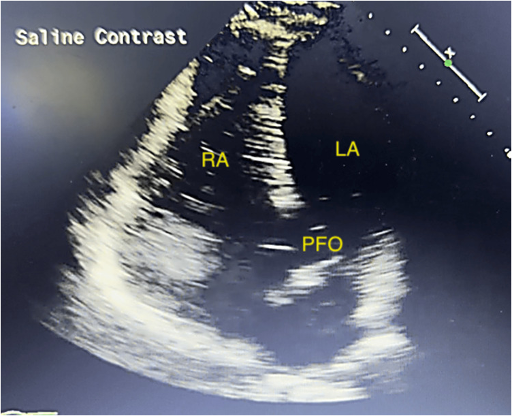
Shunt size is graded semiquantitatively by the maximum number of microbubbles in the left heart in a single frame. Trial definitions of a large shunt differ: more than 20 microbubbles in RESPECT and more than 30 in CLOSE. The count depends on the volume of contrast that reaches the right atrium and the adequacy of provocation, so a single small count does not exclude a large PFO.
| Test | Strengths | Limitations |
|---|---|---|
| Transthoracic contrast echocardiography | Noninvasive; effective Valsalva in an awake patient; screens for shunt and assesses chambers | Lower sensitivity with poor windows; limited septal anatomy; cannot rule out a PFO when negative with suboptimal provocation |
| Transesophageal echocardiography | Reference standard for PFO anatomy: tunnel length, septum secundum thickness, aneurysm, rims, Eustachian valve, thrombus, aortic atheroma | Sedation impairs Valsalva and can give false-negative results; semi-invasive |
| Transcranial Doppler | Detects microembolic signals in the middle cerebral artery; semiquantitative grading on the six-level Spencer logarithmic scale; comfortable | Does not localize the shunt; detects intrapulmonary shunts as well |
| Intracardiac echocardiography | Procedural guidance without general anesthesia | Invasive; procedural use only |
Detection of right-to-left shunting, after the 2015 ASE/SCAI guideline. Elevated left atrial pressure from left ventricular disease or mitral disease suppresses right-to-left shunting and lowers detection.
Transesophageal echocardiography, CT, cardiac magnetic resonance, and catheterization
Transesophageal echocardiography defines the anatomy that transthoracic imaging cannot. The septum is swept from 0° in 15° increments, and the bicaval view near 90° to 120° shows the fossa ovalis, both venae cavae, and the superior and inferior rims. For an ASD, the report includes the type and number of defects, maximal and minimal diameters (ideally from three-dimensional data), dynamic change through the cycle, flow direction, and the presence of an aneurysm. All six rims are reported: aortic (anterosuperior), superior vena caval, right upper pulmonary vein (posterosuperior), posterior, inferior vena caval, and atrioventricular valve (anteroinferior). For a PFO, it includes tunnel length, septum secundum thickness, septal mobility, and associated structures. Three-dimensional en face views show defect shape, fenestrations, and the relationship to the aorta.
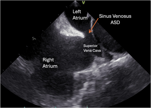
Cardiac magnetic resonance measures right ventricular volumes and function without geometric assumptions and calculates Qp/Qs from phase-contrast flow in the main pulmonary artery and ascending aorta. Contrast-enhanced magnetic resonance angiography maps every pulmonary vein. CT angiography gives the highest spatial resolution for anomalous pulmonary veins, the relationship of a sinus venosus defect to the superior vena cava and right pulmonary artery, and planning of covered-stent closure, at the cost of radiation and iodinated contrast. Both are indicated when transthoracic imaging shows unexplained right heart enlargement or when a sinus venosus defect, anomalous venous return, or unroofed coronary sinus is suspected. Lipomatous hypertrophy of the interatrial septum, which spares the fossa ovalis in a dumbbell shape, is distinguished from a mass on CT by its fat attenuation.
Catheterization, shunt quantification, and pulmonary vascular resistance. Noninvasive imaging suffices for most uncomplicated secundum defects. Catheterization is performed when pulmonary pressure is elevated by echocardiography, when noninvasive data disagree, and when closure is planned in an older patient or one with left heart disease. It measures Qp/Qs by oximetry, pulmonary vascular resistance (PVR) indexed to pulmonary flow, and the response to vasodilator testing and balloon occlusion. The oximetry run and calculations are described on the right heart catheterization and hemodynamics page. In left-to-right shunts, PVR is calculated with pulmonary flow, not systemic flow, and a falling Qp/Qs with rising PVR signals pulmonary vascular disease.
Indications for closure of an atrial septal defect
The 2025 ACC/AHA adult congenital guideline bases closure on three conditions: a hemodynamically significant shunt (Qp/Qs 1.5 or more with right ventricular dilation), an acceptable PVR, and no significant left ventricular disease. It also recommends assessment for pulmonary arterial hypertension in every adult with an unrepaired ASD and comanagement with pulmonary hypertension specialists when pulmonary arterial hypertension is present (both Class 1).
| Clinical setting | 2025 ACC/AHA | 2020 ESC |
|---|---|---|
| Qp/Qs ≥1.5, right ventricular dilation, PVR ≤2 Wood units, no significant left ventricular disease | Class 1: closure recommended to improve functional class and outcomes | Class I (PVR <3 WU), regardless of symptoms |
| Same, PVR >2 and <5 WU | Class 2a: closure reasonable to improve functional status | Class IIa for PVR 3 to <5 WU |
| PVR 5–8 WU lowered to <5 WU by targeted PAH therapy, significant left-to-right shunt | Class 2a: closure can be beneficial | Class IIb, fenestrated closure, when PVR ≥5 WU falls below 5 WU |
| PVR ≥5 WU despite targeted therapy, or desaturation on exercise | Class 2b: usefulness of fenestrated closure uncertain when PVR remains 5–8 WU on targeted therapy (significant left-to-right shunt) | Class III: closure not recommended |
| Eisenmenger physiology | Class 3: Harm | Class III |
| Unrepaired ASD with evidence of, or strong suspicion for, paradoxical embolism | Class 1: closure to prevent recurrent embolism | Class IIa: device closure regardless of size after other causes are excluded, in the absence of pulmonary arterial hypertension and left ventricular disease |
| Isolated secundum ASD suitable for a device | Class 1: transcatheter closure usually preferred to surgery, for shorter stay and recovery | Class I: device closure is the method of choice when technically suitable |
| Superior sinus venosus defect | Class 2b: transcatheter closure, when technically feasible, may be a reasonable alternative to surgery | Surgery |
| Unrepaired ASD, anatomic definition | Class 1: cardiac magnetic resonance (CMR), transesophageal echocardiography, or cardiac CT defines defect size, morphology, rims, and pulmonary venous connections | — |
| Suspected significant left ventricular diastolic dysfunction | Class 2b: balloon test occlusion before closure may be reasonable | Class I: balloon testing before closure |
| ASD with PAH in which closure is indicated | Class 2b: fenestrated repair can be considered | Class IIb: fenestrated closure when PVR ≥5 WU falls below 5 WU |
Closure recommendations for adults with an atrial septal defect. The ACC/AHA recommendations assume no significant left heart disease, defined as any condition with chronically elevated left atrial pressure of 15 mm Hg or more. The 2022 ESC/European Respiratory Society pulmonary hypertension guideline defines pulmonary arterial hypertension by a PVR above 2 WU. WU, Wood units; PAH, pulmonary arterial hypertension.
The lower US threshold reflects experience that patients with a PVR of 2 to 5 Wood units have undergone closure safely and improved. A small defect with Qp/Qs below 1.5 and a normal right ventricle has no hemodynamic indication for closure. It is followed for enlargement, rising pulmonary pressure, and atrial arrhythmia, and closed when paradoxical embolism occurs. Sinus venosus, primum, and coronary sinus defects follow the same hemodynamic logic but are repaired surgically except in selected sinus venosus cases.
Atrial septal defect closure by shunt and pulmonary vascular resistance
2025 ACC/AHA and 2020 ESC classesThe example (Qp/Qs 2.0, right ventricular dilation, PVR 3.6 Wood units, no left ventricular disease) falls in ACC/AHA Class 2a and ESC Class IIa. PVR is measured at catheterization with pulmonary flow in the denominator.
Device closure and surgical repair by defect type
Transcatheter closure is the standard treatment for a suitable secundum ASD. Under fluoroscopic and transesophageal or intracardiac echocardiographic guidance, the defect is crossed from the femoral vein and sized. Balloon sizing is performed by inflating a compliant balloon until color flow across the defect stops and measuring the waist in several planes (the stop-flow diameter). A self-expanding nitinol double-disc device is then deployed, its position and its relationship to the aorta, pulmonary veins, coronary sinus, and atrioventricular valves are confirmed, and it is released. Multifenestrated (cribriform) devices cover a fenestrated septum through a central hole, and some septa need more than one device.
| Feature | Favors transcatheter closure | Favors surgery |
|---|---|---|
| Defect type | Secundum ASD; PFO | Primum, inferior sinus venosus, and most superior sinus venosus defects; unroofed coronary sinus |
| Size | Single defect within device range | Single defect larger than 38 mm |
| Rims | Rims of 5 mm or more toward the atrioventricular valves, inferior vena cava, right upper pulmonary vein, and posterior wall; a deficient aortic rim alone is acceptable | Anterior, posterior, superior, or inferior rim less than 5 mm; absent inferior or posterior rim |
| Associated anatomy | None | Anomalous pulmonary venous drainage; multiple defects unsuitable for devices; another lesion requiring surgery |
| Rhythm | Atrial fibrillation treated by catheter ablation before or after closure | Concomitant surgical ablation (maze) planned |
| Other | Shorter stay and recovery (Class 1 preference) | Intracardiac thrombus; severe pulmonary hypertension with bidirectional shunting (closure contraindicated by either route) |
Anatomic selection for device closure, after the 2015 ASE/SCAI guideline (Table 8) and the 2025 ACC/AHA guideline.
A deficient aortic rim is present in a large share of successfully closed defects, about 40% in one report, so it does not exclude device closure. It is, however, the feature most often present in device erosion, and it is weighed with device size.
Surgical repair uses direct suture or a pericardial or synthetic patch through a median sternotomy or a minimally invasive approach on cardiopulmonary bypass. A primum defect is closed with a patch together with repair of the cleft left atrioventricular valve; late problems are left atrioventricular valve regurgitation or stenosis, subaortic obstruction, and complete heart block. A superior sinus venosus defect is repaired with a patch that baffles the right upper pulmonary vein to the left atrium, or with the Warden operation. In the Warden operation, the superior vena cava is divided above the anomalous vein and reimplanted into the right atrial appendage. Late complications are superior vena caval or pulmonary venous obstruction and sinus node dysfunction.
Transcatheter covered-stent closure of a superior sinus venosus defect places a long covered stent in the superior vena cava across the defect. The stent seals the communication and redirects the anomalous right upper pulmonary vein to the left atrium. The 2025 guideline lists it as Class 2b. Before implantation, transesophageal-guided balloon test occlusion of the cavoatrial junction confirms that the right upper pulmonary vein will continue to drain to the left atrium. Closure is then performed by an operator with adult congenital expertise.
Transseptal access for ablation, mitral transcatheter edge-to-edge repair, and left atrial appendage occlusion creates an iatrogenic ASD. Most close within weeks to months; persistence is more common after large-bore sheaths. Closure is reserved for a defect that causes right heart volume overload or right-to-left shunting with hypoxemia.
Atrial septal defect with pulmonary hypertension: treat-and-repair and Eisenmenger syndrome
Pulmonary arterial hypertension develops in a minority of adults with ASD, sometimes out of proportion to the shunt. In these patients the shunt may drive the pulmonary vascular disease, or the vascular disease may be primary and the defect coincidental. Closing a defect that decompresses a right ventricle failing against fixed resistance removes its outlet and can precipitate right heart failure. Defects associated with severe pulmonary vascular disease are therefore assessed by catheterization at an adult congenital and pulmonary hypertension center. Classification and targeted therapy of pulmonary hypertension are covered on the pulmonary hypertension page.
Treat-and-repair. In patients with a significant left-to-right shunt and a PVR of 5 to 8 Wood units, targeted pulmonary arterial hypertension therapy is started first, and catheterization is repeated. If PVR falls below 5 Wood units and the shunt remains left to right, closure can be beneficial (2025 ACC/AHA, Class 2a). The ESC guideline restricts this approach to fenestrated closure (Class IIb), which leaves a small calibrated outlet for the right ventricle. The 2025 ACC/AHA guideline lists fenestrated repair as a Class 2b option in adults with pulmonary arterial hypertension in whom closure is indicated. No fenestrated transcatheter device is available in the United States, so fenestration is created surgically. Evidence is observational. In one series, 22 adults with ASD and significant pulmonary arterial hypertension underwent transcatheter closure. In the 8 who received targeted therapy first, PVR fell from 9.6 ± 3.8 to 4.0 ± 0.8 Wood units before closure. Neither group had adverse events, and functional class improved over a mean of 52 months. Targeted therapy continues after closure because pulmonary arterial hypertension can persist or progress once the shunt is removed. Patients with persistent disease after defect correction are followed like those with idiopathic pulmonary arterial hypertension.
Vasodilator and occlusion testing. Inhaled nitric oxide during catheterization shows whether PVR falls and pulmonary flow rises, which supports reversibility. Temporary balloon occlusion of the defect measures the right atrial and pulmonary pressure response and systemic output with the shunt removed. A rise in pulmonary artery pressure or a fall in systemic output during occlusion argues against complete closure.
Eisenmenger syndrome. When PVR approaches or exceeds systemic resistance, the shunt reverses and becomes predominantly right to left, with cyanosis, secondary erythrocytosis, and multiorgan consequences. Closure is harmful (2025 ACC/AHA Class 3: Harm). Management is targeted pulmonary arterial hypertension therapy, avoidance of dehydration, iron deficiency correction, avoidance of routine phlebotomy, and prevention of paradoxical embolism, including air filters on all venous lines.
Atrial septal defect in older adults: left ventricular disease and atrial arrhythmias
Adults closed after age 40 to 60 gain exercise capacity, functional class, and right ventricular remodeling. Left ventricular diastolic dysfunction and atrial arrhythmias are more common in older patients.
Left ventricular diastolic dysfunction and left atrial stiffness. In an older patient with hypertension, coronary disease, or a stiff left atrium, the ASD acts as a relief valve for elevated left atrial pressure. Abrupt closure can raise left atrial and pulmonary venous pressure and precipitate pulmonary edema. The ESC guideline recommends balloon test occlusion before definitive closure in patients with advanced left ventricular diastolic or systolic dysfunction. A marked rise in left atrial or wedge pressure during occlusion leads to preparation with diuretics and afterload reduction, fenestrated closure, or no closure. The 2025 ACC/AHA recommendations apply to patients without significant left ventricular disease, and closure may be contraindicated when significant diastolic dysfunction is suspected. The guideline considers balloon test occlusion before closure reasonable in that setting (Class 2b). It notes that test occlusion adds little when resting left ventricular end-diastolic pressure is below 15 mm Hg and tends to overestimate the rise in wedge pressure after closure. Fenestrated patch closure is an alternative.
Atrial flutter and fibrillation. Atrial arrhythmias affect a large share of adults with unrepaired ASD after age 40, and closure in adulthood does not reliably prevent them. Catheter ablation of the cavotricuspid isthmus or pulmonary veins is performed before device closure when indicated, because a device complicates later transseptal access. Transseptal puncture through or beside a device is feasible but technically harder. Surgical patients can undergo concomitant maze or isthmus ablation. Anticoagulation decisions follow the atrial fibrillation guidelines, with the shunt as an added route for paradoxical embolism.
Patent foramen ovale and cryptogenic stroke
Paradoxical embolism is the passage of venous thrombus across a right-to-left shunt into the systemic circulation. Venous thrombi in PFO-associated stroke are often small and short-lived, and a deep vein thrombosis is found in only a minority of patients. Other proposed mechanisms are thrombus forming within the PFO tunnel or an atrial septal aneurysm, and atrial arrhythmia associated with the abnormal septum.
Because a PFO is present in about one adult in four, the central question after a stroke is whether the PFO caused it or is incidental. The evaluation establishes three things: a nonlacunar, embolic-appearing infarct (cortical, large subcortical, or retinal); the absence of a competing mechanism; and features that raise the probability of a PFO-related stroke. The SCAI guideline defines a PFO-associated stroke as an ischemic stroke with a cortical, large white matter, or retinal infarct in the presence of a PFO and with no other identified likely cause.
| Domain | Evaluation | Finding that lowers the likelihood that the PFO is causal |
|---|---|---|
| Brain imaging | MRI with diffusion-weighted imaging | Lacunar infarct (small vessel disease) |
| Vascular imaging | CT or MR angiography of head and neck | Ipsilateral atherosclerotic stenosis, dissection, vasculopathy |
| Cardiac rhythm | 12-lead electrocardiogram and telemetry or Holter monitoring in all; insertable cardiac monitor considered before closure in patients older than 65 and in younger patients at risk for atrial fibrillation (European position paper) | Atrial fibrillation or flutter |
| Cardiac structure | Transthoracic and transesophageal echocardiography with contrast | Left atrial appendage thrombus, left ventricular thrombus, endocarditis, aortic arch atheroma, cardiac tumor |
| Venous system | Lower extremity venous duplex; pelvic MR venography in selected patients | None; a deep vein thrombosis supports paradoxical embolism and changes antithrombotic therapy |
| Hematology | Thrombophilia testing when the history suggests it; malignancy evaluation when indicated | Cancer-associated hypercoagulability |
Evaluation of ischemic stroke with a PFO. The American Academy of Neurology (AAN) advisory states that a thorough evaluation for alternative mechanisms precedes any closure decision (Level B). Closure is not routinely recommended when a higher-risk alternative mechanism is found (Level B).
Embolic stroke of undetermined source (ESUS) is a nonlacunar infarct without proximal arterial stenosis of 50% or more, a major cardioembolic source, or another specific cause after a standard evaluation. All trials that showed benefit from PFO closure excluded lacunar strokes. The 2021 American Heart Association/American Stroke Association (AHA/ASA) guideline therefore treats the requirement for a cryptogenic stroke as equivalent to nonlacunar stroke of undetermined source. Clinical judgment remains necessary, because many strokes in this category have a lower-risk competing condition such as nonstenotic large-artery plaque.
RoPE score and PASCAL classification
The RoPE score was derived from pooled cohorts of patients with cryptogenic stroke to estimate the probability that a PFO, when found, is stroke related. Younger age and absence of vascular risk factors raise the score, because in such patients a PFO is less likely to be incidental. The score ranges from 0 to 10. PFO prevalence rises with the score, and the attributable fraction rises from 0% at scores of 0 to 3 to 88% at scores of 9 to 10. The 2-year risk of recurrent stroke or transient ischemic attack is about 20% at scores of 0 to 3 and about 2% at 9 to 10. Patients with low scores have other, persistent mechanisms.
| RoPE item | Points |
|---|---|
| No history of hypertension | 1 |
| No history of diabetes | 1 |
| No history of stroke or transient ischemic attack | 1 |
| Nonsmoker | 1 |
| Cortical infarct on imaging | 1 |
| Age 18–29 / 30–39 / 40–49 / 50–59 / 60–69 / 70 years or older | 5 / 4 / 3 / 2 / 1 / 0 |
RoPE score, after the original derivation. Scores range from 0 to 10.
The RoPE score uses no anatomic information. The PFO-Associated Stroke Causal Likelihood (PASCAL) classification adds the two anatomic features that predicted benefit in the trials: an atrial septal aneurysm and a large shunt. Patients are classified as unlikely (RoPE score below 7 and no high-risk feature), possible (RoPE score of 7 or more without a high-risk feature, or below 7 with one), and probable (RoPE score of 7 or more with a high-risk feature).
In the pooled individual-patient analysis of six randomized trials (3,740 patients; median follow-up 57 months), stroke incidence was 1.09% per year with medical therapy and 0.47% per year with closure (adjusted hazard ratio 0.41). Benefit varied by PASCAL category:
| PASCAL category | Hazard ratio for recurrent stroke with closure | 2-year absolute risk reduction | Absolute increase in atrial fibrillation beyond day 45 |
|---|---|---|---|
| Unlikely | 1.14 (0.53–2.46) | −0.7% | 4.41% |
| Possible | 0.38 (0.22–0.65) | 2.1% | 1.53% |
| Probable | 0.10 (0.03–0.35) | 2.1% | 0.65% |
Treatment effect of PFO closure by PASCAL category in the SCOPE pooled analysis (Kent et al., JAMA 2021). In patients classified as unlikely, closure produced no stroke reduction and the largest excess of atrial fibrillation.
RoPE score and PASCAL classification
For a nonlacunar stroke of undetermined cause with a PFOThe example, a 34-year-old nonsmoker without vascular risk factors who has a cortical infarct and an atrial septal aneurysm, scores 9 and is classified as probable. A 58-year-old smoker with hypertension and diabetes and a subcortical infarct without high-risk anatomy scores 3 and is classified as unlikely.
The scores estimate probability across groups and do not prove causation in a patient. A high RoPE score cannot overcome an identified competing mechanism, and a deep vein thrombosis or pulmonary embolism at the time of the stroke supports paradoxical embolism whatever the score.
Randomized trials of PFO closure after stroke
The first three trials (CLOSURE I, the Percutaneous Closure [PC] Trial, and the initial RESPECT analysis) enrolled patients regardless of shunt size and were neutral in their primary analyses; RESPECT became positive with extended follow-up. Three later trials enrolled patients aged 60 or younger with nonlacunar strokes, used current devices, enriched for high-risk anatomy or compared closure with antiplatelet therapy alone, and showed benefit. They differed in population, control arm, device, and follow-up, so their effect sizes are not interchangeable.
| Trial | Population | Comparison | Main result |
|---|---|---|---|
| CLOSURE I (2012) | 909 patients aged 18–60 with cryptogenic stroke or TIA | STARFlex closure vs. antiplatelet or anticoagulant therapy | No significant reduction in the 2-year composite; higher atrial fibrillation with closure |
| PC (2013) | 414 patients younger than 60 with ischemic stroke, TIA, or peripheral embolism | Amplatzer PFO Occluder vs. medical therapy | No significant reduction in the composite end point |
| RESPECT, extended follow-up (2017) | 980 patients aged 18–60; median 5.9 years | Amplatzer PFO Occluder vs. medical therapy | Recurrent ischemic stroke 18 vs. 28 (0.58 vs. 1.07 per 100 patient-years; HR 0.55); stroke of undetermined cause HR 0.38; venous thromboembolism more common with closure |
| CLOSE (2017) | 663 patients aged 16–60 with PFO plus atrial septal aneurysm or more than 30 microbubbles | Closure plus antiplatelet vs. antiplatelet; a third arm received anticoagulation | No strokes with closure vs. 14 with antiplatelet therapy (HR 0.03); atrial fibrillation 4.6% vs. 0.9% |
| REDUCE (2017) | 664 patients aged 18–59; median 3.2 years | Helex or Cardioform occluder plus antiplatelet vs. antiplatelet (2:1) | Clinical ischemic stroke 1.4% vs. 5.4% (HR 0.23); new brain infarction 5.7% vs. 11.3%; atrial fibrillation or flutter 6.6% vs. 0.4% |
| DEFENSE-PFO (2018) | 120 patients with high-risk PFO (atrial septal aneurysm, hypermobility of 10 mm or more, or separation of 2 mm or more) | Closure vs. medical therapy | 2-year composite of stroke, vascular death, or major bleeding 0% vs. 12.9%; stopped early |
Randomized trials of PFO closure after stroke. HR, hazard ratio; TIA, transient ischemic attack.
In RESPECT, the benefit was concentrated in patients with an atrial septal aneurysm or a substantial shunt. The reduction in silent infarction in REDUCE indicates ongoing subclinical embolism in the medical arm. A pooled analysis of six trials found the greatest stroke reduction in patients with both a large shunt and an atrial septal aneurysm. The 2021 AHA/ASA guideline cites a number needed to treat of 131 to prevent one recurrent stroke over 1 person-year of follow-up and 13 over 10 person-years. A 2026 individual-participant meta-analysis using PASCAL confirmed net benefit in the possible and probable categories, with no stroke benefit and more atrial fibrillation in the unlikely category. No adequately powered trial has compared closure with long-term anticoagulation, and randomized data in patients older than 60 are minimal.
Guideline recommendations and medical therapy for PFO-associated stroke
| Document | Recommendation | Strength |
|---|---|---|
| 2021 AHA/ASA secondary stroke prevention | Closure versus medical therapy decided jointly by the patient, a cardiologist, and a neurologist, considering the probability that the PFO is causal | Class 1, C-EO |
| Age 18–60, nonlacunar stroke of undetermined cause after thorough evaluation, PFO with high-risk anatomic features: transcatheter closure plus long-term antiplatelet therapy is reasonable over antiplatelet therapy alone | Class 2a, B-R | |
| Same population without high-risk features: benefit of closure not well established | Class 2b, C-LD | |
| Closure versus warfarin: comparative benefit unknown | Class 2b, C-LD | |
| 2020 AAN practice advisory | Age younger than 60, embolic-appearing infarct, no other mechanism after thorough evaluation: closure may be recommended after discussion; closure probably reduces recurrence (absolute reduction 3.4% at 5 years) at a periprocedural complication rate of 3.9% and an absolute increase in nonperiprocedural atrial fibrillation of 0.33% per year | Level C |
| 2022 SCAI PFO guideline | Age 18–60 with PFO-associated stroke: closure over antiplatelet therapy alone; a RoPE score of 7 or more may identify patients with greater benefit | Strong, moderate certainty |
| Age older than 60 with PFO-associated stroke: closure suggested over antiplatelet therapy alone | Conditional, very low certainty | |
| Age 18–60 without another indication for anticoagulation: closure plus antiplatelet therapy suggested over anticoagulation alone | Conditional, low certainty | |
| PFO-associated stroke with thrombophilia, deep vein thrombosis, or pulmonary embolism requiring lifelong anticoagulation: closure suggested in addition to anticoagulation | Conditional, very low certainty |
US recommendations for PFO closure after ischemic stroke. The 2019 European position paper supports closure in carefully selected patients aged 18 to 65 years with a cryptogenic stroke, transient ischemic attack, or systemic embolism and a high probability that the PFO is causal, and allows case-by-case closure outside that age range.
The documents agree on closure for well-selected patients aged 60 or younger and differ mainly for older patients. The AHA/ASA guideline states that closure should rarely be performed in patients older than 60 and only in very unusual circumstances. Randomized evidence is extremely limited, and serious periprocedural complications in claims data were 10.9% in this age group compared with 4.9% in younger patients. Both documents address ischemic stroke; the SCAI guideline suggests against closure after a transient ischemic attack alone.
Medical therapy. When closure is not chosen or is not indicated, antithrombotic therapy is prescribed for secondary prevention. The AAN advisory considers aspirin or anticoagulation reasonable (Level C). Anticoagulation has a theoretical advantage against venous thrombus and was compared with antiplatelet therapy in a third arm of CLOSE and in subgroups of ESUS trials, without a significant difference. Anticoagulation is indicated when a concurrent deep vein thrombosis, pulmonary embolism, or high-risk thrombophilia is present. Whether closure adds benefit to lifelong anticoagulation is unknown, and the SCAI guideline suggests closure in these patients on very low-certainty evidence. Vascular risk factors are treated in every patient, because a low RoPE score indicates that other mechanisms dominate future risk.
PFO in decompression illness, migraine, platypnea-orthodeoxia, and procedures
Decompression illness. Inert gas bubbles form in venous blood after diving and are normally filtered by the lungs. A PFO with a large right-to-left shunt allows them to reach the arterial circulation and is associated with cerebral, spinal, inner-ear, and cutaneous decompression illness. The SCAI guideline suggests against routine closure to prevent recurrent decompression illness in divers (conditional, very low certainty). The joint position statement of the South Pacific Underwater Medicine Society and the United Kingdom Sports Diving Medical Committee advises testing for a right-to-left shunt after shunt-pattern decompression illness. For a diver with a shunt, the options are to stop diving, dive conservatively to reduce venous bubble formation, or undergo closure followed by repeat shunt testing before diving resumes.
Migraine. Migraine with aura is associated with a higher prevalence of PFO and larger shunts. Three randomized trials did not meet their primary end points. MIST, a sham-controlled trial, found no difference in migraine cessation. PRIMA, in migraine with aura, found a reduction of 2.9 compared with 1.7 monthly migraine days (P = 0.17). PREMIUM, with a sham procedure, found a 50% responder rate of 38% (45 of 117) with closure and 32% (33 of 103) with sham. Closure reduced headache days by 3.4 compared with 2.0 per month, and complete remission was more frequent with closure (8.5% vs. 1%). Secondary end points favoring patients with aura generate hypotheses for further trials. The SCAI guideline suggests against routine closure for migraine (conditional, moderate certainty), and PFO closure is not a treatment for migraine outside trials.
Platypnea-orthodeoxia syndrome. Platypnea-orthodeoxia is dyspnea and arterial desaturation in the upright position that improve when the patient lies down. Right-to-left shunting through a PFO or ASD occurs at normal right atrial pressure when an anatomic change redirects inferior vena caval flow toward the septum in the upright posture. Causes include aortic root dilation or elongation, kyphoscoliosis, a prominent Eustachian valve, right pneumonectomy, and diaphragmatic paralysis. The diagnosis is made by desaturation on standing with a contrast echocardiogram performed upright, which shows shunting that may be absent supine. When other causes of hypoxemia are excluded, closure is suggested (SCAI, conditional, very low certainty) and usually abolishes the desaturation.
Refractory hypoxemia in critical illness. A PFO produces right-to-left shunting and refractory hypoxemia in right ventricular infarction, acute pulmonary embolism, severe tricuspid regurgitation, mechanical ventilation with high positive end-expiratory pressure, and left ventricular assist device implantation (as left atrial pressure falls). A PFO with thrombus straddling the foramen in acute pulmonary embolism is a high-risk finding discussed on the pulmonary embolism page.
Noncardiac surgery and procedures. In a cohort of 150,198 adults undergoing noncardiac surgery, a preoperatively diagnosed PFO was associated with perioperative ischemic stroke within 30 days in 3.2% compared with 0.5% of patients without a PFO (adjusted odds ratio 2.66). The observational design does not support routine screening for or closure of a PFO before surgery. Neurosurgery in the sitting position carries a risk of paradoxical venous air embolism, and a large PFO is evaluated before such procedures. Laparoscopic gas insufflation and orthopedic procedures with fat embolism are other recognized triggers.
Intravenous lines and filters. Air filters on all intravenous lines prevent paradoxical air embolism in patients with right-to-left shunting, because a small air bolus that would lodge harmlessly in the lungs reaches the cerebral circulation. Inferior vena cava filters have no trial evidence for preventing paradoxical embolism and are used under the usual venous thromboembolism indications, chiefly a contraindication to anticoagulation.
Pacemaker and defibrillator leads. In a multicenter cohort of 202 patients with intracardiac shunts, transvenous leads were associated with a more than 2-fold increased risk of systemic thromboembolism (hazard ratio 2.6), and antiplatelet or warfarin prescription did not appear protective. An interatrial shunt is closed before transvenous lead implantation when feasible, or epicardial leads are used.
Complications of transcatheter closure
Procedural complications are uncommon and include vascular access injury, air embolism, cardiac perforation with tamponade, arrhythmia, and device malposition. Later complications are specific to the device.
| Complication | Frequency and timing | Recognition and management |
|---|---|---|
| Device embolization | About 0.1%–0.4%; mostly early; more common with ASD devices; risk rises with undersizing and deficient rims | Echocardiography or fluoroscopy; percutaneous snare retrieval, or surgery when the device is lodged across a valve |
| Device erosion | About 0.1%–0.3% with the Amplatzer septal occluder; most within 72 hours to the first week; cases reported more than 6 years later | Chest pain, syncope, or new pericardial effusion; tamponade, aortic–atrial fistula; urgent surgery. Risk factors: oversizing, absent aortic rim, high superior defect, device deforming at the aortic root, early effusion |
| New atrial fibrillation or flutter | Higher after closure (4.6% vs. 0.9% in CLOSE; 6.6% vs. 0.4% in REDUCE); most within 45 days; often transient | Rhythm monitoring for palpitations; anticoagulation by standard criteria when persistent |
| Device-related thrombus | Uncommon; early, before endothelialization | Transesophageal echocardiography; anticoagulation until resolution |
| Residual shunt | Common early and declines with endothelialization; persistent large residual shunt is associated with recurrent stroke | Contrast echocardiography at follow-up; a second device for a significant residual shunt after PFO closure |
| Atrioventricular block | Rare; with large ASD devices | 12-lead electrocardiogram after closure |
| Wire frame fracture | Reported with the Helex occluder | Fluoroscopy without contrast when device stability is in doubt |
| Metal hypersensitivity | Rare; nitinol contains nickel, including in ePTFE-covered devices | History of severe nickel reaction reviewed before device choice |
Complications of transcatheter ASD and PFO closure, after the 2015 ASE/SCAI guideline and the closure trials.
Antithrombotic therapy, endocarditis prophylaxis, and follow-up after closure
A septal occluder is covered by platelets and fibrin within hours and is incorporated by fibrous tissue and endothelium over approximately 3 to 6 months. Antithrombotic therapy and surveillance are concentrated in this period. After PFO closure for stroke, the trials and the AHA/ASA recommendation pair the device with long-term antiplatelet therapy. The SCAI guideline lists the regimen beyond 1 month of dual antiplatelet therapy as a knowledge gap and makes no recommendation. A common regimen is aspirin and clopidogrel for the first month followed by aspirin alone. After ASD device closure, antiplatelet therapy, most often aspirin, is given at least until endothelialization is complete. Patients with atrial fibrillation or venous thromboembolism continue anticoagulation.
Antibiotic prophylaxis before dental procedures that manipulate gingival tissue is reasonable during the first 6 months after closure with a device or patch (2021 AHA scientific statement). It continues indefinitely when a residual shunt lies at or next to the device or patch. An unrepaired ASD or PFO without prosthetic material does not require prophylaxis.
| Time after device closure | Assessment |
|---|---|
| Before discharge | Transthoracic echocardiogram for device position, residual shunt, pericardial effusion, and impingement; 12-lead electrocardiogram |
| 1 week | Repeat transthoracic echocardiogram after an Amplatzer septal occluder (erosion surveillance) |
| 1, 6, and 12 months | Clinical review, electrocardiogram, transthoracic echocardiogram; contrast study for residual right-to-left shunt after PFO closure |
| Thereafter | By 2025 ACC/AHA physiological stage, adult congenital cardiology review with electrocardiogram and transthoracic echocardiogram occurs every 36 to 60 months in stage A and every 12 to 24 months in stage B. Review occurs every 6 to 12 months in stage C and every 3 to 6 months in stage D, with yearly electrocardiogram and echocardiogram. Repeat CMR or CT has no fixed interval and is used for a specific question such as suspected erosion. The ESC guideline keeps regular follow-up for residual shunt, pulmonary hypertension, arrhythmia, or closure after age 40. |
Surveillance after transcatheter closure, after the 2015 ASE/SCAI guideline and the 2020 ESC guideline. Surgical patients follow the same principles, with attention to sinus node function and venous obstruction after sinus venosus repair and to left atrioventricular valve function after primum repair.
The right ventricle shrinks rapidly in the first month after the shunt is abolished, and long-standing dilation regresses more slowly and may not normalize. Persistent right ventricular enlargement after closure suggests a residual shunt, an unrecognized anomalous pulmonary vein, pulmonary hypertension, or intrinsic right ventricular disease. New exertional symptoms after closure in an older patient suggest unmasked left ventricular diastolic dysfunction.
Exercise and sport. The 2015 AHA/ACC eligibility statement allows participation in all competitive sports for athletes with hemodynamically insignificant ASD and for those 3 to 6 months after closure without pulmonary hypertension, arrhythmia, or ventricular dysfunction. The 2025 ACC/AHA guideline considers competitive sports reasonable after a comprehensive evaluation by an adult congenital specialist (Class 2a). Pulmonary hypertension and arrhythmia require individual assessment. Scuba diving with an unclosed PFO or ASD carries the risk of decompression illness described above.
Pregnancy with an atrial septal defect or PFO
Pregnancy is usually well tolerated in women with an ASD without pulmonary hypertension. In the modified World Health Organization (mWHO) classification of the 2018 ESC pregnancy guideline, a successfully repaired ASD is class I and an unoperated ASD is class II. The added volume load of pregnancy can unmask right heart failure or atrial arrhythmias in women with a large shunt. The hypercoagulable state of pregnancy and the postpartum period, together with straining at delivery, raises the risk of paradoxical embolism. Measures to reduce the risk are early mobilization, compression stockings, prophylactic anticoagulation in women with additional risk factors, and air filters on intravenous lines during labor. Vaginal delivery is the default, and routine cesarean delivery without an obstetric indication or high-risk cardiac condition has no benefit and may cause harm (2025 ACC/AHA, Class 3: No Benefit). A hemodynamically significant ASD is ideally closed before conception. Pulmonary arterial hypertension, including Eisenmenger syndrome, is mWHO class IV: pregnancy is contraindicated, and termination is discussed if pregnancy occurs.
A PFO does not alter obstetric management. A pregnant woman with a stroke and a PFO is evaluated for venous thrombosis and other causes in the same way as a nonpregnant patient.
Sources of error in the diagnosis of interatrial shunts
- Apical four-chamber dropout of the thin fossa ovalis interpreted as a secundum defect: the subcostal views and color Doppler confirm or exclude it.
- Right ventricular dilation with an intact fossa ovalis attributed to pulmonary hypertension or cardiomyopathy: sinus venosus defect, anomalous pulmonary venous return, and unroofed coronary sinus are excluded with transesophageal echocardiography, CT, or cardiac magnetic resonance.
- A negative bubble study after a weak Valsalva maneuver, under sedation, or with elevated left atrial pressure: without an effective maneuver, which moves the septum toward the left atrium at release, a negative study does not exclude a PFO.
- Inferior vena caval flow and a prominent Eustachian valve washing contrast away from the fossa ovalis: a femoral or leg vein injection reaches the septum.
- Late bubbles from a pulmonary arteriovenous malformation attributed to a PFO, or an intracardiac shunt delayed by respiration attributed to an intrapulmonary shunt: bubbles seen crossing the septum or entering from the pulmonary veins localize the shunt.
- Pseudocontrast, a faint smokelike swirl entering the left atrium from the pulmonary veins during Valsalva strain and release, mistaken for microbubbles: it appears without or before right atrial opacification and lacks the bright discrete echoes of agitated saline. True shunt bubbles follow right atrial opacification.
- Qp/Qs from outflow tract diameters measured off axis or in the presence of significant semilunar regurgitation: the squared error alters the ratio, and the result is checked against right ventricular size.
- A large defect with Qp/Qs near 1 interpreted as a small shunt: balanced resistances indicate pulmonary vascular disease or ventricular disease and lead to catheterization.
- PVR calculated with systemic flow in a left-to-right shunt: pulmonary flow is the correct denominator, and the error overstates resistance when Qp exceeds Qs.
- A PFO found during a stroke evaluation accepted as the cause before atrial fibrillation, large-artery disease, small-vessel disease, and aortic arch atheroma are excluded.
Self-assessment
A 52-year-old woman has exertional dyspnea and a fixed split S2. Transthoracic echocardiography shows a dilated right ventricle and a 22-mm secundum atrial septal defect with a deficient aortic rim and adequate remaining rims. Catheterization shows Qp/Qs 2.0 and pulmonary vascular resistance 3.6 Wood units with a normal wedge pressure. What does the 2025 ACC/AHA guideline recommend, and does the aortic rim exclude device closure?
Cardiovascular Medicine Board ReviewReferences
- Gurvitz M, Krieger EV, Fuller S, et al. 2025 ACC/AHA/HRS/ISACHD/SCAI guideline for the management of adults with congenital heart disease: a report of the American College of Cardiology/American Heart Association Joint Committee on Clinical Practice Guidelines. Circulation. 2026;153:e115–e251. doi:10.1161/CIR.0000000000001402
- Baumgartner H, De Backer J, Babu-Narayan SV, et al. 2020 ESC guidelines for the management of adult congenital heart disease. Eur Heart J. 2021;42:563–645. doi:10.1093/eurheartj/ehaa554
- Stout KK, Daniels CJ, Aboulhosn JA, et al. 2018 AHA/ACC guideline for the management of adults with congenital heart disease. Circulation. 2019;139:e698–e800. doi:10.1161/CIR.0000000000000603
- Kleindorfer DO, Towfighi A, Chaturvedi S, et al. 2021 guideline for the prevention of stroke in patients with stroke and transient ischemic attack: a guideline from the American Heart Association/American Stroke Association. Stroke. 2021;52:e364–e467. doi:10.1161/STR.0000000000000375
- Messé SR, Gronseth GS, Kent DM, et al. Practice advisory update summary: patent foramen ovale and secondary stroke prevention: report of the Guideline Subcommittee of the American Academy of Neurology. Neurology. 2020;94:876–885. doi:10.1212/WNL.0000000000009443
- Kavinsky CJ, Szerlip M, Goldsweig AM, et al. SCAI guidelines for the management of patent foramen ovale. J Soc Cardiovasc Angiogr Interv. 2022;1:100039. doi:10.1016/j.jscai.2022.100039
- Silvestry FE, Cohen MS, Armsby LB, et al. Guidelines for the echocardiographic assessment of atrial septal defect and patent foramen ovale: from the American Society of Echocardiography and Society for Cardiac Angiography and Interventions. J Am Soc Echocardiogr. 2015;28:910–958. doi:10.1016/j.echo.2015.05.015
- Pristipino C, Sievert H, D'Ascenzo F, et al. European position paper on the management of patients with patent foramen ovale. General approach and left circulation thromboembolism. Eur Heart J. 2019;40:3182–3195. doi:10.1093/eurheartj/ehy649
- Humbert M, Kovacs G, Hoeper MM, et al. 2022 ESC/ERS guidelines for the diagnosis and treatment of pulmonary hypertension. Eur Heart J. 2022;43:3618–3731. doi:10.1093/eurheartj/ehac237
- Mas JL, Derumeaux G, Guillon B, et al. Patent foramen ovale closure or anticoagulation vs. antiplatelets after stroke (CLOSE). N Engl J Med. 2017;377:1011–1021. doi:10.1056/NEJMoa1705915
- Søndergaard L, Kasner SE, Rhodes JF, et al. Patent foramen ovale closure or antiplatelet therapy for cryptogenic stroke (REDUCE). N Engl J Med. 2017;377:1033–1042. doi:10.1056/NEJMoa1707404
- Saver JL, Carroll JD, Thaler DE, et al. Long-term outcomes of patent foramen ovale closure or medical therapy after stroke (RESPECT). N Engl J Med. 2017;377:1022–1032. doi:10.1056/NEJMoa1610057
- Lee PH, Song JK, Kim JS, et al. Cryptogenic stroke and high-risk patent foramen ovale: the DEFENSE-PFO trial. J Am Coll Cardiol. 2018;71:2335–2342. doi:10.1016/j.jacc.2018.02.046
- Furlan AJ, Reisman M, Massaro J, et al. Closure or medical therapy for cryptogenic stroke with patent foramen ovale (CLOSURE I). N Engl J Med. 2012;366:991–999. doi:10.1056/NEJMoa1009639
- Meier B, Kalesan B, Mattle HP, et al. Percutaneous closure of patent foramen ovale in cryptogenic embolism (PC trial). N Engl J Med. 2013;368:1083–1091. doi:10.1056/NEJMoa1211716
- Kent DM, Ruthazer R, Weimar C, et al. An index to identify stroke-related vs incidental patent foramen ovale in cryptogenic stroke. Neurology. 2013;81:619–625. doi:10.1212/WNL.0b013e3182a08d59
- Kent DM, Saver JL, Kasner SE, et al. Heterogeneity of treatment effects in an analysis of pooled individual patient data from randomized trials of device closure of patent foramen ovale after stroke. JAMA. 2021;326:2277–2286. doi:10.1001/jama.2021.20956
- Saver JL, Kent DM, Kasner SE, et al. Patent foramen ovale closure in stroke and the PASCAL classification system. JAMA Neurol. 2026;83:242–249. doi:10.1001/jamaneurol.2025.5446
- Mas JL, Saver JL, Kasner SE, et al. Association of atrial septal aneurysm and shunt size with stroke recurrence and benefit from patent foramen ovale closure. JAMA Neurol. 2022;79:1175–1179. doi:10.1001/jamaneurol.2022.3248
- Kent DM, Wang AY. Patent foramen ovale and stroke: a review. JAMA. 2025;334:1463–1473. doi:10.1001/jama.2025.10946
- Hagen PT, Scholz DG, Edwards WD. Incidence and size of patent foramen ovale during the first 10 decades of life: an autopsy study of 965 normal hearts. Mayo Clin Proc. 1984;59:17–20. doi:10.1016/s0025-6196(12)60336-x
- Lechat P, Mas JL, Lascault G, et al. Prevalence of patent foramen ovale in patients with stroke. N Engl J Med. 1988;318:1148–1152. doi:10.1056/NEJM198805053181802
- Tobis JM, Charles A, Silberstein SD, et al. Percutaneous closure of patent foramen ovale in patients with migraine: the PREMIUM trial. J Am Coll Cardiol. 2017;70:2766–2774. doi:10.1016/j.jacc.2017.09.1105
- Mattle HP, Evers S, Hildick-Smith D, et al. Percutaneous closure of patent foramen ovale in migraine with aura, a randomized controlled trial (PRIMA). Eur Heart J. 2016;37:2029–2036. doi:10.1093/eurheartj/ehw027
- Dowson A, Mullen MJ, Peatfield R, et al. Migraine Intervention With STARFlex Technology (MIST) trial. Circulation. 2008;117:1397–1404. doi:10.1161/CIRCULATIONAHA.107.727271
- Smart D, Mitchell S, Wilmshurst P, Turner M, Banham N. Joint position statement on persistent foramen ovale (PFO) and diving. South Pacific Underwater Medicine Society (SPUMS) and the United Kingdom Sports Diving Medical Committee (UKSDMC). Diving Hyperb Med. 2015;45:129–131. PMID 26165538
- Ng PY, Ng AKY, Subramaniam B, et al. Association of preoperatively diagnosed patent foramen ovale with perioperative ischemic stroke. JAMA. 2018;319:452–462. doi:10.1001/jama.2017.21899
- Khairy P, Landzberg MJ, Gatzoulis MA, et al. Transvenous pacing leads and systemic thromboemboli in patients with intracardiac shunts: a multicenter study. Circulation. 2006;113:2391–2397. doi:10.1161/CIRCULATIONAHA.106.622076
- Kijima Y, Akagi T, Takaya Y, et al. Treat and repair strategy in patients with atrial septal defect and significant pulmonary arterial hypertension. Circ J. 2016;80:227–234. doi:10.1253/circj.CJ-15-0599
- Murphy JG, Gersh BJ, McGoon MD, et al. Long-term outcome after surgical repair of isolated atrial septal defect: follow-up at 27 to 32 years. N Engl J Med. 1990;323:1645–1650. doi:10.1056/NEJM199012133232401
- Konstantinides S, Geibel A, Olschewski M, et al. A comparison of surgical and medical therapy for atrial septal defect in adults. N Engl J Med. 1995;333:469–473. doi:10.1056/NEJM199508243330801
- Attie F, Rosas M, Granados N, et al. Surgical treatment for secundum atrial septal defects in patients >40 years old: a randomized clinical trial. J Am Coll Cardiol. 2001;38:2035–2042. doi:10.1016/s0735-1097(01)01635-7
- Wilson WR, Gewitz M, Lockhart PB, et al. Prevention of viridans group streptococcal infective endocarditis: a scientific statement from the American Heart Association. Circulation. 2021;143:e963–e978. doi:10.1161/CIR.0000000000000969
- Regitz-Zagrosek V, Roos-Hesselink JW, Bauersachs J, et al. 2018 ESC guidelines for the management of cardiovascular diseases during pregnancy. Eur Heart J. 2018;39:3165–3241. doi:10.1093/eurheartj/ehy340
- Van Hare GF, Ackerman MJ, Evangelista JK, et al. Eligibility and disqualification recommendations for competitive athletes with cardiovascular abnormalities: Task Force 4: congenital heart disease. Circulation. 2015;132:e281–e291. doi:10.1161/CIR.0000000000000240
- Diller GP, Gatzoulis MA, Broberg CS, et al. Coronavirus disease 2019 in adults with congenital heart disease: a position paper from the ESC working group of adult congenital heart disease, and the International Society for Adult Congenital Heart Disease. Eur Heart J. 2021;42:1858–1865. doi:10.1093/eurheartj/ehaa960