Library › Pharmacology › Antiarrhythmic drugs
Antiarrhythmic drugs
The pharmacology, selection, dosing, initiation, monitoring, interactions, proarrhythmia, and organ toxicity of antiarrhythmic drugs in adults.
Antiarrhythmic drugs are chosen for safety first and efficacy second. Flecainide and propafenone are limited to hearts without prior myocardial infarction, scar, or significant structural disease. Dofetilide and sotalol are started with at least 3 days of continuous electrocardiographic (ECG) monitoring at a dose set by creatinine clearance and the corrected QT interval (QTc). Amiodarone is the most effective drug for maintaining sinus rhythm and the one with the most organ toxicity, so it carries a fixed surveillance schedule. No antiarrhythmic drug other than a beta-blocker has reduced mortality in structural heart disease, and several have increased it.
The Vaughan Williams classification and its limits
The Vaughan Williams scheme, built between 1970 and 1984, sorts antiarrhythmic drugs by their dominant action on the cardiac action potential. Class I drugs block the fast sodium current and are subdivided by their effect on action potential duration. Class II drugs are beta-adrenergic antagonists. Class III drugs prolong the action potential and refractoriness, mainly by blocking potassium currents. Class IV drugs are the nondihydropyridine calcium channel blockers. Digoxin, adenosine, and magnesium were later grouped as a miscellaneous "class V."
The scheme survives because it predicts each drug's principal toxicity as well as its effect. Sodium channel blockers slow conduction and widen the QRS complex. Potassium channel blockers lengthen the QT interval and cause torsades de pointes. Beta-blockers and calcium channel blockers slow the sinus node and atrioventricular (AV) node and depress contractility.
The classes have limits. Most drugs act on more than one target. Amiodarone has actions in all four classes, sotalol is a class II and class III drug, and propafenone and dronedarone also block beta-receptors. Active metabolites can belong to a different class than the parent drug: N-acetylprocainamide (NAPA), the metabolite of procainamide, is a class III agent. The classes say nothing about the arrhythmia mechanism a drug suppresses, and they describe drug action at normal heart rates and resting membrane potential, not in ischemic or remodeled tissue. The 1991 Sicilian Gambit proposed a spreadsheet of each drug's actions on channels, receptors, and pumps, matched to "vulnerable parameters" of each arrhythmia mechanism. It proved too complex for routine use, but it established the principle of matching drug action to mechanism.
| Class | Primary action | ECG effect | Drugs | Characteristic toxicity |
|---|---|---|---|---|
| IA | Moderate sodium channel block with potassium (IKr) block; intermediate binding kinetics | QRS and QT prolongation | Quinidine, procainamide, disopyramide | Torsades de pointes; quinidine diarrhea and cinchonism; procainamide lupus and agranulocytosis; disopyramide anticholinergic effects and negative inotropy |
| IB | Weak sodium channel block with fast kinetics, preferential binding to inactivated channels; action potential shortened or unchanged | Little change at normal rates | Lidocaine, mexiletine | Tremor, dizziness, confusion, seizures; nausea with mexiletine |
| IC | Marked sodium channel block with slow kinetics | PR and QRS prolongation, most at fast rates | Flecainide, propafenone | Ventricular proarrhythmia in scar; 1:1 atrial flutter; conduction block; Brugada pattern |
| II | Beta-adrenergic receptor block | Sinus slowing, PR prolongation | Metoprolol, carvedilol, bisoprolol, propranolol, nadolol, esmolol | Bradycardia, AV block, bronchospasm with nonselective agents, fatigue |
| III | Potassium channel block (IKr, with IKs and others for amiodarone); prolonged refractoriness | QT prolongation | Amiodarone, dronedarone, sotalol, dofetilide, ibutilide | Torsades (sotalol, dofetilide, ibutilide); organ toxicity (amiodarone) |
| IV | L-type calcium channel block in nodal tissue | PR prolongation, sinus slowing | Verapamil, diltiazem | Bradycardia, AV block, hypotension, negative inotropy |
| Other | Vagal and Na⁺/K⁺-ATPase effects; A1 receptor activation; late sodium current block | Varies | Digoxin, adenosine, magnesium, ranolazine | Digoxin toxicity; transient asystole and bronchospasm with adenosine |
The Vaughan Williams classification with its later additions. IKr, rapid component of the delayed rectifier potassium current; IKs, slow component. Classification follows the dominant action; several drugs act in more than one class.
The modernized classification of 2018
In 2018, a revision from Oxford and collaborating groups kept the four Vaughan Williams classes and expanded them to describe every known pharmacological target, including drugs in development. Its practical value is that it places newer agents (ivabradine, ranolazine, ryanodine receptor modulators) in a logical position and separates drugs that act on the same current through different mechanisms.
| Class | Target | Examples |
|---|---|---|
| 0 | Hyperpolarization-activated cyclic nucleotide-gated (HCN) channel; the funny current (If) of the sinus node | Ivabradine |
| Ia, Ib, Ic | Peak sodium current, divided by binding kinetics as in the original scheme | Quinidine, procainamide, disopyramide; lidocaine, mexiletine; flecainide, propafenone |
| Id | Late sodium current | Ranolazine |
| II | Autonomic modulation: beta-blockers (IIa), beta-agonists (IIb), muscarinic M2 antagonists (IIc) and agonists (IId), adenosine A1 agonists (IIe) | Beta-blockers; isoproterenol; atropine; digoxin (vagal effect); adenosine |
| III | Potassium channel blockers and openers: nonselective, IKr-selective, IKs, ultrarapid IKur, acetylcholine-activated IKACh, ATP-sensitive channels | Amiodarone, dronedarone, sotalol, dofetilide, ibutilide, vernakalant (atrial-predominant) |
| IV | Calcium handling: surface L-type channel (IVa), ryanodine receptor RyR2 (IVb), sarcoplasmic reticulum uptake (IVc), sodium-calcium exchanger (IVd), phosphokinase and phosphorylase (IVe) | Verapamil, diltiazem; flecainide and propafenone (RyR2 effect) |
| V | Mechanosensitive channels | None in clinical use |
| VI | Gap junction channels | Carbenoxolone (experimental) |
| VII | Upstream targets: structural and electrophysiological remodeling | Renin-angiotensin system inhibitors, omega-3 fatty acids, statins |
Modernized classification of antiarrhythmic drugs (Lei and colleagues, Circulation 2018). Class VII comprises drugs whose antiarrhythmic effect, where present, arises from modification of the substrate.
US guidelines list recommendations by drug name. The revised scheme explains why flecainide suppresses catecholaminergic polymorphic ventricular tachycardia (a class IVb action on RyR2) and why ranolazine and mexiletine act in long QT syndrome type 3 (late sodium current block).
The cardiac action potential and the currents antiarrhythmic drugs target
Working atrial and ventricular myocytes and Purkinje fibers have a fast-response action potential. Its upstroke depends on the sodium current, which determines conduction velocity. Sinus and AV nodal cells have a slow-response action potential. Their upstroke depends on the L-type calcium current, and their spontaneous phase 4 depolarization makes them pacemakers. This difference explains why sodium channel blockers slow conduction in muscle and the His-Purkinje system and spare the AV node, and why calcium channel blockers and beta-blockers act on the nodes.
| Phase | Event | Dominant currents | Drugs acting on this phase |
|---|---|---|---|
| 0 | Rapid depolarization (upstroke) | Peak INa (NaV1.5) in muscle and Purkinje fibers; ICa,L in nodal cells | Class I in muscle; verapamil and diltiazem in nodes |
| 1 | Early rapid repolarization (notch) | Transient outward Ito | Quinidine (Ito block, the basis of its use in Brugada syndrome) |
| 2 | Plateau | ICa,L inward balanced by IKr and IKs; late INa | Ranolazine and mexiletine (late INa); calcium channel blockers |
| 3 | Final repolarization | IKr (hERG), IKs; IKur in atria | Class III drugs; class IA drugs |
| 4 | Resting potential or diastolic depolarization | IK1 in muscle; If and ICa in pacemaker cells; IKACh with vagal tone | Beta-blockers, ivabradine, adenosine, digoxin |
Phases of the cardiac action potential. hERG, the gene product (KV11.1) that carries IKr and the off-target binding site of most QT-prolonging drugs.
Atrial myocytes have a shorter action potential than ventricular myocytes, a less negative resting potential, and currents largely confined to the atrium (IKur and IKACh). These differences are the basis of atrial-selective drug development; vernakalant is the one agent with atrial-predominant action in clinical use outside the United States.
Arrhythmia mechanisms and how antiarrhythmic drugs interrupt them
Re-entry requires a circuit with unidirectional block and a path of slow conduction, so that tissue ahead of the advancing wavefront has recovered excitability by the time the wavefront arrives. The wavelength of re-entry is the product of conduction velocity and refractory period. Re-entry stops when the wavelength exceeds the path length. Class III drugs lengthen the wavelength by prolonging refractoriness. Sodium channel blockers have opposing effects: they slow conduction (which shortens the wavelength and can stabilize re-entry) and can convert unidirectional block into bidirectional block (which terminates it). Which effect predominates depends on the substrate, and in scarred ventricular myocardium the conduction-slowing effect can create new re-entrant circuits. This is the mechanism of class IC proarrhythmia after myocardial infarction.
Enhanced or abnormal automaticity arises when phase 4 depolarization accelerates (catecholamines, ischemia, hypokalemia) or when partially depolarized myocytes fire spontaneously. Beta-blockers, calcium channel blockers in nodal tissue, and sodium channel blockers in depolarized tissue suppress it.
Triggered activity is driven by afterdepolarizations. Early afterdepolarizations arise during phases 2 and 3 when repolarization is prolonged. They are favored by bradycardia and pauses, initiate torsades de pointes, and are the mechanism of class III and class IA proarrhythmia. Delayed afterdepolarizations follow full repolarization and are caused by spontaneous calcium release from the sarcoplasmic reticulum, as in digoxin toxicity and catecholaminergic polymorphic ventricular tachycardia. They respond to beta-blockade, calcium channel blockade, and flecainide.
Atrial fibrillation (AF) combines all three. Pulmonary vein triggers (automaticity and triggered activity) initiate it, and multiple wavelet or rotor re-entry in a remodeled atrium sustains it. Electrical remodeling shortens atrial refractoriness within days of continuous fibrillation, which is one reason drug efficacy falls as AF persists. The atrial fibrillation hub describes these mechanisms in full.
Sodium channel block: state dependence and use dependence
Sodium channels cycle among resting (closed), open, and inactivated states with each beat. The modulated receptor hypothesis holds that class I drugs bind with higher affinity to open or inactivated channels than to resting channels and unbind during diastole. Two consequences follow.
Use dependence. Block accumulates when the interval between beats is too short for the drug to unbind. The faster the heart rate, the greater the steady-state block and the wider the QRS complex. Class IC drugs unbind slowly, so they produce measurable QRS widening even at normal heart rates and marked widening during tachycardia. Class IB drugs bind and unbind quickly, so block dissipates within each diastole at normal rates and accumulates only at fast rates or in depolarized tissue. Class IA drugs are intermediate.
Voltage dependence. Depolarized cells, such as ischemic myocytes and those near a scar border, spend more time in the inactivated state. Lidocaine and mexiletine bind preferentially to inactivated channels, so they act selectively on ischemic tissue and have little effect on normal atrium. This explains why lidocaine is useful for ischemic ventricular arrhythmias and ineffective for atrial fibrillation.
Use dependence has clinical consequences. QRS widening during exercise or tachycardia is the first sign of excessive class IC effect, which is why exercise testing is used after flecainide or propafenone is started or increased. A QRS increase of more than about 25% over baseline is generally taken as a sign of excessive sodium channel block. Use dependence also explains 1:1 flutter. When a class IC drug slows the flutter rate from 300 to about 200 to 220 beats/min, the AV node can conduct every impulse. The rapid ventricular rate then widens the QRS further.
Potassium channel block, reverse use dependence, and repolarization reserve
Class III drugs lengthen refractoriness by blocking repolarizing potassium currents, principally IKr. Nearly every drug that causes torsades de pointes blocks the hERG channel that carries IKr, whether or not it is an antiarrhythmic.
Reverse use dependence. Pure IKr blockers (sotalol, dofetilide, ibutilide) prolong the action potential most at slow heart rates and least at fast rates. Their antiarrhythmic effect is therefore weakest during tachycardia, and their proarrhythmic effect is greatest during bradycardia and after pauses. Torsades on these drugs characteristically begins with a short-long-short sequence and often occurs at night. Amiodarone is an exception. It blocks multiple currents, prolongs repolarization relatively uniformly across the ventricular wall, and shows little reverse use dependence, so it prolongs the QT substantially with a low incidence of torsades.
Repolarization reserve. Repolarization is carried by redundant currents, so loss of one is usually compensated. Torsades occurs when several insults combine: a QT-prolonging drug plus hypokalemia, hypomagnesemia, bradycardia, female sex, structural heart disease, a second QT-prolonging drug, an inhibitor of the drug's clearance, or a subclinical congenital channel variant. Hypokalemia is a particularly potent amplifier because low extracellular potassium paradoxically reduces IKr and increases drug binding to hERG. The mechanisms and risk factors are described in the long QT syndrome and QT prolongation hub.
The QT response to class III drugs is concentration dependent for sotalol and dofetilide, both of which are cleared by the kidney. A fall in creatinine clearance raises the concentration and the QT without any change in the prescribed dose. For this reason both drugs are dosed by creatinine clearance, and kidney function is rechecked whenever the patient's clinical state changes.
Beta-blockers and calcium channel blockers as antiarrhythmic drugs
Beta-blockers are the only antiarrhythmic drugs that consistently reduce sudden death and total mortality, in patients after myocardial infarction and in heart failure with reduced ejection fraction (HFrEF). They suppress catecholamine-driven automaticity and triggered activity, slow AV nodal conduction, and raise the ventricular fibrillation threshold. They are first-line therapy for rate control in atrial fibrillation, for most supraventricular tachycardias, for idiopathic outflow-tract ventricular tachycardia (VT), and for long QT syndrome types 1 and 2. They are also first-line for catecholaminergic polymorphic VT, where nonselective agents (nadolol, propranolol) are preferred. In electrical storm, a beta-blocker is part of initial treatment.
Verapamil and diltiazem block the L-type calcium channel in the sinus and AV nodes. They control the ventricular rate in atrial fibrillation and flutter, terminate and prevent AV nodal re-entrant and orthodromic AV re-entrant tachycardia, and suppress verapamil-sensitive fascicular VT and some outflow-tract VTs. They are avoided in HFrEF because of negative inotropy, and they are harmful in pre-excited atrial fibrillation and in wide-complex tachycardia of uncertain origin. Their pharmacology is covered in the calcium channel blocker hub, and combined use with class I or III drugs is discussed below under bradycardia and drug interactions.
Class IA drugs: quinidine, procainamide, and disopyramide
Class IA drugs combine moderate sodium channel block with IKr block. They slow conduction and prolong repolarization, and they cause torsades de pointes, sometimes at low concentrations and early in therapy. Their role is now confined to specific settings.
Quinidine blocks INa, IKr, and the transient outward current Ito, and it has vagolytic and alpha-blocking effects. Ito block restores the epicardial action potential dome in Brugada syndrome. On this basis quinidine is used for recurrent ventricular fibrillation or electrical storm in Brugada syndrome, early repolarization syndrome, idiopathic ventricular fibrillation, and short QT syndrome. Oral quinidine sulfate is given in divided doses (200 to 600 mg every 6 to 12 hours in ventricular arrhythmia regimens); the intravenous gluconate formulation is no longer available in the United States. Diarrhea is the most frequent reason for stopping it. Cinchonism (tinnitus, hearing loss, blurred vision, headache, confusion), immune thrombocytopenia, hepatitis, and a lupus-like syndrome also occur. Quinidine is a strong inhibitor of cytochrome P450 2D6 (CYP2D6) and of P-glycoprotein, and it roughly doubles digoxin concentrations. Its vagolytic effect can accelerate AV conduction during atrial flutter. "Quinidine syncope" was the original description of drug-induced torsades.
Procainamide is now used almost only intravenously in the United States. It is acetylated in the liver to NAPA, which has class III activity and is cleared by the kidney, so slow acetylators accumulate parent drug and patients with kidney disease accumulate NAPA. Intravenous loading is 10 to 17 mg/kg at 20 to 50 mg/min, followed by 1 to 4 mg/min. The infusion stops for hypotension, QRS widening of more than 50%, or termination of the arrhythmia. Procainamide is reasonable for hemodynamically stable monomorphic VT (2017 American Heart Association/American College of Cardiology/Heart Rhythm Society [AHA/ACC/HRS] guideline, Class 2a). In the PROCAMIO trial of tolerated wide-QRS tachycardia, procainamide terminated the arrhythmia more often within 40 minutes (67% versus 38%) and caused fewer major cardiac adverse events (9% versus 41%) than amiodarone. It is also used for pre-excited atrial fibrillation and, less often, for pharmacologic cardioversion of atrial fibrillation (Class 2b in the 2023 ACC/AHA/ACCP/HRS guideline, from the American College of Cardiology, American Heart Association, American College of Clinical Pharmacy, and Heart Rhythm Society). Long-term use produces a positive antinuclear antibody in most patients and drug-induced lupus in a substantial minority, typically with antihistone antibodies and without renal involvement. Agranulocytosis occurs at a rate of about 0.5%, with 20% to 25% mortality in reported cases, so the US label requires weekly blood counts during the first 3 months of prolonged therapy.
Disopyramide has strong negative inotropic and anticholinergic effects. The negative inotropy is used therapeutically in obstructive hypertrophic cardiomyopathy, where disopyramide added to a beta-blocker or nondihydropyridine calcium channel blocker reduces the outflow gradient and symptoms (2024 AHA/ACC, Class 1). The same effect makes it hazardous in reduced ejection fraction. Anticholinergic effects (urinary retention, constipation, dry mouth, angle-closure glaucoma, confusion) limit its use in older adults. Disopyramide is also used for vagally mediated atrial fibrillation. It can enhance AV conduction during atrial flutter, so it is combined with an AV nodal blocker. The usual adult dose is 400 to 800 mg/day in divided doses (150 mg every 6 hours for most adults, 100 mg every 6 hours below 50 kg). With creatinine clearance of 40 mL/min or less, 100 mg is given every 8, 12, or 24 hours according to the degree of kidney impairment.
Class IB drugs: lidocaine and mexiletine
Class IB drugs have fast binding kinetics and preferential affinity for inactivated sodium channels, so they act on depolarized, ischemic, or rapidly firing ventricular tissue and have negligible effect on atrial arrhythmias. They shorten or do not change the action potential duration and rarely cause torsades.
Lidocaine is given intravenously: 1 to 1.5 mg/kg, repeated at 0.5 to 0.75 mg/kg every 5 to 10 minutes to a total of 3 mg/kg, then 1 to 4 mg/min. It has a high hepatic extraction ratio, so clearance falls with reduced hepatic blood flow in heart failure, shock, and old age; the maintenance infusion is reduced in these settings. Its active metabolites accumulate after about 24 hours. Neurotoxicity (paresthesia, slurred speech, confusion, tremor, seizures) is the dose-limiting effect. Lidocaine is used for shock-refractory ventricular fibrillation (VF) or pulseless VT, with amiodarone as the alternative, and for recurrent ischemic VT and electrical storm. Prophylactic lidocaine in acute myocardial infarction reduced ventricular fibrillation in older trials without improving survival.
Mexiletine is the oral analog of lidocaine. The label starting dose is 200 mg every 8 hours with food or antacid, adjusted in 50 to 100 mg steps at intervals of at least 2 to 3 days. Most patients respond to 200 to 300 mg every 8 hours, and the total dose does not exceed 1,200 mg/day. A 400 mg loading dose is used when rapid control is needed. About 90% is metabolized in the liver by CYP2D6 and CYP1A2, so liver disease, marked right-sided heart failure, ciprofloxacin, and fluvoxamine raise its concentration, and smoking lowers it. Kidney failure usually needs no adjustment. Gastrointestinal upset and neurologic effects (tremor, dizziness, ataxia, blurred vision) limit tolerability. The label describes seizures in about 2 per 1,000 patients in its compassionate-use program and warns about drug reaction with eosinophilia and systemic symptoms (DRESS). Mexiletine is added to amiodarone for recurrent scar-related VT and is used as gene-directed therapy in long QT syndrome type 3, where it shortens the QT by blocking the late sodium current (covered in the long QT hub).
Class IC drugs: flecainide and propafenone
Flecainide and propafenone are potent sodium channel blockers with slow dissociation kinetics. They slow conduction in the atria, the His-Purkinje system, accessory pathways, and the ventricles, prolong the PR and QRS intervals, and have little effect on the QT interval beyond the QRS widening. In hearts without prior infarction or significant structural disease they are effective and well tolerated for atrial fibrillation, supraventricular tachycardia, and idiopathic ventricular ectopy.
Flecainide is well absorbed, about one-third cleared unchanged by the kidney, and partly metabolized by CYP2D6, with a half-life of 12 to 27 hours in patients. Steady state takes 3 to 5 days, so doses are increased no more often than every 4 days and no loading dose is used for maintenance therapy. For paroxysmal atrial fibrillation and paroxysmal supraventricular tachycardia the starting dose is 50 mg every 12 hours, increased in 50 mg steps to a maximum of 300 mg/day. For sustained VT in a structurally normal heart the starting dose is 100 mg every 12 hours, with a maximum of 400 mg/day. With creatinine clearance of 35 mL/min/1.73 m² or less, the starting dose is 100 mg once daily or 50 mg twice daily with plasma level monitoring. Flecainide is contraindicated with preexisting second- or third-degree AV block or bifascicular block without a pacemaker, and in cardiogenic shock. Dizziness and visual disturbance are the most frequent noncardiac effects.
Propafenone also has beta-blocking activity, which is clinically relevant in CYP2D6 poor metabolizers, in whom the parent drug accumulates. Its active metabolite, 5-hydroxypropafenone, is formed by CYP2D6. The immediate-release dose is 150 mg every 8 hours, increased at intervals of at least 3 to 4 days to 225 or 300 mg every 8 hours (maximum 900 mg/day). The extended-release form is given at 225 to 425 mg every 12 hours. The US label contraindicates propafenone in heart failure, cardiogenic shock, sinus or AV conduction disease without a pacemaker, known Brugada syndrome, bradycardia, marked hypotension, bronchospastic disease, and marked electrolyte imbalance. Simultaneous inhibition of CYP2D6 and CYP3A4 is avoided because the combination raises concentrations to proarrhythmic levels. Altered taste, nausea, dizziness, and first-degree AV block are the most common adverse effects; the label also describes agranulocytosis in the first 2 months, reversible positive antinuclear antibody titers, and exacerbation of myasthenia gravis.
Class IC safety rules. Four rules govern their use.
- Flecainide and propafenone are not used after myocardial infarction or with significant structural heart disease, including heart failure with reduced ejection fraction (HFrEF; left ventricular ejection fraction [LVEF] 40% or less). Both are harmful in these patients (2023 ACC/AHA/ACCP/HRS and 2017 AHA/ACC/HRS, Class 3: Harm). Ventricular scar or fibrosis also excludes them, because the 2023 Class 2a recommendation is limited to hearts without it. Coronary disease without infarction, ischemia, or scar is assessed individually, and active ischemia is usually excluded with stress testing before either drug is started.
- An AV nodal blocker (beta-blocker or nondihydropyridine calcium channel blocker) is given with them in atrial fibrillation or flutter to reduce the risk of 1:1 conduction of slowed atrial flutter. The protection is incomplete, as the case shown in the proarrhythmia section illustrates.
- The QRS duration is monitored at rest and at higher heart rates after initiation and dose changes.
- Conduction disease, sinus node dysfunction without a pacemaker, Brugada syndrome or a Brugada-pattern ECG, and severe kidney or liver impairment are contraindications or require dose reduction.
The Cardiac Arrhythmia Suppression Trial and other mortality trials
The Cardiac Arrhythmia Suppression Trial (CAST) tested the hypothesis that suppressing asymptomatic ventricular ectopy after myocardial infarction would prevent sudden death. Patients 6 days to 2 years after infarction whose ectopy was suppressed by encainide, flecainide, or moricizine during open-label titration were randomized to the effective drug or placebo. The encainide and flecainide arms were stopped early. Among 1,498 patients followed for a mean of 10 months, 43 patients receiving active drug and 16 receiving placebo died of arrhythmia. Another 17 and 5 died of nonarrhythmic cardiac causes, mostly acute infarction with shock. Total mortality was 63 of 755 patients on active drug and 26 of 743 on placebo (8.3% versus 3.5%). The moricizine arm (CAST II) was later stopped for excess early mortality and no long-term benefit.
CAST established three principles. Suppression of an arrhythmia marker is not a valid surrogate for survival. Sodium channel block that slows conduction in scarred myocardium can create sustained re-entry, especially during ischemia and at faster heart rates. Antiarrhythmic drugs are prescribed to treat a documented, symptomatic, or dangerous arrhythmia, not to suppress asymptomatic premature beats. The CAST boxed warning appears in the US labels of flecainide, propafenone, mexiletine, procainamide, and disopyramide.
| Trial | Population | Drug versus placebo | Principal result | Consequence |
|---|---|---|---|---|
| CAST (1991) | 1,498 patients after myocardial infarction with asymptomatic ectopy suppressed by the drug | Encainide or flecainide | Arrhythmic death 43 versus 16; total mortality 8.3% versus 3.5% | Class IC drugs contraindicated after infarction and in structural heart disease |
| SWORD (1996) | 3,121 patients with a left ventricular ejection fraction (LVEF) of 40% or less after recent or remote infarction | d-sotalol (pure IKr blocker without beta-blockade) | Death 5.0% versus 3.1% (relative risk 1.65); excess presumed arrhythmic death | Pure IKr block does not prevent sudden death and can increase it |
| DIAMOND-CHF (1999) | 1,518 patients with heart failure and LVEF 35% or less | Dofetilide | Death 41% versus 42% (neutral); torsades 3.3% | Dofetilide is mortality-neutral in HFrEF when dosed by kidney function and started in hospital |
| SCD-HeFT (2005) | 2,521 patients with NYHA class II–III heart failure and LVEF 35% or less | Amiodarone versus placebo versus ICD | Deaths 28% amiodarone, 29% placebo (hazard ratio 1.06), 22% ICD | Amiodarone does not replace an ICD for primary prevention |
| ANDROMEDA (2008) | 627 patients hospitalized with heart failure and severe left ventricular dysfunction | Dronedarone | Death 8.1% versus 3.8% at a median of 2 months (hazard ratio 2.13) | Dronedarone contraindicated in recently decompensated or class IV heart failure |
| ATHENA (2009) | 4,628 patients with paroxysmal or persistent AF and risk factors | Dronedarone | Cardiovascular hospitalization or death 31.9% versus 39.4% (hazard ratio 0.76); total mortality 5.0% versus 6.0% (not significant) | Basis of the US indication to reduce AF hospitalization |
| PALLAS (2011) | 3,236 patients with high-risk permanent AF | Dronedarone | Cardiovascular death 21 versus 10; stroke 23 versus 10; heart failure hospitalization 43 versus 24 | Dronedarone contraindicated in permanent AF |
Placebo-controlled mortality and outcome trials of antiarrhythmic drugs. CAST and SWORD figures are from the primary publications; SCD-HeFT randomized patients to amiodarone, placebo, or a single-chamber ICD. ICD, implantable cardioverter-defibrillator; NYHA, New York Heart Association.
A 2019 Cochrane review of 59 randomized trials (20,981 participants) of drugs for maintaining sinus rhythm after cardioversion found that sotalol increased all-cause mortality compared with placebo or no treatment (relative risk 2.23), with a possible increase for quinidine. Dofetilide and dronedarone showed little or no difference in mortality, and data for flecainide, propafenone, and disopyramide were too sparse for a reliable estimate. Every drug increased withdrawal for adverse effects. The 2023 ACC/AHA/ACCP/HRS guideline rates sotalol for atrial fibrillation as Class 2b.
Amiodarone: pharmacology, loading, and maintenance
Amiodarone is a benzofuran derivative that blocks IKr, IKs, Ito, inactivated sodium channels, and L-type calcium channels, and it is a noncompetitive alpha- and beta-adrenergic antagonist. It slows the sinus rate and AV conduction, prolongs the QT, and raises the defibrillation threshold. It is relatively safe in structural heart disease and HFrEF because it has little negative inotropic effect orally and a low incidence of torsades. Intravenous amiodarone acts within hours mainly through AV nodal slowing and beta-blockade; its full class III effect develops over days to weeks of oral loading.
Its pharmacokinetics explain most of its clinical behavior. Oral bioavailability is incomplete and variable and increases with food. It is extremely lipophilic, with a volume of distribution of thousands of liters, and it accumulates in fat, liver, lung, skin, and thyroid. It is metabolized by CYP3A and CYP2C8 to the active metabolite desethylamiodarone. The US label gives a mean terminal half-life of 58 days (range 15 to 142 days) for amiodarone and 36 days (range 14 to 75 days) for desethylamiodarone. Effects, toxicity, and drug interactions therefore begin slowly and persist for months after the drug is stopped. Neither amiodarone nor its metabolite is removed by dialysis, and no dose change is needed for kidney disease. By weight, amiodarone is about 37% iodine, so a 200 mg tablet delivers far more iodine than the daily requirement, which underlies its thyroid effects.
| Setting | Regimen | Notes |
|---|---|---|
| Shock-refractory VF or pulseless VT | 300 mg intravenous or intraosseous bolus; 150 mg for persisting VF | Lidocaine is the alternative (2025 AHA, Class 2b) |
| Hemodynamically tolerated VT or recurrent VT/VF | 150 mg intravenously over 10 minutes, then 1 mg/min for 6 hours (360 mg), then 0.5 mg/min for 18 hours (540 mg); supplemental 150 mg boluses for breakthrough | Hypotension and bradycardia; phlebitis with peripheral concentrations above 2 mg/mL |
| Pharmacologic cardioversion of AF | 300 mg or 5 to 7 mg/kg intravenously, then 1,200 to 3,000 mg by continuous infusion over 24 hours (2023 ACC/AHA/ACCP/HRS, Table 22). This use is off-label: the US injection label covers VF and unstable VT, gives about 1,050 mg in the first 24 hours, and associates mean daily doses above 2,100 mg with hypotension | Conversion is delayed (8 to 12 hours or longer); acceptable in structural heart disease |
| Oral loading, atrial fibrillation | 400 to 800 mg/day in 2 to 4 divided doses until a total of about 6 to 10 g has been given over 1 to 4 weeks | Faster loading increases bradycardia and gastrointestinal effects |
| Oral loading, ventricular arrhythmia (US label) | 800 to 1,600 mg/day for 1 to 3 weeks, then 600 to 800 mg/day for 1 month | Label regimen for life-threatening ventricular arrhythmias; divided doses with meals at 1,000 mg/day or more |
| Maintenance | 100 to 200 mg daily for AF; 200 to 400 mg daily for ventricular arrhythmia | Toxicity is related to dose and cumulative exposure; the lowest effective dose is used |
Amiodarone regimens from the US labels, the 2023 ACC/AHA/ACCP/HRS AF guideline, the 2017 AHA/ACC/HRS ventricular arrhythmia guideline, and the 2025 AHA adult advanced life support guideline. Doses are for adults.
Amiodarone is indicated by its US label only for life-threatening recurrent ventricular arrhythmias, but it is used widely for atrial fibrillation, particularly in HFrEF, hypertrophic cardiomyopathy, and when other drugs have failed or are contraindicated. Hypokalemia, hypomagnesemia, and hypocalcemia are corrected before it is started.
Amiodarone organ toxicity
Toxicity increases with the daily dose, the cumulative dose, and the duration of therapy, and it can appear after the drug is stopped. Most effects are reversible if detected early; pulmonary toxicity and optic neuropathy can be permanent or fatal.
| Organ | Frequency and presentation | Evaluation | Management |
|---|---|---|---|
| Thyroid: hypothyroidism | Up to 10% (US label); more common in iodine-sufficient regions, women, and patients with thyroid autoantibodies; insidious onset | Raised thyrotropin (TSH) with low free thyroxine (T4). In the first 3 months, a modest TSH rise with high-normal T4 is an expected iodine effect | Levothyroxine; amiodarone continued when needed |
| Thyroid: thyrotoxicosis | About 2% (US label), more in iodine-deficient regions; can appear months after the drug is stopped; presents as new or recurrent AF, weight loss, worsening heart failure, or VT | Suppressed TSH with high free T4. Type 1 (iodine-induced hormone synthesis in an abnormal gland) and type 2 (destructive thyroiditis in a normal gland) are separated by history, color-flow Doppler, and uptake studies | Type 1: thionamides. Type 2: oral glucocorticoids tapered over months. Mixed or uncertain: both. Thyroidectomy for refractory cases. Whether amiodarone is stopped is decided jointly by the cardiologist and endocrinologist |
| Lung | Interstitial or organizing pneumonitis, rarely fibrosis or acute respiratory distress syndrome after surgery; rates up to 17% in some early high-dose series, with about 10% of cases fatal (US label); risk rises with higher dose, longer exposure, older age, and preexisting lung disease | New cough, dyspnea, or fever; chest radiograph or CT with bilateral interstitial or ground-glass opacities, sometimes high-attenuation lesions; a fall in diffusing capacity from baseline; exclusion of heart failure and infection | Discontinuation; glucocorticoids for moderate or severe disease, often for months |
| Liver | Asymptomatic transaminase elevation is common; clinical hepatitis and cirrhosis are uncommon. Intravenous amiodarone can cause acute hepatocellular injury within days | Transaminases at baseline and every 6 months | Dose reduced or drug stopped if transaminases exceed 3 times normal or double in a patient with an elevated baseline (US label); stopped for clinical liver injury |
| Eye | Corneal microdeposits in nearly all long-term users, rarely affecting vision (halos, glare); optic neuropathy or neuritis is rare but can cause permanent visual loss | Ophthalmologic examination for visual symptoms; baseline examination when preexisting visual impairment is present | Microdeposits need no treatment; optic neuropathy leads to discontinuation |
| Skin | Photosensitivity is common; blue-gray discoloration of sun-exposed skin develops with long exposure and high cumulative dose and fades slowly, sometimes incompletely | Clinical | Sun protection; dose reduction |
| Nervous system | Tremor, ataxia, sleep disturbance, peripheral neuropathy | Clinical | Dose reduction; neuropathy can persist |
| Heart | Sinus bradycardia, AV block, QT prolongation; torsades is uncommon | ECG | Dose reduction, pacing when the drug is essential |
| Other | Nausea and constipation during loading; epididymitis; hypotension and phlebitis with intravenous use | Clinical | Divided doses with meals; central line for prolonged infusion |
Organ toxicity of amiodarone, from the US label, the 2007 HRS practical guide, and the 2018 European Thyroid Association guideline.
Thyroid function tests on amiodarone. Amiodarone inhibits peripheral conversion of T4 to triiodothyronine (T3) and blocks pituitary deiodinase. Clinically euthyroid patients therefore commonly have a high-normal or mildly raised free T4, a low-normal T3, a raised reverse T3, and, during the first months, a transiently raised TSH. A suppressed TSH with a clearly raised free T4 indicates thyrotoxicosis, which is suspected whenever arrhythmia control deteriorates. Amiodarone-induced thyrotoxicosis increases the risk of ventricular arrhythmia and death, particularly in patients with reduced ejection fraction.
Amiodarone surveillance schedule
| Test | Baseline | During therapy |
|---|---|---|
| Thyroid function (TSH, free T4) | Yes | At 3 to 6 months, then every 6 months; at any change in arrhythmia control, weight, or heart failure status |
| Liver transaminases | Yes | Every 6 months |
| Chest radiograph | Yes | Annually (HRS practical guide) and for new cough, dyspnea, or fever; the US label advises history, examination, and chest radiograph every 3 to 6 months |
| Pulmonary function with diffusing capacity | Yes (US label and HRS guide) | For unexplained dyspnea or radiographic change, compared with baseline |
| 12-lead ECG | Yes | At the end of loading, then at least annually; heart rate, PR, QRS, and QTc |
| Ophthalmologic examination | If visual impairment is present | For visual symptoms |
| Drug interaction review | Yes (warfarin, digoxin, statins, other QT-prolonging drugs) | At every new prescription; international normalized ratio (INR) weekly during loading for patients on warfarin |
Monitoring of long-term amiodarone, after the 2007 HRS practical guide and the US label. Monitoring continues for several months after discontinuation for thyroid and pulmonary toxicity and for interactions.
Dronedarone: efficacy, contraindications, and safety
Dronedarone is a noniodinated benzofuran derivative of amiodarone with a methanesulfonyl group that reduces lipophilicity. It blocks multiple potassium, sodium, and calcium currents and has antiadrenergic activity. Its half-life is 13 to 19 hours, and steady state is reached in about a week. It lacks the iodine-related thyroid toxicity and the tissue accumulation of amiodarone, and it is less effective than amiodarone (DIONYSOS).
The US indication is to reduce the risk of hospitalization for atrial fibrillation in patients in sinus rhythm with a history of paroxysmal or persistent AF, based on ATHENA. The dose is fixed at 400 mg twice daily with the morning and evening meals. The 2023 ACC/AHA/ACCP/HRS guideline rates dronedarone as reasonable to reduce AF recurrence in patients without recent decompensated heart failure or severe left ventricular dysfunction (Class 2a), including patients with coronary disease. The guideline rates it harmful in NYHA class III–IV heart failure or after decompensated heart failure within the previous 4 weeks, and in permanent AF with cardiovascular risk factors (Class 3: Harm). The US label sets a narrower heart failure limit. Its boxed warning contraindicates dronedarone in NYHA class IV heart failure and in symptomatic heart failure with recent decompensation requiring hospitalization, and the label also contraindicates permanent AF. The 2024 European Society of Cardiology (ESC) guideline gives dronedarone a Class I recommendation for long-term rhythm control, including in heart failure with mildly reduced or preserved ejection fraction, ischemic heart disease, and valve disease.
| Contraindication (US label) | Basis |
|---|---|
| Permanent AF (sinus rhythm will not or cannot be restored) | PALLAS: doubled cardiovascular death, stroke, and heart failure hospitalization |
| Symptomatic heart failure with recent decompensation requiring hospitalization, or NYHA class IV | ANDROMEDA: doubled mortality, mainly from worsening heart failure |
| Second- or third-degree AV block or sick sinus syndrome without a pacemaker; heart rate below 50 beats/min | Sinus and AV nodal depression |
| QTc above 500 ms or PR above 280 ms | Repolarization and conduction effects |
| Strong CYP3A inhibitors (ketoconazole, itraconazole, voriconazole, cyclosporine, clarithromycin, nefazodone, ritonavir, telithromycin) and erythromycin | Dronedarone is a CYP3A substrate; exposure rises severalfold |
| Other QT-prolonging drugs, including class I and III antiarrhythmics | Torsades risk |
| Liver or lung toxicity from previous amiodarone; severe hepatic impairment | Postmarketing hepatocellular injury, including liver failure requiring transplant |
Contraindications to dronedarone from the US prescribing information. Pregnancy is excluded before dronedarone is started because of embryo-fetal toxicity.
Dronedarone inhibits tubular creatinine secretion. Serum creatinine rises early in many patients (an increase of 10% or more in 51% of dronedarone recipients versus 21% on placebo in the label's pooled data) without a change in glomerular filtration. The US label describes a rise of about 0.1 mg/dL with rapid onset, a plateau after 7 days, and reversal after discontinuation. A continuing rise suggests true kidney injury, which has occurred, usually with heart failure or hypovolemia. Dronedarone prolongs the Fridericia-corrected QTc (QTcF) by about 15 ms at the recommended dose. It is a moderate CYP3A and CYP2D6 inhibitor and a P-glycoprotein inhibitor, which is the basis of its interactions with digoxin, simvastatin, and dabigatran. Liver enzymes are measured periodically, especially in the first 6 months, and patients are told to report symptoms of liver injury. The label requires the cardiac rhythm to be determined at least every 3 months; if AF is present, dronedarone is stopped or the patient is cardioverted.
Dofetilide: in-hospital initiation and dose adjustment
Dofetilide is a selective IKr blocker without beta-blocking, sodium channel, or negative inotropic effects. About 80% of an oral dose is excreted in the urine, mostly unchanged, by glomerular filtration and cationic tubular secretion; the remainder is metabolized by CYP3A4. Its half-life is about 10 hours. It is approved for conversion of atrial fibrillation and flutter and for maintenance of sinus rhythm. With amiodarone, it is one of two drugs recommended for rhythm control in HFrEF (2023 ACC/AHA/ACCP/HRS, Class 2a), on the basis of mortality neutrality in DIAMOND-CHF and DIAMOND-MI. In DIAMOND-CHF, dofetilide restored sinus rhythm by 1 month in 12% of patients with AF at baseline (1% on placebo) and reduced recurrence once sinus rhythm was restored (hazard ratio 0.35).
Torsades de pointes is dose related and concentrated in the first 3 days. In the label's pooled supraventricular arrhythmia trials, the incidence was 0.8%, falling from 3.1% to 0.4% after dosing by kidney function was introduced; in DIAMOND-CHF it was 3.3%. Initiation or reinitiation in a facility that provides creatinine clearance calculation, continuous ECG monitoring, and resuscitation for at least 3 days is mandatory (US boxed warning; 2023 ACC/AHA/ACCP/HRS, Class 1). The US Food and Drug Administration (FDA) eliminated the original dofetilide Risk Evaluation and Mitigation Strategy (REMS) in 2016; prescriber and pharmacy enrollment is no longer required, and the labeled initiation algorithm is unchanged.
- Baseline QTc (or QT if the heart rate is below 60 beats/min) is measured as the average of 5 to 10 beats. Dofetilide is contraindicated if it exceeds 440 ms, or 500 ms with a ventricular conduction abnormality. Patients with heart rates below 50 beats/min have not been studied.
- Creatinine clearance is calculated by the Cockcroft-Gault formula with actual body weight (multiplied by 0.85 for women).
- The starting dose is 500 µg twice daily for creatinine clearance above 60 mL/min, 250 µg twice daily for 40 to 60 mL/min, and 125 µg twice daily for 20 to under 40 mL/min. Dofetilide is contraindicated below 20 mL/min.
- Potassium is in the normal range and magnesium is repleted before the first dose; trial patients were maintained at 3.6 to 4.0 mEq/L or higher.
- The QTc is measured 2 to 3 hours after the first dose. If it has risen by more than 15% from baseline or exceeds 500 ms (550 ms with a conduction abnormality), the dose is reduced from 500 to 250 µg twice daily, from 250 to 125 µg twice daily, or from 125 µg twice daily to 125 µg once daily.
- The QTc is measured 2 to 3 hours after each subsequent in-hospital dose (doses 2 to 5). No further downward titration is made: if the QTc exceeds 500 ms (550 ms with a conduction abnormality) at any time after the second dose, dofetilide is stopped.
- Monitoring continues for at least 3 days, or at least 12 hours after electrical or pharmacologic conversion to sinus rhythm, whichever is longer. Electrical cardioversion is considered if AF persists 24 hours after initiation.
Other class I and III drugs are withdrawn for at least 3 plasma half-lives before dofetilide. After amiodarone, dofetilide is started only when the amiodarone level is below 0.3 µg/mL or amiodarone has been stopped for at least 3 months. Dofetilide is contraindicated with verapamil, hydrochlorothiazide (alone or with triamterene), and the renal cation transport inhibitors cimetidine, trimethoprim (alone or with sulfamethoxazole), and ketoconazole; prochlorperazine, dolutegravir, and megestrol are also not used. When an interacting drug must be started, dofetilide is stopped at least 2 days beforehand. A patient who misses a dose does not double the next dose. Raising a dose that was set below the algorithm's recommendation requires readmission for 3 days.
Sotalol: oral initiation, renal dosing, and the intravenous loading protocol
Sotalol is a racemic mixture. Both isomers block IKr; the l-isomer is responsible for nonselective beta-blockade. At low doses the beta-blocking effect predominates, and the class III effect and QT prolongation increase with dose and concentration. Sotalol is not metabolized and not protein bound, is excreted unchanged by the kidney, and has a half-life of about 12 hours. It is indicated for life-threatening ventricular arrhythmias and for maintenance of sinus rhythm in symptomatic atrial fibrillation or flutter. For AF it is a Class 2b option in the 2023 ACC/AHA/ACCP/HRS guideline. For recurrent VT in ischemic or nonischemic cardiomyopathy with an ICD it is, with amiodarone, a first-line drug (2017 AHA/ACC/HRS, Class 1 ischemic, Class 2a nonischemic). In SAFE-T, sotalol and amiodarone converted persistent AF at similar rates, amiodarone maintained sinus rhythm longer overall (median 487 versus 74 days), and the two drugs were similar in patients with ischemic heart disease.
The US label contraindicates sotalol with sinus bradycardia, sick sinus syndrome, or second- or third-degree AV block without a pacemaker. Other contraindications are congenital or acquired long QT syndrome, cardiogenic shock, decompensated heart failure, serum potassium below 4 mEq/L, bronchial asthma or related bronchospastic disease, and, for the AF indication, a baseline QT above 450 ms.
Oral initiation. Other antiarrhythmics are withdrawn for 2 to 3 half-lives. Patients are hospitalized for at least 3 days, or until steady state, with continuous ECG monitoring. Potassium and magnesium are normalized and creatinine clearance is calculated. The starting dose is 80 mg twice daily with normal kidney function. It is increased by 80 mg/day every 3 days if the QTc remains below 500 ms, with the QTc measured 2 to 4 hours after each increase. Most patients with AF respond to 120 mg twice daily. For ventricular arrhythmia the usual range is 160 to 320 mg/day, and doses up to 480 to 640 mg/day have been used for refractory life-threatening arrhythmias. A QT of 500 ms or more leads to dose reduction, a longer interval, or discontinuation.
| Oral sotalol: creatinine clearance (mL/min) | Dosing interval |
|---|---|
| Above 60 | 12 hours |
| 30 to 59 | 24 hours |
| 10 to 29 | 36 to 48 hours |
| Below 10 | Individualized |
Oral sotalol dosing interval by kidney function, from the Betapace/Betapace AF tablet label (Table 1). The bands are creatinine clearance estimated by the Cockcroft-Gault formula. In kidney impairment, dose escalation follows at least 5 doses at the chosen interval. Sotalol is partly removed by dialysis, and the label gives no dialysis regimen.
| Intravenous sotalol: individual glomerular filtration rate, GFR (mL/min) | Load, initiation to 80 mg | Load, initiation to 120 mg | Load, escalation 80 to 120 mg | Load, escalation 120 to 160 mg | Minimum delay to first oral dose | Oral interval after the load |
|---|---|---|---|---|---|---|
| 90 or more | 60 mg | 90 mg | 75 mg | 90 mg | 4 hours | 12 hours |
| 60 to under 90 | 82.5 mg | 125 mg | 82.5 mg | 105 mg | 4 hours | 12 hours |
| 30 to under 60 | 75 mg | 112.5 mg | 82.5 mg | 105 mg | 6 hours | 24 hours |
| 10 to under 30 | 75 mg | 112.5 mg | 82.5 mg | 105 mg | 12 hours | 48 hours |
| Below 10 | Not established | |||||
Intravenous sotalol loading by kidney function, from the sotalol hydrochloride injection label (Table 2, revised July 2025). The injection label defines its bands by individual glomerular filtration rate (GFR; estimated GFR × body surface area ÷ 1.73), which Cockcroft-Gault clearance approximates. Each load is infused over 1 hour. The 80 mg target is the recommended starting dose. After a load at a GFR of 10 to under 30 mL/min, the injection label sets the oral interval at 48 hours; the tablet label gives 36 to 48 hours for a creatinine clearance of 10 to 29 mL/min.
Intravenous sotalol. The intravenous formulation can replace oral therapy. With a GFR of 60 mL/min or more, 75 mg intravenously replaces 80 mg orally, 112.5 mg replaces 120 mg, and 150 mg replaces 160 mg. Doses are lower at lower GFR, and each dose is infused over 5 hours at the oral interval. Its loading protocol, approved in 2020, brings the drug to steady-state concentration within the first day and shortens the hospital stay compared with the 3-day oral protocol. Baseline QTc above 450 ms (JT interval, the QT interval minus the QRS duration, above 330 ms if the QRS exceeds 100 ms) is a contraindication. The loading dose is infused over 1 hour with the QTc measured every 15 minutes. If the QTc exceeds 500 ms or rises by 20% from baseline, the infusion is stopped. After the infusion, the QTc is checked at the expected peak, 2 to 4 hours after the first oral dose in all patients and after the second oral dose when GFR is 60 mL/min or more. If reinitiation at 80 mg is desired after a failed load, the label specifies a wait of at least 1 day (GFR above 60 mL/min), 3 days (above 30 to 60 mL/min), or 7 days (above 10 to 30 mL/min).
Sotalol's beta-blockade adds bradycardia, fatigue, and bronchospasm, and it can worsen heart failure during initiation. Torsades is more likely with female sex, reduced creatinine clearance, high dose, bradycardia, prior sustained VT or ventricular fibrillation, atrial fibrillation with sinus node dysfunction, and heart failure. Antacids containing aluminum or magnesium reduce absorption if taken within 2 hours.
Dofetilide and sotalol initiation calculator
Starting dose from creatinine clearance and QTc
Tikosyn, Betapace, and sotalol injection prescribing informationCreatinine clearance = (140 − age) × actual weight in kg ÷ (72 × serum creatinine in mg/dL), multiplied by 0.85 for women, as specified in the dofetilide label and used for sotalol. The calculator applies the labeled thresholds and gives no estimate where a label is silent. The ventricular conduction exception (QTc limits of 500 and 550 ms) applies to dofetilide only.
Ibutilide and vernakalant for pharmacologic cardioversion
The atrial fibrillation hub covers the choice between electrical and pharmacologic cardioversion, anticoagulation around cardioversion, and the full cardioversion drug table. The 2023 ACC/AHA/ACCP/HRS guideline rates ibutilide, intravenous amiodarone, and pill-in-the-pocket flecainide or propafenone as reasonable (Class 2a) and intravenous procainamide as possible (Class 2b). Pharmacologic conversion avoids sedation, and it works best in AF of recent onset.
Ibutilide is an intravenous class III drug that blocks IKr and enhances the slow inward sodium current. The dose is 1 mg over 10 minutes for patients weighing 60 kg or more (0.01 mg/kg below 60 kg), repeated once 10 minutes after the end of the first infusion if the arrhythmia persists. The infusion stops when the arrhythmia terminates or if VT or marked QT prolongation develops. It converts atrial flutter more often than atrial fibrillation. In the label's comparison with intravenous sotalol, 2 mg of ibutilide converted 70% of flutter and 43% of fibrillation. Sustained polymorphic VT requiring cardioversion occurred in 1.7% of patients in registration studies, 5.4% of those with a history of heart failure and 0.8% of those without. Nonsustained polymorphic VT occurred in 2.7% and nonsustained monomorphic VT in 4.9%. Hypokalemia and hypomagnesemia are corrected beforehand. Ibutilide is avoided in patients with prior polymorphic VT, a long QT interval, or a markedly reduced ejection fraction. Continuous ECG monitoring is maintained for at least 4 hours or until the QTc returns to baseline. Pretreatment with ibutilide also increases the success of electrical cardioversion. Ibutilide is used in stable pre-excited atrial fibrillation (2015 ACC/AHA/HRS, Class 1, with procainamide) because it slows accessory pathway conduction.
Vernakalant blocks atrial-predominant currents (IKur, IKACh) and frequency-dependent sodium current. It is approved in Europe and other regions and is not approved in the United States. In the AVRO trial of AF lasting 3 to 48 hours, vernakalant (3 mg/kg over 10 minutes, then 2 mg/kg after 15 minutes if needed) converted 51.7% of patients within 90 minutes, compared with 5.2% for amiodarone. The median time to conversion in responders was 11 minutes. Its main adverse effects are hypotension, bradycardia, dysgeusia, and sneezing. Its European label contraindicates hypotension, NYHA class III–IV heart failure, recent acute coronary syndrome, severe aortic stenosis, and QT prolongation. The 2024 ESC guideline recommends intravenous vernakalant for pharmacologic cardioversion of recent-onset AF, excluding patients with a recent acute coronary syndrome, HFrEF, or severe aortic stenosis. This recommendation is one of the principal differences from US practice.
Pill-in-the-pocket flecainide or propafenone
A single oral dose of flecainide or propafenone taken by the patient soon after symptom onset can terminate recent-onset paroxysmal atrial fibrillation outside the hospital. The 2023 ACC/AHA/ACCP/HRS guideline rates it as reasonable (Class 2a) for selected patients with infrequent, symptomatic episodes and no structural heart disease, after the strategy has been shown to be safe in a monitored setting.
| Element | Specification |
|---|---|
| Candidates | Infrequent, symptomatic, hemodynamically stable episodes of paroxysmal AF; no prior myocardial infarction, significant structural heart disease, HFrEF, conduction disease, sinus node dysfunction, or Brugada pattern; appropriate anticoagulation according to stroke risk |
| Flecainide dose | 200 mg if under 70 kg; 300 mg if 70 kg or more |
| Propafenone dose | 450 mg if under 70 kg; 600 mg if 70 kg or more |
| AV nodal protection | A beta-blocker or nondihydropyridine calcium channel blocker taken with or before the dose, or as regular therapy, to reduce the risk of 1:1 conduction of slowed atrial flutter |
| First use | Supervised with ECG monitoring to exclude hypotension, bradycardia on conversion, QRS widening, and flutter with rapid conduction |
| Expected effect | Conversion within about 3 to 8 hours; patients rest until conversion or symptom resolution and seek care if symptoms persist or new symptoms appear |
Pill-in-the-pocket protocol, from the 2023 ACC/AHA/ACCP/HRS guideline (Table 22) and the protocol of the Alboni trial.
In the trial that established the strategy, 210 patients without significant structural heart disease whose AF had been converted safely in hospital took flecainide or propafenone at home for subsequent episodes. Over 15 months, 165 patients had 618 episodes. Treatment was successful in 94% of treated episodes, with symptom resolution after a mean of 113 minutes, and emergency department visits and hospitalizations fell compared with the previous year. One patient developed atrial flutter with a rapid ventricular rate, and 11 had noncardiac side effects.
Selection of antiarrhythmic drugs by structural substrate
The first question in choosing an antiarrhythmic drug is which drugs are safe for the patient's heart. Efficacy differences among the safe options are modest, except that amiodarone is more effective than the others. The atrial fibrillation hub presents the 2023 ACC/AHA/ACCP/HRS selection scheme for AF. The table extends it to other determinants of safety: kidney function, the QT interval, conduction disease, and inherited arrhythmia syndromes.
| Substrate | Preferred or reasonable | Avoided or harmful | Principal reason |
|---|---|---|---|
| No prior infarction, structural heart disease, scar, or fibrosis | Flecainide or propafenone with an AV nodal blocker (2a); dronedarone (2a); dofetilide (2a); sotalol (2b); amiodarone when others fail or are not appropriate | None | Class IC drugs are effective and well tolerated in a normal heart |
| Coronary disease or prior infarction with preserved ejection fraction | Dronedarone (2a); dofetilide (2a); sotalol (2b); amiodarone | Flecainide and propafenone after infarction or with significant structural heart disease (3: Harm), and class IA drugs; in coronary disease without infarction, ischemia, or scar, flecainide and propafenone are assessed individually | CAST: conduction slowing in scar creates re-entry |
| HFrEF (LVEF 40% or less) | Amiodarone or dofetilide (2a) | Flecainide, propafenone (3: Harm); dronedarone with NYHA class III–IV symptoms or decompensation within 4 weeks (2023 ACC/AHA/ACCP/HRS, 3: Harm; US label contraindication: NYHA class IV, or symptomatic heart failure with recent decompensation requiring hospitalization); disopyramide; sotalol generally avoided | Negative inotropy and proarrhythmia; amiodarone and dofetilide are mortality-neutral |
| Marked left ventricular hypertrophy | Amiodarone, dronedarone; dofetilide or sotalol with careful QT monitoring | Class IC drugs in marked hypertrophy | Hypertrophy prolongs repolarization and lowers repolarization reserve |
| Hypertrophic cardiomyopathy | Amiodarone, sotalol, dofetilide; disopyramide with an AV nodal blocker (also for obstruction) | Flecainide and propafenone unless an ICD is present | Scar and hypertrophy; ICD backup |
| Chronic kidney disease or dialysis | Amiodarone; dronedarone with creatinine monitoring; flecainide and propafenone at reduced dose with levels or ECG monitoring | Dofetilide below 20 mL/min; sotalol below 10 mL/min without individualized dosing; procainamide (NAPA accumulation) | Renal clearance of dofetilide, sotalol, and NAPA |
| Baseline QT prolongation or prior torsades | Amiodarone with caution; ablation | Dofetilide (QTc above 440 ms), sotalol (above 450 ms), ibutilide, class IA drugs | Repolarization reserve already reduced |
| Sinus node dysfunction or conduction disease without a pacemaker | Pacing before antiarrhythmic therapy when the drug is needed | All antiarrhythmic drugs, especially class IC, sotalol, amiodarone, dronedarone | Bradycardia, pauses, AV block (ESC 2024, Class III without pacing) |
| Brugada syndrome or Brugada-pattern ECG | Quinidine for recurrent ventricular arrhythmia | Flecainide, propafenone, procainamide, and other sodium channel blockers | Sodium channel block accentuates the type 1 pattern and can trigger ventricular fibrillation |
| Pre-excited atrial fibrillation | Electrical cardioversion; ibutilide or intravenous procainamide when stable | Adenosine, beta-blockers, verapamil, diltiazem, digoxin, and intravenous amiodarone (2023 ACC/AHA/ACCP/HRS, Class 3: Harm) | AV nodal blockade increases conduction over the accessory pathway |
| Permanent atrial fibrillation | Rate control | Dronedarone (3: Harm); continued rhythm-control drugs without a rhythm goal | PALLAS |
Antiarrhythmic drug selection by substrate. Classes of recommendation are from the 2023 ACC/AHA/ACCP/HRS AF guideline where shown; the hypertrophic cardiomyopathy entries follow the 2024 AHA/ACC guideline and the pre-excitation entries the 2015 ACC/AHA/HRS supraventricular tachycardia guideline. Disopyramide is avoided in HFrEF because of its negative inotropy.
Within the safe options, choice depends on the arrhythmia, kidney function, interactions, preference about hospitalization for initiation, and tolerance of side effects.
Efficacy for maintaining sinus rhythm and comparison with catheter ablation
Antiarrhythmic drugs reduce recurrence of atrial fibrillation without eliminating it. Recurrence within a year is common with every drug, and success is usually defined as fewer and shorter episodes with improved symptoms.
| Trial | Comparison | Principal result |
|---|---|---|
| CTAF (2000), 403 patients | Amiodarone versus sotalol or propafenone | AF recurrence over a mean of 16 months: 35% with amiodarone, 63% with sotalol or propafenone |
| AFFIRM drug substudy (2003) | First drug in the rhythm-control arm | Success at 1 year: amiodarone 62% versus class I 23%; amiodarone 60% versus sotalol 38%; nearly 80% in sinus rhythm with serial drug therapy |
| SAFE-T (2005), 665 patients with persistent AF | Amiodarone versus sotalol versus placebo | Median time to recurrence 487, 74, and 6 days; similar efficacy in ischemic heart disease |
| DIONYSOS (2010), 504 patients | Dronedarone versus amiodarone | Recurrence at 12 months 63.5% versus 42.0%; fewer thyroid, neurologic, skin, and ocular events with dronedarone |
| EAST-AFNET 4 (2020), 2,789 patients with AF diagnosed within 1 year | Early rhythm control (mostly antiarrhythmic drugs) versus usual care | Lower composite of cardiovascular death, stroke, and hospitalization for heart failure or acute coronary syndrome |
| AVANT GUARD (2026), previously untreated persistent AF | First-line pulsed field ablation versus antiarrhythmic drugs (2:1) | Treatment success at 12 months 56% versus 30% (hazard ratio for treatment failure 0.46) |
Comparative efficacy of antiarrhythmic drugs and first-line ablation for atrial fibrillation. Definitions of success differ among trials.
EAST-AFNET 4 showed that rhythm control started within a year of diagnosis improves outcomes even when it is delivered mostly with drugs (flecainide, amiodarone, and dronedarone were the most frequent). The earlier rate-versus-rhythm trials, which enrolled patients later in their disease and relied on amiodarone and sotalol, showed no advantage of rhythm control. For symptomatic paroxysmal AF, catheter ablation is more effective than drugs and is a first-line option (2023 ACC/AHA/ACCP/HRS, Class 1 in selected patients). AVANT GUARD extended randomized evidence for first-line ablation to persistent AF. Antiarrhythmic drugs are also used for a short period after ablation to reduce early recurrence. The strategy decision, trial evidence on rate and rhythm control, and ablation outcomes are covered in the atrial fibrillation hub.
Antiarrhythmic drugs for ventricular arrhythmias, cardiac arrest, and ICD shocks
Drug therapy for ventricular arrhythmias reduces recurrences and ICD therapies; it does not replace an ICD for the prevention of sudden death. The acute treatment algorithms, the oral drug table for long-term suppression, and the management of electrical storm are in the ventricular tachycardia and sudden cardiac death hub.
Recurrent VT and ICD shocks. In ischemic heart disease with recurrent ventricular arrhythmias causing symptoms or ICD shocks despite optimal device programming and a beta-blocker, amiodarone or sotalol is useful to suppress recurrence (2017 AHA/ACC/HRS, Class 1); in nonischemic cardiomyopathy the same approach is reasonable (Class 2a). Amiodarone slows VT and can bring its rate below the ICD detection zone, and it raises the defibrillation threshold, so device programming is reviewed after it is started or increased.
Ablation versus escalation of drugs. In VANISH2, 416 patients with prior infarction, an ICD, and clinically significant VT were randomized to first-line catheter ablation or antiarrhythmic drugs (sotalol or amiodarone). Over a median of 4.3 years, the composite of death, VT storm, appropriate shock, or treated sustained VT occurred in 50.7% after ablation and 60.6% with drugs (hazard ratio 0.75). Drug-related adverse events included one death from pulmonary toxicity and nonfatal events in 21.6% of the drug group. The 2022 ESC guideline recommends catheter ablation over escalation of antiarrhythmic drugs for recurrent monomorphic VT in ischemic heart disease despite chronic amiodarone.
Stable monomorphic VT. Intravenous procainamide is reasonable (2017 AHA/ACC/HRS, Class 2a), and intravenous amiodarone or sotalol may be considered (Class 2b). Synchronized cardioversion is used for instability or drug failure.
Cardiac arrest. For ventricular fibrillation or pulseless VT persisting after defibrillation, amiodarone (300 mg, then 150 mg) or lidocaine (1 to 1.5 mg/kg, then 0.5 to 0.75 mg/kg) may be considered (2025 AHA, Class 2b). In ALPS, survival to discharge was 24.4% with amiodarone, 23.7% with lidocaine, and 21.0% with placebo, a difference that was not significant; survival was higher with either drug in bystander-witnessed arrest. Magnesium is given for torsades and is not beneficial for other ventricular fibrillation. Dosing within the arrest algorithm is covered in the adult cardiac arrest hub.
Primary prevention. Amiodarone was neutral for mortality in SCD-HeFT. It may be considered to prevent sudden death in nonischemic cardiomyopathy when an ICD cannot be implanted (2017 AHA/ACC/HRS, Class 2b). Suppression of asymptomatic premature ventricular complexes or nonsustained VT with class I drugs is not indicated.
Ranolazine. In the RAID trial of 1,012 high-risk ICD recipients, ranolazine 1,000 mg twice daily did not significantly reduce the composite of VT, ventricular fibrillation, or death (hazard ratio 0.84, P = 0.117), although it reduced recurrent VT or ventricular fibrillation requiring ICD therapy in a prespecified secondary analysis.
Antiarrhythmic drugs in inherited arrhythmia syndromes
Several inherited syndromes are treated with drugs chosen for a specific channel effect, and several common antiarrhythmics are dangerous in them.
| Syndrome | Drug therapy | Drugs avoided | Mechanism |
|---|---|---|---|
| Long QT syndrome types 1 and 2 | Beta-blocker (nadolol or propranolol preferred) | All QT-prolonging drugs, including class IA and III antiarrhythmics | Reduced IKs or IKr; adrenergic triggers |
| Long QT syndrome type 3 | Beta-blocker plus mexiletine; ranolazine and flecainide studied | QT-prolonging drugs | Gain of function in late INa |
| Brugada syndrome | Quinidine for recurrent ICD shocks or when an ICD is declined; isoproterenol for electrical storm; prompt treatment of fever | Sodium channel blockers (flecainide, propafenone, procainamide, ajmaline), tricyclic antidepressants, and some anesthetics | Loss of INa; Ito-mediated loss of the epicardial dome |
| Catecholaminergic polymorphic VT | Nonselective beta-blocker; flecainide added for breakthrough arrhythmia | Catecholamines | RyR2 or calsequestrin variants; diastolic calcium release |
| Short QT syndrome | Quinidine; isoproterenol for storm | None | Gain of function in potassium currents; quinidine lengthens the QT |
| Early repolarization syndrome and idiopathic ventricular fibrillation | Isoproterenol for storm; quinidine for recurrence | None | Ito-mediated phase 2 re-entry |
Antiarrhythmic drug therapy in inherited arrhythmia syndromes, after the 2017 AHA/ACC/HRS and 2022 ESC ventricular arrhythmia guidelines. Diagnosis, risk stratification, and device therapy are covered in the ventricular tachycardia hub and the long QT hub.
In a randomized crossover trial in catecholaminergic polymorphic VT, flecainide added to beta-blockade reduced exercise-induced ventricular arrhythmias, which supports its use despite the general restriction of class IC drugs to structurally normal hearts. The restriction does not apply here because the heart is structurally normal and the target is RyR2. A procainamide or ajmaline challenge is used diagnostically in suspected Brugada syndrome. Flecainide can unmask a type 1 pattern in patients treated for atrial fibrillation, so the ECG is reviewed after it is started.
Antiarrhythmic drugs in supraventricular tachycardia and pre-excitation
AV nodal re-entrant tachycardia and orthodromic AV re-entrant tachycardia are terminated acutely by vagal maneuvers and adenosine, followed by intravenous beta-blockers, verapamil, or diltiazem. For recurrent supraventricular tachycardia in patients who are not candidates for or decline ablation, the 2015 ACC/AHA/HRS guideline supports beta-blockers or nondihydropyridine calcium channel blockers first, and flecainide or propafenone in patients without structural or ischemic heart disease (Class 2a). Sotalol, dofetilide, and amiodarone are reserved for patients in whom other drugs fail or are contraindicated. Catheter ablation is the preferred long-term treatment for most patients.
In patients with a manifest accessory pathway, atrial fibrillation can conduct to the ventricles at very high rates over the pathway, producing an irregular, wide, and varying QRS complex that can degenerate into ventricular fibrillation. Drugs that block the AV node (adenosine, beta-blockers, verapamil, diltiazem, digoxin) and intravenous amiodarone can increase pathway conduction and are harmful (2023 ACC/AHA/ACCP/HRS, Class 3: Harm). Unstable patients receive synchronized cardioversion; stable patients receive ibutilide or intravenous procainamide (Class 1), followed by ablation of the pathway.
Antiarrhythmic drugs in acute coronary syndromes, heart failure, and after cardiac surgery
Acute coronary syndromes. Ventricular fibrillation and polymorphic VT in acute myocardial infarction are treated with defibrillation, revascularization, correction of potassium and magnesium, and beta-blockade; amiodarone or lidocaine is used for recurrence. Prophylactic lidocaine or high-dose amiodarone to prevent VT in suspected infarction is potentially harmful (2017 AHA/ACC/HRS, Class 3: Harm). Class IC drugs are contraindicated after infarction. Atrial fibrillation complicating infarction is treated with rate control and, when rhythm control is needed, amiodarone or cardioversion.
Heart failure. Amiodarone and dofetilide are the rhythm-control drugs for HFrEF. Dronedarone, flecainide, propafenone, disopyramide, verapamil, and diltiazem worsen heart failure in this setting, and sotalol's beta-blockade can precipitate decompensation during initiation. The mechanisms by which antiarrhythmic drugs worsen heart failure are covered in the drug-induced cardiovascular disease hub. In CASTLE-AF, catheter ablation reduced the composite of death and heart failure hospitalization compared with medical therapy in selected patients with HFrEF and AF, which shifts many of these patients away from long-term drug therapy.
After cardiac surgery. In patients at high risk of postoperative atrial fibrillation, short-term prophylactic amiodarone or a beta-blocker is reasonable (2023 ACC/AHA/ACCP/HRS, Class 2a). When postoperative AF occurs, rate control, rhythm-control drugs, or both are recommended; amiodarone is used most often because of its safety in structural heart disease, and therapy is usually stopped after a few weeks once sinus rhythm is stable. The atrial fibrillation hub covers postoperative AF in full.
Digoxin, adenosine, ranolazine, and other agents outside the four classes
Digoxin slows AV nodal conduction through vagal enhancement and Na⁺/K⁺-ATPase inhibition and is used for rate control in atrial fibrillation, particularly with heart failure or hypotension. It does not convert AF or prevent recurrence. Amiodarone, dronedarone, propafenone, quinidine, and verapamil raise digoxin concentrations. Its dosing, levels, and toxicity are covered in the digoxin hub.
Adenosine activates A1 receptors and the acetylcholine-sensitive potassium current in the sinus and AV nodes, producing transient AV block that terminates AV node-dependent tachycardias and exposes atrial flutter or atrial tachycardia. Its effect lasts seconds. Dipyridamole and heart transplantation increase sensitivity, and methylxanthines reduce it. It can precipitate atrial fibrillation and, in pre-excitation, rapid pathway conduction.
Ranolazine inhibits the late sodium current and, at higher concentrations, IKr. It is approved only for chronic angina. Trials in atrial fibrillation (combined with low-dose dronedarone) and in ICD recipients have not established an arrhythmia indication; the antiarrhythmic data are reviewed in the antianginal drugs hub.
Magnesium sulfate (2 g intravenously) suppresses early afterdepolarizations and is first-line therapy for torsades de pointes regardless of the serum level; it is also used with ibutilide and dofetilide initiation and in digoxin toxicity. Ivabradine blocks the sinus node funny current, slows the sinus rate without negative inotropy, and is used for inappropriate sinus tachycardia and in HFrEF; it is not an antiarrhythmic for atrial or ventricular arrhythmias and can increase the risk of atrial fibrillation.
Proarrhythmia: torsades de pointes, 1:1 atrial flutter, and incessant VT
Proarrhythmia is a new arrhythmia, or a worsening of an existing one, caused by an antiarrhythmic drug. Its form follows the drug class.
| Form | Drugs | Mechanism | Typical timing and setting | Management |
|---|---|---|---|---|
| Torsades de pointes | Dofetilide, sotalol, ibutilide, quinidine, procainamide, disopyramide; rarely amiodarone and dronedarone | Early afterdepolarizations with QT prolongation; reverse use dependence | First days of therapy, dose increase, falling kidney function, hypokalemia, bradycardia, pauses | Drug stopped; magnesium; potassium to 4.0 mEq/L or more; pacing or isoproterenol for pause-dependent torsades; defibrillation |
| 1:1 atrial flutter | Flecainide, propafenone; class IA drugs (vagolytic effect) | Slowing of the flutter rate to about 200 beats/min allows 1:1 AV conduction; use-dependent QRS widening at the fast rate | Exertion, missed AV nodal blocker | Cardioversion if unstable; AV nodal blockade; ablation of typical flutter |
| Incessant or slower wide-complex VT | Flecainide, propafenone, class IA drugs; any sodium channel blocker at toxic levels | Conduction slowing in scar stabilizes re-entry; sinusoidal VT in overdose | Structural heart disease, high levels, ischemia, hyperkalemia | Drug stopped; sodium bicarbonate or hypertonic saline; cardioversion is often ineffective until levels fall |
| Bradycardia and AV block | Amiodarone, dronedarone, sotalol, beta-blockers, verapamil, diltiazem, digoxin; class IC drugs in His-Purkinje disease | Sinus and AV nodal depression; infranodal block | Conversion pauses in tachy-brady syndrome, combinations, older adults | Dose reduction or withdrawal; pacing when the drug is essential |
| Increased defibrillation or pacing threshold | Amiodarone (defibrillation); flecainide, propafenone (pacing) | Altered excitability | After initiation or dose increase in device patients | Device testing and reprogramming |
Forms of antiarrhythmic proarrhythmia and their management. Torsades treatment follows the 2017 AHA/ACC/HRS guideline (potassium repletion to 4.0 mEq/L or more and magnesium to normal values, Class 1).
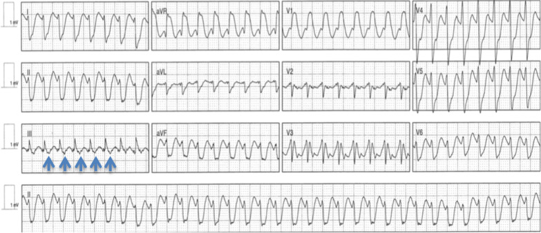
Risk factors for torsades on antiarrhythmic drugs, in addition to those described under repolarization reserve, are older age, recent conversion from atrial fibrillation (with its post-conversion pauses), heart failure or left ventricular hypertrophy, reduced kidney function for renally cleared drugs, a baseline QTc at the upper limit, and rapid intravenous administration. Most drug-induced torsades occurs in patients with two or more risk factors, which is why initiation protocols correct electrolytes, calculate kidney function, and monitor the QT at the expected peak concentration. The thresholds for stopping QT-prolonging drugs, monitoring standards in hospital, and the acute treatment of torsades are covered in the long QT hub.
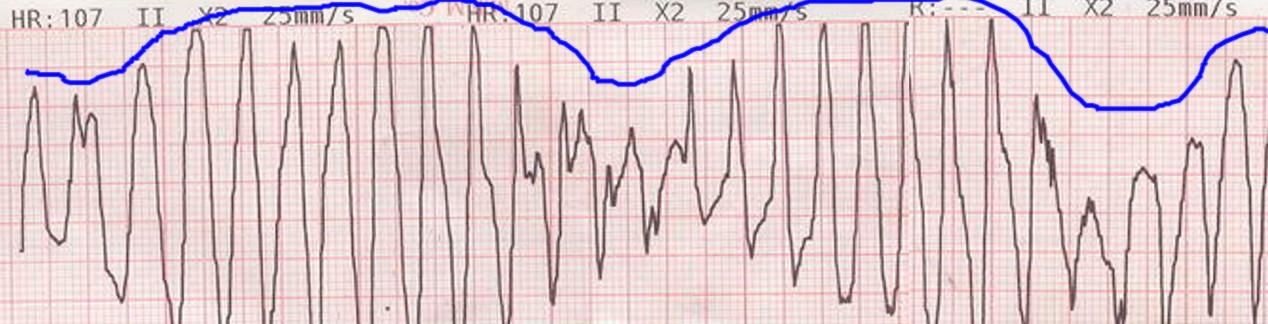

Bradycardia, conduction block, and device interactions
All antiarrhythmic drugs can depress sinus node function or AV conduction, and sinus node dysfunction is often first revealed when an antiarrhythmic converts atrial fibrillation and a long pause follows. Amiodarone and dronedarone slow the sinus rate in most patients. Sotalol causes dose-related bradycardia. Class IC drugs impair His-Purkinje conduction and can produce infranodal block in patients with bundle branch disease, which is why flecainide is contraindicated with bifascicular block without a pacemaker. Combinations multiply these effects: amiodarone with a beta-blocker, digoxin, verapamil, or diltiazem, and amiodarone with sofosbuvir-containing hepatitis C regimens. The latter combination has caused symptomatic bradycardia requiring pacing and at least one death.
When an antiarrhythmic drug is essential and bradycardia limits it, permanent pacing allows the drug to continue (tachy-brady syndrome is a common example). Drug-induced AV block that resolves after withdrawal still predicts later conduction disease in some patients. The bradycardia and AV block hub discusses drug-related block and the decision to pace.
In patients with devices, amiodarone raises the defibrillation threshold and slows VT, so the detection zone and shock safety margin are reassessed. Flecainide and propafenone raise pacing and sensing thresholds and can cause loss of capture at high levels or with hyperkalemia; propafenone's label advises monitoring and reprogramming pacemakers and defibrillators during and after therapy.
Antiarrhythmic drug overdose and toxicity
Sodium channel blocker toxicity (flecainide, propafenone, class IA drugs, and by analogy tricyclic antidepressants and cocaine) produces progressive PR and QRS widening, a terminal R wave in aVR, bradycardia, hypotension, wide-complex tachycardia that can be sinusoidal, and seizures. Hypertonic sodium bicarbonate (1 to 2 mEq/kg boluses, repeated to narrow the QRS, with arterial pH kept near 7.45 to 7.55) increases the sodium gradient and reduces drug binding. Hypertonic saline is an alternative, and intravenous lipid emulsion and venoarterial extracorporeal membrane oxygenation have been used for refractory cases. Other sodium channel blockers are avoided, and electrical cardioversion is often ineffective until drug levels fall. Flecainide toxicity can occur at usual doses when kidney function deteriorates or a CYP2D6 inhibitor is added.
Class III and IA toxicity presents as QT prolongation and torsades. The drug is stopped, magnesium is given, potassium is repleted, and pause-dependent torsades is treated with temporary transvenous pacing at a rate above 70 beats/min, increased as needed to suppress pauses, or with isoproterenol. Sotalol overdose adds beta-blockade (bradycardia, hypotension, hypoglycemia); hemodialysis lowers sotalol concentrations because the drug is not protein bound, and hypotension can slow its initial elimination.
Amiodarone rarely causes acute toxicity after oral overdose because absorption is slow and distribution vast. Rapid intravenous administration causes hypotension and bradycardia, and acute hepatocellular injury can follow intravenous loading. It is not removed by dialysis. Lidocaine toxicity is neurologic (perioral numbness, confusion, seizures) before it is cardiac, and lipid emulsion is used for severe local anesthetic systemic toxicity.
Drug interactions: warfarin, digoxin, direct oral anticoagulants, statins, and CYP pathways
Antiarrhythmic drugs interact through three mechanisms: inhibition of cytochrome P450 enzymes (amiodarone, dronedarone, quinidine, propafenone), inhibition of P-glycoprotein and renal transporters (amiodarone, dronedarone, quinidine, propafenone; dofetilide and procainamide as victims of transporter inhibitors), and pharmacodynamic addition of QT prolongation or nodal depression. The full interaction tables, including transplant drugs, antivirals, and anti-infectives, are in the cardiovascular drug interactions and dosing hub.
| Interaction | Mechanism | Effect | Labeled or recommended action |
|---|---|---|---|
| Amiodarone + warfarin | CYP2C9 and CYP3A inhibition | Prothrombin time increases by about 100% after 3 to 4 days; the effect builds over weeks and persists for months after withdrawal | Warfarin reduced by one-third to one-half; INR at least weekly during loading (US label). See the warfarin hub |
| Amiodarone + digoxin | P-glycoprotein inhibition and reduced clearance | Digoxin concentration roughly doubles | Digoxin halved or stopped; level and kidney function rechecked |
| Dronedarone + digoxin | P-glycoprotein inhibition | Higher digoxin exposure; more sudden death with digoxin in PALLAS and ANDROMEDA | Digoxin stopped or halved; level monitored |
| Amiodarone + simvastatin or lovastatin | CYP3A inhibition | Myopathy and rhabdomyolysis | Simvastatin 20 mg/day maximum; lovastatin 40 mg/day maximum |
| Dronedarone + simvastatin | CYP3A inhibition | Myopathy | Simvastatin 10 mg/day maximum |
| Dronedarone or amiodarone + dabigatran or other direct oral anticoagulants (DOACs) | P-glycoprotein (and CYP3A) inhibition | Higher anticoagulant exposure | Dabigatran 75 mg twice daily with dronedarone at creatinine clearance 30 to 50 mL/min; other DOAC adjustments as listed in the atrial fibrillation hub |
| Dofetilide + verapamil, hydrochlorothiazide, cimetidine, trimethoprim, ketoconazole, prochlorperazine, megestrol, dolutegravir | Renal cation transport inhibition, higher absorption (verapamil), hypokalemia (thiazide) | Higher dofetilide concentration and QT | Contraindicated or not used |
| Propafenone + CYP2D6 inhibitor and CYP3A4 inhibitor together | Loss of both metabolic pathways | Proarrhythmic propafenone concentrations | Simultaneous use avoided |
| Flecainide + amiodarone | CYP2D6 inhibition | Flecainide levels can double | Flecainide dose reduced (amiodarone label: other antiarrhythmics reduced by 30% to 50%) |
| Quinidine + digoxin or CYP2D6 substrates | P-glycoprotein and CYP2D6 inhibition | Digoxin roughly doubles; metabolism of metoprolol and codeine is altered | Digoxin reduced; alternatives sought |
| Mexiletine + ciprofloxacin, fluvoxamine; smoking | CYP1A2 inhibition or induction | Higher or lower mexiletine levels | Dose adjusted to effect and tolerance |
| Any two QT-prolonging drugs | Additive IKr block | Torsades | Combination avoided; contraindicated with dronedarone and dofetilide |
| Amiodarone or dronedarone + beta-blocker, verapamil, diltiazem, digoxin, ivabradine | Additive nodal depression | Bradycardia, AV block | Low starting doses; ECG after initiation |
Common antiarrhythmic drug interactions, from the US labels of amiodarone, dronedarone, dofetilide, flecainide, propafenone, and mexiletine.
Antiarrhythmic drugs in kidney and liver disease
Kidney function determines the dose of dofetilide, sotalol, procainamide, disopyramide, and flecainide, and it changes the safety of all of them. Creatinine clearance by the Cockcroft-Gault formula is the measure used in the dofetilide and sotalol labels. The estimated glomerular filtration rate from laboratory reports is not interchangeable with it in older, small, or obese patients. A kidney function calculator on the interactions hub compares the estimates.
| Drug | Renal elimination | Adjustment for reduced kidney function | Hepatic impairment |
|---|---|---|---|
| Dofetilide | About 80% renal | 500 / 250 / 125 µg twice daily for creatinine clearance above 60 / 40 to 60 / 20 to under 40 mL/min; contraindicated below 20 | No adjustment for mild to moderate impairment; severe impairment not studied |
| Sotalol | Nearly complete, unchanged | Tablets: interval 12 / 24 / 36 to 48 hours for creatinine clearance above 60 / 30 to 59 / 10 to 29 mL/min; individualized below 10. Injection: oral interval after the load 12 / 24 / 48 hours for individual GFR 60 or more / 30 to under 60 / 10 to under 30 mL/min. Partly dialyzable | No adjustment |
| Flecainide | About one-third unchanged | Starting dose 100 mg once daily or 50 mg twice daily at 35 mL/min/1.73 m² or less, with plasma levels | Reduced dose with plasma levels |
| Propafenone | Minimal (metabolites renal) | No specific adjustment; monitoring for toxicity | Substantial reduction because bioavailability rises |
| Procainamide | Parent and NAPA renal | Reduced maintenance; NAPA accumulates; avoided in severe kidney disease when possible | Reduced dose |
| Disopyramide | About half renal | 400 mg/day above 40 mL/min with impairment; 100 mg every 8, 12, or 24 hours for 30 to 40, 15 to 30, and below 15 mL/min | 400 mg/day |
| Mexiletine | About 10% unchanged | Usual dose in kidney failure | Lower dose and close monitoring |
| Quinidine | Partly renal | Modest reduction | Reduced dose |
| Amiodarone | Negligible; not dialyzable | None | Used with caution; enzymes monitored |
| Dronedarone | Minimal | None; early creatinine rise expected from secretion inhibition | Contraindicated in severe impairment |
| Lidocaine | Metabolites renal | Metabolites accumulate with prolonged infusion | Reduced infusion rate (also in heart failure and shock) |
Antiarrhythmic dosing in kidney and liver disease, from the US prescribing information. Creatinine clearance by Cockcroft-Gault with actual body weight for dofetilide and sotalol.
In dialysis, amiodarone is the most commonly used rhythm-control drug, and dofetilide is contraindicated. Sotalol has been used with individualized dosing after dialysis under monitoring, because the label provides no regimen. Hyperkalemia, common in kidney disease, increases sodium channel block and raises pacing thresholds; hypokalemia after dialysis increases torsades risk with class III drugs.
Antiarrhythmic drugs in pregnancy and lactation
Electrical cardioversion is safe in all trimesters and is preferred for unstable or drug-resistant arrhythmias. Drug choice follows the 2023 HRS consensus statement on arrhythmias in pregnancy and the 2023 ACC/AHA/ACCP/HRS AF guideline.
| Drug | Use in pregnancy | Principal concern |
|---|---|---|
| Metoprolol, propranolol | First-line for rate control and many arrhythmias | Fetal growth restriction and neonatal bradycardia or hypoglycemia; atenolol avoided |
| Flecainide | Reasonable for maintenance of sinus rhythm in structurally normal hearts (AF guideline Class 2a); used for fetal tachycardia | Same structural restrictions as outside pregnancy |
| Sotalol | Reasonable for maintenance of sinus rhythm in AF without structural heart disease (2023 ACC/AHA/ACCP/HRS AF guideline, pregnancy section, Class 2a; outside pregnancy the same guideline rates sotalol for AF Class 2b); considered safe for ventricular arrhythmia in the 2023 HRS statement | QT monitoring; renal clearance increases during pregnancy |
| Quinidine | Long history of safe use | QT prolongation; gastrointestinal intolerance |
| Procainamide (intravenous) | Acute cardioversion of AF or treatment of stable VT (AF guideline Class 2b for cardioversion) | Hypotension; lupus with long-term use |
| Lidocaine | Acceptable for acute ventricular arrhythmia | Neonatal depression at high levels |
| Mexiletine | With caution | Limited data |
| Amiodarone | Avoided except for life-threatening arrhythmias resistant to other therapy | Fetal and neonatal hypothyroidism or hyperthyroidism, goiter, growth restriction, bradycardia, neurodevelopmental effects; avoided in breastfeeding |
| Dronedarone | Not used | Embryo-fetal toxicity in animal studies; pregnancy excluded before initiation |
| Dofetilide | Limited data; used only when needed | Requires in-hospital initiation |
Antiarrhythmic drugs in pregnancy, after the 2023 HRS consensus statement and the 2023 ACC/AHA/ACCP/HRS AF guideline. Ventricular arrhythmias in pregnancy are also discussed on the ventricular tachycardia hub.
Pregnancy increases the volume of distribution, renal clearance, and hepatic metabolism of many drugs, so levels fall during gestation and rise after delivery. Dose changes are therefore checked against the ECG (QRS and QTc) and clinical effect in each trimester and postpartum.
Antiarrhythmic drugs in older adults
Older adults have more atrial fibrillation, more structural and conduction disease, lower creatinine clearance (often concealed by a normal serum creatinine because of low muscle mass), and more interacting drugs. They also have more torsades on class III drugs and more bradycardia and falls with all antiarrhythmics. The 2023 Beers Criteria advise avoiding amiodarone as first-line therapy for atrial fibrillation unless the patient has heart failure or substantial left ventricular hypertrophy. They also advise avoiding dronedarone in permanent AF and in severe or recently decompensated heart failure. The 2023 update removed disopyramide from the list because of low use in the United States; its negative inotropic and anticholinergic effects still make it a poor choice in older adults. Rate control is adequate for many older patients with few symptoms. Rhythm control is chosen when symptoms, heart failure, or early disease favor it, as in EAST-AFNET 4, whose participants had a mean age of 70 years.
In older adults, creatinine clearance is calculated at every dose decision, doses start low, initiation includes ECG monitoring for pauses after conversion, and the medication list is reviewed for additive nodal blockers and QT-prolonging drugs. Cognitive impairment and polypharmacy make strict dosing schedules, such as dofetilide's twice-daily regimen without doubling for missed doses, harder to follow.
Monitoring schedule after initiation
| Drug | At initiation | Early follow-up | Long term |
|---|---|---|---|
| Dofetilide | In hospital at least 3 days; QTc 2 to 3 hours after each dose; creatinine clearance; potassium, magnesium | ECG, creatinine, potassium, magnesium within weeks of discharge and after any interacting drug or illness | Kidney function and QTc every 3 months; dose reduced if creatinine clearance falls |
| Sotalol | In hospital at least 3 days (or intravenous load); QTc 2 to 4 hours after doses and increases; creatinine clearance; potassium at least 4 mEq/L | ECG and creatinine after each dose change | ECG, creatinine, potassium, and magnesium every 3 to 6 months and with illness |
| Flecainide, propafenone | ECG for PR and QRS; first pill-in-the-pocket dose monitored | ECG at steady state (3 to 5 days after each dose change); exercise test for use-dependent QRS widening | ECG periodically and after dose or kidney changes; flecainide levels in kidney or liver disease; new ischemia or reduced ejection fraction leads to withdrawal |
| Dronedarone | ECG; creatinine; liver enzymes; pregnancy excluded; digoxin and statin adjustment | Creatinine in the first weeks (the secretory rise plateaus by 7 days); liver enzymes in the first 6 months | Cardiac rhythm at least every 3 months; stopped if AF becomes permanent or heart failure decompensates |
| Mexiletine | ECG; liver function | Tolerance and levels where available | Blood counts and liver tests when symptoms arise; neurologic and gastrointestinal effects reviewed |
| Quinidine, procainamide, disopyramide | In-hospital ECG monitoring with QTc for the oral class IA drugs | QTc; for procainamide, blood counts weekly for 3 months; for quinidine, platelet count | Antinuclear antibodies for procainamide; anticholinergic effects for disopyramide |
Monitoring after initiation of antiarrhythmic drugs, from the US labels, the 2023 ACC/AHA/ACCP/HRS guideline, the 2007 HRS practical guide, and the 2017 AHA practice standards for in-hospital ECG monitoring. The standards recommend QTc monitoring for every patient started on dofetilide, ibutilide, sotalol, disopyramide, procainamide, or quinidine. Amiodarone monitoring is tabulated in the amiodarone surveillance schedule.
Sources of error in antiarrhythmic drug prescribing
- Starting flecainide or propafenone in a patient whose infarction is evident only on imaging or ECG risks lethal proarrhythmia. Continuing either drug once infarction, significant structural heart disease, or reduced ejection fraction is diagnosed carries the same risk.
- Omitting the AV nodal blocker, or stopping it later, in a patient on a class IC drug for atrial fibrillation or flutter permits 1:1 conduction of slowed flutter.
- Dosing dofetilide or sotalol from a laboratory estimated glomerular filtration rate, or failing to recalculate Cockcroft-Gault clearance when kidney function changes, can give a dose too high for the true clearance.
- Measuring the QTc with the Bazett correction during the tachycardia of atrial fibrillation, or before the expected peak concentration, misjudges the response to dofetilide and sotalol.
- Combining dofetilide with a contraindicated drug, or adding a second QT-prolonging drug to any class III agent, raises the QT and the risk of torsades.
- Starting amiodarone without reducing the warfarin and digoxin doses, or forgetting that the interaction persists for months after the drug is stopped, causes over-anticoagulation and digoxin toxicity.
- Attributing new atrial fibrillation, weight loss, or ventricular arrhythmia on amiodarone to the underlying disease, or new dyspnea to heart failure, delays the diagnosis of thyrotoxicosis or pulmonary toxicity.
- Reading the expected early TSH rise or high-normal free T4 on amiodarone as thyroid disease leads to unnecessary treatment.
- Continuing dronedarone after AF becomes permanent or heart failure decompensates raises mortality, and in permanent AF also stroke and heart failure hospitalization.
- Interpreting the early creatinine rise on dronedarone as kidney injury stops a useful drug, and dismissing a continuing rise as the secretory effect misses true kidney injury.
- Using an antiarrhythmic drug in a patient with sinus node dysfunction or conduction disease without pacing backup, or overlooking post-conversion pauses, risks symptomatic bradycardia and syncope.
- Treating asymptomatic premature ventricular complexes after infarction with class I drugs increases mortality, as CAST showed.
- Giving AV nodal blockers or intravenous amiodarone in pre-excited atrial fibrillation increases conduction over the accessory pathway and can cause ventricular fibrillation.
- Prescribing sodium channel blockers to a patient with a Brugada-pattern ECG, or overlooking a type 1 pattern unmasked by flecainide, can trigger ventricular fibrillation.
- Omitting device reprogramming after amiodarone slows VT below the detection rate, or after a class IC drug raises pacing thresholds, leaves VT undetected or causes loss of capture.
Self-assessment
A 68-year-old woman weighing 62 kg with persistent atrial fibrillation and an LVEF of 30% is admitted to start dofetilide. Serum creatinine is 1.3 mg/dL, potassium 4.3 mEq/L, and baseline QTc 430 ms with a QRS of 96 ms. What is her starting dose, and what happens if the QTc 2 to 3 hours after the first dose is 505 ms?
Cardiovascular Medicine Board ReviewReferences
- Joglar JA, Chung MK, Armbruster AL, et al. 2023 ACC/AHA/ACCP/HRS guideline for the diagnosis and management of atrial fibrillation: a report of the American College of Cardiology/American Heart Association Joint Committee on Clinical Practice Guidelines. Circulation. 2024;149:e1–e156. Corrections: Circulation. 2024;149:e167, e936, and e1413. doi:10.1161/CIR.0000000000001193
- Al-Khatib SM, Stevenson WG, Ackerman MJ, et al. 2017 AHA/ACC/HRS guideline for management of patients with ventricular arrhythmias and the prevention of sudden cardiac death. Circulation. 2018;138:e272–e391. Correction: Circulation. 2018;138:e419–e420. doi:10.1161/CIR.0000000000000549
- Page RL, Joglar JA, Caldwell MA, et al. 2015 ACC/AHA/HRS guideline for the management of adult patients with supraventricular tachycardia. Circulation. 2016;133:e506–e574. Correction: Circulation. 2016;134:e234–e235. doi:10.1161/CIR.0000000000000311
- Van Gelder IC, Rienstra M, Bunting KV, et al. 2024 ESC guidelines for the management of atrial fibrillation developed in collaboration with the European Association for Cardio-Thoracic Surgery (EACTS). Eur Heart J. 2024;45:3314–3414. Correction: Eur Heart J. 2025;46:4349. doi:10.1093/eurheartj/ehae176
- Zeppenfeld K, Tfelt-Hansen J, de Riva M, et al. 2022 ESC guidelines for the management of patients with ventricular arrhythmias and the prevention of sudden cardiac death. Eur Heart J. 2022;43:3997–4126. doi:10.1093/eurheartj/ehac262
- Ommen SR, Ho CY, Asif IM, et al. 2024 AHA/ACC/AMSSM/HRS/PACES/SCMR guideline for the management of hypertrophic cardiomyopathy. Circulation. 2024;149:e1239–e1311. doi:10.1161/CIR.0000000000001250
- Joglar JA, Kapa S, Saarel EV, et al. 2023 HRS expert consensus statement on the management of arrhythmias during pregnancy. Heart Rhythm. 2023;20:e175–e264. doi:10.1016/j.hrthm.2023.05.017
- Drew BJ, Ackerman MJ, Funk M, et al. Prevention of torsade de pointes in hospital settings: a scientific statement from the American Heart Association and the American College of Cardiology Foundation. Circulation. 2010;121:1047–1060. doi:10.1161/CIRCULATIONAHA.109.192704
- Goldschlager N, Epstein AE, Naccarelli GV, et al. A practical guide for clinicians who treat patients with amiodarone: 2007. Heart Rhythm. 2007;4:1250–1259. doi:10.1016/j.hrthm.2007.07.020
- Bartalena L, Bogazzi F, Chiovato L, et al. 2018 European Thyroid Association (ETA) guidelines for the management of amiodarone-associated thyroid dysfunction. Eur Thyroid J. 2018;7:55–66. doi:10.1159/000486957
- 2023 American Geriatrics Society Beers Criteria Update Expert Panel. American Geriatrics Society 2023 updated AGS Beers Criteria for potentially inappropriate medication use in older adults. J Am Geriatr Soc. 2023;71:2052–2081. doi:10.1111/jgs.18372
- Lei M, Wu L, Terrar DA, Huang CL. Modernized classification of cardiac antiarrhythmic drugs. Circulation. 2018;138:1879–1896. doi:10.1161/CIRCULATIONAHA.118.035455
- Vaughan Williams EM. A classification of antiarrhythmic actions reassessed after a decade of new drugs. J Clin Pharmacol. 1984;24:129–147. doi:10.1002/j.1552-4604.1984.tb01822.x
- Task Force of the Working Group on Arrhythmias of the European Society of Cardiology. The Sicilian gambit: a new approach to the classification of antiarrhythmic drugs based on their actions on arrhythmogenic mechanisms. Circulation. 1991;84:1831–1851. doi:10.1161/01.CIR.84.4.1831
- Hondeghem LM, Katzung BG. Antiarrhythmic agents: the modulated receptor mechanism of action of sodium and calcium channel-blocking drugs. Annu Rev Pharmacol Toxicol. 1984;24:387–423. doi:10.1146/annurev.pa.24.040184.002131
- Roden DM. Drug-induced prolongation of the QT interval. N Engl J Med. 2004;350:1013–1022. doi:10.1056/NEJMra032426
- Echt DS, Liebson PR, Mitchell LB, et al. Mortality and morbidity in patients receiving encainide, flecainide, or placebo: the Cardiac Arrhythmia Suppression Trial. N Engl J Med. 1991;324:781–788. doi:10.1056/NEJM199103213241201
- Waldo AL, Camm AJ, deRuyter H, et al. Effect of d-sotalol on mortality in patients with left ventricular dysfunction after recent and remote myocardial infarction (SWORD). Lancet. 1996;348:7–12. doi:10.1016/s0140-6736(96)02149-6
- Torp-Pedersen C, Møller M, Bloch-Thomsen PE, et al. Dofetilide in patients with congestive heart failure and left ventricular dysfunction (DIAMOND-CHF). N Engl J Med. 1999;341:857–865. doi:10.1056/NEJM199909163411201
- Køber L, Bloch Thomsen PE, Møller M, et al. Effect of dofetilide in patients with recent myocardial infarction and left-ventricular dysfunction: a randomised trial (DIAMOND-MI). Lancet. 2000;356:2052–2058. doi:10.1016/S0140-6736(00)03402-4
- Køber L, Torp-Pedersen C, McMurray JJ, et al. Increased mortality after dronedarone therapy for severe heart failure (ANDROMEDA). N Engl J Med. 2008;358:2678–2687. doi:10.1056/NEJMoa0800456
- Connolly SJ, Camm AJ, Halperin JL, et al. Dronedarone in high-risk permanent atrial fibrillation (PALLAS). N Engl J Med. 2011;365:2268–2276. doi:10.1056/NEJMoa1109867
- Hohnloser SH, Crijns HJ, van Eickels M, et al. Effect of dronedarone on cardiovascular events in atrial fibrillation (ATHENA). N Engl J Med. 2009;360:668–678. doi:10.1056/NEJMoa0803778
- Le Heuzey JY, De Ferrari GM, Radzik D, et al. A short-term, randomized, double-blind, parallel-group study to evaluate the efficacy and safety of dronedarone versus amiodarone in patients with persistent atrial fibrillation: the DIONYSOS study. J Cardiovasc Electrophysiol. 2010;21:597–605. doi:10.1111/j.1540-8167.2010.01764.x
- Singh BN, Singh SN, Reda DJ, et al. Amiodarone versus sotalol for atrial fibrillation (SAFE-T). N Engl J Med. 2005;352:1861–1872. doi:10.1056/NEJMoa041705
- Roy D, Talajic M, Dorian P, et al. Amiodarone to prevent recurrence of atrial fibrillation (CTAF). N Engl J Med. 2000;342:913–920. doi:10.1056/NEJM200003303421302
- AFFIRM First Antiarrhythmic Drug Substudy Investigators. Maintenance of sinus rhythm in patients with atrial fibrillation: an AFFIRM substudy of the first antiarrhythmic drug. J Am Coll Cardiol. 2003;42:20–29. doi:10.1016/s0735-1097(03)00559-x
- Valembois L, Audureau E, Takeda A, et al. Antiarrhythmics for maintaining sinus rhythm after cardioversion of atrial fibrillation. Cochrane Database Syst Rev. 2019;9:CD005049. doi:10.1002/14651858.CD005049.pub5
- Alboni P, Botto GL, Baldi N, et al. Outpatient treatment of recent-onset atrial fibrillation with the "pill-in-the-pocket" approach. N Engl J Med. 2004;351:2384–2391. doi:10.1056/NEJMoa041233
- Camm AJ, Capucci A, Hohnloser SH, et al. A randomized active-controlled study comparing the efficacy and safety of vernakalant to amiodarone in recent-onset atrial fibrillation (AVRO). J Am Coll Cardiol. 2011;57:313–321. doi:10.1016/j.jacc.2010.07.046
- Ellenbogen KA, Stambler BS, Wood MA, et al. Efficacy of intravenous ibutilide for rapid termination of atrial fibrillation and atrial flutter: a dose-response study. J Am Coll Cardiol. 1996;28:130–136. doi:10.1016/0735-1097(96)00121-0
- Kirchhof P, Camm AJ, Goette A, et al. Early rhythm-control therapy in patients with atrial fibrillation (EAST-AFNET 4). N Engl J Med. 2020;383:1305–1316. doi:10.1056/NEJMoa2019422
- Wazni OM, Chun KRJ, Nair DG, et al. Pulsed field ablation as initial therapy for persistent atrial fibrillation (AVANT GUARD). N Engl J Med. 2026;394:2407–2418. doi:10.1056/NEJMoa2600929
- Sapp JL, Tang ASL, Parkash R, et al. Catheter ablation or antiarrhythmic drugs for ventricular tachycardia (VANISH2). N Engl J Med. 2025;392:737–747. doi:10.1056/NEJMoa2409501
- Bardy GH, Lee KL, Mark DB, et al. Amiodarone or an implantable cardioverter-defibrillator for congestive heart failure (SCD-HeFT). N Engl J Med. 2005;352:225–237. doi:10.1056/NEJMoa043399
- Kudenchuk PJ, Brown SP, Daya M, et al. Amiodarone, lidocaine, or placebo in out-of-hospital cardiac arrest (ALPS). N Engl J Med. 2016;374:1711–1722. doi:10.1056/NEJMoa1514204
- Ortiz M, Martín A, Arribas F, et al. Randomized comparison of intravenous procainamide vs. intravenous amiodarone for the acute treatment of tolerated wide QRS tachycardia: the PROCAMIO study. Eur Heart J. 2017;38:1329–1335. doi:10.1093/eurheartj/ehw230
- Zareba W, Daubert JP, Beck CA, et al. Ranolazine in high-risk patients with implanted cardioverter-defibrillators: the RAID trial. J Am Coll Cardiol. 2018;72:636–645. doi:10.1016/j.jacc.2018.04.086
- Kannankeril PJ, Moore JP, Cerrone M, et al. Efficacy of flecainide in the treatment of catecholaminergic polymorphic ventricular tachycardia: a randomized clinical trial. JAMA Cardiol. 2017;2:759–766. doi:10.1001/jamacardio.2017.1320
- Naccarelli GV, Rackley J, Boriani G. New recommendations for rhythm control: what has changed in the 2023 ACC/AHA/ACCP/HRS and 2024 ESC guidelines for atrial fibrillation, and where does dronedarone fit in? Am Heart J Plus. 2025;60:100645. doi:10.1016/j.ahjo.2025.100645
- Tiver KD, Martin DK, Quah J, Lahiri A, Ganesan AN. Failure of diltiazem to prevent 1:1 conduction of atrial flutter: a case report. J Med Case Rep. 2023;17:319. doi:10.1186/s13256-023-03947-5
- Marrouche NF, Brachmann J, Andresen D, et al. Catheter ablation for atrial fibrillation with heart failure (CASTLE-AF). N Engl J Med. 2018;378:417–427. doi:10.1056/NEJMoa1707855
- US Food and Drug Administration. Tikosyn (dofetilide) capsules: prescribing information. Label effective April 2026. DailyMed
- US Food and Drug Administration. Betapace and Betapace AF (sotalol hydrochloride) tablets: prescribing information (label effective December 2024); sotalol hydrochloride injection: prescribing information (revised July 2025). DailyMed (tablets); DailyMed (injection)
- US Food and Drug Administration. Multaq (dronedarone) tablets: prescribing information. Label effective January 2026. DailyMed
- US Food and Drug Administration. Prescribing information for amiodarone hydrochloride tablets (2024), flecainide acetate tablets (2024), propafenone hydrochloride tablets (2023), mexiletine hydrochloride capsules (2024), disopyramide phosphate capsules (2024), procainamide hydrochloride injection (2023), and ibutilide fumarate injection (2025). DailyMed
- Sandau KE, Funk M, Auerbach A, et al. Update to practice standards for electrocardiographic monitoring in hospital settings: a scientific statement from the American Heart Association. Circulation. 2017;136:e273–e344. doi:10.1161/CIR.0000000000000527
- Merino JL, Tamargo J, Blomström-Lundqvist C, et al. Practical compendium of antiarrhythmic drugs: a clinical consensus statement of the European Heart Rhythm Association of the European Society of Cardiology. Europace. 2025;27:euaf076. doi:10.1093/europace/euaf076