Library › Vascular disease › Peripheral artery disease
Lower extremity peripheral artery disease
Atherosclerotic lower extremity peripheral artery disease in adults, from screening and physiologic testing through medical therapy, exercise, revascularization, chronic limb-threatening ischemia, and acute limb ischemia.
Lower extremity peripheral artery disease is diagnosed by a resting ankle-brachial index (ABI) of 0.90 or less, or by a toe-brachial index (TBI) of 0.70 or less when the ankle arteries are noncompressible. Every patient receives a high-intensity statin and risk factor control, and symptomatic patients receive antiplatelet therapy. Structured exercise is first-line treatment for claudication, and revascularization is reserved for lifestyle-limiting symptoms that persist despite it, for chronic limb-threatening ischemia, and for acute limb ischemia with a salvageable limb.
Definition and the four clinical subsets of peripheral artery disease
Lower extremity peripheral artery disease (PAD) is atherosclerotic obstruction of the arteries supplying the legs, from the infrarenal aorta to the pedal arteries. It is a manifestation of systemic atherosclerosis and a clinical form of atherosclerotic cardiovascular disease (ASCVD). The 2024 American College of Cardiology/American Heart Association (ACC/AHA) multisociety guideline organizes patients with objectively confirmed PAD into four clinical subsets. The subset sets the urgency, the tests, and the goals of treatment, and patients move between subsets over time.
| Clinical subset | Definition | Clinical features | Urgency and goal |
|---|---|---|---|
| Asymptomatic PAD | Objectively confirmed PAD without leg symptoms | 20% to 59% of patients with confirmed PAD; many limit activity below the ischemic threshold and have walking impairment | Cardiovascular risk reduction and foot care |
| Chronic symptomatic PAD | Claudication or other ischemic exertional leg symptoms, absent at rest | Most common clinically evident subset; symptoms resolve within 10 minutes of rest | Risk reduction, improved walking performance and quality of life |
| Chronic limb-threatening ischemia (CLTI) | Ischemic rest pain, nonhealing wound, or gangrene present for more than 2 weeks with objective PAD | Develops in 11% to 20% of patients with known PAD and accounts for most PAD-related amputations. Historical 1-year mortality is 25% to 35%, and amputation occurs in up to 30%. | Prompt multispecialty evaluation and revascularization when feasible |
| Acute limb ischemia (ALI) | Sudden decrease in limb perfusion, symptoms present for less than 2 weeks, threatening limb viability | Pain, pallor, pulselessness, poikilothermia, paresthesia, and paralysis. Incidence is 0.8 per 100 patient-years in a contemporary trial of symptomatic PAD. | Emergency assessment, heparin, and revascularization of a salvageable limb |
Adapted from the 2024 ACC/AHA guideline, Table 4. CLTI replaces the older term critical limb ischemia, which implied a single perfusion threshold.
Epidemiology, risk factors, and health disparities in peripheral artery disease
PAD affects an estimated 10 to 12 million adults older than 40 years in the United States. Worldwide estimates range from at least 113 million to as many as 236 million people. In a systematic review of population studies that defined PAD by an ABI of 0.90 or less, prevalence rose steadily with age to about 21% at 80 to 84 years in high-income countries, where women had a slightly higher prevalence than men up to 75 years of age.
Smoking and diabetes are the strongest risk factors: 80% to 90% of patients revascularized for severe limb symptoms are current smokers, and diabetes carries an adjusted hazard ratio of about 5.5 for amputation. Hypertension, dyslipidemia, chronic kidney disease, elevated lipoprotein(a), and family history complete the profile. The guideline defines the population at increased risk, in whom history, examination, and ABI screening are directed.
| Patients at increased risk of PAD |
|---|
| Age 65 years or older |
| Age 50 to 64 years with risk factors for atherosclerosis (diabetes, history of smoking, dyslipidemia, hypertension), chronic kidney disease, or a family history of PAD |
| Age younger than 50 years with diabetes and one additional risk factor for atherosclerosis |
| Known atherosclerotic disease in another vascular bed (coronary, carotid, subclavian, renal, or mesenteric artery stenosis, or abdominal aortic aneurysm) |
From the 2024 ACC/AHA guideline, Table 5. The guideline also lists PAD-related risk amplifiers that raise the risk of major adverse cardiovascular events (MACE), major adverse limb events (MALE), or both: age 75 years or older, diabetes, ongoing smoking or other tobacco use, chronic kidney disease, end-stage kidney disease, polyvascular disease, microvascular disease, and depression. Incorporating them into treatment decisions is a Class 1 recommendation.
Health disparities. The 2024 guideline added a section on health disparities and recommends that clinicians and health systems identify and limit them (Class 1). Estimated lifetime risk of PAD is about 30% in Black men and 28% in Black women, compared with about 19% in White and 22% in Hispanic adults. Black patients present more often with CLTI. They receive statins, antiplatelet therapy, and supervised exercise less often and have a 2- to 4-fold higher risk of amputation, more often without an attempt at revascularization. American Indian and Hispanic populations and rural residents also have higher amputation rates. Women present 10 to 20 years later than men, with more atypical symptoms and more advanced disease. They are more likely to undergo above-knee amputation and have higher periprocedural mortality.
Atherosclerotic and nonatherosclerotic causes of limb ischemia
Atherosclerosis causes the large majority of chronic lower extremity arterial disease in adults. Its distribution follows risk factors. Smokers more often have aortoiliac and femoropopliteal disease, whereas diabetes and chronic kidney disease favor infrapopliteal (tibial) disease with heavy medial calcification, the pattern most often associated with CLTI and noncompressible ankle arteries. The superficial femoral artery in the adductor canal is the most frequently involved segment.
Nonatherosclerotic disease is considered when symptoms begin before about 50 years of age, risk factors are absent, or the distribution is atypical, because each entity has a different treatment.
| Condition | Typical patient and distribution | Distinguishing features |
|---|---|---|
| Thromboangiitis obliterans (Buerger disease) | Young smokers; segmental occlusion of small and medium arteries and veins of the legs and arms | Digital ulcers, migratory thrombophlebitis, corkscrew collaterals; complete cessation of tobacco and nicotine is the only disease-modifying treatment |
| Popliteal artery entrapment syndrome | Young athletic adults | Compression by the medial gastrocnemius head; pulses fall with active plantar flexion; MRI or provocative duplex; surgical release |
| Cystic adventitial disease | Middle-aged men, popliteal artery | Mucinous cyst in the adventitia narrows the lumen; smooth tapered stenosis ("scimitar sign") on angiography; resection or cyst evacuation |
| Fibromuscular dysplasia | Predominantly women; renal and carotid arteries most often, iliac arteries less often | Nonatherosclerotic, noninflammatory; "string of beads" pattern; associated dissection and aneurysm |
| Large-vessel vasculitis | Takayasu arteritis in younger women; giant cell arteritis after age 50 | Smooth long stenoses, raised inflammatory markers, arm claudication; immunosuppression before revascularization |
| Embolic and aneurysmal disease | Atrial fibrillation, left ventricular thrombus, aortic or popliteal aneurysm | Sudden onset, blue toe with palpable pedal pulses after atheroembolism, contralateral popliteal aneurysm |
| Iatrogenic and traumatic injury | Femoral access, radiation, trauma | Dissection, thrombosis, pseudoaneurysm, or radiation arteritis in the treated field |
Renal artery stenosis and fibromuscular dysplasia of the renal arteries are discussed with secondary causes of hypertension.
Pathophysiology of exertional and rest ischemia
Flow through a stenosis falls steeply once the lumen is narrowed by about 50% in diameter, because resistance varies inversely with the fourth power of the radius and energy is lost to turbulence at the exit of the lesion. At rest, collateral channels and distal arteriolar dilation maintain perfusion pressure, so many patients with occlusions have no symptoms. During walking, calf muscle oxygen demand rises, distal resistance falls, and the fixed proximal lesion prevents a matching rise in flow. Ankle pressure falls, the muscle becomes ischemic, and claudication follows.
The limitation is not purely hemodynamic. Chronic ischemia damages skeletal muscle mitochondria, causes myofiber atrophy and denervation, and impairs endothelium-dependent vasodilation, which explains why walking performance correlates only loosely with the ABI and why exercise training improves walking without changing the ABI.
In CLTI, resting perfusion no longer meets the metabolic needs of the skin and distal tissues, typically because disease involves several levels in series (for example, femoropopliteal and tibial occlusions together). Rest pain begins in the forefoot, worsens with elevation, and improves with dependency, because gravity adds hydrostatic pressure to the distal perfusion pressure. Healing a wound requires more perfusion than maintaining intact skin, and diabetes adds neuropathy, impaired collateral formation, and susceptibility to infection.
Natural history, cardiovascular risk, and polyvascular disease
The dominant risk in PAD is cardiovascular. In the ABI Collaboration meta-analysis of 16 population cohorts, the relation between ABI and mortality was reverse J-shaped, with the lowest risk at an ABI of 1.11 to 1.40. Ten-year cardiovascular mortality was 18.7% in men with an ABI of 0.90 or less and 4.4% in men with a normal ABI (hazard ratio 4.2), and 12.6% compared with 4.1% in women (hazard ratio 3.5). The excess risk persisted after adjustment for the Framingham risk score. A noncompressible ABI above 1.40 also marks increased mortality and is not a normal result.
Asymptomatic PAD carries a systemic risk well above that of people with a normal ABI. Event rates rise stepwise with each additional affected vascular bed. In EUCLID, myocardial infarction occurred in 4.9% of patients with symptomatic PAD over a median of 30 months despite antiplatelet therapy. Polyvascular disease, diabetes, heart failure, and a high-risk limb presentation (CLTI, prior amputation, or prior revascularization) identify the patients with the highest event rates and the largest absolute benefit from low-dose rivaroxaban with aspirin.
Most patients with claudication remain stable or improve with exercise and risk factor control; progression to CLTI is more likely with diabetes, continued smoking, chronic kidney disease, and a low ABI. ALI is uncommon (1.7% over 30 months in a contemporary trial) and concentrates in patients with prior revascularization, atrial fibrillation, or an ABI of 0.60 or less. A foot ulcer in a patient with PAD is associated with a 40% mortality at 5 years.
Claudication, atypical leg symptoms, and their differential diagnosis
Typical claudication is aching, cramping, fatigue, or tightness in a muscle group that occurs consistently during walking, does not start at rest, worsens uphill or at a faster pace, and resolves within about 10 minutes of standing still. The symptomatic muscle group lies one level below the obstruction. Calf claudication indicates femoropopliteal disease, thigh and buttock claudication indicates aortoiliac disease, and foot claudication suggests tibial or pedal disease. Bilateral buttock and thigh claudication with erectile dysfunction and absent femoral pulses is the Leriche syndrome of aortoiliac occlusion.
Classic claudication is the minority presentation. Many patients describe leg fatigue, weakness, or numbness while walking, or discomfort that needs more than 10 minutes to resolve, and the 2024 guideline includes these "other nonjoint-related exertional leg symptoms" in chronic symptomatic PAD. Neuropathy, reduced activity, and arthritis further blunt the history.
| Condition | Location and character | Relation to exercise and rest | Effect of position and other clues |
|---|---|---|---|
| Arterial claudication | Calf, thigh, or buttock; cramping or fatigue | Reproducible at a consistent distance; relieved within minutes of standing still | No positional requirement; diminished pulses; abnormal ABI |
| Spinal stenosis (neurogenic claudication) | Often bilateral buttocks and posterior legs; pain, paresthesia, weakness | Variable distance; also provoked by standing; slow relief | Relieved by sitting or lumbar flexion (leaning on a shopping cart); worse with spinal extension; often easier uphill |
| Nerve root compression | Sharp, lancinating pain radiating down the leg | Induced by sitting, standing, or walking; often present at rest | Positional change improves it; history of back disease |
| Hip or knee arthritis | Lateral hip, thigh, or knee; aching | Variable onset; not quickly relieved | Improved by unloading the joint; reduced range of motion |
| Venous claudication | Whole leg, worse in the calf; tight, bursting pain | After walking; subsides slowly | Relieved by elevation; prior iliofemoral deep vein thrombosis, edema |
| Chronic exertional compartment syndrome | Calf muscles; tight, bursting pain | After strenuous exercise such as running; subsides very slowly | Muscular athletes; elevated compartment pressures |
| Symptomatic popliteal (Baker) cyst | Behind the knee and down the calf; swelling, tenderness | With exercise and at rest | Not intermittent; palpable mass, duplex confirms |
Adapted from the 2024 ACC/AHA guideline, Table 7. A normal resting and exercise ABI excludes hemodynamically significant PAD as the cause of exertional leg symptoms.
Vascular examination of the legs and feet
The examination is performed with shoes and socks removed and is repeated regularly in patients at increased risk (Class 1). Blood pressure is measured in both arms. A systolic difference of 15 to 20 mm Hg or more suggests subclavian or innominate stenosis, and the higher arm pressure is used for the ABI. Femoral, popliteal, dorsalis pedis, and posterior tibial pulses are palpated and graded 0 (absent), 1 (diminished), 2 (normal), or 3 (bounding). Examiners agree well on absent pulses and poorly on diminished ones, and an isolated absent dorsalis pedis pulse can be a normal variant. A widened or prominent popliteal pulse suggests a popliteal aneurysm.
Bruits over the abdomen, flank, or groin indicate turbulent flow. The skin shows hair loss, shiny atrophic skin, thickened nails, coolness, and delayed capillary refill. In the elevation-dependency (Buerger) test, pallor of the sole when the leg of a supine patient is elevated indicates severe ischemia. Dependent rubor with slow venous filling when the leg is lowered reflects maximal arteriolar dilation in a chronically ischemic foot. The feet are inspected between the toes, at the heels, and at pressure points for fissures, calluses, ulcers, and gangrene.
| Feature | Arterial (ischemic) ulcer | Venous ulcer | Neuropathic ulcer |
|---|---|---|---|
| Location | Toes, heel, lateral malleolus, pressure points, interdigital spaces | Gaiter area, above the medial malleolus | Plantar pressure zones, metatarsal heads |
| Appearance | Punched-out, dry, pale or necrotic base, little granulation | Shallow, irregular, moist, granulating, exudative | Surrounded by callus, often deep |
| Pain | Painful unless neuropathy coexists; worse with elevation | Aching, relieved by elevation | Painless |
| Surrounding skin and pulses | Cool, hairless, absent pulses | Edema, hemosiderin, lipodermatosclerosis; pulses present | Warm, dry skin; loss of protective sensation; pulses may be present |
Neuroischemic ulcers are the most common pattern in diabetes. A wound that persists despite adequate perfusion raises the alternative diagnoses in guideline Table 8: vasculitic, inflammatory, infectious, malignant, and drug-induced ulcers.
Ankle-brachial index: technique and reporting categories
The resting ABI is the first diagnostic test in a patient whose history or examination suggests PAD (Class 1). It is the ratio of the higher of the dorsalis pedis and posterior tibial systolic pressures in each leg to the higher of the two brachial systolic pressures. Compared with angiographic stenosis, the resting ABI has a sensitivity of 69% to 79% and a specificity of 83% to 99% in symptomatic patients. Sensitivity is lower in diabetes and chronic kidney disease, where medial calcification raises ankle pressures.
The AHA scientific statement on ABI measurement standardizes the method. The patient rests supine for 5 to 10 minutes with the head and heels supported. Cuffs are sized so that the bladder encircles at least 40% of the limb circumference, and the ankle cuff is wrapped straight with its lower edge 2 cm above the medial malleolus. Systolic pressures are recorded with a handheld continuous-wave Doppler probe in each arm and in the dorsalis pedis and posterior tibial arteries of each ankle. The sequence is the same for every patient (for example, right arm, right posterior tibial, right dorsalis pedis, left posterior tibial, left dorsalis pedis, left arm). If the first arm pressure exceeds the second by 10 mm Hg or more, it is repeated at the end.
| Resting ABI | 2024 ACC/AHA category | Interpretation and next step |
|---|---|---|
| >1.40 | Noncompressible | Medial calcification (diabetes, chronic kidney disease, older age). Not interpretable; toe pressure and TBI with waveforms are required (Class 1). Associated with increased mortality. |
| 1.00–1.40 | Normal | Exertional symptoms with a normal resting ABI lead to a treadmill exercise ABI (Class 1). |
| 0.91–0.99 | Borderline | Intermediate cardiovascular risk; exercise ABI when symptoms suggest PAD (Class 1). |
| ≤0.90 | Abnormal | Diagnostic of PAD. Lower values indicate more severe disease, and an ABI below 0.40 meets the Global Vascular Guidelines hemodynamic criterion for ischemic rest pain. |
Reporting categories from the 2024 ACC/AHA guideline (Class 1). A decrease in ABI of more than 0.15 on serial testing indicates significant progression. A systolic difference of 15 mm Hg or more between arms is itself associated with PAD and with higher cardiovascular mortality.
Ankle-brachial index, toe-brachial index, and WIfI ischemia grade from limb pressures
The WIfI ischemia grade comes from toe pressure when one is entered, because the Global Vascular Guidelines prefer it in CLTI, and from the ABI otherwise. Indices are rounded to two decimals before categorization.
ABI and TBI
Systolic pressures in mm HgABI = higher ankle pressure (dorsalis pedis or posterior tibial) ÷ higher brachial pressure. TBI = great-toe pressure ÷ higher brachial pressure; 0.70 or less is abnormal. The example leg on the right (ABI 0.53) is diagnostic of PAD; the left ankle arteries are noncompressible (ABI 1.50), and a toe pressure of 42 mm Hg gives a TBI of 0.30 and WIfI ischemia grade 1. The WIfI system was published with a noncompressibility threshold of 1.30; the 2024 ACC/AHA threshold of 1.40 is used here.
Toe pressures, exercise ABI, segmental pressures, waveforms, and tissue perfusion
Toe pressure and toe-brachial index. Digital arteries rarely calcify, so a toe pressure measured with a small cuff and a photoplethysmographic sensor remains valid when the ankle arteries are noncompressible. Toe pressure and TBI with waveforms are performed in suspected PAD when the resting ABI is above 1.40 (Class 1), and a TBI of 0.70 or less establishes the diagnosis. An absolute toe pressure below 30 mm Hg indicates severe ischemia and is associated with major amputation and poor wound healing. In CLTI, ABI alone is inadequate. Nearly one-quarter of patients with CLTI have an ABI of 0.90 to 1.40, and only 58% of patients with abnormal toe pressures have an abnormal ABI. Toe pressure or TBI with waveforms, transcutaneous oxygen pressure (TcPO₂), and skin perfusion pressure are reasonable to establish CLTI (Class 2a) and to estimate wound-healing potential (Class 2a). The Global Vascular Guidelines require ischemic rest pain to be accompanied by an ABI below 0.40, ankle pressure below 50 mm Hg, toe pressure below 30 mm Hg, TcPO₂ below 30 mm Hg, or flat or minimally pulsatile pulse volume recordings, equivalent to WIfI ischemia grade 3.
Exercise treadmill ABI. A patient with exertional nonjoint leg symptoms and a normal or borderline resting ABI undergoes treadmill exercise ABI testing (Class 1). Ankle and brachial pressures are repeated within 1 to 5 minutes after exercise and until recovery. Criteria vary; the AHA statement accepts a postexercise fall in ankle pressure of more than 30 mm Hg or in ABI of more than 20%. In patients with an abnormal resting ABI, the exercise test documents claudication onset time, peak walking time, and the nature of symptoms, providing a baseline against which exercise therapy and revascularization are judged (Class 2a).
Segmental pressures and pulse volume recordings (PVR). Cuffs from thigh to ankle localize disease by the pressure drop between adjacent levels (Class 2a); a low high-thigh pressure indicates aortoiliac or common femoral disease. Pulse volume recordings, independent of arterial compressibility, flatten distal to an obstruction.
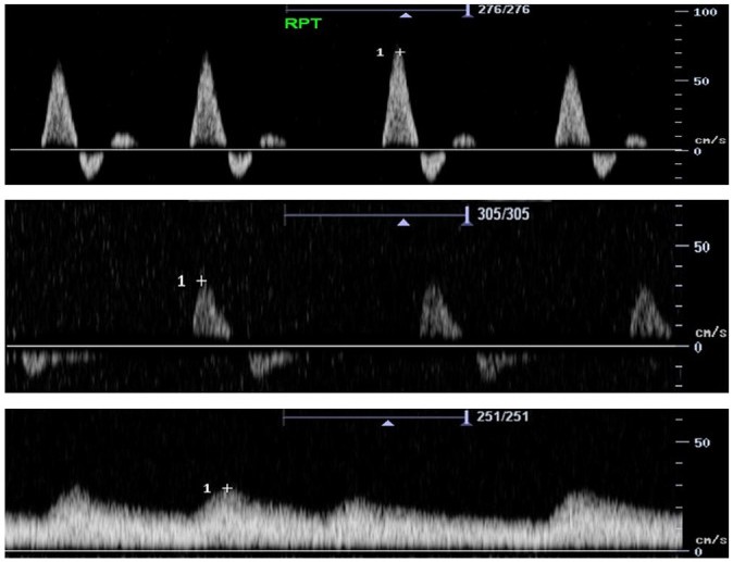
Doppler waveforms. The normal peripheral artery waveform is triphasic: a sharp systolic upstroke, brief early diastolic flow reversal reflecting high peripheral resistance, and late forward flow. Distal to a significant stenosis, the reversal disappears and the waveform becomes biphasic, then monophasic with a delayed upstroke, a rounded peak, and continuous diastolic flow. A monophasic ankle signal with a normal-appearing ABI exposes pressures falsely elevated by calcification.
Screening for peripheral artery disease
Screening with the resting ABI, with or without pulse volume recordings or Doppler waveforms, is reasonable in patients at increased risk of PAD (Class 2a) and is not recommended in patients who are not at increased risk and have no suggestive findings (Class 3: No Benefit). The rationale is that asymptomatic PAD identifies a patient with clinical ASCVD who qualifies for intensive risk reduction and foot care. Population yield below 50 years of age without risk factors is about 1%, and in a national survey about 95% of participants with an abnormal ABI had at least one atherosclerotic risk factor.
The US Preventive Services Task Force reached a different conclusion in 2018. It found the evidence insufficient to assess the balance of benefits and harms of ABI screening in asymptomatic adults (I statement), largely because two trials of aspirin in people with a low screening ABI showed no benefit. The ACC/AHA recommendation applies to defined high-risk groups in whom the result changes treatment intensity.
Duplex ultrasound, CT angiography, MR angiography, and catheter angiography
Anatomic imaging is obtained when revascularization is being considered. Duplex ultrasound, CT angiography (CTA), MR angiography (MRA), or catheter angiography is useful in functionally limiting claudication with an inadequate response to guideline-directed management and therapy (GDMT) including structured exercise (Class 1) and in CLTI (Class 1). Noninvasive imaging may be considered to establish the diagnosis when physiologic testing is inconclusive (Class 2b). CTA, MRA, or catheter angiography performed solely for anatomic assessment in a patient who is not a revascularization candidate exposes the patient to risk without benefit (Class 3: Harm).
| Modality | Strengths | Limitations |
|---|---|---|
| Duplex ultrasound | No contrast or radiation; hemodynamic data; surveillance; vein mapping | Operator-dependent; limited by bowel gas and dense calcification. A peak systolic velocity ratio of about 2.0 or more indicates stenosis of 50% or more. |
| CT angiography | Rapid aorta-to-foot imaging; defines calcification, aneurysm, and access vessels | Iodinated contrast and radiation; blooming from calcium overestimates stenosis in tibial arteries |
| MR angiography | No ionizing radiation; good tibial and pedal visualization; noncontrast sequences available | Overestimates stenosis; stent artifact; gadolinium and nephrogenic systemic fibrosis with some agents in advanced kidney disease; device and claustrophobia limitations |
| Catheter (digital subtraction) angiography | Reference standard; pressure gradients; treatment in the same session; carbon dioxide as an alternative contrast agent | Access-site bleeding, pseudoaneurysm, atheroembolism, contrast exposure, and radiation |
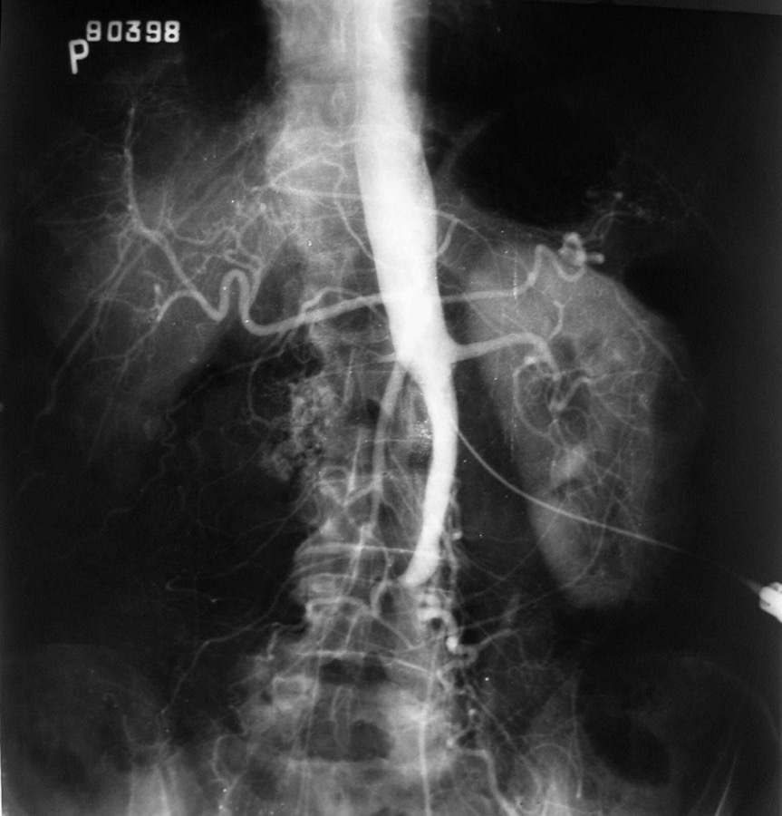

Contrast-associated kidney injury. Patients with PAD often have diabetes and chronic kidney disease. Strategies that lower the risk of contrast-associated kidney injury are duplex ultrasound or noncontrast MRA as the first test, minimal contrast volume, carbon dioxide angiography for the aortoiliac and femoropopliteal segments, withholding nephrotoxic drugs, and intravenous isotonic fluid in high-risk patients. In advanced kidney disease, diagnostic angiography combined with treatment in one session can use less contrast than CTA followed by a separate procedure. PRESERVE randomized 5,177 high-risk patients undergoing coronary or noncoronary angiography. Neither intravenous sodium bicarbonate compared with saline nor oral acetylcysteine compared with placebo reduced death, dialysis, or persistent kidney injury at 90 days. When the estimated glomerular filtration rate is below 30 mL/min/1.73 m², MRA uses noncontrast sequences or gadolinium agents with a low reported risk of nephrogenic systemic fibrosis.
Clinical classification: Fontaine and Rutherford
The Fontaine and Rutherford classifications grade chronic limb ischemia by symptoms, and both separate claudication from CLTI. Rutherford added objective hemodynamic criteria. Neither grades wound extent or infection, which the WIfI system addresses.
| Fontaine stage | Rutherford grade and category | Clinical description | Rutherford objective criteria |
|---|---|---|---|
| I | Grade 0, category 0 | Asymptomatic | Normal treadmill or reactive hyperemia test |
| IIa | Grade I, category 1 | Mild claudication | Completes treadmill exercise; postexercise ankle pressure above 50 mm Hg but at least 20 mm Hg below resting |
| IIb | Grade I, category 2 | Moderate claudication | Between categories 1 and 3 |
| Grade I, category 3 | Severe claudication | Cannot complete treadmill exercise; postexercise ankle pressure below 50 mm Hg | |
| III | Grade II, category 4 | Ischemic rest pain | Resting ankle pressure <40 mm Hg, flat or barely pulsatile ankle or forefoot PVR, toe pressure <30 mm Hg |
| IV | Grade III, category 5 | Minor tissue loss: nonhealing ulcer, focal gangrene with diffuse pedal ischemia | Resting ankle pressure <60 mm Hg, flat or barely pulsatile ankle or forefoot PVR, toe pressure <40 mm Hg |
| Grade III, category 6 | Major tissue loss above the transmetatarsal level; functional foot no longer salvageable | As category 5 |
Rutherford criteria from the 1997 revised reporting standards (treadmill test at 2 mph on a 12% grade for 5 minutes). PVR, pulse volume recording. Fontaine stage II is subdivided into mild (IIa) and moderate to severe (IIb) claudication.
WIfI threatened-limb staging
The Society for Vascular Surgery (SVS) WIfI system grades the threatened limb on three axes: wound extent (W), ischemia (I), and foot infection (fI), each from 0 to 3. Perfusion is one of three determinants of limb threat, which suits the diabetic foot, where a moderate perfusion deficit with a deep infected wound threatens the limb as much as severe ischemia. Expert consensus assigned each of the 64 combinations to one of four clinical stages of 1-year amputation risk and to an estimated benefit of revascularization. Stage correlates with amputation, wound healing time, and amputation-free survival. The Global Vascular Guidelines endorse WIfI staging for every patient with suspected CLTI, repeated after revascularization, foot surgery, or treatment of infection, and the 2024 ACC/AHA guideline recommends objective classification of the threatened limb when primary amputation or palliative care is considered (Class 1) and finds retrospective review of institutional outcomes with limb-threat classification tools useful (Class 2a).
| Grade | Wound (W) | Ischemia (I): ABI · ankle pressure · toe pressure or TcPO₂ | Foot infection (fI) |
|---|---|---|---|
| 0 | No ulcer, no gangrene (ischemic rest pain) | ≥0.80 · >100 mm Hg · ≥60 mm Hg | No symptoms or signs of infection |
| 1 | Small, shallow ulcer on the distal leg or foot, no exposed bone unless limited to the distal phalanx; no gangrene. Salvageable with simple digital amputation or skin coverage. | 0.60–0.79 · 70–100 mm Hg · 40–59 mm Hg | Local infection of skin and subcutaneous tissue only, erythema >0.5 to ≤2 cm around the wound |
| 2 | Deeper ulcer with exposed bone, joint, or tendon, generally not involving the heel, or shallow heel ulcer; gangrene limited to the digits. Salvageable with multiple digital or standard transmetatarsal amputation. | 0.40–0.59 · 50–70 mm Hg · 30–39 mm Hg | Local infection with erythema >2 cm or involving deeper structures (abscess, osteomyelitis, septic arthritis, fasciitis), without systemic signs |
| 3 | Extensive deep ulcer of the forefoot or midfoot, or deep full-thickness heel ulcer with or without calcaneal involvement; extensive gangrene. Salvageable only with complex reconstruction or nontraditional amputation. | ≤0.39 · <50 mm Hg · <30 mm Hg | Local infection with systemic inflammatory response syndrome |
From the SVS WIfI system and the Global Vascular Guidelines (Tables 3.2 to 3.5). Toe pressure or TcPO₂ is used when the ABI is noncompressible. Stage 5 (not in the matrix) denotes an unsalvageable foot.
WIfI clinical stage
Amputation risk and revascularization benefitStage 1 very low, stage 2 low, stage 3 moderate, and stage 4 high estimated 1-year amputation risk, from the consensus matrix of the SVS WIfI system. The example, W1 I2 fI1, is stage 3 with a high estimated benefit of revascularization. Revascularization benefit assumes that infection is controlled.
GLASS anatomic staging and the PLAN framework
The Global Vascular Guidelines introduced the Global Limb Anatomic Staging System (GLASS) to describe infrainguinal disease in CLTI by its complexity for endovascular treatment. The treating physician selects a target arterial path, the route of in-line flow from groin to ankle, usually through the least diseased tibial artery. The femoropopliteal and infrapopliteal segments of that path are graded 0 to 4, and severe calcification (more than 50% of the circumference, or bulky "coral reef" plaque) raises a segment grade by one. A matrix combines the two grades into stage I, II, or III. Inflow disease is staged separately, and a pedal modifier (P0 to P2) describes the arteries crossing the ankle; P2 denotes that no named artery crosses the ankle into the foot.
| Grade | Femoropopliteal segment | Infrapopliteal segment (target artery) |
|---|---|---|
| 0 | Mild or no significant (<50%) disease | Mild or no significant disease |
| 1 | Superficial femoral artery (SFA) disease <1/3 of length (<10 cm); may include a single focal occlusion <5 cm that is not flush; popliteal mild or no disease | Focal tibial stenosis <3 cm |
| 2 | SFA disease 1/3 to 2/3 (10–20 cm); occlusion <1/3 (10 cm), not flush; focal popliteal stenosis <2 cm not involving the trifurcation | Stenosis of 1/3 of vessel length; may include focal occlusion <3 cm; not involving the tibioperoneal trunk or tibial origin |
| 3 | SFA disease >2/3 (>20 cm); any flush occlusion <20 cm or nonflush occlusion 10–20 cm; popliteal stenosis 2–5 cm not involving the trifurcation | Disease up to 2/3 of vessel length; occlusion up to 1/3 (may include the tibial origin, not the tibioperoneal trunk) |
| 4 | SFA occlusion >20 cm; popliteal disease >5 cm or extending into the trifurcation; any popliteal occlusion | Diffuse stenosis >2/3 of vessel length; occlusion >1/3 (may include the origin); any tibioperoneal trunk occlusion if the anterior tibial is not the target |
| Femoropopliteal grade | IP 0 | IP 1 | IP 2 | IP 3 | IP 4 |
|---|---|---|---|---|---|
| 4 | III | III | III | III | III |
| 3 | II | II | II | III | III |
| 2 | I | II | II | II | III |
| 1 | I | I | II | II | III |
| 0 | Not applicable | I | I | II | III |
GLASS grades and stage matrix from the Global Vascular Guidelines (Figs 5.2 and 5.3, Table 5.3). Expected outcomes of endovascular treatment: stage I, technical failure <10% and 1-year limb-based patency >70%; stage II, technical failure <20% and patency 50% to 70%; stage III, technical failure >20% or patency <50%. SFA, superficial femoral artery; IP, infrapopliteal grade.
Aortoiliac inflow disease is staged I (iliac stenoses, occlusion of the common or external iliac artery but not both, infrarenal aortic stenosis) or II (aortic or combined iliac occlusion, diffuse or small iliac arteries, aneurysm, diffuse in-stent restenosis), with suffix B for common femoral stenosis above 50%.
GLASS is one axis of the PLAN framework: Patient risk, Limb severity (WIfI), and ANatomic complexity (GLASS). An average-risk patient has an anticipated periprocedural mortality below 5% and a 2-year survival above 50%. For average-risk patients with advanced limb threat and complex (GLASS III) disease, the Global Vascular Guidelines favor bypass with autologous vein; less complex anatomy, intermediate limb threat, or high patient risk favors endovascular treatment.
Antiplatelet and low-dose anticoagulant therapy
Antithrombotic therapy in PAD prevents MACE and MALE, which includes ALI and major amputation. In asymptomatic PAD, trials of aspirin after a low screening ABI showed no net benefit. In CAPRIE, clopidogrel reduced the composite of ischemic stroke, myocardial infarction, or vascular death compared with aspirin 325 mg (5.32% compared with 5.83% per year, relative reduction 8.7%), with the largest relative benefit in the PAD subgroup. In EUCLID, ticagrelor was not superior to clopidogrel (10.8% compared with 10.6%), and ALI occurred in 1.7% of each group.
| Setting | 2024 ACC/AHA recommendation | Class |
|---|---|---|
| Symptomatic PAD | Single antiplatelet therapy (clopidogrel 75 mg or aspirin 75–325 mg daily) | 1 |
| Symptomatic PAD, not at high bleeding risk | Rivaroxaban 2.5 mg twice daily plus low-dose aspirin to reduce MACE and MALE | 1 |
| After endovascular or surgical revascularization | Antiplatelet therapy; rivaroxaban 2.5 mg twice daily plus low-dose aspirin | 1 |
| After endovascular revascularization | Dual antiplatelet therapy (P2Y12 inhibitor plus low-dose aspirin) for at least 1 to 6 months | 2a |
| After revascularization in a patient on full-intensity anticoagulation for another indication | Adding single antiplatelet therapy, if not at high bleeding risk | 2a |
| Asymptomatic PAD | Single antiplatelet therapy | 2a |
| After bypass with a prosthetic graft | Dual antiplatelet therapy for at least 1 month | 2b |
| Symptomatic PAD without recent revascularization | Benefit of dual antiplatelet therapy uncertain | 2b |
| Symptomatic PAD | Benefit of vorapaxar added to antiplatelet therapy uncertain | 2b |
| PAD without another indication | Full-intensity oral anticoagulation to reduce MACE and MALE | 3: Harm |
Dual-pathway inhibition. In the PAD population of COMPASS (7,470 patients with stable PAD of the legs or carotid arteries), rivaroxaban 2.5 mg twice daily plus aspirin 100 mg reduced cardiovascular death, myocardial infarction, or stroke compared with aspirin alone (5% compared with 7%, hazard ratio 0.72) and MALE including major amputation (1% compared with 2%, hazard ratio 0.54), at the cost of more major bleeding (3% compared with 2%, hazard ratio 1.61). The excess was mostly gastrointestinal, with no increase in fatal or critical-organ bleeding. VOYAGER PAD randomized 6,564 patients within 10 days of lower extremity revascularization. The same regimen reduced the composite of ALI, major amputation, myocardial infarction, ischemic stroke, or cardiovascular death at 3 years (17.3% compared with 19.9%, hazard ratio 0.85), mainly through fewer ALI events. Thrombolysis in Myocardial Infarction (TIMI) major bleeding was not significantly increased (2.65% compared with 1.87%), whereas International Society on Thrombosis and Haemostasis (ISTH) major bleeding was (5.94% compared with 4.06%). Both trials excluded patients at high bleeding risk.
Vorapaxar. Vorapaxar, an antagonist of the platelet thrombin receptor (protease-activated receptor-1), was tested in 3,787 patients with symptomatic PAD in TRA2°P-TIMI 50. It did not reduce cardiovascular death, myocardial infarction, or stroke (11.3% compared with 11.9%), but it reduced hospitalization for ALI (2.3% compared with 3.9%, hazard ratio 0.58) and peripheral revascularization (18.4% compared with 22.2%), with more moderate or severe bleeding. Vorapaxar is contraindicated after stroke, transient ischemic attack, or intracranial hemorrhage, and its net benefit in PAD is uncertain (Class 2b).
Full-intensity anticoagulation. In WAVE, warfarin added to antiplatelet therapy did not reduce cardiovascular events (12.2% compared with 13.3%) and tripled life-threatening bleeding (4.0% compared with 1.2%).
Lipid, blood pressure, diabetes, and smoking targets in peripheral artery disease
Lipids. High-intensity statin therapy aimed at a reduction in low-density lipoprotein (LDL) cholesterol of 50% or more is indicated in every patient with PAD (Class 1). Adding ezetimibe or a proprotein convertase subtilisin/kexin type 9 (PCSK9) inhibitor is reasonable when LDL cholesterol remains 70 mg/dL or higher on maximally tolerated statin therapy (Class 2a). Goals, sequencing, and drug choice are covered on the dyslipidemia and LDL-lowering page.
Blood pressure. Antihypertensive therapy is recommended to reduce MACE (Class 1), with a goal below 130/80 mm Hg (Class 1). Angiotensin-converting enzyme inhibitors and angiotensin receptor blockers are recommended selectively to reduce MACE (Class 1). Beta-blockers do not worsen claudication or limb outcomes and are not contraindicated in PAD; they are used whenever coronary disease, heart failure, or arrhythmia calls for them. Treatment details are on the hypertension page.
Diabetes. Glucagon-like peptide-1 receptor agonists (liraglutide, semaglutide) and sodium-glucose cotransporter 2 inhibitors (canagliflozin, dapagliflozin, empagliflozin) are recommended in PAD with type 2 diabetes to reduce MACE (Class 1). Glycemic control may improve limb outcomes (Class 2b); in EUCLID, each 1% increase in hemoglobin A1c was associated with a 14% higher relative risk of MACE. About half of patients with CLTI have diabetes, and diabetes care is coordinated by the team (Class 1).
Smoking and tobacco. Tobacco users are advised at every visit to quit (Class 1) and are offered a plan that combines varenicline, bupropion, or nicotine replacement with counseling or referral (Class 1). Avoidance of secondhand smoke is recommended (Class 1). Continued smoking accelerates progression and lowers the patency of every type of revascularization.
Other measures. Annual influenza vaccination is recommended (Class 1), and a diet rich in vegetables, fruits, legumes, nuts, whole grains, and fish is beneficial (Class 2a). B-vitamin supplementation for homocysteine, vitamin D supplementation, and chelation therapy do not reduce MACE (Class 3: No Benefit).
Supervised exercise therapy and structured community exercise
Structured exercise is the foundation of treatment for walking impairment in PAD. Supervised exercise therapy (SET) is recommended in chronic symptomatic PAD (Class 1), as is a structured community-based program with behavioral change techniques (Class 1). SET or a structured community program is offered as initial treatment of functionally limiting claudication (Class 1) and after revascularization (Class 1). Nonwalking structured programs such as arm ergometry or recumbent stepping are beneficial (Class 2a), whereas the usefulness of unstructured advice to "go and walk" is uncertain (Class 2b). In a Cochrane review of 32 trials with 1,835 participants, exercise programs improved maximal and pain-free walking distance compared with usual care.
| Element | Supervised exercise therapy | Structured community-based program |
|---|---|---|
| Setting | Hospital or outpatient facility, often within a cardiac rehabilitation program | Home, neighborhood, or fitness facility |
| Supervision | Directly supervised by a physician or advanced practice provider; delivered by exercise physiologists or nurses | Self-directed with professional prescription and periodic review; health coaching or activity monitors |
| Session and duration | At least 30 to 45 minutes of exercise per 60-minute session, at least 3 times per week, for at least 12 weeks | Regimen similar to SET, progressed by distance or speed |
| Intensity | Intermittent treadmill walking to moderate or maximal claudication, alternating with rest, increased as symptoms improve | Usually walking-based; higher intensity more effective than low intensity |
| Coverage | Medicare covers up to 36 sessions over 12 weeks for symptomatic PAD | Not a defined benefit |
Adapted from the 2024 ACC/AHA guideline, Table 14. Despite Class 1 status and Medicare coverage, 1.3% of Medicare beneficiaries with newly diagnosed claudication enrolled in SET during the first 19 months of coverage, and 5.1% of those enrolled completed all 36 sessions.
Exercise improves walking at least as much as revascularization for claudication. In CLEVER (111 patients with aortoiliac disease), peak walking time at 6 months increased by 5.8 minutes with supervised exercise, 3.7 minutes with stenting, and 1.2 minutes with medical care alone; stenting produced greater gains in patient-reported quality of life. In ERASE, endovascular revascularization plus supervised exercise increased maximal walking distance more than exercise alone (an additional 282 m at 12 months). Walking performance and quality of life are measured at baseline and during follow-up (Class 1) with treadmill claudication onset and peak walking time, the 6-minute walk test, and questionnaires such as the Walking Impairment Questionnaire and the Vascular Quality of Life Questionnaire.
Cilostazol and other drugs for claudication
Cilostazol, a phosphodiesterase-3 inhibitor with antiplatelet and vasodilator effects, is recommended to improve leg symptoms and walking distance in claudication (Class 1). In a Cochrane review of 16 trials with 3,972 participants, cilostazol increased initial claudication distance by a mean of about 26 m compared with placebo, without a demonstrated reduction in cardiovascular events. The usual dose is 100 mg twice daily, taken at least 30 minutes before or 2 hours after breakfast and dinner, reduced to 50 mg twice daily with strong or moderate inhibitors of CYP3A4 (such as ketoconazole, itraconazole, erythromycin, or diltiazem) or with inhibitors of CYP2C19 (such as ticlopidine, fluconazole, or omeprazole). Benefit appears over 2 to 4 weeks and may take up to 12 weeks, so a 3-month trial precedes a judgment of failure. Headache, diarrhea, and palpitations are common.
Cilostazol is contraindicated in heart failure of any severity (Class 3: Harm). The prohibition derives from increased mortality with oral milrinone, another phosphodiesterase-3 inhibitor, in severe heart failure. Cilostazol may also reduce restenosis after femoropopliteal endovascular therapy (Class 2b).
Pentoxifylline does not improve claudication (Class 3: No Benefit), and chelation therapy has no role (Class 3: No Benefit).
Revascularization for claudication by anatomic segment
Revascularization for claudication treats symptoms and does not prevent amputation or cardiovascular events. Its benefits are weighed against procedural risk, restenosis, and repeat procedures in a shared decision (Class 1). It is reasonable when functionally limiting claudication responds inadequately to GDMT including structured exercise (Class 2a). It is not recommended after an adequate response to that therapy (Class 3: No Benefit), and revascularization of asymptomatic PAD solely to prevent progression is harmful (Class 3: Harm). Asymptomatic iliofemoral disease may be treated when access is needed for transcatheter aortic valve replacement, mechanical circulatory support, or endovascular aneurysm repair (Class 2a).
| Segment | 2024 ACC/AHA recommendation | Class | Considerations |
|---|---|---|---|
| Aortoiliac | Endovascular revascularization effective; surgery reasonable if perioperative risk is acceptable and technical factors favor it | 1 / 2a | Most durable endovascular results |
| Common femoral | Surgical endarterectomy reasonable, especially when endovascular treatment would compromise the profunda femoris; endovascular treatment may be considered at high surgical risk or with favorable anatomy | 2a / 2b | Endarterectomy 7-year primary patency 78.5% in a large series |
| Femoropopliteal | Endovascular revascularization effective; surgery reasonable in selected anatomy | 1 / 2a | Patency lower than aortoiliac, worse with long lesions, occlusions, and poor runoff; popliteal bypass uses autogenous vein in preference to prosthetic graft (Class 1) |
| Isolated infrapopliteal | Effectiveness of endovascular or surgical revascularization unknown | 2b | Uncommon cause of claudication; tibial intervention is generally reserved for CLTI |
Endovascular devices. Options include plain balloon angioplasty, drug-coated balloons, bare-metal, drug-eluting, and covered stents, atherectomy, and intravascular lithotripsy, selected by lesion location, length, and calcification. In the femoropopliteal segment, paclitaxel-coated balloons and stents reduce restenosis compared with uncoated devices. A 2018 meta-analysis of randomized trials reported higher late mortality after paclitaxel-coated femoropopliteal devices, which led to US Food and Drug Administration warnings. After review of larger patient-level data with longer follow-up, the FDA concluded in July 2023 that the data do not support an excess mortality risk. Atherectomy is used as vessel preparation for heavily calcified or bulky lesions; randomized evidence that it improves clinical outcomes over angioplasty is lacking.
Surgical options. Aortobifemoral bypass, endarterectomy, femoropopliteal and femorotibial bypass, and extra-anatomic (axillofemoral or femorofemoral) bypass are the open procedures, and hybrid procedures combine common femoral endarterectomy with iliac or femoral stenting. Great saphenous vein is the preferred infrainguinal conduit because prosthetic grafts have lower long-term patency.
Chronic limb-threatening ischemia: team care and revascularization strategy
CLTI is defined in the Global Vascular Guidelines as PAD with ischemic rest pain, gangrene, or a lower limb ulcer of more than 2 weeks' duration, with objective hemodynamic evidence of ischemia; venous, traumatic, embolic, and nonatherosclerotic causes are excluded. Suspected CLTI is referred urgently to a vascular specialist, and WIfI and GLASS staging precede the choice of procedure.
The 2024 ACC/AHA guideline recommends evaluation and care by a multispecialty team with expertise in endovascular and surgical revascularization, wound care, podiatric and foot surgery, infectious disease, diabetes, and rehabilitation (Class 1). Surgical, endovascular, or hybrid revascularization is recommended when feasible to minimize tissue loss, heal wounds, relieve pain, and preserve a functional limb (Class 1). The team evaluates revascularization options before any nonemergency amputation (Class 1). Great saphenous vein mapping by duplex ultrasound is recommended in patients who are candidates for both strategies (Class 1). Bypass to the popliteal or infrapopliteal arteries uses autogenous vein when available (Class 1), and prosthetic or cadaveric conduits can be effective when it is not (Class 2a). Restoration of in-line flow, or maximal perfusion, to the wound bed is beneficial (Class 2a). With rest pain alone and multilevel disease, treating inflow disease first is reasonable (Class 2a), whereas tissue loss usually requires inflow and outflow treatment together.
| Trial | Population | Primary outcome | Result |
|---|---|---|---|
| BEST-CLI, cohort 1 (2022) | Infrainguinal CLTI with an adequate single-segment great saphenous vein (1,830 patients enrolled in the two cohorts) | MALE (above-ankle amputation or major reintervention) or death | 42.6% with surgery compared with 57.4% with endovascular therapy at a median of 2.7 years; hazard ratio 0.68, driven by fewer reinterventions |
| BEST-CLI, cohort 2 (2022) | Infrainguinal CLTI needing an alternative conduit | Same | 42.8% compared with 47.7%; hazard ratio 0.79, not significant (underpowered) |
| BASIL-2 (2023) | 345 patients with CLTI requiring infrapopliteal revascularization | Major amputation or death | 63% with vein bypass first compared with 53% with best endovascular treatment first; adjusted hazard ratio 1.35, driven by more deaths after bypass |
The trials differ in anatomy (BEST-CLI mostly femoropopliteal with or without tibial disease; BASIL-2 infrapopliteal), patient risk, conduit, and endovascular technique, and they are complementary. In BEST-CLI, an adequate vein was a great saphenous vein of at least 3 mm.
The guideline lists factors that shape the first strategy (Table 16). Anatomy favoring surgery includes disease of both the common femoral artery and the profunda origin, multilevel occlusions, long-segment disease involving the below-knee popliteal and tibial arteries, and lesions whose endovascular treatment would compromise a later bypass. Absence of usable vein (for example, after harvest for coronary bypass) and high perioperative risk from coronary disease, heart failure, lung disease, chronic kidney disease, or frailty favor endovascular treatment. Patient preference after shared decision-making completes the decision (Class 1).
Wound care, infection, offloading, and amputation in chronic limb-threatening ischemia
Infection. Foot infection is treated promptly with antibiotics, debridement, and drainage (Class 1). Deep infection, abscess, and osteomyelitis are evaluated with probing of the wound to bone, plain radiographs, and MRI. Severe infection with systemic toxicity is drained or amputated urgently before revascularization.
Wound care and offloading. Wound care after revascularization aims at complete healing (Class 1): serial debridement, a moist wound bed with exudate control, treatment of biofilm, nutrition, glycemic control, and smoking cessation. Pressure offloading is recommended for diabetic foot ulcers (Class 1) and may be considered for nondiabetic ulcers (Class 2b), and customized footwear is recommended after a diabetic foot ulcer has healed (Class 1). Negative-pressure wound therapy has the best supporting evidence among adjuncts. Hyperbaric oxygen may be considered after revascularization for nonhealing diabetic foot ulcers (Class 2b); the Global Vascular Guidelines advise against it in limbs with uncorrected severe ischemia. Cell and gene therapies remain investigational.
No-option CLTI. When no revascularization is possible and the foot has no outflow, the usefulness of prostanoids is uncertain (Class 2b), arterial intermittent pneumatic compression may be considered (Class 2b), and deep venous arterialization, which routes arterial flow into the pedal veins, may be considered for limb preservation (Class 2b).
Amputation. Primary amputation is indicated when life over limb is the prevailing consideration and the limb is the source of instability, such as overwhelming infection or metabolic derangement (Class 1). It is also appropriate for nonambulatory or bedbound patients, limited life expectancy, or tissue destruction that leaves no functional foot, decided with the patient and family (Class 1). The multispecialty team selects the most distal level that will heal and preserve function (Class 1), and minor (inframalleolar) amputation is followed by a customized program of wound care, offloading, and therapeutic footwear (Class 1).
Preventive foot care. Every patient with PAD receives foot self-care education, foot inspection at every visit, and a comprehensive foot evaluation at least annually (all Class 1). Therapeutic footwear is recommended for patients at high risk, such as those with a prior ulcer or amputation, deformity, neuropathy, or poorly controlled diabetes (Class 1).
Acute limb ischemia: causes, categories, and emergency treatment
ALI is a sudden decrease in limb perfusion, present for less than 2 weeks, that threatens viability. Skeletal muscle tolerates complete ischemia for about 4 to 6 hours, so delay determines outcome. The classic findings are the six Ps: pain, pallor, pulselessness, poikilothermia, paresthesia, and paralysis. Sensory loss and motor weakness are the findings that change urgency.
Causes. In patients with known PAD, thrombosis of a stenotic native artery, bypass graft, or stent is the most common cause, and collaterals often blunt the presentation. Embolism from the left atrium in atrial fibrillation or from left ventricular thrombus produces abrupt symptoms with a normal contralateral pulse examination and lodges at bifurcations, most often the common femoral. Other causes are popliteal aneurysm, dissection, trauma and access-site injury, paradoxical embolism, heparin-induced thrombocytopenia, antiphospholipid syndrome, cancer and its therapy, vasculitis, and viral infection including COVID-19.
| Category | Prognosis | Sensory loss | Muscle weakness | Arterial Doppler | Venous Doppler |
|---|---|---|---|---|---|
| I. Viable | Not immediately threatened | None | None | Audible | Audible |
| IIa. Marginally threatened | Salvageable with prompt treatment | Minimal, limited to the toes | None | Often inaudible | Audible |
| IIb. Immediately threatened | Salvageable with immediate revascularization | More than the toes, with rest pain | Mild to moderate | Inaudible | Audible |
| III. Irreversible | Major tissue loss or permanent nerve damage inevitable | Profound, anesthetic | Profound paralysis, rigor | Inaudible | Inaudible |
Rutherford acute limb ischemia categories, as adopted in the 2024 ACC/AHA guideline. Handheld continuous-wave Doppler replaces unreliable pulse palpation; loss of the venous signal marks a limb that may be nonsalvageable.
Initial management. Emergency evaluation by a clinician who can assess viability and deliver treatment is recommended (Class 1). Viability is assessed clinically without noninvasive imaging (Class 1), except after complex prior revascularization (Class 2b) or with suspected aortic dissection. Intravenous unfractionated heparin is given at diagnosis unless contraindicated, regardless of cause or level of occlusion (Class 1); a direct thrombin inhibitor is substituted in heparin-induced thrombocytopenia.
Revascularization. In a salvageable limb, endovascular or surgical revascularization, including catheter-directed thrombolysis, is indicated (Class 1). Category IIb limbs need immediate revascularization, IIa limbs prompt revascularization, and category I limbs allow time for imaging to plan an urgent procedure. Surgical balloon catheter embolectomy suits a fresh embolus at a bifurcation; thrombosis on a diseased artery often needs bypass or patch angioplasty. Endovascular options are catheter-directed thrombolysis and pharmacomechanical or aspiration thrombectomy. TOPAS randomized 544 patients with occlusion of 14 days or less. Amputation-free survival at 1 year was similar with intra-arterial urokinase and with surgery (65.0% and 69.9%), and thrombolysis reduced open operations. Major hemorrhage was more frequent with thrombolysis (12.5% compared with 5.5%), including intracranial hemorrhage in 1.6%. Thrombolysis is most effective for occlusions of less than 14 days, including graft thrombosis, and endovascular or surgical treatment of the culprit lesion it unmasks can be useful (Class 2a). Revascularization of a category III limb is harmful (Class 3: Harm), because reperfusion of necrotic muscle can cause multiorgan failure; primary amputation is performed.
Reperfusion injury and compartment syndrome. Reperfusion releases potassium, myoglobin, and acid, causing hyperkalemia, arrhythmia, acidosis, and pigment nephropathy, and muscle edema raises compartment pressure, most often in the lower leg. Pain on passive stretch, tense compartments, and rising creatine kinase lead to fasciotomy (Class 1). Prophylactic fasciotomy is reasonable for category IIa and IIb limbs (Class 2a), and concurrent amputation can be beneficial after prolonged ischemia (Class 2a).
Cause and secondary prevention. A history and examination directed at the cause (Class 1) and testing for a cardiac source, including electrocardiography, rhythm monitoring, and echocardiography, are useful (Class 2a). Long-term anticoagulation follows embolism from atrial fibrillation, and thrombosis on underlying PAD treated by revascularization is followed by the antithrombotic regimens recommended after any lower extremity revascularization.
Peripheral artery disease in older adults, kidney disease, and diabetes
Older adults. In patients 75 years or older, assessment for geriatric syndromes such as frailty, sarcopenia, malnutrition, and mobility impairment is useful before revascularization and in setting goals (Class 2a). Frailty predicts lower survival after revascularization for CLTI and favors less invasive strategies or palliation in some patients.
Chronic kidney disease and dialysis. Kidney disease raises MACE (hazard ratio about 1.45) and is associated with tibial calcification, noncompressible ABI, CLTI, and amputation. Revascularization for CLTI in patients with kidney disease is associated with lower mortality and amputation than no revascularization, and endovascular treatment carries lower in-hospital mortality than surgery in this group. COMPASS excluded patients with severe kidney disease.
Diabetes. Up to 20% of patients with diabetes older than 40 years, 30% older than 50 years, and 70% older than 70 years have PAD. Neuropathy masks symptoms, and infection is the most frequent trigger of limb loss. Toe pressure with TBI is the routine perfusion test.
Follow-up and surveillance after revascularization
Every patient with PAD has longitudinal follow-up with assessment of limb symptoms, functional status, pulses, the feet, and risk factor control (Class 1), and long-term GDMT is recommended to prevent MACE and MALE (Class 1). Care is coordinated among the clinicians involved (Class 1), and telehealth is an alternative when it matches the urgency of symptoms (Class 2a). After any revascularization, new symptoms or signs lead to ABI and arterial duplex ultrasound (Class 1).
| Revascularization | Surveillance without new symptoms | Class |
|---|---|---|
| Infrainguinal autogenous vein bypass | ABI and arterial duplex ultrasound within 1 to 3 months, at 6 and 12 months, then annually | 2a |
| Endovascular procedure | ABI and arterial duplex ultrasound within 1 to 3 months, at 6 and 12 months, then annually | 2a |
| Infrainguinal prosthetic bypass | Effectiveness of routine ABI and duplex surveillance uncertain | 2b |
| Any revascularization with new symptoms or signs | ABI and arterial duplex ultrasound | 1 |
From the 2024 ACC/AHA guideline (Section 12). A failing vein graft found by duplex surveillance is easier to salvage than an occluded one.
The Global Vascular Guidelines define loss of limb-based patency after CLTI revascularization as occlusion, critical stenosis, or reintervention on the target arterial path, or a fall in ABI of 0.15 or more or in TBI of 0.10 or more with recurrent or unresolved symptoms. Restaging with WIfI after revascularization and after treatment of infection identifies limbs that remain at high risk.
European guidance compared with the 2024 ACC/AHA guideline
The 2024 European Society of Cardiology (ESC) guideline on peripheral arterial and aortic diseases agrees on ABI as the first test and on structured exercise, statins, and antiplatelet therapy, and it differs in several points. It sets an LDL cholesterol goal below 55 mg/dL (1.4 mmol/L) with at least a 50% reduction for atherosclerotic PAD (Class 1) and a systolic blood pressure goal of 120 to 129 mm Hg. It assigns rivaroxaban 2.5 mg twice daily plus aspirin a Class 2a recommendation in symptomatic PAD with high ischemic and non-high bleeding risk and after lower limb revascularization, compared with Class 1 in the US guideline. It recommends duplex ultrasound as the first-line imaging test, considers drug-eluting treatment the first-choice endovascular strategy for femoropopliteal lesions (Class 2a), and allows aspirin in asymptomatic PAD only with diabetes (Class 2b), compared with Class 2a single antiplatelet therapy in all asymptomatic PAD in the US guideline.
Self-assessment
A 71-year-old man with type 2 diabetes and stage 4 chronic kidney disease has a 2-week-old ulcer over the first metatarsal head with exposed tendon and no cellulitis. Brachial systolic pressures are 142 and 138 mm Hg, ankle pressures on the affected side are 238 and 226 mm Hg, and the great-toe pressure is 34 mm Hg. What are the ABI, the TBI, and the WIfI classification, and what is the next step?
Cardiovascular Medicine Board ReviewReferences
- Gornik HL, Aronow HD, Goodney PP, et al. 2024 ACC/AHA/AACVPR/APMA/ABC/SCAI/SVM/SVN/SVS/SIR/VESS guideline for the management of lower extremity peripheral artery disease: a report of the American College of Cardiology/American Heart Association Joint Committee on Clinical Practice Guidelines. Circulation. 2024;149:e1313–e1410. doi:10.1161/CIR.0000000000001251
- Mazzolai L, Teixido-Tura G, Lanzi S, et al. 2024 ESC guidelines for the management of peripheral arterial and aortic diseases. Eur Heart J. 2024;45:3538–3700. doi:10.1093/eurheartj/ehae179
- Conte MS, Bradbury AW, Kolh P, et al. Global vascular guidelines on the management of chronic limb-threatening ischemia. J Vasc Surg. 2019;69(6S):3S–125S.e40. doi:10.1016/j.jvs.2019.02.016
- Mills JL Sr, Conte MS, Armstrong DG, et al. The Society for Vascular Surgery lower extremity threatened limb classification system: risk stratification based on wound, ischemia, and foot infection (WIfI). J Vasc Surg. 2014;59:220–234.e2. doi:10.1016/j.jvs.2013.08.003
- Rutherford RB, Baker JD, Ernst C, et al. Recommended standards for reports dealing with lower extremity ischemia: revised version. J Vasc Surg. 1997;26:517–538. doi:10.1016/S0741-5214(97)70045-4
- Aboyans V, Criqui MH, Abraham P, et al. Measurement and interpretation of the ankle-brachial index: a scientific statement from the American Heart Association. Circulation. 2012;126:2890–2909. doi:10.1161/CIR.0b013e318276fbcb
- Ankle Brachial Index Collaboration; Fowkes FG, Murray GD, Butcher I, et al. Ankle brachial index combined with Framingham risk score to predict cardiovascular events and mortality: a meta-analysis. JAMA. 2008;300:197–208. doi:10.1001/jama.300.2.197
- Criqui MH, Aboyans V. Epidemiology of peripheral artery disease. Circ Res. 2015;116:1509–1526. doi:10.1161/CIRCRESAHA.116.303849
- Song P, Rudan D, Zhu Y, et al. Global, regional, and national prevalence and risk factors for peripheral artery disease in 2015: an updated systematic review and analysis. Lancet Glob Health. 2019;7:e1020–e1030. doi:10.1016/S2214-109X(19)30255-4
- US Preventive Services Task Force; Curry SJ, Krist AH, Owens DK, et al. Screening for peripheral artery disease and cardiovascular disease risk assessment with the ankle-brachial index: US Preventive Services Task Force recommendation statement. JAMA. 2018;320:177–183. doi:10.1001/jama.2018.8357
- CAPRIE Steering Committee. A randomised, blinded, trial of clopidogrel versus aspirin in patients at risk of ischaemic events (CAPRIE). Lancet. 1996;348:1329–1339. doi:10.1016/S0140-6736(96)09457-3
- Hiatt WR, Fowkes FG, Heizer G, et al. Ticagrelor versus clopidogrel in symptomatic peripheral artery disease (EUCLID). N Engl J Med. 2017;376:32–40. doi:10.1056/NEJMoa1611688
- Anand SS, Bosch J, Eikelboom JW, et al. Rivaroxaban with or without aspirin in patients with stable peripheral or carotid artery disease: an international, randomised, double-blind, placebo-controlled trial (COMPASS). Lancet. 2018;391:219–229. doi:10.1016/S0140-6736(17)32409-1
- Bonaca MP, Bauersachs RM, Anand SS, et al. Rivaroxaban in peripheral artery disease after revascularization (VOYAGER PAD). N Engl J Med. 2020;382:1994–2004. doi:10.1056/NEJMoa2000052
- Bonaca MP, Scirica BM, Creager MA, et al. Vorapaxar in patients with peripheral artery disease: results from TRA2°P-TIMI 50. Circulation. 2013;127:1522–1529. doi:10.1161/CIRCULATIONAHA.112.000679
- Warfarin Antiplatelet Vascular Evaluation Trial Investigators; Anand S, Yusuf S, Xie C, et al. Oral anticoagulant and antiplatelet therapy and peripheral arterial disease (WAVE). N Engl J Med. 2007;357:217–227. doi:10.1056/NEJMoa065959
- Murphy TP, Cutlip DE, Regensteiner JG, et al. Supervised exercise versus primary stenting for claudication resulting from aortoiliac peripheral artery disease: six-month outcomes from the Claudication: Exercise Versus Endoluminal Revascularization (CLEVER) study. Circulation. 2012;125:130–139. doi:10.1161/CIRCULATIONAHA.111.075770
- Fakhry F, Spronk S, van der Laan L, et al. Endovascular revascularization and supervised exercise for peripheral artery disease and intermittent claudication: a randomized clinical trial (ERASE). JAMA. 2015;314:1936–1944. doi:10.1001/jama.2015.14851
- Lane R, Harwood A, Watson L, Leng GC. Exercise for intermittent claudication. Cochrane Database Syst Rev. 2017;12:CD000990. doi:10.1002/14651858.CD000990.pub4
- Brown T, Forster RB, Cleanthis M, et al. Cilostazol for intermittent claudication. Cochrane Database Syst Rev. 2021;6:CD003748. doi:10.1002/14651858.CD003748.pub5
- Katsanos K, Spiliopoulos S, Kitrou P, et al. Risk of death following application of paclitaxel-coated balloons and stents in the femoropopliteal artery of the leg: a systematic review and meta-analysis of randomized controlled trials. J Am Heart Assoc. 2018;7:e011245. doi:10.1161/JAHA.118.011245
- Farber A, Menard MT, Conte MS, et al. Surgery or endovascular therapy for chronic limb-threatening ischemia (BEST-CLI). N Engl J Med. 2022;387:2305–2316. doi:10.1056/NEJMoa2207899
- Bradbury AW, Moakes CA, Popplewell M, et al. A vein bypass first versus a best endovascular treatment first revascularisation strategy for patients with chronic limb threatening ischaemia who required an infra-popliteal, with or without an additional more proximal infra-inguinal revascularisation procedure to restore limb perfusion (BASIL-2): an open-label, randomised, multicentre, phase 3 trial. Lancet. 2023;401:1798–1809. doi:10.1016/S0140-6736(23)00462-2
- Weisbord SD, Gallagher M, Jneid H, et al. Outcomes after angiography with sodium bicarbonate and acetylcysteine (PRESERVE). N Engl J Med. 2018;378:603–614. doi:10.1056/NEJMoa1710933
- Creager MA, Kaufman JA, Conte MS. Clinical practice. Acute limb ischemia. N Engl J Med. 2012;366:2198–2206. doi:10.1056/NEJMcp1006054
- Ouriel K, Veith FJ, Sasahara AA. A comparison of recombinant urokinase with vascular surgery as initial treatment for acute arterial occlusion of the legs (TOPAS). N Engl J Med. 1998;338:1105–1111. doi:10.1056/NEJM199804163381603
- Normahani P, Agrawal R, Sounderajah V, et al. Arterial spectral waveform analysis in the prediction of diabetic foot ulcer healing. Perfusion. 2021;36:751–756. doi:10.1177/0267659120957849
- Divakaran S, Carroll BJ, Chen S, Shen C, Bonaca MP, Secemsky EA. Supervised exercise therapy for symptomatic peripheral artery disease among Medicare beneficiaries between 2017 and 2018: participation rates and outcomes. Circ Cardiovasc Qual Outcomes. 2021;14:e007953. doi:10.1161/CIRCOUTCOMES.121.007953
- Clark CE, Taylor RS, Shore AC, Ukoumunne OC, Campbell JL. Association of a difference in systolic blood pressure between arms with vascular disease and mortality: a systematic review and meta-analysis. Lancet. 2012;379:905–914. doi:10.1016/S0140-6736(11)61710-8